Status of Plant Protein-Based Green Scaffolds for Regenerative Medicine Applications

 ,
,
Abstract
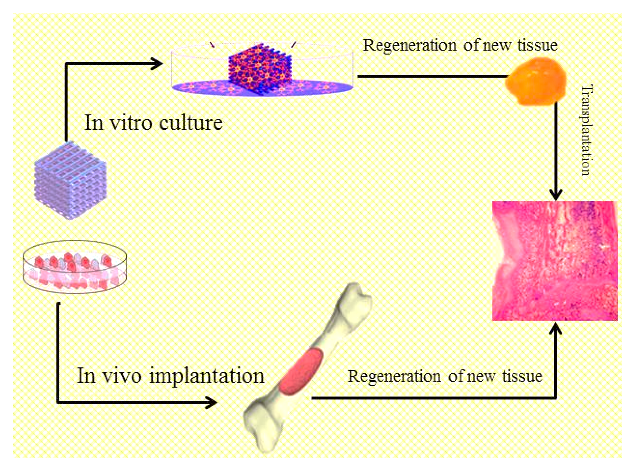
:1. Introduction
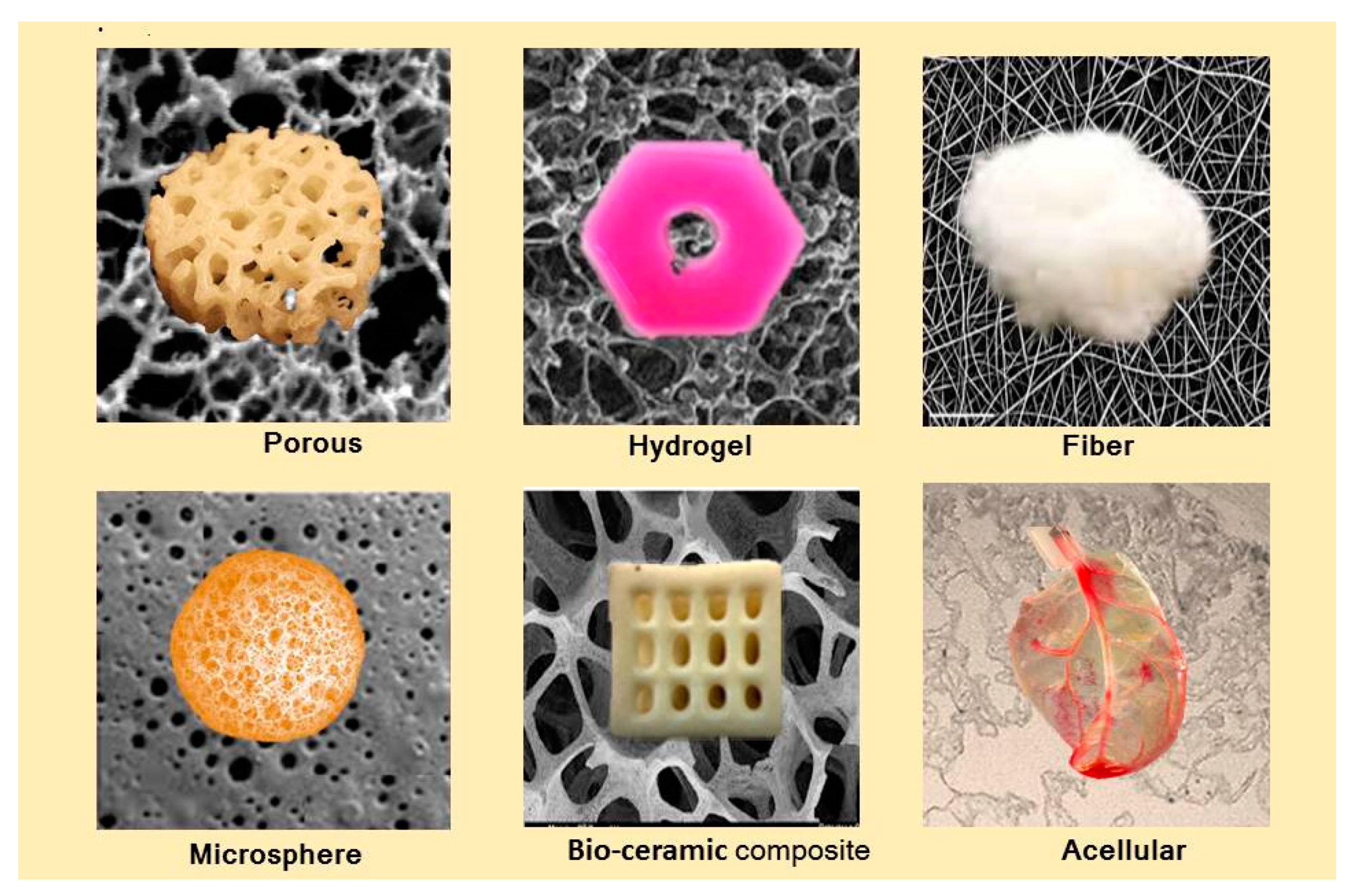
2. Various Forms of Scaffolds for Tissue Engineering Applications
2.1. Porous Scaffolds
2.2. Fibrous Scaffolds
2.3. Scaffolds Based on Hydrogels
2.4. Microsphere Scaffolds
2.5. Polymer/Bioceramic Composite Scaffolds
2.6. Acellular Scaffolds
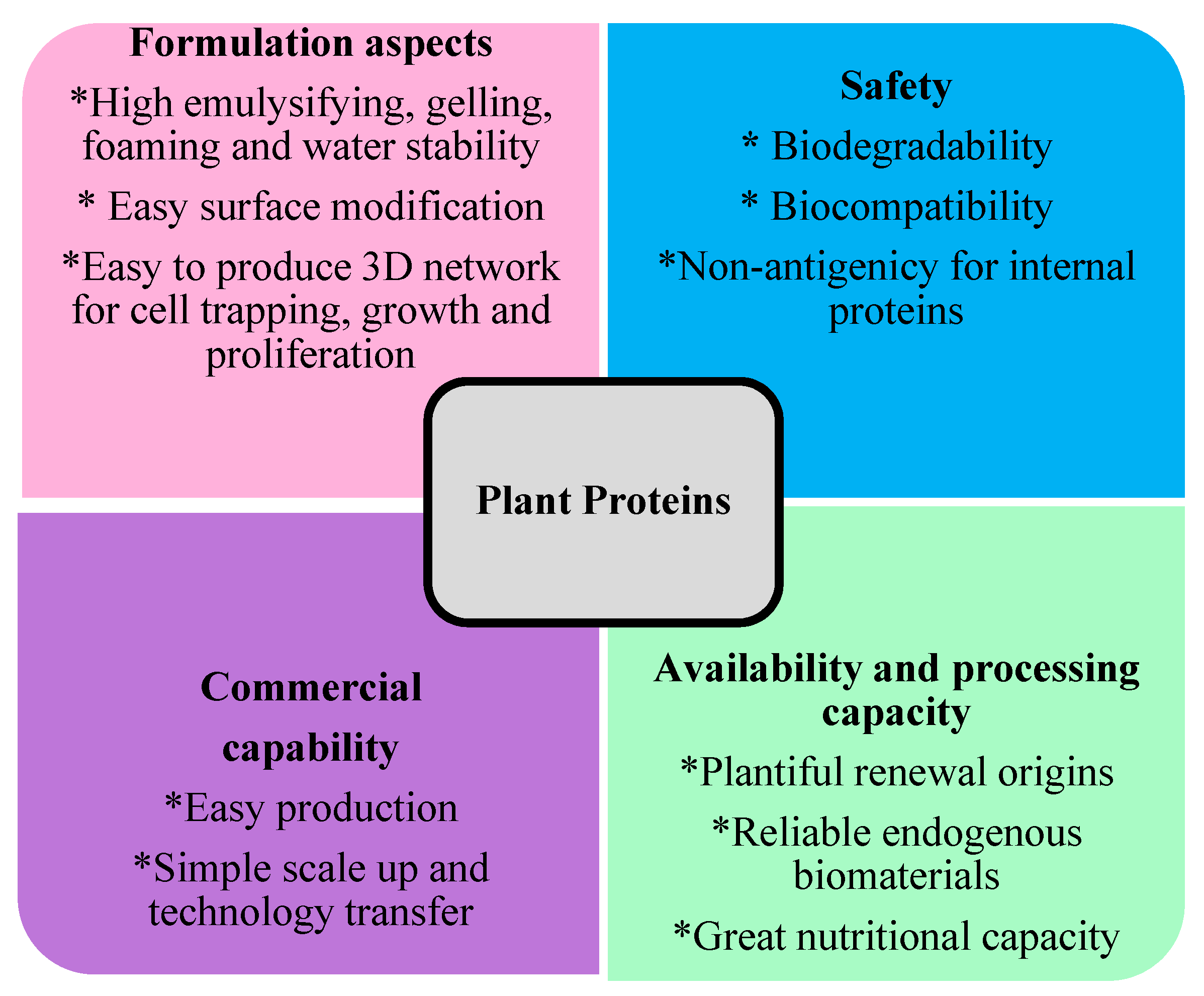
3. Plant Proteins
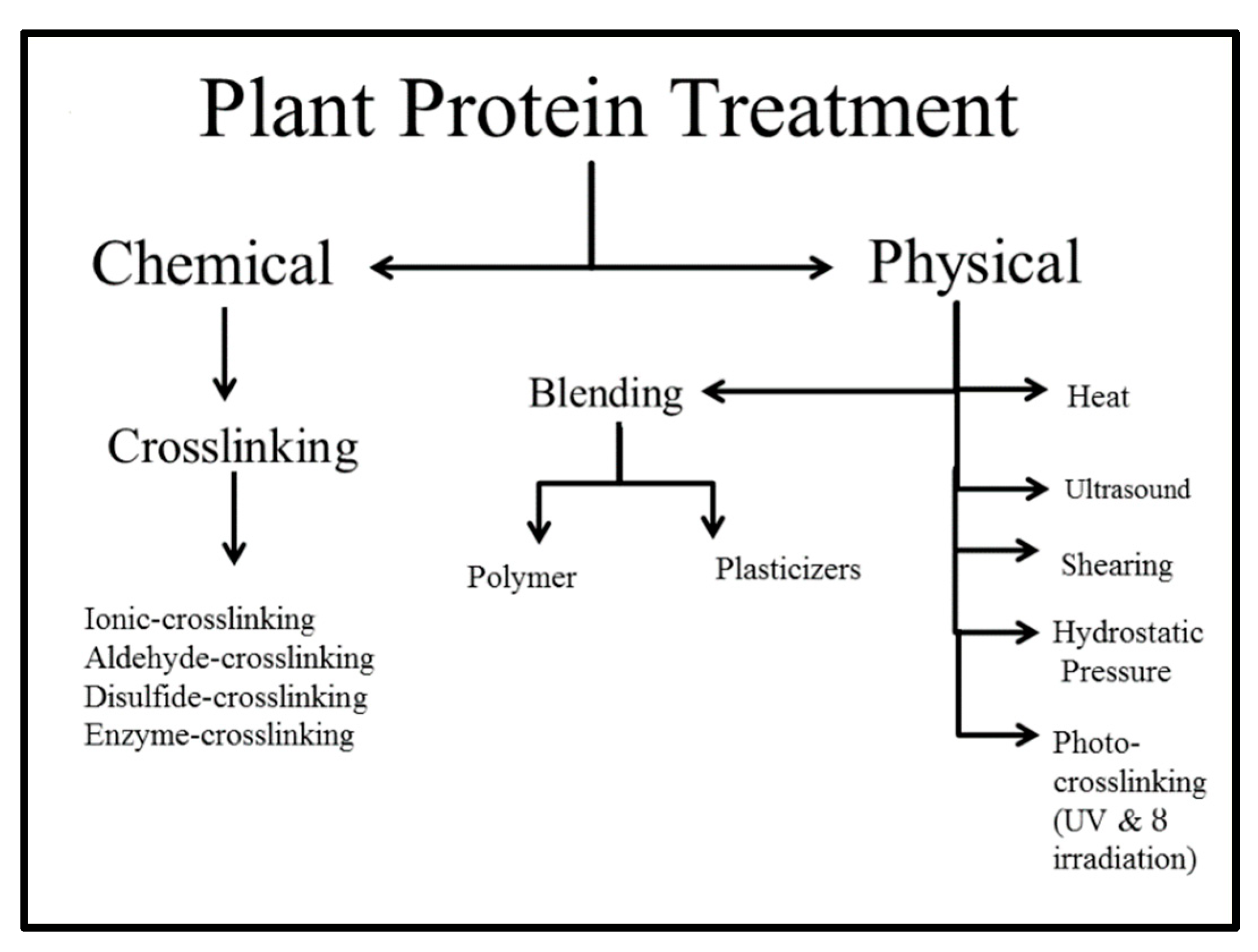
3.1. Chemical and Physical Treatment
3.2. Plantprotein-Based Nanocomposites
3.3. Plant Protein-Based Electrospun Nanofibers and Films/Natural Extracts
4. Plant Protein-Based Green Scaffolds for Tissue Engineering
4.1. Soy Protein
4.1.1. Soy Protein Porous Scaffolds
4.1.2. Soy Protein Fibrous Scaffolds
4.1.3. Soy Protein Hydrogel Scaffolds
4.2. Zein Protein
4.2.1. Zein Porous Scaffolds
4.2.2. Zein Fibrous Scaffolds
4.3. Wheat Gluten (Gliadin, Glutenin) Protein
Wheat Gluten Scaffolds
4.4. Camelina Scaffolds
4.5. Aloe Vera (AV) Scaffolds
5. Conclusions
6. Future Perspectives
Author Contributions
Funding
Acknowledgments
Conflicts of Interest
References
- Shalak, R.; Fox, C.F. Preface. In Tissue Engineering; Shalak, R., Fox, F.C., Eds.; Alan, R. Liss, Inc.: New York, NY, USA, 1988; pp. 26–29. [Google Scholar]
- Engler, A.J.; Sen, S.; Sweeney, H.L.; Discher, D.E. Matrix elasticity directs stem cell lineage specification. Cell 2006, 126, 677–689. [Google Scholar] [CrossRef] [PubMed]
- Alaribe, F.N.; Manoto, S.L.; Motaung, S. Scaffolds from biomaterials: Advantages and limitations in boneand tissue engineering. Biol. Sect. Cell Mol. Biol. 2016, 71, 353–366. [Google Scholar]
- Mohammadinejad, R.; Shavandi, A.; Raie, D.S.; Sangeetha, J.; Soleimani, M.; Hajibehzad, S.S.; Thangadurai, D.; Hospet, R.; Popoola, J.O.; Arzani, A. Plant molecular farming: Production of metallic nanoparticles and therapeutic proteins using green factories. Green Chem. 2019, 21, 1845–1865. [Google Scholar] [CrossRef]
- Sheikhi, A.; de Rutte, J.; Haghniaz, R.; Akouissi, O.; Sohrabi, A.; di Carlo, D.; Khademhosseini, A. Microfluidic-enabled bottom-up hydrogels from annealable naturally-derived protein microbeads. Biomaterials 2019, 192, 560–568. [Google Scholar] [CrossRef] [PubMed]
- Shi, W.; Dumont, M.J.; Ly, E.B. Synthesis and properties of canola protein-based superabsorbent hydrogels. Eur. Polym. J. 2014, 54, 172–180. [Google Scholar] [CrossRef]
- Malafaya, P.B.; Silva, G.A.; Reis, R.L. Natural–origin polymers as carriers and scaffolds for biomolecules and cell delivery in tissue engineering applications. Adv. Drug Deliv. Rev. 2007, 59, 207–233. [Google Scholar] [CrossRef] [PubMed]
- Rinoldi, C.; Costantini, M.; Kijen’ska-Gawron’ska, E.; Testa, S.; Fornetti, E.; Heljak, M.; Ćwiklin’ska, M.; Buda, R.; Baldi, J.; Cannata, S. Tendon tissue engineering: Effects of mechanical and biochemical stimulation on stem cell alignment on cell-laden hydrogel yarns. Adv. Healthc. Mater. 2019, 8, 1801218. [Google Scholar] [CrossRef] [PubMed]
- Sani, E.S.; Kheirkhah, A.; Rana, D.; Sun, Z.; Foulsham, W.; Sheikhi, A.; Khademhosseini, A.; Dana, R.; Annabi, N. Sutureless repair of corneal injuries using naturally derived bioadhesive hydrogels. Sci. Adv. 2019, 5, eaav1281. [Google Scholar] [CrossRef] [Green Version]
- Mohammadinejad, R.; Karimi, S.; Iravani, S.; Varma, R.S. Plant-derivednanostructures: Types and applications. Green Chem. 2016, 18, 20–52. [Google Scholar] [CrossRef]
- Hurtado-López, P.; Murdan, S. An investigation into the adjuvanticity and immunogenicity of zein microspheres being researched as drug and vaccine carriers. J. Pharm. Pharm. 2006, 58, 769–774. [Google Scholar] [CrossRef] [Green Version]
- Reddy, N.; Jiang, Q.; Yang, Y. Novel wheat protein films as substrates for tissue engineering. J. Biomater. Sci. Polym. Ed. 2011, 22, 2063–2077. [Google Scholar] [CrossRef] [PubMed]
- Tansaz, S.; Singh, R.; Cicha, I.; Boccaccini, A. Soy protein-based composite hydrogels:physico-chemical characterization and in vitro cytocompatibility. Polymers 2018, 10, 1159. [Google Scholar] [CrossRef] [PubMed]
- Lin, H.H.; Hsieh, F.Y.; Tseng, C.S.; Hsu, S.H. Preparation and characterization of a biodegradable polyurethane hydrogel and the hybrid gel with soy protein for 3D cell-laden bioprinting. J. Mater. Chem. B 2016, 4, 6694–6705. [Google Scholar] [CrossRef]
- Prustya, K.; Biswala, A.; Biswal, S.B.; Swain, S.K. Synthesis of soy protein/polyacrylamide nanocomposite hydrogels for delivery of ciprofloxacin drug. Mater. Chem. Phys. 2019, in press. [Google Scholar] [CrossRef]
- Chien, K.B.; Chung, E.J.; Shah, R.N. Investigation of soy protein hydrogels for biomedical applications: Materials characterization, drug release, and biocompatibility. J. Biomater. Appl. 2014, 28, 1085–1096. [Google Scholar] [CrossRef]
- El-Lakany, S.A.; Abd-Elhamid, A.I.; Kamoun, E.A.; El-Fakharany, E.M.; Samy, W.M.; Elgindy, N.A. α-Bisabolol-loaded cross-linked zein nanofibrous 3d-scaffolds for accelerating wound healing and tissue regeneration in rats. Int. J. Nanomed. 2019, 14, 8251–8270. [Google Scholar] [CrossRef]
- Ullah, S.; Hashmi, M.; Qamar Khan, M.; Kharaghani, D.; Saito, Y.; Yamamoto, T.; Kim, I.S. Silver sulfadiazine loaded zein nanofiber mats as a novel wound dressing. RSC Adv. 2019, 1. in progress. [Google Scholar] [CrossRef]
- Kimna, C.; Tamburaci, S.; Tihminlioglu, F. Novel zein-based multilayer wound dressing membranes with controlled release of gentamicin. J. Biomed. Mater. Res. B 2018, 107, 2. [Google Scholar] [CrossRef]
- Reddy, N.; Yang, Y. Potential of plant proteins for medical applications. Trends. Biotechnol. 2011, 29, 490–498. [Google Scholar] [CrossRef]
- Bowes, J.H.; Elliott, R.G.; Moss, J.A. The composition of collagen and acid-soluble collagen of bovine skin. Biochem. J. 1956, 62, 353.b1. [Google Scholar] [CrossRef]
- Rombouts, I.; Lamberts, L.; Celus, I.; Lagrain, B.; Brijs, K.; Delcour, J.A. Wheat gluten amino acid composition analysis by high-performance anion-exchange chromatography with integrated pulsed amperometric detection. J. Chromatogr. A 2009, 1216, 5557–5562. [Google Scholar] [CrossRef] [PubMed]
- Sen, K.; Babu, K.M. Studies on Indian silk. I. macrocharacterization and analysis of amino acid composition. J. Appl. Polym. Sci. 2004, 92, 1080–1097. [Google Scholar] [CrossRef]
- Arangoa, M.; Campanero, M.; Popineau, Y.; Irache, J. Research note-evaluation and characterization of gliadin nanoparticles and isolates by reversed-phase HPLC. J. Cereal Sci. 2000, 31, 223–228. [Google Scholar] [CrossRef]
- Hurtado-López, P.; Murdan, S. Formulation and characterisation of zein microspheres as delivery vehicles. J. Drug Del. Sci. Tech. 2005, 15, 267–272. [Google Scholar] [CrossRef]
- Zhong, Q.; Jin, M. Zein nanoparticles produced by liquid–liquid dispersion. Food Hydrocoll. 2009, 23, 2380–2387. [Google Scholar] [CrossRef]
- Selling, G.W.; Woods, K.K.; Sessa, D.; Biswas, A. Electrospun zein fibers using glutaraldehyde as the crosslinking reagent: Effect of time and temperature. Macromol. Chem. Phys. 2008, 209, 1003–1011. [Google Scholar] [CrossRef]
- Vaz, C.M.; van Doeveren, P.F.; Reis, R.L.; Cunha, A.M. Soy matrix drug delivery systems obtained by melt-processing techniques. Biomacromolecules 2003, 4, 1520–1529. [Google Scholar] [CrossRef]
- Woerdeman, D.L.; Ye, P.; Shenoy, S.; Parnas, R.S.; Wnek, G.E.; Trofimova, O. Electrospun fibers from wheat protein: Investigation of the interplay between molecular structure and the fluid dynamics of the electrospinning process. Biomacromolecules 2005, 6, 707–712. [Google Scholar] [CrossRef]
- Xu, W.; Yang, Y. Drug loading onto and release from wheat gluten fibers. J. Appl. Polym. Sci. 2010, 116, 708–717. [Google Scholar] [CrossRef]
- Yang, Y.; Wang, L.; Li, S. Formaldehyde-free zein fiber—Preparation and investigation. J. Appl. Polym. Sci. 1996, 59, 433–441. [Google Scholar] [CrossRef]
- Vega-Lugo, A.; Lim, L. Electrospinning of soy protein isolate nanofibers. J. Biobased Mater. 2008, 2, 223–230. [Google Scholar] [CrossRef]
- Jiang, Q.; Reddy, N.; Yang, Y. Cytocompatible cross-linking of electrospun zein fibers for the development of water-stable tissue engineering scaffolds. Acta Biomater. 2010, 6, 4042–4051. [Google Scholar] [CrossRef] [PubMed] [Green Version]
- Jiang, H.; Zhao, P.; Zhu, K. Fabrication and characterization of zein-based nanofibrous scaffolds by an electrospinning method. Macromol. Biosci. 2007, 7, 517–525. [Google Scholar] [CrossRef] [PubMed]
- Yao, C.; Li, X.; Song, T. Electrospinning and crosslinking of zein nanofiber mats. J. Appl. Polym. Sci. 2006, 103, 380–385. [Google Scholar] [CrossRef]
- Jiang, Q.; Yang, Y. Water-stable electrospun zein fibers for potential drug delivery. J. Biomater. Sci. Polym. Ed. 2011, 22, 1393–1408. [Google Scholar] [CrossRef]
- Afratis, N.A.; Sagi, I. Novel approaches for extracellular matrix targeting in disease treatment. Methods Mol. Biol. 2019, 1952, 261–275. [Google Scholar]
- Chaudhari, A.; Vig, K.; Baganizi, D.; Sahu, R.; Dixit, S.; Dennis, V.; Singh, S.R.; Pillai, S. Future prospects for scaffolding methods and biomaterials in skin tissue engineering: A Review. Int. J. Mol. Sci. 2016, 17, 1974. [Google Scholar] [CrossRef]
- Kumar Saini, R.; Prasad Bagri, L.; Bajpai, A. Nano-silver hydroxyapatite based antibacterial 3D scaffolds of gelatin/alginate/poly (vinyl alcohol) for bone tissue engineering applications. Colloids Surf. B 2019, 177, 211–218. [Google Scholar] [CrossRef]
- Sundaramurthi, D.; Krishnan, U.M.; Sethuraman, S. Electrospun nanofibers as scaffolds for skin tissue engineering. Polym. Rev. 2014, 54, 348–376. [Google Scholar] [CrossRef]
- Norouzi, M.; Boroujeni, S.M.; Omidvarkordshouli, N.; Soleimani, M. Advances in skin regeneration: Application of electrospun scaffolds. Adv. Healthc. Mater. 2015, 4, 1114–1133. [Google Scholar] [CrossRef]
- Du, H.; Liu, W.; Zhang, M.; Si, C.; Zhang, X.; Li, B. Cellulose nanocrystals and cellulose nanofibrils based hydrogels for biomedical applications. Carbohydr. Polym. 2019, 209, 130–144. [Google Scholar] [CrossRef] [PubMed]
- Lee, H.; Yang, G.H.; Kim, M.; Lee, J.; Huh, J.; Kim, G. Fabrication of micro/nanoporous collagen/dECM/silk-fibroin biocomposite scaffolds using a low temperature 3D printing process for bone tissue regeneration. Mater. Sci. Eng. C 2018, 84, 140–147. [Google Scholar] [CrossRef] [PubMed]
- Lee, J.; Yeo, M.; Kim, W.; Koo, Y.; Kim, G.H. Development of a tannic acid cross-linking process for obtaining 3D porous cell-laden collagen structure. Int. J. Biol. Macromol. 2018, 110, 497–503. [Google Scholar] [CrossRef] [PubMed]
- Yang, X.; Lu, Z.; Wu, H.; Li, W.; Zheng, L.; Zhao, J. Collagen-alginate as bioink for three-dimensional (3D) cell printing based cartilage tissue engineering. Mater. Sci. Eng. C 2018, 83, 195–201. [Google Scholar] [CrossRef] [PubMed]
- Kim, W.J.; Yun, H.-S.; Kim, G.H. An innovative cell-laden α-TCP/collagen sca_old fabricated using a two-step printing process for potential application in regenerating hard tissues. Sci. Rep. 2017, 7, 3181. [Google Scholar] [CrossRef] [PubMed]
- Aragón, J.; Salerno, S.; De Bartolo, L.; Irusta, S.; Mendoza, G. Polymeric electrospun scaffolds for bone morphogenetic protein 2 delivery in bone tissue engineering. J. Colloid Interface Sci. 2018, 531, 126–137. [Google Scholar] [CrossRef]
- Chen, P.; Liu, L.; Pan, J.; Mei, J.; Li, C.; Zheng, Y. Biomimetic composite scaffold of hydroxyapatite/gelatin-chitosan core-shell nanofibers for bone tissue engineering. Mater. Sci. Eng. C 2019, 97, 325–335. [Google Scholar] [CrossRef]
- Aragón, J.; Feoli, S.; Irusta, S.; Mendoza, G. Composite scaffold obtained by electro-hydrodynamic technique for infection prevention and treatment in bone repair. Int. J. Pharm. 2019, 557, 162–169. [Google Scholar] [CrossRef]
- Elrayah, A.; Xiao, D.; Suliman, E.; Weng, J. A simple method to prepare hybrid hydroxyapatite scaffold mimicking nature bone. Ceram. Int. 2019, 45, 18931–18936. [Google Scholar] [CrossRef]
- Hu, X.; Li, W.; Li, L.; Lu, Y.; Wang, Y.; Parungao, R.; Zheng, S.; Liu, T.; Nie, Y.; Wang, H.; et al. A biomimetic cartilage gradient hybrid scaffold for functional tissue engineering of cartilage. Tissue Cell. 2019, 58, 84–92. [Google Scholar] [CrossRef]
- Jaganathan, S.K.; Prasath Mani, M.; Ayyar, M.; Rathanasamy, R. Biomimetic electrospun polyurethane matrix composites with tailor made properties for bone tissue engineering scaffolds. Polym. Test. 2019, 78, 105955. [Google Scholar] [CrossRef]
- Olad, A.; Bakht Khosh Hagh, H.; Mirmohseni, A.; Farshi Azhar, F. Graphene oxide and montmorillonite enriched natural polymeric scaffold for bone tissue engineering. Ceram. Int. 2019, 45, 15609–15619. [Google Scholar] [CrossRef]
- Jiang, L.B.; Su, D.H.; Liu, P.; Ma, Y.Q.; Shao, Z.Z.; Dong, J. Shape-memory collagen scaffold for enhanced cartilage regeneration: Native collagen versus denatured collagen. Osteoarthr. Cart. 2018, 26, 1389–1399. [Google Scholar] [CrossRef] [PubMed]
- Liu, Y.; Lim, J.; Teoh, S. Review: Development of clinically relevant scaffolds for vascularised bone tissue engineering. Biotechnol. Adv. 2013, 31, 688–705. [Google Scholar] [CrossRef]
- Almetwally, A.A.; El-Sakhawy, M.; Elshakankery, M.H.; Kasem, M.H. Technology of nano-fibers: Production techniques and properties—Critical review. J. Text. Assoc. 2017, 78, 5–14. [Google Scholar]
- Elliott, W.H.; Bonani, W.; Maniglio, D.; Motta, A.; Tan, W.; Migliaresi, C. Silk hydrogels of tunable structure and viscoelastic properties using different chronological orders of genipin and physical cross-linking. ACS Appl. Mater. Interfaces 2015, 7, 12099–12108. [Google Scholar] [CrossRef]
- Azizi, S.; Mohamad, R.; Abdul Rahim, R.; Mohammadinejad, R.; Bin Ariff, A. Hydrogel beads bio-nanocomposite based on Kappa-Carrageenan and green synthesized silver nanoparticles for biomedical applications. Int. J. Biol. Macromol. 2017, 104, 423–431. [Google Scholar] [CrossRef]
- Moniri, M.; Boroumand Moghaddam, A.; Azizi, S.; Abdul Rahim, R.; Wan Zuhainis, S.; Navaderi, M.; Mohamad, R. In vitro molecular study of wound healing using biosynthesized bacteria nanocellulose/silver nanocomposite assisted by bioinformatics databases. Int. J. Nanomed. 2018, 13, 5097. [Google Scholar] [CrossRef]
- Abdul Munim, S.; Raza, Z.A. Poly(lactic acid) based hydrogels: Formation, characteristics and biomedical applications. J. Porous. Mat. 2019, 26, 881–901. [Google Scholar] [CrossRef]
- Cabodi, M.; Choi, N.W.; Gleghorn, J.P.; Lee, C.S.; Bonassar, L.J.; Stroock, A.D. A Microfluidic Biomaterial. J. Am. Chem. Soc. 2005, 127, 13788–13789. [Google Scholar] [CrossRef]
- Lee, Y.P.; Liu, H.Y.; Lin, P.C.; Lee, Y.H.; Yu, L.R.; Hsieh, C.C.; Shih, P.J.; Shih, W.P.; Wang, I.J.; Yen, J.Y.; et al. Facile fabrication of superporous and biocompatible hydrogel scaffolds for artificial corneal periphery. Colloids Surf. B 2019, 175, 26–35. [Google Scholar] [CrossRef]
- Brunelle, A.R.; Horner, C.B.; Low, K.; Ico, G.; Nam, J. Electrospun thermosensitive hydrogel scaffold for enhanced chondrogenesis of human mesenchymal stem cells. Acta Biomater. 2018, 66, 166–176. [Google Scholar] [CrossRef] [PubMed] [Green Version]
- Celie, K.; Toyoda, Y.; Dong, X.; Morrison, K.A.; Zhang, P.; Asanbe, O.; Jin, J.L.; Hooper, R.C.; Zanotelli, M.R.; Kaymakcalan, O.; et al. Microstructured hydrogel scaffolds containing differential density interfaces promote rapid cellular invasion and vascularization. Acta Biomater. 2019, 91, 144–158. [Google Scholar] [CrossRef] [PubMed]
- Della Giustina, G.; Gandin, A.; Brigo, L.; Panciera, T.; Giulitti, S.; Sgarbossa, P.; D’Alessandro, D.; Trombi, L.; Danti, S.; Brusatin, G. Polysaccharide hydrogels for multiscale 3D printing of pullulan scaffolds. Mater. Des. 2019, 165, 107566. [Google Scholar] [CrossRef]
- Huang, L.; Du, X.; Fan, S.; Yang, G.; Shao, H.; Li, D.; Caod, C.; Zhu, Y.; Zhua, M.; Zhang, Y. Bacterial cellulose nanofibers promote stress and fidelity of 3D-printed silk based hydrogel scaffold with hierarchical pores. Carbohydr. Polym. 2019, 221, 146–156. [Google Scholar] [CrossRef] [PubMed]
- Zhang, J.; Allardyce, B.J.; Rajkhowa, R.; Kalita, S.; Dilley, R.J.; Wang, X.; Liu, X. Silk particles, microfibres and nanofibres: A comparative study of their functions in 3D printing hydrogel scaffolds. Mater. Sci. Eng. C 2019, 103, 109784. [Google Scholar] [CrossRef] [PubMed]
- Singh, M.; Morris, C.P.; Ellis, R.J.; Detamore, M.S.; Berkland, C. Microsphere-based seamless scaffolds containing macroscopic gradients of encapsulated factors for tissue engineering. Tissue. Eng. Part. C. Methods 2008, 14, 299–309. [Google Scholar] [CrossRef]
- Stephens, D. Investigation of the in vitro release of gentamicin from a polyanhydride matrix. J. Control. Release 2000, 63, 305–317. [Google Scholar] [CrossRef]
- Yao, J.; Radin, S.; Leboy, P.S.; Ducheyne, P. The effect of bioactive glass content on synthesis and bioactivity of composite poly (lactic-co-glycolic acid)/bioactive glass substrate for tissue engineering. Biomaterials 2005, 26, 1935–1943. [Google Scholar] [CrossRef]
- Brown, J.L.; Nair, L.S.; Laurencin, C.T. Solvent/non-solvent sintering: A novel route to create porous microsphere scaffolds for tissue regeneration. J. Biomed. Mater. Res. B 2008, 86B, 396–406. [Google Scholar] [CrossRef]
- Jaklenec, A.; Wan, E.; Murray, M.E.; Mathiowitz, E. Novel scaffolds fabricated from protein-loaded microspheres for tissue engineering. Biomaterials 2008, 29, 185–192. [Google Scholar] [CrossRef] [PubMed]
- Berkland, C.; Kim, K.; Pack, D.W. PLG microsphere size controls drug release rate through several competing factors. Pharm. Res. 2003, 20, 1055–1062. [Google Scholar] [CrossRef] [PubMed]
- Rezwan, K.; Chen, Q.; Blaker, J.; Boccaccini, A.R. Biodegradable and bioactive porous polymer/inorganic composite scaffolds for bone tissue engineering. Biomaterials 2006, 27, 3413–3431. [Google Scholar] [CrossRef] [PubMed]
- Pina, S.; Ribeiro, V.P.; Marques, C.F.; Maia, F.R.; Silva, T.H.; Reis, R.L.; Oliveira, J.M. Scaffolding strategies for tissue engineering and regenerative medicine applications. Materials 2019, 12, 1824. [Google Scholar] [CrossRef]
- Dinesh Kumar, S.; Ekanthamoorthy, J.B.; Senthil Kumar, K. Study of development and applications of bioactive materials and methods in bone tissue engineering. Biomed. Res. 2015, 26, S55–S61. [Google Scholar]
- Türk, S.; Altınsoy, I.; Çelebi Efe, G.; Ipek, M.; Özacar, M.; Bindal, C. 3D porous collagen/functionalized multiwalled carbon nanotube/chitosan/hydroxyapatite composite scaffolds for bone tissue engineering. Mater. Sci. Eng. C 2018, 92, 757–768. [Google Scholar] [CrossRef]
- Li, N.; Zhou, L.; Xie, W.; Zeng, D.; Cai, D.; Wang, H.; Zhou, C.; Wang, J.; Li, L. Alkaline phosphatase enzyme-induced biomineralization of chitosan scaffolds with enhanced osteogenesis for bone tissue engineering. Chem. Eng. J. 2019, 371, 618–630. [Google Scholar] [CrossRef]
- Nabavinia, M.; Khoshfetrat, A.B.; Naderi-Meshkin, H. Nano-hydroxyapatite-alginate-gelatin microcapsule as a potential osteogenic building block for modular bone tissue engineering. Mater. Sci. Eng. C 2019, 97, 67–77. [Google Scholar] [CrossRef]
- Shaheen, T.; Montaser, A.; Li, S. Effect of cellulose nanocrystals on scaffolds comprising chitosan, alginate and hydroxyapatite for bone tissue engineering. Int. J. Biol. Macromol. 2019, 121, 814–821. [Google Scholar] [CrossRef]
- Torgbo, S.; Sukyai, P. Fabrication of microporous bacterial cellulose embedded with magnetite and hydroxyapatite nanocomposite scaffold for bone tissue engineering. Mater. Chem. Phys. 2019, 237, 121868. [Google Scholar] [CrossRef]
- Chan, B.P.; Leong, K.W. Scaffolding in tissue engineering: General approaches and tissue-specific considerations. Eur. Spine J. 2008, 17, 467–479. [Google Scholar] [CrossRef] [PubMed]
- Chen, F.; Yoo, J.J.; Atala, A. Acellular collagen matrix as a possible “off the shelf” biomaterial for urethral repair. Urology 1999, 54, 407–410. [Google Scholar] [CrossRef]
- Dahms, S.E.; Piechota, H.J.; Nunes, L.; Dahiya, R.; Lue, T.F.; Tanagho, E.A. Free ureteral replacement in rats: Regeneration of ureteral wall components in the acellular matrix graft. Urology 1997, 50, 818–825. [Google Scholar] [CrossRef]
- Gilbert, T.; Sellaro, T.; Badylak, S. Decellularization of tissues and organs. Biomaterials 2006, 27, 3675–3683. [Google Scholar] [CrossRef] [PubMed]
- Wilczek, P.; Paulina, G.; Karolina, J.; Martyna, M.; Grazyna, W.; Roman, M.; Aldona, M.; Anna, S.; Aneta, S. Biomechanical and morphological stability of acellular scaffolds for tissue-engineered heart valves depends on different storage conditions. J. Mater. Sci. Mater. Med. 2018, 29, 106. [Google Scholar] [CrossRef] [Green Version]
- Pashos, N.C.; Scarrittm, M.E.; Eagle, Z.R.; Gimble, J.M.; Chaffin, A.E.; Bunnell, B.A. Characterization of an acellular scaffold for a tissue engineering approach to the nipple-areolar complex reconstruction. Cells Tissues Organs. 2017, 203, 183–193. [Google Scholar] [CrossRef]
- Esmaeili Pourfarhangi, K.; Mashayekhan, S.; Asl, S.G.; Hajebrahimi, Z. Construction of scaffolds composed of acellular cardiac extracellular matrix for myocardial tissue engineering. Biologicals 2018, 53, 10–18. [Google Scholar] [CrossRef]
- Wang, F.; Maeda, Y.; Zachar, V.; Ansari, T.; Emmersen, J. Regeneration of the oesophageal muscle layer from oesophagus acellular matrix scaffold using adipose-derived stem cells. Biochem. Biophys. Res. Commun. 2018, 503, 271–277. [Google Scholar] [CrossRef]
- Xing, H.; Ren, X.; Yin, H.; Sun, C.; Jiang, T. Construction of a NT-3 sustained-release system cross-linked with an acellular spinal cord scaffold and its effects on differentiation of cultured bone marrow mesenchymal stem cells. Mater. Sci. Eng. C 2019, 104, 109902. [Google Scholar] [CrossRef]
- Zwirner, J.; Ondruschka, B.; Scholze, M.; Schulze-Tanzil, G.; Hammer, N. Mechanical and morphological description of human acellular dura mater as a scaffold for surgical reconstruction. J. Mech. Behav. Biomed. Mater. 2019, 96, 38–44. [Google Scholar]
- Del Bakhshayesh, A.R.; Mostafavi, E.; Alizadeh, E.; Asadi, N.; Akbarzadeh, A.; Davaran, S. fabrication of three-dimensional scaffolds based on nano-biomimetic collagen hybrid constructs for skin tissue engineering. ACS Omega 2018, 3, 8605–8611. [Google Scholar] [CrossRef] [PubMed]
- Kolahreez, D.; Morshed, M. Fabrication of porous three-dimensional fibroin structures through a freezing process. J. Appl. Polym. Sci. 2018, 135, 13. [Google Scholar] [CrossRef]
- Zhang, Y.; Jiang, L.L.; Zheng, T.Z.; Sha, L.; Wang, J.Z.; Dong, H.C.; Song, K.D.; Liu, T.Q. Development of decellularized meniscus extracellular matrix and gelatin/chitosan scaffolds for meniscus tissue engineering. Bio-Med. Mater. Eng. 2019, 30, 125–132. [Google Scholar]
- Siracusa, V.; Rocculi, P.; Romani, S.; Dalla Rosa, M. Biodegradable polymers for food packaging: A review. Trends Food Sci. Technol. 2008, 19, 634–643. [Google Scholar] [CrossRef]
- Yue, H.B.; Cui, Y.D.; Shuttleworth, P.S.; Clark, J.H. Preparation and characterization of bioplastics made from cotton seed protein. Green Chem. 2012, 14, 2009–2016. [Google Scholar] [CrossRef]
- Moreno, M.A.; Orqueda, M.E.; Gómez-Mascaraque, L.G.; Isla, M.I.; López-Rubio, A. Crosslinked electrospun zein-based food packaging coatings containing bioactive chilto fruit extracts. Food Hydrocoll. 2019, 95, 496–505. [Google Scholar] [CrossRef]
- Hernández-Muñoz, P.; Villalobos, R.; Chiralt, A. Effect of cross-linking using aldehydes on properties of glutenin-rich films. Food Hydrocoll. 2004, 18, 403–411. [Google Scholar] [CrossRef]
- Orliac, O.; Rouilly, A.; Silvestre, F.; Rigal, L. Effects of additives on the mechanical properties, hydrophobicity and water uptake of thermo-moulded films produced from sunflower protein isolate. Polymers 2002, 43, 5417–5425. [Google Scholar] [CrossRef]
- Jiang, Y.; Tang, C.; Wen, Q.; Li, L.; Yang, X. Effect of processing parameters on the properties of transglutaminase-treated soy protein isolate films. Innov. Food Sci. Emerg. Technol. 2007, 8, 218–225. [Google Scholar] [CrossRef]
- Lee, H.A.; Choi, S.J.; Moon, T.W. Characteristics of sodium caseinate- and soy protein isolate-stabilized emulsion-gels formed by microbial transglutaminase. J. Food Sci. 2006, 71, C352–C357. [Google Scholar] [CrossRef]
- Tang, C.; Wu, H.; Chen, Z.; Yang, X. Formation and properties of glycinin-rich and β-conglycinin-rich soy protein isolate gels induced by microbial transglutaminase. Food. Res. Int. 2006, 39, 87–97. [Google Scholar] [CrossRef]
- Zhang, M.; Wang, P.; Zou, M.; Yang, R.; Tian, M.; Gu, Z. Microbial transglutaminase-modified protein network and its importance in enhancing the quality of high-fiber tofu with okara. Food Chem. 2019, 289, 169–176. [Google Scholar] [CrossRef] [PubMed]
- Bourtoom, T.; Chinnan, M. Improvement of water barrier property of rice starch-chitosan composite film incorporated with lipids. Food Sci. Technol. Int. 2009, 15, 149–158. [Google Scholar] [CrossRef]
- Friess, W. Collagen–biomaterial for drug delivery. Eur. J. Pharm. Biopharm. 1998, 45, 113–136. [Google Scholar] [CrossRef]
- Lee, M.; Lee, S.; Song, K.B. Effect of γ-irradiation on the physicochemical properties of soy protein isolate films. Prev. Nutr. Food Sci. 2005, 72, 35–40. [Google Scholar] [CrossRef]
- Boy, R.; Bourham, M.; Kotek, R. Blend films of cellulose and soy protein isolate prepared from gamma irradiated solutions. Eur. J. Eng. Appl. Sci. 2018, 1, 78–83. [Google Scholar]
- Lee, S.; Lee, M.; Song, K. Effect of gamma-irradiation on the physicochemical properties of gluten films. Food Chem. 2005, 92, 621–625. [Google Scholar] [CrossRef]
- Song, X.; Zhou, C.; Fu, F.; Chen, Z.; Wu, Q. Effect of high-pressure homogenization on particle size and film properties of soy protein isolate. Ind. Crops Prod. 2013, 43, 538–544. [Google Scholar] [CrossRef]
- Cho, S.; Park, J.; Batt, H.P.; Thomas, R.L. Edible films made from membrane processed soy protein concentrates. LWT J. Food Sci. Technol. 2007, 40, 418–423. [Google Scholar] [CrossRef]
- Kim, K.M.; Weller, C.L.; Hanna, M.A.; Gennadios, A. Heat curing of soy protein films at selected temperatures and pressures. LWT J. Food Sci. Technol. 2002, 35, 140–145. [Google Scholar] [CrossRef]
- Perez-Gago, M.; Nadaud, P.; Krochta, J. Water vapor permeability, solubility, and tensile properties of heat-denatured versus native whey protein films. J. Food Sci. 1999, 64, 1034–1037. [Google Scholar] [CrossRef]
- Zubeldía, F.; Ansorena, M.R.; Marcovich, N.E. Wheat gluten films obtained by compression molding. Polym. Test. 2015, 43, 68–77. [Google Scholar] [CrossRef]
- Chen, L.; Remondetto, G.; Rouabhia, M.; Subirade, M. Kinetics of the breakdown of cross-linked soy protein films for drug delivery. Biomaterials 2008, 29, 3750–3756. [Google Scholar] [CrossRef] [PubMed]
- Koshy, R.R.; Mary, S.K.; Thomas, S.; Pothan, L.A. Environment friendly green composites based on soy protein isolate—A review. Food Hydrocoll. 2015, 50, 174–192. [Google Scholar] [CrossRef]
- Balny, C.; Masson, P. Effects of high pressure on proteins. Food Rev. Int. 2009, 9, 611–628. [Google Scholar] [CrossRef]
- Kajiyama, N.; Isobe, S.; Uemura, K.; Noguchi, A. Changes of soy protein under ultra-high hydraulic pressure. Int. J. Food Sci. Technol. 2007, 30, 147–158. [Google Scholar] [CrossRef]
- Deng, L.; Li, Y.; Feng, F.; Zhang, H. Study on wettability, mechanical property and biocompatibility of electrospun gelatin/zein nanofibers cross-linked by glucose. Food Hydrocoll. 2019, 87, 1–10. [Google Scholar] [CrossRef]
- Shi, C.; Xi, S.; Han, Y.; Zhang, H.; Liu, J.; Li, Y. Structure, rheology and electrospinning of zein and poly(ethylene oxide) in aqueous ethanol solutions. Chin. Chem. Lett. 2019, 30, 305–310. [Google Scholar] [CrossRef]
- Zhang, L.; Liu, Z.; Wang, X.; Dong, S.; Sun, Y.; Zhao, Z. The properties of chitosan/zein blend film and effect of film on quality of mushroom (Agaricus bisporus). Postharvest Biol. Technol. 2019, 155, 47–56. [Google Scholar] [CrossRef]
- Chen, G.; Dong, S.; Zhao, S.; Li, S.; Chen, Y. Improving functional properties of zein film via compositing with chitosan and cold plasma treatment. Ind. Crops Prod. 2019, 129, 318–326. [Google Scholar] [CrossRef]
- Jiang, W.; Zhou, Z.; Wang, D.; Zhou, X.; Tao, R.; Yang, Y.; Shia, Y.; Zhang, G.; Wang, D.; Zhou, Z. Transglutaminase catalyzed hydrolyzed wheat gliadin grafted with chitosan oligosaccharide and its characterization. Carbohyd. Polym. 2016, 153, 105–114. [Google Scholar] [CrossRef] [PubMed]
- Perez, V.; Felix, M.; Romero, A.; Guerrero, A. Characterization of pea protein-based bioplastics processed by injection moulding. Food. Bioprod. Process. 2016, 97, 100–108. [Google Scholar] [CrossRef]
- Saenghirunwattana, P.; Noomhorm, A.; Rungsardthong, V. Mechanical properties of soy protein based “green” composites reinforced with surface modified cornhusk fiber. Ind. Crops Prod. 2014, 60, 144–150. [Google Scholar] [CrossRef]
- Nur Hanani, Z.A.; Roos, Y.H.; Kerry, J.P. Use and application of gelatin as potential biodegradable packaging materials for food products. Int. J. Biol. Macromol. 2014, 71, 94–102. [Google Scholar] [CrossRef]
- Khalsa, A.; Kim, J.T.; Netravali, A.N. Sisal fiber reinforced Green composite using soy flour resin modified with sorbitol, agar and NB416 microfibers. Mater. Res. 2009, 1, 10–20. [Google Scholar]
- Fahem, A.H.; Kadhim, M.; Ibrahim, A. Effect of type and concentration of plasticizer on mechanical properties of protein edible films. IJME 2018, 9, 1493–1503. [Google Scholar]
- Kashiri, M.; López-Carballo, G.; Hernández-Muñoz, P.; Gavara, R. Antimicrobial packaging based on a LAE containing zein coating to control foodborne pathogens in chicken soup. Int. J. Food Microbiol. 2019, 306, 108272. [Google Scholar] [CrossRef]
- Hong, S.I.; Choi, W.Y.; Cho, S.Y.; Jung, S.H.; Shin, B.Y.; Park, H.J. Mechanical properties and biodegradability of poly-ε-caprolactone/soy protein isolate blends compatibilized by coconut oil. Polym. Degrad. Stabil. 2009, 94, 1876–1881. [Google Scholar] [CrossRef]
- Rahman, M.M.; Netravali, A.N.; Tiimob, B.J.; Apalangya, V.; Rangari, V.K. Bio-inspired“green” nanocomposite using hydroxyapatite synthesized from eggshell waste and soy protein. J. Appl. Polym. Sci. 2016, 133, 43477–43480. [Google Scholar] [CrossRef]
- Rahman, M.M.; Netravali, A.N.; Tiimob, B.J.; Rangari, V.K. Bioderived “green” composite from soy protein and eggshell nanopowder. ACS Sustain. Chem. Eng. 2014, 2, 2329–2337. [Google Scholar] [CrossRef]
- Han, Y.; Wang, L. Improved water barrier and mechanical properties of soy protein isolate films by incorporation of SiO2 nanoparticles. RSC Adv. 2016, 6, 112317–112324. [Google Scholar] [CrossRef]
- Zhang, C.; Zhang, W.; Mao, L.; Zhao, Y.; Yu, S. Biomimetic mineralization of zein/calcium phosphate nanocomposite nanofibrous mats for bone tissue scaffolds. CrystEngComm 2014, 16, 9513–9519. [Google Scholar] [CrossRef]
- Felix, M.; Martinez, I.; Romero, A.; Partal, P.; Guerrero, A. Effect of pH and nanoclay content on the morphology and physicochemical properties of soy protein/montmorillonite nanocomposite obtained by extrusion. Compos. Part. B-Eng. 2018, 140, 197–203. [Google Scholar] [CrossRef]
- Davarpanah, Z.; Keramat, J.; Hamdami, N.; Shahedi, M.; Behzad, T. Dispersion of silicate layers in zein/Montmorillonite composite films using two sonication methods. J. Agric. Sci. Technol. 2016, 18, 1523–1530. [Google Scholar]
- Wu, Q.; Sundborg, H.; Andersson, R.L.; Peuvot, K.; Guex, L.; Nilsson, F.; Hedenqvist, M.S.; Olsson, R.T. Conductive biofoams of wheat gluten containing carbon nanotubes, carbon black or reduced graphene oxide. RSC Adv. 2017, 7, 18260–18269. [Google Scholar] [CrossRef] [Green Version]
- Yu, Z.; Dhital, R.; Wang, W.; Sun, L.; Zeng, W.; Mustapha, A.; Lin, M. Development of multifunctional nanocomposites containing cellulose nanofibrils and soy proteins as food packaging materials. Food Packag. Shelf Life 2019, 21, 100366. [Google Scholar] [CrossRef]
- Bagheri, V.; Ghanbarzadeh, B.; Ayaseh, A.; Ostadrahimi, A.; Ehsani, A.; Alizadeh-Sani, M.; Adun, P.A. The optimization of physico-mechanical properties of bionanocomposite films based on gluten/ carboxymethyl cellulose/ cellulose nanofiber using response surface methodology. Polym. Test. 2019, 78, 105989. [Google Scholar] [CrossRef]
- Zhang, Y.; Lee, M.W.; An, S.; Sinha-Ray, S.; Khansari, S.; Joshi, B.; Hong, S.; Hong, J.H.; Kim, J.J.; Pourdeyhimi, B.; et al. Antibacterial activity of photocatalytic electrospun titania nanofiber mats and solution-blown soy protein nanofiber mats decorated with silver nanoparticles. Catal. Commun. 2013, 34, 35–40. [Google Scholar] [CrossRef]
- Krishnaveni, T.; Ramasubbu, A. Synthesis and characterization of biomimetic hydroxy apatite-silver impregnated soy protein isolate nanocomposites for dental implantations. Asian J. Chem. 2017, 29, 2634–2638. [Google Scholar] [CrossRef]
- Qu, L.; Chen, G.; Dong, S.; Huo, Y.; Yin, Z.; Li, S.; Chen, Y. Improved mechanical and antimicrobial properties of zein/chitosan films by adding highly dispersed nano-TiO2. Ind. Crops Prod. 2019, 130, 450–458. [Google Scholar] [CrossRef]
- Tang, S.; Wang, Z.; Li, W.; Li, M.; Deng, Q.; Wang, Y.; Li, C.; Chu, P.K. Ecofriendly and biodegradable soybean protein isolate films incorporated with zno nanoparticles for food packaging. ACS Appl. Biol. Mater. 2019, 2, 2202–2207. [Google Scholar] [CrossRef]
- Azizi, S.; Mohamad, R.; Mahdavi Shahri, M. Green microwave-assisted combustion synthesis of zinc oxide nanoparticles with citrullus colocynthis (l.) schrad: Characterization and biomedical applications. Molecules 2017, 22, 301. [Google Scholar] [CrossRef] [PubMed]
- Azizi, S.; Mahdavi Shahri, M.; Rahman, H.; Abdul Rahim, R.; Rasedee, A.; Mohamad, R. Green synthesis palladium nanoparticles mediated by white tea (Camellia sinensis) extract with antioxidant, antibacterial, and antiproliferative activities toward the human leukemia (MOLT-4) cell line. Int. J. Nanomed. 2017, 12, 8841–8853. [Google Scholar] [CrossRef] [PubMed]
- Azizi, S.; Mohamad, R.; Rahim, R.A.; Moghaddam, A.B.; Moniri, M.; Ariff, A.; Zuhainis Saad, W.; Namvab, F. ZnO-Ag core shell nanocomposite formed by green method using essential oil of wild ginger and their bactericidal and cytotoxic effects. Appl. Surf. Sci. 2016, 384, 517–524. [Google Scholar] [CrossRef] [Green Version]
- Antunesa, M.D.; Dannenberg, G.S.; Fiorentini, A.M.; Pinto, V.; Lim, L.; Zavareze, E.R.; Dias, A.R. Antimicrobial electrospun ultrafine fibers from zein containing eucalyptus essential oil/cyclodextrin inclusion complex. Int. J. Biol. Macromol. 2017, 104, 874–882. [Google Scholar] [CrossRef]
- Aytac, Z.; Ipek, S.; Durgun, E.; Uyar, T. Antioxidant electrospun zein nanofibrous web encapsulating quercetin/cyclodextrin inclusion complex. J. Mater. Sci. 2018, 53, 1527–1539. [Google Scholar] [CrossRef]
- Yeum, J.H.; Park, S.M.; Yang, S.B.; Sabina, Y.; Kim, Y.H.; Shin, J.C. Novel natural polymer/medicinal plant extract electrospun nanofiber for cosmeceutical application. Nanofiber Res. Reach. New Heights 2016. [Google Scholar] [CrossRef]
- Wang, L.; Xue, J.; Zhang, Y. Preparation and characterization of curcumin loaded caseinate/zein nanocomposite film using pH-driven method. Ind. Crops Prod. 2019, 130, 71–80. [Google Scholar] [CrossRef]
- Wang, L.; Mu, R.; Li, Y.; Lin, L.; Lin, Z.; Pang, J. Characterization and antibacterial activity evaluation of curcumin loaded konjac glucomannan and zein nanofibril films. LWT Food Sci. Technol. 2019, 113, 108293. [Google Scholar] [CrossRef]
- Vahedikia, N.; Garavand, F.; Tajeddin, B.; Cacciotti, I.; Jafari, S.M.; Omidi, T.; Zahedi, Z. Biodegradable zein film composites reinforced with chitosan nanoparticles and cinnamon essential oil: Physical, mechanical, structural and antimicrobial attributes. Colloids Surf. B 2019, 177, 25–32. [Google Scholar] [CrossRef]
- Kashiri, M.; Maghsoudlo, Y.; Khomeiri, M. Incorporating Zataria multiflora Boiss. essential oil and sodium bentonite nano-clay open a new perspective to use zein films as bioactive packaging materials. Food Sci. Technol. Int. 2017, 23, 582–596. [Google Scholar] [CrossRef] [PubMed]
- Erdogan, I.; Demir, M.; Bayraktar, O. Olive leaf extract as a crosslinking agent for the preparation of electrospun zein fibers. J. Appl. Polym. Sci. 2014, 132, 41338. [Google Scholar] [CrossRef]
- Xue, F.; Gu, Y.; Wang, Y.; Li, C.; Adhikari, B. Encapsulation of essential oil in emulsion based edible films prepared by soy protein isolate-gum acacia conjugates. Food Hydrocoll. 2019, 96, 178–189. [Google Scholar] [CrossRef]
- Ansorena, M.R.; Zubeldía, F.; Marcovich, N.E. Active wheat gluten films obtained by thermoplastic processing. LWT Food Sci. Technol. 2016, 69, 47–54. [Google Scholar] [CrossRef]
- Rosso, F.; Marino, G.; Giordano, A.; Barbarisi, M.; Parmeggiani, D.; Barbarisi, A. Smart materials as scaffolds for tissue engineering. J. Cell Physiol. 2005, 203, 465–470. [Google Scholar] [CrossRef]
- Iravani, S.; Varma, R.S. Plants and plant-based polymers as scaffolds for tissue engineering. Green Chem. 2019. [Google Scholar] [CrossRef]
- Ivanov, D.S.; Lević, J.D.; Sredanović, S.A. Fatty acid composition of various soybean products. Food. Feed. Res. 2010, 2, 65–70. [Google Scholar]
- Singh, P.; Kumar, R.; Sabapathy, S.N.; Bawa, A.S. Functional and edible uses of soy protein products. Compr. Rev. Food. Sci. 2008, 7, 14–28. [Google Scholar] [CrossRef]
- Silva, G.A.; Vaz, C.M.; Coutinho, O.P.; Cunha, A.M.; Reis, R.L. In vitro degradation and cytocompatibility evaluation of novel soy and sodium caseinate-based membrane biomaterials. J. Mater. Sci. Mater. Med. 2003, 14, 1055–1066. [Google Scholar] [CrossRef] [Green Version]
- Lin, L.; Perets, A.; Har-el, Y.E.; Varma, D.; Li, M.; Lazarovici, P.; Woerdeman, D.L.; Lelkes, P.I. Alimentary ‘green’ proteins as electrospun scaffolds for skin regenerative engineering. J. Tissue Eng. Regen. Med. 2012, 7, 994–1008. [Google Scholar] [CrossRef]
- Hofland, G.W.; de Rijke, A.; Thiering, R.; Van der Wielen, L.A.; Witkamp, G. Isoelectric precipitation of soybean protein using carbon dioxide as a volatile acid. J. Chromatogr. B Biomed. Sci. Appl. 2000, 743, 357–368. [Google Scholar] [CrossRef]
- Cho, D.; Nnadi, O.; Netravali, A.; Joo, Y.L. Electrospun hybrid soy protein/PVA fibers. Macromol. Mater. Eng. 2010, 295, 763–773. [Google Scholar] [CrossRef]
- Ramji, K.; Shah, R.N. Electrospun soy protein nanofiber scaffolds for tissue regeneration. J. Biomater. Appl. 2014, 29, 411–422. [Google Scholar] [CrossRef] [PubMed]
- Tian, K.; Shao, Z.; Chen, X. Natural electroactive hydrogel from soy protein isolation. Biomacromolecules 2010, 11, 3638–3643. [Google Scholar] [CrossRef] [PubMed]
- Percival, N.J. Classification of wounds and their management. Surgery 2002, 20, 114–117. [Google Scholar] [CrossRef]
- Reddy, N.; Yang, Y. Soyprotein fibers with high strength and water stability for potential medical applications. Biotechnol. Prog. 2009, 25, 1796–1802. [Google Scholar] [CrossRef] [PubMed]
- Reddy, N.; Yang, Y. Self-crosslinked gliadin fibers with high strength and water stability for potential medical applications. J. Mater. Sci. Mater. Med. 2007, 19, 2055–2061. [Google Scholar] [CrossRef]
- Silva, S.; Oliveira, J.M.; Mano, J.F.; Reis, R.L. Physicochemical characterization of novel chitosan-soy protein/teos porous hybrids for tissue engineering applications. Mater. Sci. Forum 2006, 514–516, 1000–1004. [Google Scholar] [CrossRef]
- Kokubo, T. Apatite formation on surfaces of ceramics, metals and polymers in body environment. Acta Mater. 1998, 46, 2519–2527. [Google Scholar] [CrossRef]
- Rodrigues, M.T.; Leonor, I.B.; Gröen, N.; Viegas, C.A.; Dias, I.R.; Caridade, S.G.; Mano, J.F.; Gomes, M.E.; Reis, R.L. Bone marrow stromal cells on a three-dimensional bioactive fiber mesh undergo osteogenic differentiation in the absence of osteogenic media supplements: The effect of silanol groups. Acta Biomater. 2014, 10, 4175–4185. [Google Scholar] [CrossRef]
- Luo, L.; Wang, X.; Zhang, Y.; Liu, Y.; Chang, P.R.; Wang, Y.; Chen, Y. Physical properties and biocompatibility of cellulose/soy protein isolate membranes coagulated from acetic aqueous solution. J. Biomater. Sci. Polym. Ed. 2008, 19, 479–496. [Google Scholar] [CrossRef] [PubMed]
- Luo, L.; Zhang, Y.; Wang, X.; Wan, Y.; Chang, P.R.; Anderson, D.P.; Chen, Y. Preparation, characterization, and in vitro and in vivo evaluation of cellulose/soy protein isolate composite sponges. J. Biomater. Appl. 2010, 24, 503–526. [Google Scholar] [CrossRef] [PubMed]
- Luo, L.; Gan, L.; Liu, Y.; Tian, W.; Tong, Z.; Wang, X.; Huselstein, C.; Chen, Y. Construction of nerve guide conduits from cellulose/soy protein composite membranes combined with schwann cells and pyrroloquinoline quinone for the repair of peripheral nerve defect. Biochem. Biophys. Res. Commun. 2015, 457, 507–513. [Google Scholar] [CrossRef] [PubMed]
- Chen, Y.; Chang, J.; Cheng, C.; Tsai, F.; Yao, C.; Liu, B. An in vivo evaluation of a biodegradable genipin-cross-linked gelatin peripheral nerve guide conduit material. Biomaterials 2005, 26, 3911–3918. [Google Scholar] [CrossRef]
- Zhao, Y.; He, M.; Zhao, L.; Wang, S.; Li, Y.; Gan, L.; Li, M.; Xu, L.; Chang, P.R.; Anderson, D.P.; et al. Epichlorohydrin-cross-linked hydroxyethyl cellulose/soy protein isolate composite films as biocompatible and biodegradable implants for tissue engineering. ACS Appl. Mater. Interfaces 2016, 8, 2781–2795. [Google Scholar] [CrossRef]
- Guan, J.; Porter, D.; Tian, K.; Shao, Z.; Chen, X. Morphology and mechanical properties of soy protein scaffolds made by directional freezing. J. Appl. Polym. Sci. 2010, 118, 1658–1665. [Google Scholar] [CrossRef]
- Chien, K.B.; Shah, R.N. Novel soy protein scaffolds for tissue regeneration: Material characterization and interaction with human mesenchymal stem cells. Acta Biomater. 2012, 8, 694–703. [Google Scholar] [CrossRef]
- Chien, K.B.; Makridakis, E.; Shah, R.N. Three-dimensional printing of soy protein scaffolds for tissue regeneration. Tissue Eng. Part C Methods 2013, 19, 417–426. [Google Scholar] [CrossRef]
- Chien, K.B.; Aguado, B.A.; Bryce, P.J.; Shah, R.N. In vivo acute and humoral response to three-dimensional porous soy protein scaffolds. Acta Biomater. 2013, 9, 8983–8990. [Google Scholar] [CrossRef]
- Barkay-Olami, H.; Zilberman, M. Novel porous soy protein-based blend structures for biomedical applications: Microstructure, mechanical, and physical properties. J. Biomed. Mater. Res. B 2015, 104, 1109–1120. [Google Scholar] [CrossRef]
- Huang, J.; Huang, K.; You, X.; Liu, G.; Hollett, G.; Kang, Y.; Gu, Z.; Wu, J. Evaluation of tofu as a potential tissue engineering scaffold. J. Mater. Chem. B 2018, 6, 1328–1334. [Google Scholar] [CrossRef]
- Jiang, T.; Carbone, E.J.; Lo, K.W.H.; Laurencin, C.T. Electrospinning of polymer nanofibers for tissue regeneration. Prog. Polym. Sci. 2015, 46, 1–24. [Google Scholar] [CrossRef] [Green Version]
- Xu, H.; Cai, S.; Sellers, A.; Yang, Y. Intrinsically water-stable electrospun three-dimensional ultrafine fibrous soy protein scaffolds for soft tissue engineering using adipose derived mesenchymal stem cells. RSC Adv. 2014, 4, 15451–15457. [Google Scholar] [CrossRef] [Green Version]
- Wongkanya, R.; Chuysinuan, P.; Pengsuk, C.; Techasakul, S.; Lirdprapamongkol, K.; Svasti, J.; Nooeaid, P. Electrospinning of alginate/soy protein isolated nanofibers and their release characteristics for biomedical applications. J. Sci. Adv. Mater. Dev. 2017, 2, 309–316. [Google Scholar] [CrossRef]
- Tansaz, S.; Liverani, L.; Vester, L.; Boccaccini, A.R. Soy protein meets bioactive glass: Electrospun composite fibers for tissue engineering applications. Mater. Lett. 2017, 199, 143–146. [Google Scholar] [CrossRef]
- Chuysinuan, P.; Pengsuk, C.; Lirdprapamongkol, K.; Techasakul, S.; Svasti, J.; Nooeaid, P. Enhanced structural stability and controlled drug release of hydrophilic antibiotic-loaded alginate/soy protein isolate core-sheath fibers for tissue engineering applications. Fiber. Polym. 2019, 20, 1–10. [Google Scholar] [CrossRef]
- Thirugnanaselvam, M.; Gobi, N.; Arun Karthick, S. SPI/PEO blended electrospun martrix for wound healing. Fibers Polym. 2013, 14, 965–969. [Google Scholar] [CrossRef]
- Silva, R.; Bulut, B.; Roether, J.A.; Kaschta, J.; Schubert, D.W.; Boccaccini, A.R. Sonochemical processing and characterization of composite materials based on soy protein and alginate containing micron-sized bioactive glass particles. J. Mol. Struct. 2014, 1073, 87–96. [Google Scholar] [CrossRef]
- Zhijiang, C.; Ping, X.; Shiqi, H.; Cong, Z. Soy protein nanoparticles modified bacterial cellulose electrospun nanofiber membrane scaffold by ultrasound-induced self-assembly technique: Characterization and cytocompatibility. Cellulose 2019, 26, 6133–6150. [Google Scholar] [CrossRef]
- Zhao, Y.; Wang, Z.; Zhang, Q.; Chen, F.; Yue, Z.; Zhang, T.; Zhang, T.; Deng, H.; Huselstein, C.; Anderson, D.P.; et al. Accelerated skin wound healing by soy protein isolate–modified hydroxypropyl chitosan composite films. Int. J. Biol. Macromol. 2018, 118, 1293–1302. [Google Scholar] [CrossRef]
- Zhao, Y.; He, M.; Jin, H.; Zhao, L.; Du, Q.; Deng, H.; Tian, W.; Li, Y.; Lv, X.; Chen, Y. Construction of highly biocompatible hydroxyethyl cellulose/soy protein isolate composite sponges for tissue engineering. Chem. Eng. J. 2018, 341, 402–413. [Google Scholar] [CrossRef]
- Tansaz, S.; Schulte, M.; Kneser, U.; Mohn, D.; Stark, W.; Roether, J.A.; Cicha, I.; Boccaccini, A. Soy protein isolate/bioactive glass composite membranes: Processing and properties. Eur. Polym. J. 2018, 106, 232–241. [Google Scholar] [CrossRef] [Green Version]
- Sousa, F.F.O.; Luzardo-Alvarez, A.; Blanco-Mendez, J.; Martin-Pastor, M. NMR techniques in drug delivery: Application to zein protein complexes. Int. J. Pharm. 2012, 439, 41–48. [Google Scholar] [CrossRef] [PubMed]
- Corradini, E.; Souto de Medeiros, E.; Carvalho, A.J.; Curvelo, A.A.; Mattoso, L.H. Mechanical and morphological characterization of starch/zein blends plasticized with glycerol. J. Appl. Polym. Sci. 2006, 101, 4133–4139. [Google Scholar] [CrossRef]
- Liu, X.; Sun, Q.; Wang, H.; Zhang, L.; Wang, J. Microspheres of corn protein, zein, for an ivermectin drug delivery system. Biomaterials 2005, 26, 109–115. [Google Scholar] [CrossRef]
- Wang, H.; Lin, Z.; Liu, X.; Sheng, S.; Wang, J. Heparin-loaded zein microsphere film and hemocompatibility. J. Control. Release 2005, 105, 120–131. [Google Scholar] [CrossRef]
- Qu, Z.; Wang, H.; Tang, T.; Zhang, X.; Wang, J.; Dai, K. Evaluation of the zein/inorganics composite on biocompatibility and osteoblastic differentiation. Acta Biomater. 2008, 4, 1360–1368. [Google Scholar] [CrossRef]
- Wang, H.; Gong, S.; Lin, Z.; Fu, J.; Xue, S.; Huang, J.; Wang, J. In vivo biocompatibility and mechanical properties of porous zein scaffolds. Biomaterials 2007, 28, 3952–3964. [Google Scholar] [CrossRef]
- Gong, S.; Wang, H.; Sun, Q.; Xue, S.; Wang, J. Mechanical properties and in vitro biocompatibility of porous zein scaffolds. Biomaterials 2006, 27, 3793–3799. [Google Scholar] [CrossRef]
- Xu, Y.; Wu, J.; Chen, Y.; Liu, J.; Li, N.; Yang, F. The use of zein and shuanghuangbu for periodontal tissue engineering. Int. J. Oral Sci. 2010, 2, 142–148. [Google Scholar] [CrossRef]
- Wu, F.; Wei, J.; Liu, C.; O’Neill, B.; Ngothai, Y. Fabrication and properties of porous scaffold of zein/PCL biocomposite for bone tissue engineering. Compos. Part. B-Eng. 2012, 43, 2192–2197. [Google Scholar] [CrossRef]
- Wang, G.; Yang, H.; Wu, W.; Zhang, P.; Wang, J. Design and optimization of a biodegradable porous zein conduit using microtubes as a guide for rat sciatic nerve defect repair. Biomaterials 2017, 131, 145–159. [Google Scholar] [CrossRef] [PubMed]
- Jiang, Q.; Reddy, N.; Zhang, S.; Roscioli, N.; Yang, Y. Water-stable electrospun collagen fibers from a non-toxic solvent and crosslinking system. J. Biomed. Mater. Res. A. 2012, 101, 1237–1247. [Google Scholar] [CrossRef] [PubMed]
- Li, Y.; Lim, L.; Kakuda, Y. Electrospun zein fibers as carriers to stabilize (−)-epigallocatechin gallate. J. Food Sci. 2009, 74, C233–C240. [Google Scholar] [CrossRef]
- Xu, H.; Liu, P.; Mi, X.; Xu, L.; Yang, Y. Potent and regularizable crosslinking of ultrafine fibrous protein scaffolds for tissue engineering using a cytocompatible disaccharide derivative. J. Mater. Chem. B 2015, 3, 3609–3616. [Google Scholar] [CrossRef]
- Zhang, M.; Liu, Y.; Jia, Y.; Han, H.; Sun, D. Preparation and evaluation of electrospun zein/HA fibers based on two methods of adding HA nanoparticles. J. Bionic Eng. 2014, 11, 115–124. [Google Scholar] [CrossRef]
- Figueira, D.R.; Miguel, S.P.; De Sá, K.D.; Correia, I.J. Production and characterization of polycaprolactone- hyaluronic acid/chitosan-zein electrospun bilayer nanofibrous membrane for tissue regeneration. Int. J. Biol. Macromol. 2016, 93, 1100–1110. [Google Scholar] [CrossRef] [PubMed]
- Zhijiang, C.; Qin, Z.; Xianyou, S.; Yuanpei, L. Zein/poly(3-hydroxybutyrate-co-4-hydroxybutyrate) electrospun blend fiber scaffolds: Preparation, characterization and cytocompatibility. Mater. Sci. Eng. C 2017, 71, 797–806. [Google Scholar] [CrossRef]
- Liao, N.; Joshi, M.K.; Tiwari, A.P.; Park, C.; Kim, C.S. Fabrication, characterization and biomedical application of two-nozzle electrospun polycaprolactone/zein-calcium lactate composite nonwoven mat. J. Mech. Behav. Biomed. Mater. 2016, 60, 312–323. [Google Scholar] [CrossRef]
- Dippold, D.; Tallawi, M.; Tansaz, S.; Roether, J.; Boccaccini, A. Novel electrospun poly(glycerol sebacate)–zein fiber mats as candidate materials for cardiac tissue engineering. Eur. Polym. J. 2016, 75, 504–513. [Google Scholar] [CrossRef]
- Vogt, L.; Liverani, L.; Roether, J.; Boccaccini, A. Electrospun zein fibers incorporating poly(glycerol sebacate) for soft tissue engineering. Nanomaterials 2018, 8, 150. [Google Scholar] [CrossRef] [PubMed]
- Zhang, M.; Zeng, J.; Hu, L.; Liu, G.; Ma, Y.; Tang, Y.; Sun, D. Three-dimensional coating of porous zein/PLLA coaxial nanofiber membranes on surfaces of calcium phosphate cement. Ceram. Int. 2017, 43, 11039–11047. [Google Scholar] [CrossRef]
- Pedram Rad, Z.; Mokhtari, J.; Abbasi, M. Fabrication and characterization of PCL/zein/gum arabic electrospun nanocomposite scaffold for skin tissue engineering. Mater. Sci. Eng. C 2018, 93, 356–366. [Google Scholar] [CrossRef] [PubMed]
- Pedram Rad, Z.; Mokhtari, J.; Abbasi, M. Calendula officinalis extract/PCL/Zein/Gum arabic nanofibrous bio-composite scaffolds via suspension, two-nozzle and multilayer electrospinning for skin tissue engineering. Int. J. Biol. Macromol. 2019, 135, 530–543. [Google Scholar] [CrossRef] [PubMed]
- Babaei, M.; Ghaee, A.; Nourmohammadi, I. Poly (sodium 4-styrene sulfonate)-modified hydroxyapatite nanoparticles in zein-based scaffold as a drug carrier for vancomycin. Mater. Sci. Eng. C 2019, 100, 874–885. [Google Scholar] [CrossRef]
- Lian, H.; Liu, X.; Meng, Z. Enhanced mechanical and osteogenic differentiation performance of hydroxyapatite/zein composite for bone tissue engineering. J. Mater. Sci. 2019, 54, 719–729. [Google Scholar] [CrossRef]
- Shahbazarab, Z.; Teimouri, A.; Chermahini, A.N.; Azadi, M. Fabrication and characterization of nanobiocomposite scaffold of zein/chitosan/nanohydroxyapatite prepared by freeze-drying method for bone tissue engineering. Int. J. Biol. Macromol. 2018, 108, 1017–1027. [Google Scholar] [CrossRef]
- Fereshteh, Z.; Nooeaid, P.; Fathi, M.; Bagri, A.; Boccaccini, A.R. Mechanical properties and drug release behavior of PCL/zein coated 45S5 bioactive glass scaffolds for bone tissue engineering application. Data Brief 2015, 4, 524–528. [Google Scholar] [CrossRef] [Green Version]
- Salerno, A.; Zeppetelli, S.; Maio, E.D.; Iannace, S.; Netti, P.A. Novel 3D porous multi-phase composite scaffolds based on PCL, thermoplastic zein and ha prepared via supercritical CO2 foaming for bone regeneration. Compos. Sci. Technol. 2010, 70, 1838–1846. [Google Scholar] [CrossRef]
- Zhou, P.; Xia, Y.; Cheng, X.; Wang, P.; Xie, Y.; Xu, S. Enhanced bone tissue regeneration by antibacterial and osteoinductive silica-HACC-zein composite scaffolds loaded with rhBMP-2. Biomaterials 2014, 35, 10033–10045. [Google Scholar] [CrossRef]
- Dong, J.; Sun, Q.; Wang, J. Basic study of corn protein, zein, as a biomaterial in tissue engineering, surface morphology and biocompatibility. Biomaterials 2004, 25, 4691–4697. [Google Scholar] [CrossRef]
- Dhandayuthapani, B.; Poulose, A.C.; Nagaoka, Y.; Hasumura, T.; Yoshida, Y.; Maekawa, T.; Kumar, D.S. Biomimetic smart nanocomposite: In vitrobiological evaluation of zein electrospun fluorescent nanofiber encapsulated CdS quantum dots. Biofabrication 2012, 4, 025008. [Google Scholar] [CrossRef] [PubMed]
- Tu, J.; Wang, H.; Li, H.; Dai, K.; Wang, J.; Zhang, X. The in vivo bone formation by mesenchymal stem cells in zein scaffolds. Biomaterials 2009, 30, 4369–4376. [Google Scholar] [CrossRef] [PubMed]
- Lin, Y.X.; Ding, Z.Y.; Zhou, X.B.; Li, S.T.; Xie, D.M.; Li, Z.Z.; Sun, G.D. In vitro and in vivo evaluation of the developed PLGA/HAP/zein scaffolds for bone-cartilage interface regeneration. Biomed. Env. Sci. 2015, 28, 1–12. [Google Scholar]
- Wang, J.-Y. Natural polymer, zein for tissue regeneration. J. Tissue. Sci. Eng. 2017, 8, 2–12. [Google Scholar] [CrossRef]
- Babitha, S.; Annamalai, M.; Dykas, M.M.; Saha, S.; Poddar, K.; Venugopal, J.R.; Ramakrishna, S.; Venkatesan, T.; Korrapati, P.S. Fabrication of a biomimetic ZeinPDA nanofibrous scaffold impregnated with BMP-2 peptide conjugated TiO2 nanoparticle for bone tissue engineering. J. Tissue Eng. Regen. Med. 2017, 12, 991–1001. [Google Scholar] [CrossRef]
- Hum, J.; Naseri, S.; Boccaccini, A.R. Bioactive glass combined with zein as composite material for the application in bone tissue engineering. Biomed. Glasses 2018, 4, 72–81. [Google Scholar] [CrossRef]
- Li, R.; Ma, Y.; Zhang, Y.; Zhang, M.; Sun, D. Potential of rhBMP-2 and dexamethasone-loaded zein/PLLA scaffolds for enhanced in vitro osteogenesis of mesenchymal stem cells. Colloids Surf. B 2018, 169, 384–394. [Google Scholar] [CrossRef]
- Yang, F.; Miao, Y.; Wang, Y.; Zhang, L.; Lin, X. Electrospun zein/gelatin scaffold-enhanced cell attachment and growth of human periodontal ligament stem cells. Materials 2017, 10, 1168. [Google Scholar] [CrossRef]
- Mamidi, N.; Romo, I.L.; Leija Gutiérrez, H.M.; Barrera, E.V.; Elías-Zúñiga, A. Development of forcespun fiber-aligned scaffolds from gelatin–zein composites for potential use in tissue engineering and drug release. MRS Commun. 2018, 8, 885–892. [Google Scholar] [CrossRef]
- Jing, L.; Wang, X.; Liu, H.; Lu, Y.; Bian, J.; Sun, J.; Huang, D. Zein increases the cytoaffinity and biodegradability of scaffolds 3d-printed with zein and poly(ε-caprolactone) composite ink. ACS Appl. Mater. Interfaces 2018, 10, 18551–18559. [Google Scholar] [CrossRef] [PubMed]
- El-Rashidy, A.A.; Waly, G.; Gad, A.; Roether, J.A.; Hum, J.; Yang, Y.; Detsch, R.; Hashem, A.A.; Sami, I.; Goldmann, W.H.; et al. Antibacterial activity and biocompatibility of zein scaffolds containing silver-doped bioactive glass. Biomed. Mater. 2018, 13, 065006. [Google Scholar] [CrossRef] [PubMed]
- Dong, J.; Asandei, A.D.; Parnas, R.S. Aqueous electrospinning of wheat gluten fibers with thiolated additives. Polymer 2010, 51, 3164–3172. [Google Scholar] [CrossRef]
- Elli, L. Gliadin cytotoxicity and in vitro cell cultures. Toxicol. Lett. 2003, 146, 1–8. [Google Scholar] [CrossRef]
- Cai, S.; Xu, H.; Jiang, Q.; Yang, Y. Novel 3D electrospun scaffolds with fibers oriented randomly and evenly in three dimensions to closely mimic the unique architectures of extracellular matrices in soft tissues: Fabrication and mechanism study. Langmuir 2013, 29, 2311–2318. [Google Scholar] [CrossRef]
- Xu, H.; Cai, S.; Sellers, A.; Yang, Y. Electrospun ultrafine fibrous wheat glutenin scaffolds with three-dimensionally random organization and water stability for soft tissue engineering. J. Biotechnol. 2014, 184, 179–186. [Google Scholar] [CrossRef]
- Zhao, Y.; Jiang, Q.; Xu, H.; Reddy, N.; Xu, L.; Yang, Y. Cytocompatible and water-stable camelina protein films for tissue engineering. J. Biomed. Mater. Res. B 2013, 102, 729–736. [Google Scholar] [CrossRef] [PubMed]
- Odo, B.; Ekenyem, B.; Nwamo, A. Effects of aloe vera as leaf protein concentrate on growth performance of cockerels. Int. J. Poult. Sci. 2010, 9, 426–428. [Google Scholar] [CrossRef]
- Jithendra, P.; Rajam, A.M.; Kalaivani, T.; Mandal, A.B.; Rose, C. Preparation and characterization of aloe vera blended collagen-chitosan composite scaffold for tissue engineering applications. ACS Appl. Mater. Interfaces 2013, 5, 7291–7298. [Google Scholar] [CrossRef] [PubMed]
- Kim, D.K.; Sim, B.R.; Khang, G. Nature-derived aloe vera gel blended silk fibroin film scaffolds for cornea endothelial cell regeneration and transplantation. ACS Appl. Mater. Interfaces 2016, 8, 15160–15168. [Google Scholar] [CrossRef]
- Selvakumar, M.; Pawar, H.S.; Francis, N.K.; Das, B.; Dhara, S.; Chattopadhyay, S. Retraction of “excavating the role of aloe vera wrapped mesoporous hydroxyapatite frame ornamentation in newly architectured polyurethane scaffolds for osteogenesis and guided bone regeneration with microbial protection”. ACS Appl. Mater. Interfaces 2018, 10, 12068. [Google Scholar] [CrossRef] [PubMed]
- Suganya, S.; Venugopal, J.; Agnes Mary, S.; Ramakrishna, S.; Lakshmi, B.S.; Giri Dev, V.R. Aloe vera incorporated biomimetic nanofibrous scaffold: A regenerative approach for skin tissue engineering. Iran. Polym. J. 2014, 23, 237–248. [Google Scholar] [CrossRef]
- López Angulo, D.E.; Do Amaral Sobral, P.J. Characterization of gelatin/chitosan scaffold blended with aloe vera and snail mucus for biomedical purpose. Int. J. Biol. Macromol. 2016, 92, 645–653. [Google Scholar] [CrossRef] [PubMed]
- Suganya, S.; Venugopal, J.; Ramakrishna, S.; Lakshmi, B.S.; Giri, V.R. Aloe vera/silk fibrin/hydroxyapatite incorporated electronspun nanofibrous scaffold for enhanced osteogenesis. J. Biomater. Tissue Eng. 2014, 4, 9–19. [Google Scholar] [CrossRef]
- Carter, P.; Rahman, S.M.; Bhattarai, N. Facile fabrication of Aloe vera containing PCL nanofibers for barrier membrane application. J. Biomater. Sci. Polym. Ed. 2016, 27, 692–708. [Google Scholar] [CrossRef] [PubMed]
- Isfandiary, A.; Widiyanti, P.; Hikmawati, D. Composite of chitosancollagen-Aloe vera for scaffolds application on skin tissue. J. Biomim. Biomater. Tissue Eng. 2017, 32, 82–89. [Google Scholar] [CrossRef]


| Property | |||||||||||
|---|---|---|---|---|---|---|---|---|---|---|---|
| Protein | Molecular Weight (kDa) | Isoelectric Point | Platform | Solubility | Wet Mechanical Properties | ||||||
| Micro/Nano Particles | Micro/Nano Fibers | Micro/Nano Film | Hydrogel | Water | Ethanol | Organic Solvents | |||||
| Soy | 25–120 | 4.5–4.8 | ✓ | ✓ | ✓ | ✓ | × | × | × | Good | |
| Zein | 19–25 | 6 | ✓ | ✓ | ✓ | × | × | ✓ | ✓ | Fair | |
| Wheat gluten | Gliadin | 25–55 | 6.5 | ✓ | ✓ | ✓ | × | × | ✓ | ✓ | Fair |
| Gluten | 35–100 | 6 | ✓ | ✓ | ✓ | × | × | × | × | Good | |
| Glutenin | 32–130 | 6.8–7.0 | ✓ | ✓ | ✓ | × | × | × | × | Good | |
| Collagen | 300 | 4.7 | ✓ | ✓ | ✓ | × | ✓ | ✓ | ✓ | Poor | |
| Silk | 250–450 | 3.8–3.9 | ✓ | ✓ | ✓ | × | × | × | ✓ | Excellent | |
| Ref | [21,22,23] | [21,22,23] | [24,25,26] | [27,28,29] | [30,31] | [32] | [27,33] | [11,34,35,36] | |||
| Scaffold Types | Advantages | Disadvantages |
|---|---|---|
| Porous | High porosity provides a proper environment for extracellular matrix (ECM) secretion and nutrient materials to cells. Pore sizes exact to cell types avoid clustering of the cells, therefore preventing necrotic center formation. | Homogenous distribution of the cells is confined by a porous nature. Diverse pore sizes are needed for the particular cell types and are, thus, time consuming. |
| Fibrous | A highly microporous structure is best appropriate for adhesion, proliferation, and differentiation of cells. Slight inflammatory reaction upon implantation. | Surface functionalization is needed to make the nanofibers of these scaffolds. |
| Hydrogel | Extremely biocompatible and controlled biodegradation degree. | Low mechanical strength owing to soft structures. |
| Microsphere | Easily produced with controlled physical features suitable for slow or rapid drug delivery. Provides greater cell attachment and migration characteristics. | Microsphere sintering approaches are sometimes not compatible to the cells and decreases cell viability. |
| Composite | Highly biodegradable and offer mechanical strength. Greater absorbability. | Acidic derivatives are generated upon degradation. Insignificant cell affinity. Require tedious efforts to develop composite scaffolds. |
| Acellular | Natural ECM is maintained and consequently normal anatomical features are retained. Slight inflammatory and immune response with greater mechanical strength. | Partial decellularization is needed to avoid immune reactions. |
| Scaffold Structure | Method | Cells/Factors/Animal Model | Type | TE | Ref |
|---|---|---|---|---|---|
| Collagen and denatured collagen (DCol) | Solution casting-freezing-thawing | Rabbit chondrocytes seeding | 3D porous | Cartilage | [54] |
| Collagen (Col)/carbon nanotube (CNT)/chitosan(CS)/hydroxyapatite (HAP) | Freezing and lyophilization | - | 3D porous | Bone | [77] |
| Poly(lactic acid) (PLLA)/ polycaprolactone (PCL), and collagen type I | Freeze-drying | Adipose tissue-derived mesenchymal stem cells seeding | 3D porous | Skin | [92] |
| Silk fibroin | Freezing | - | 3D porous (sponge) | - | [93] |
| Decellularized extracellular matrix (dECM)/gelatin/chitosan | - | Bone marrow mesenchymal stem cell (BMSC) seeding | Porous | Meniscus | [94] |
| Collagen/dECM/silk fibroin (SF) | 3D printing | Pre-osteoblast MC3T3-E1 cells | 3D micro-nanoporous | Bone | [43] |
| Collagen | 3D Cell-printing | MC3T3-E1 | 3D porous | - | [44] |
| Collagen type I/agarose with sodium alginate | 3D printing | Primary chondrocytes | 3D porous hydrogel | Cartilage | [45] |
| α-TCP/collagen | 3D printing combined with a cell-printing | MC3T3-E1 cells | 3D porous | Bone | [46] |
| Polycaprolactone/polyvinyl acetate (PCL/PVAc)/poly(lactic-co-glycolic acid)- one morphogenetic protein 2 (PLGA-BMP2) | Electrospinning and electrospraying | Osteogenic and osteoconductive markers (OCN and OPN) | 3D porous core-shell nanofibers | Bone | [47] |
| Hydroxyapatite/gelatin-chitosan | Coaxial electrospinning | Human osteoblast like cell line (MG-63) | 3D porous nanofibers | Bone | [48] |
| Polycaprolactone (PCL) nanofibres/poly (lactic-co-glycolicacid) (PLGA) particles | Electrospinning | - | 3D porous nanofibers | Bone | [49] |
| Hydroxyapatite (HA), 5CuHA and 5MgHA | Sol-gel and physio-chemical mixing | - | 3D porous fibers | Bone | [50] |
| Chitosan/Sodium β-glycerophosphate/Gelatin (Cs/GP/Gel) | Freeze-drying | P3 bone mesenchymal stem cells (BMSCs) | 3D porous | Cartilage | [51] |
| Polyurethane (PU), rosemary (RM) oil, and copper sulphate (CuSO4) | Electrospinning | Fibroblast cells | Two-dimensional (2D) porous nanofiber | Bone | [52] |
| Chitosan (CS)/nano-hydroxyapatite (n-HAP) | Solution casting/Freeze-drying | MC3T3-E1 cells | Porous polymer/bioceramic | Bone | [78] |
| Alginate/gelatin/nano-hydroxyapatite (n-HAP) | - | MG63 cells | Hydrogel | Bone | [79] |
| Chitosan-gelatin (CS-Gel)/graphene oxide (GO) and Montmorillonite (MMT) | Freeze-drying | Human osteoblast cells | Porous | Bone | [53] |
| Gelatin, alginate, and poly (vinyl alcohol)/silver hydroxyapatite | Cryogelation | MC3T3-E1 preosteoblast cells | 3D porous spongy | Bone | [39] |
| Chitosan/alginate/hydroxyapatite/nanocrystalline cellulose | Freeze-drying | Fibroblast cells | 3D porous | Bone | [80] |
| Bacterial cellulose (BC)/magnetite (Fe3O4)/hydroxyapatite (HA) nanoparticles | Ultrasonic irradiation | Mouse fibroblast L929 cells and osteoblast (MC3T3-E1 cell line) | 3D microporous | Bone | [81] |
| Nipple-areolar complex (NAC) tissue | Decellularization | Bone marrow-derived mesenchymal stem cells (BMSCs) | Acellular | NAC | [85] |
| Decellularized myocardium extracellular matrix (ECM) and chitosan (CS) | Frozen and lyophilized | Human cardiac progenitor cells (hCPCs) | 3D macroporous cardiac | Myocardial | [88] |
| Decellularized pig oesophagi | Decellularization | Human aortic smooth muscle cells (hASMCs) or human adipose-derived stem cells (hASCs) | Esophageal acellular | Esophageal muscle layers | [89] |
| Acellular spinal | Decellularization | Rat bone marrow mesenchymal stem cells/Neurotrophic factor 3 (NT-3) | Acellular spinal | Spinal cord | [90] |
| Human dura mater | Acellularized | - | Acellular dura mater | - | [91] |
| Poly(ethylene glycol) (PEG/poloxamer) (P407) | Photo-polymerization | Wistar rat thigh | Hydrogel | Artificial cornea periphery | [62] |
| Poly(ethyleneglycol)-poly(N-isopropylacrylamide) (PEGPNIPAAm), /poly(e-caprolactone) (PCL) | Free-radical polymerization | Human mesenchymal stem cells (hMSCs) | Hydride hydrogel | Cartilage | [63] |
| 1% collagen microspheres and 0.3% collagen bulk | - | Human umbilical vein endothelial cells (HUVECs)/Eight-week old male C57/BL6 mice | Microspheres hydrogel | Dermal | [64] |
| Methacrylathed pullulan | Multiscale light assisted 3D printing | Epithelial and mesenchymal cells | 2D and 3D hydrogel | - | [65] |
| Silk fibroin (SF)/gelatin/bacterial cellulose nanofibers (BCNFs) | 3D printing and lyophilization | L929 cells | Hydrogel | - | [66] |
| Chitosan/silk (particles, micro and nanofibers) | 3D printing | Human fibroblasts | Hydrogel | soft tissue | [67] |
| Fiber | Strength, MPa | Elongation, % | Modulus, GPa | Ref. |
|---|---|---|---|---|
| Soy protein | 145 ± 10 | 8 ± 2 | 6.5 ± 1.7 | [167] |
| Zein | 36 ± 60 | 1.8 ± 5.0 | - | [30] |
| Wheat gluten | 115 ± 7 | 23 ± 2.7 | 5 ± 0.2 | [168] |
| Gliadin | 120 ± 10 | 25 ± 3.2 | 4.2 ± 0.4 | [168] |
| SPI Fibrous Scaffolds | Method | Tensile Strength (MPa) | Young’s Modulus | Elongation (%) | Ref. |
|---|---|---|---|---|---|
| SPI (7 wt.%)/PEO(3wt.%) | ES | 0.06 ± 0.01 | 110 ± 6 KPa | - | [164] |
| SPI (12 wt.%)/PEO(3wt.%) | ES | 0.17 ± 0.006 | 171 ± 21 KPa | - | [164] |
| Hydrated SPI (5, 6, 7, 8%)/PEO (0.05%) | ES | 0.1 | - | - | [161] |
| SPI (10 wt.%)/PEO(4 wt.%) (40:60) | ES | 2.3 | - | 9 | [188] |
| Soy protein fiber | WS | 145 ± 10 | 6.5 ± 1.7 GPa | 8 ± 2 | [166] |
| Scaffold Structure | TE Application | Method | Encapsulated/Seeded Cell Type (Source) | Animal Model | Ref |
|---|---|---|---|---|---|
| SPI/micron-sized 45S5 bioactive glass (BG) | - | Electrospinning | Mouse embryonic fibroblast (MEF) cells | - | [186] |
| Tetracycline-loaded alginate/soy protein isolate (TCH-Alg/SPI)/polycaprolactone (PCL) | - | Co-axial electrospinning | Human dermal fibroblasts | - | [187] |
| Soy protein modified bacterial cellulose (BC) | bone | Electrospinning, ultrasound-induced self-assembly | MG-63 cells | - | [190] |
| Hydroxypropyl chitosan (HPCS)/soy protein isolate (SPI) | Skin | Crosslinking, solution casting, and evaporation | L929 cells | Rat | [191] |
| Ethylene glycol diglycidyl ether (EGDE)-crosslinked hydroxyethyl cellulose (HEC)/soy protein isolate (SPI) | Skin | Blending, crosslinking and freeze-drying | L929 cells | - | [192] |
| Soy protein isolate/bioactive glass | Skin | Solvent-casting | Mouse embryonic fibroblast (MEF) cells | - | [193] |
| Scaffold Structure | TE Application | Method | Encapsulated/Seeded Cell Type (Source) | Animal Model | Reference |
|---|---|---|---|---|---|
| PCL/zein/gum arabic (GA) | Skin | Electrospinning | Fibroblast L929 cell | - | [214] |
| PCL/zein/GA/Calendula officinalis | Skin | Suspension, multilayer and two-nozzle electrospinning | Fibroblast L929 cell | - | [215] |
| Zein/(PSS-modified HAP) nanoparticles | Bone | - | MG-63 cell | - | [216] |
| HAP/zein | Bone | Solvothermal | Mouse bone marrow mesenchymal stem cells (MSCs) | - | [217] |
| Zein/chitosan/nanohydroxyapatite (nHAP) | Bone | Freeze-drying | MG-63 cell | - | [218] |
| PCL/zein coated 45S5 bioactive glass | Bone | Foam replication | - | - | [219] |
| Poly(ε-caprolactone)-thermoplastic zein/hydroxyapatite particles | Bone | scCO2 foaming | Osteoblast-like MG63 and hMSCs | - | [220] |
| Zein/calcium phosphate | Bone | Electrospinning/biomimetic mineralization process | Adipose-derived stem cells (ASCs) | [133] | |
| rhBMP-2-loaded silica/HACC/zein | Bone | Solvent casting/Salt-leaching | hMSCs | - | [221] |
| Zein films | - | Solvent casting | Human liver cells (HL-7702) and mice fibroblast cells (NIH3T3) | - | [222] |
| CdS /zein | - | Electrospinning | MSCs and fibroblasts | - | [223] |
| Zein | Bone | Salt-leaching | MSCs | Rabbit | [224] |
| Zein/oleic acid Zein/citric acid | Bone | Salt-leaching porogen (Mannitol) | MSCs | Rabbit | [197] |
| PLGA/HAP/zein | Cartilage | Electrospinning | hUC-MSCs | - | [225] |
| Zein | Bone | - | HUVECs and MSCs | Rabbit | [226] |
| Zein polydopamine/bone morphogenic protein-2 (BMP-2) peptide conjugated TiO2 | Bone | Electrospinning | Human fetal osteoblast | - | [227] |
| Zein/45S5 bioactive glass | Bone | Salt leaching | - | - | [228] |
| Zein/PLLA | Bone | Electrospinning | MSCs | - | [229] |
| Zein/gelatin | - | Electrospinning | Human periodontal ligament stem cells | - | [230] |
| Zein/gelatin | - | Force-spinning | Human fibroblasts | - | [231] |
| Poly(ε-caprolactone) (PCL)/zein | - | Electrohydrody- namic printing | Mice embryonic fibroblast (NIH/3T3) and human non small lung cancer cell (H1299) | - | [232] |
| Zein/silver-doped bioactive glass | Bone | - | MG-63 cells | - | [233] |
© 2019 by the authors. Licensee MDPI, Basel, Switzerland. This article is an open access article distributed under the terms and conditions of the Creative Commons Attribution (CC BY) license (http://creativecommons.org/licenses/by/4.0/).
Share and Cite
Jahangirian, H.; Azizi, S.; Rafiee-Moghaddam, R.; Baratvand, B.; Webster, T.J. Status of Plant Protein-Based Green Scaffolds for Regenerative Medicine Applications. Biomolecules 2019, 9, 619. https://doi.org/10.3390/biom9100619
Jahangirian H, Azizi S, Rafiee-Moghaddam R, Baratvand B, Webster TJ. Status of Plant Protein-Based Green Scaffolds for Regenerative Medicine Applications. Biomolecules. 2019; 9(10):619. https://doi.org/10.3390/biom9100619
Chicago/Turabian StyleJahangirian, Hossein, Susan Azizi, Roshanak Rafiee-Moghaddam, Bahram Baratvand, and Thomas J. Webster. 2019. "Status of Plant Protein-Based Green Scaffolds for Regenerative Medicine Applications" Biomolecules 9, no. 10: 619. https://doi.org/10.3390/biom9100619
APA StyleJahangirian, H., Azizi, S., Rafiee-Moghaddam, R., Baratvand, B., & Webster, T. J. (2019). Status of Plant Protein-Based Green Scaffolds for Regenerative Medicine Applications. Biomolecules, 9(10), 619. https://doi.org/10.3390/biom9100619