Article

Effect of Functional Groups on Protein Adsorption Performance of Membrane Adsorbers

State Key Laboratory of Chemical Resource Engineering, Beijing Key Laboratory of Membrane Science and Technology, Beijing University of Chemical Technology, Beijing 100029, China
*
Author to whom correspondence should be addressed.
Separations 2023, 10(3), 211; https://doi.org/10.3390/separations10030211
Submission received: 24 February 2023 / Revised: 15 March 2023 / Accepted: 16 March 2023 / Published: 18 March 2023
(This article belongs to the Section Materials in Separation Science)

Abstract

:
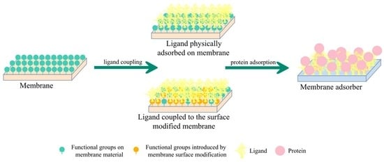
Functional groups on support membranes are important for the preparation of high-performance membrane adsorbers (MAs). In this work, the effects of different functional groups on support membranes on the adsorption performance of MAs were studied through surface modifications such as alkali treatment, oxidation treatment and polydopamine (PDA) deposition. Our experiment results indicate that chemical bonds are prone to form between C-F on PVDF membrane and amine groups on PEI, which serves to improve the protein adsorption capacity. Furthermore, PDA deposition has the potential to enhance protein adsorption capacity. Introducing a PDA layer on a support membrane with uniform pore size distribution shows potential to improve protein adsorption capacity.

Graphical Abstract

1. Introduction

Chromatography is a crucial important technique in the high-resolution separation and analysis of complex mixtures [1,2]. Membrane chromatography (MC) has attracted considerable attention in recent years due to its unique characteristics such as low pressure drop, fast transport rate and easy scale-up, etc. [3,4].
The widespread implementation of MC technology is mainly impeded by poor binding capacity, which is associated with low ligand coupling density (LigC) [5]. To overcome this challenge, Wang et al. [6] and Chenette et al. [7] introduced bromine groups onto the membrane surface to couple ligands, leading to binding capacities of 126.6 mg/mL and 97 mg/mL. Liu et al. [8] and Ma et al. [9] introduced epoxy groups onto the membrane surface, resulting in a significant improvement in protein binding capacity. Khan et al. [5] introduced carboxylic groups onto the membrane surface, with a resultant protein adsorption capacity of up to 286 mg/mL. The above research demonstrates that the degree of MA performance improvement varies with different techniques, indicating that the influencing factors contributing to MA performance remain elusive.
From the above discussion, in our consideration, the introduced functional groups and the resulting variation in membrane surface chemistry may play an important role. Functional groups on the membrane serving as ligand coupling sites may affect LigC, and the effect of functional groups on binding capacity needs to be further studied.
Decreasing the effect of any processes within MC that prevent biomolecule adsorption is considered a method to improve the sustainable performance of membranes in the water treatment process. Ho et al. [10] used various functional groups to investigate the influencing factors of protein binding properties and found that membrane surface chemistry plays a key role. Saeki et al. [11] demonstrated that biomolecule adsorption performance is dependent on the types of polymers. Most research confirms that membrane surface chemistry can affect the properties of hydrophobicity and the zeta potential of the membrane, which are regarded as the key factors of biomolecule adsorption performance [12,13]. In the field of membrane fouling prevention, the mechanism of biomolecule adsorbing onto the membrane has been widely illustrated.
Here, this mechanism can be employed to study the influence of functional groups on the MAs adsorption performance. In this work, the effect of functional groups on binding capacity enhancement was investigated. Dendritic PEI was taken as the ligand, and four kinds of microfiltration membranes with different functional groups were explored, including polyethersulfone (PES), polyvinylidene fluoride (PVDF), polyamide 6 (PA6) and mixed cellulose (MCE). Alkali, oxidation and PDA deposition treatment were applied to investigate the effect of new functional groups introduced by membrane surface modification. The PDA deposition modified PVDF membrane demonstrated favorable protein adsorption capacity, and the preparation conditions were optimized to obtain a favorable adsorption/desorption capacity.

2. Materials and Methods

2.1. Materials

Commercially available polyethersulfone (PES), polyvinylidene fluoride (PVDF) and polyamide 6 (PA6), mixed cellulose (MCE) membranes with a pore size of 0.45 μm were purchased from Tianjin JinTeng Experimental Equipment (Tianjin, China). Dopamine hydrochloride and polyethyleneimine (PEI) (molecular weight (Mw) 1800, 10,000, 70,000) were obtained from Aladdin Reagent Co., Ltd. (Shanghai, China), and PEI (Mw 750,000) was obtained from Chongqing Polytech Biotechnology Co., Ltd. (Chongqing, China). Bovine serum albumin (BSA) and Trimethylolaminomethane (Tris) were obtained from Shanghai Yuanye Biotechnology Co., Ltd. (Shanghai, China). Coomassie brilliant blue G250 was obtained from Shanghai Maclean’s Biochemical Technology (Shanghai, China). 25 wt.% Glutaraldehyde (C5H8O2) was obtained from Fuchen Chemical Reagent Co., Ltd. (Tianjin, China). The deionized water was lab homemade. All chemicals used in this study were analytical reagents.

2.2. Preparation and Characterization of MA

2.2.1. Ligand Physically Adsorbed on Membrane

To investigate the effect of different functional groups on the binding performances of MAs, herein, PES, PVDF, PA6 and MCE membranes were immersed in the ligand solution (2 g/L, PEI Mw 750,000) for 20 h [14], then washed with deionized water three times and tested for protein adsorption performance.
The MAs prepared by this method were defined as PEI@PES, PEI@PVDF, PEI@PA6 and PEI@MCE, respectively.

2.2.2. Alkali Treatment

Functional groups on the membrane can be modified by changing pH values and chemical reactions [15,16]; alkali treatment and oxidation treatment were used to try to introduce different groups onto the membrane surface.
PES, PVDF and PA6 membranes were immersed in 12 M NaOH solution, heated at 80 °C for 2 h, and subsequently washed with deionized water. The membranes were then immersed in 25 wt.% glutaraldehyde for about 5 s and put in a 50 °C vacuum drying oven for 10 min, followed by immersing the membranes in PEI solution (15 g/L, PEI Mw 70,000) for 1 min. To maintain the stability of the MCE membrane, the process of alkali immersing was only 1 min at room temperature, and the glutaraldehyde crosslinking process was carried out at room temperature; the following processes were the same as above mentioned. All the membranes were washed with deionized water three times, after which the protein adsorption performance was tested.
The MAs prepared by this method were defined as PEI@PES-alk, PEI@PVDF-alk, PEI@PA6-alk and PEI@MCE-alk, respectively.

2.2.3. Oxidation Treatment

PES, PVDF and PA6 membranes were immersed in 3 M H2O2 solutions at room temperature for about one week, ensuring adequate oxidation, and subsequently washed with deionized water. The following processes were the same as the processes of alkali treatment. To maintain the stability of the MCE membrane, the process of glutaraldehyde crosslinking was carried out at room temperature.
The MAs prepared by this method were defined as PEI@PES-oxi, PEI@PVDF-oxi, PEI@PA6-oxi and PEI@MCE-oxi, respectively.

2.2.4. PDA deposition Modification

The preparation procedure of PEI@Mm-PDA is illustrated in Figure 1. PES, PVDF, PA6 and MCE membranes were immersed in appropriate amounts of dopamine solution (2 mg/mL, 10 mM Tris-HCl), stirred at a low speed for a certain period at 25 °C to form a PDA deposition layer in the membrane, and then washed with deionized water for 2 h. The PDA deposited membrane was immersed in PEI aqueous solution, reacted at 60 °C for 8 h, and then washed with deionized water for 1 h, after which it was tested for protein adsorption/desorption performance. pHpoly and Tpoly were defined as the pH value and temperature of the dopamine polymerization, respectively, and CPEI and MPEI were defined as the concentration and molecular weight of PEI, respectively.
The MAs prepared by this method were defined as PEI@PES-PDA, PEI@PVDF-PDA, PEI@PA6-PDA and PEI@MCE-PDA, respectively.

2.3. Characterization Methods

An X-ray photoelectron spectrometer (XPS, Thermo Scientific K-Alpha, Thermo Fisher Scientific, Beijing, China) was employed to determine the chemical compositions on the surface of the MA and analyze the change in surface element content after modification. The Zeta potential of the MA was measured using a Zeta potential meter (SurPASS, Anton Paar GmbH, Austria, Shanghai, China), and the Zeta potentials of the MA at different pH values (pH meter, PHS-25, Shanghai INESA Science and Technology Scientific Instrument Co., Ltd. Shanghai, China) were determined.
A scanning electron microscope (SEM, TM-3000, Hitachi High-Technologies Group, Shanghai, China) was used to observe the morphology of the pristine membrane surface.

2.4. MA Performance Testing

BSA was used as the model media to characterize the adsorption performance and the ligand coupling density of the MAs. The membrane (Vm ≈ 0.04 mL) was placed in 15 mL of adsorption solution (1 mg/mL BSA, 20 mM Tris-HCl, pH ≈ 7.5) after shaking for 24 h [17] and then taken out. The obtained solution was centrifuged, and the protein concentration in the supernatant was measured using the Coomassie brilliant blue method [5]. The static protein adsorption capacity of the MA was obtained by calculating the difference between the initial and final amount of BSA in the adsorption solution according to Ref. [5].
The MA after adsorption of protein was put into 10 mL of desorption solution (1000 mM NaCl, 20 mM Tris-HCl, pH ≈ 7.5), shaken and desorbed for 24 h, and then taken out. The following steps were the same as those in the static protein adsorption capacity test. The static protein desorption rate (or protein recovery) was estimated using the ratio of the amount of desorbed BSA and the adsorbed amount of BSA in the adsorption solution according to Ref. [18].

3. Results and Discussion

3.1. Protein Adsorption/Desorption Performance on Different Membrane Functional Groups

As shown in Figure 2, the membranes contained O=S=O and benzene (PES), C-F (PVDF), -CONH- (PA6), and -OH and -COOH (MCE). Alkali and oxidation treatments can change the functional groups on membranes, such as introducing -OH, -COOH, etc. [15,16]; Fan et al. [19] reported that PDA deposition modification can also introduce abundant functional groups on a membrane. In this paper, alkali, oxidation and PDA deposition treatment were used to modify the functional groups on the membranes and their influences on protein adsorption capacity were investigated. Among these functional groups, O=S=O and C-F were difficult to dissociate as -OH or -COOH, compared with -CONH-, -OH and -COOH.
Similar to other research [20], PEI was used as ligand sample to detect LigC. Dendritic PEI molecules contain primary amine, secondary amine and tertiary amine groups as depicted in Figure 3.

3.1.1. Effect of Membrane Functional Groups

As shown in Figure 4, a high protein adsorption capacity of 84.2 mg/mL was obtained by PEI@PVDF, followed by PEI@MCE, PEI@PES and PEI@PA6. The PVDF membrane tends to adsorb PEI that contains amine groups, leading to a high protein adsorption capacity of PEI@PVDF; this result has also been evidenced during membrane fouling [21]. Hydrogen bonds tend to form between amine groups and atoms characterized by small radius, large electronegativity, partial negative charge and containing solitary electron pairs, such as F, O, and N. In this work, hydrogen bonds were liable to form between amine groups on PEI and the functional groups on different membrane materials [22,23] that contain F, O, and N, as shown in Figure 2. Kolev et al. [24] confirmed that the potential of hydrogen bonds forming may vary with the functional groups of organic molecules. As shown in Figure 4, the protein adsorption capacities of PEI@PES/PA6/MCE were similar, containing O=S=O, -CONH-, -OH and -COOH. It is clear from Figure 4 that the tendency to form hydrogen bonds between functional groups and amine groups can be ranked as C-F>-OH, -COOH> O=S=O≈-CONH-. The protein adsorption capacity of PEI@MCE was a bit higher than PEI@PES and PEI@PA6, which may be the positive influence of plenty of -OH and -COOH in the MCE membrane. The -CONH- in PA6 and S=O=S in PES did not generate incentive effects on protein adsorption capacity improvement.
The effect of the functional groups on PES, PA6 and MCE on protein adsorption capacity was further examined using alkali- and oxidation-treated membranes.

3.1.2. Effect of New Functional Groups Introduced by Alkali/Oxidation Treatment

Membrane surface functional groups can be modified by pH and also chemical reactions; alkali treatment and oxidation treatment were used in an attempt to introduce different groups onto the membrane surface.
As has been reported in the literature, the chemical stability of cellulose membranes is poor, and the membrane morphology may be destroyed by chemical treatment [19,25]. In this section, different from the other membrane materials, MCE membrane was immersed for only 1 min in NaOH solution and the glutaraldehyde crosslink process was carried out at room temperature in PEI@MCE-alk/oxi preparation, aiming to avoid the crumbling of the MCE membrane.
Comparing the protein adsorption capacity of the membrane material itself and the results after alkali and oxidation treatment shown in Figure 5, it seems that new functional groups were generated by alkali treatment and oxidation treatment, such as -OH, -COOH and -NH2 [15,26,27]. The functional groups generated reacted with glutaraldehyde and were further coupled with PEI. Specifically, after being treated with NaOH or H2O2, the -CONH- in PA6 membrane breaks to form -NH2 and -COOH [12], and the intermolecular hydrogen bond in MCE membrane is disrupted to exposure more reactive -OH [26]; alternatively, -OH may be oxidized as -COOH, and the CF2-CH2 in the PVDF membrane and O=S=O in the PES membrane may break to introduce -OH and -COOH [27,28].
As depicted in Figure 5a, the protein adsorption capacity of PEI@Mm-alk was not improved compared with PEI@Mm, which indicates that the functional groups introduced by alkali treatment did not produce a favorable influence on protein binding capacity.
Different from the result of alkali treatment, it can be seen from Figure 5b that the protein adsorption capacity of PA6 membrane was slightly improved after oxidation treatment, while those of the PES, PVDF, MCE membranes were not. Plenty of -OH and -COOH on the MCE membrane led to the insignificant effect of functional groups introduced by oxidation treatment on protein adsorption capacity enhancement. The similar results of PES and PVDF were consistent with the strong chemical stability of PES and PVDF membranes reported in Refs. [29,30], which shows that the oxidation stability of PES and PVDF membranes is good and both of them are excellent membrane materials.
The results shown in Figure 5a,b suggest that adopting oxidation treatment to modify these kinds of membrane materials is more feasible than alkali treatment.

3.1.3. Effect of New Functional Groups Introduced by PDA Deposition Treatment

In the membrane filtration process, functional groups and pore size distribution may affect biomolecule binding performance seriously [31]. Research has found that coating an intermediate layer on/in the membrane may introduce functional groups and change the membrane pore size distribution, and the modification effect would vary with the intermediate layer [5,32]. In this work, we tried to introduce functional groups through depositing PDA onto membrane surfaces for PEI coupling and investigate their effect on the enhancement of the protein adsorption capacity of the MAs.
The results shown in Figure 5c imply the functional groups introduced by PDA deposition were preferable to alkali treatment and oxidation treatment with regard to protein adsorption capacity improvement, and the trends of PEI@Mm and PEI@Mm-PDA were similar, suggesting that hydrogen bonds may play an important role in PDA layer adhesion compared to other possible deposition forces [32]. As depicted in Figure 1, a variety of functional groups on the PDA layer, such as -OH and amino groups, may form hydrogen bonds with the functional groups on the membrane. Analogous to the discussion in Section 3.1.1, compared to other functional groups on PES, PA6 and MCE membranes, the C-F on PVDF membrane may facilitate hydrogen bond formation, which may promote PDA deposition, resulting in the significant enhancement of the protein adsorption capacity from 84.2 mg/mL to 165.3 mg/mL.
The membrane pore size distribution may affect PDA deposition, resulting in the variation in protein adsorption performance of the MAs. As shown in Figure 6, the pore size distribution of the PVDF membrane was more even than the others, which may induce a more uniform PDA layer deposited on the membrane surface. This uniform PDA layer would be favorable for the PEI coupled on the membrane and thus facilitate the increment of LigC. Overall, the introduction of functional groups by a deposited PDA layer on a uniform PVDF membrane may be an effective method to improve the protein adsorption capacity of MAs.

3.2. Optimization of the Preparation Conditions of the PEI@PVDF-PDA

Apart from the functional groups, the preparation conditions of MAs, such as pH value and the temperature of the PDA deposition process, as well as the molecular weight and the concentration of PEI, etc., may affect their protein adsorption capacity. It is necessary to study the above-mentioned factors to fabricate a high-performance MA.
The cost of raw materials for manufacturing MAs is still relatively high for MC; though MAs possess a disposable nature, it would be desirable to operate high-binding-capacity MAs with reversible protein adsorption. The protein desorption rate was investigated to obtain a MA with the potential to be applied in MC.

3.2.1. Effect of the pH Value of the Dopamine Polymerization Process

As the pHpoly value increased from 5.4 to 9.5, the protein adsorption capacity of the MAs gradually increased slightly and then stabilized (Figure 7a). The protein desorption rate decreased as the pHpoly value increased from 5.4 to 8.7, then stabilized (Figure 7a).
Under certain dopamine polymerization time and temperature, the process of dopamine polymerization proceeded forward as pHpoly increased and leveled off at pHpoly > 8.7, which was consistent with the reference’s reports [33]. With the increase in pHpoly value, the PDA layer was denser and the density of the grafted PEI was higher, which was mainly manifested by the gradual increase in the protein adsorption capacity of the MAs as shown in Figure 7a. Further increasing the pHpoly value from 8.7 to 9.5, there were no incentive effects on the polymerization reaction, so the protein adsorption capacity was basically stable. Relatively high PEI coupling density may trigger a steric hindrance effect and increase protein desorption difficulty, which was reflected in the decrease in protein desorption rate with the increase in pHpoly value. Similar to the protein adsorption at pHpoly = 8.7~9.5, when dopamine polymerization reached a certain level, the effect of steric hindrance did not further increase the difficulty of protein desorption.
Considering both the protein adsorption and desorption performance of the MAs were almost unchanged when the pHpoly value was higher than 8.7, the optimal pH value of the dopamine self-polymerization process was 8.7.

3.2.2. Effect of the Temperature of Dopamine Polymerization Process

The effect of PDA deposition temperature on the protein adsorption/desorption performance of the MA is shown in Figure 7b.
The protein adsorption capacity of MA grew as Tpoly rose from 20 °C to 35 °C, and a protein absorption capacity of MA as high as about 166 mg/mL was achieved in the Tpoly of 25~35 °C. As Tpoly further increased from 35 °C to 60 °C, the protein adsorption capacity of the MA was decreased from 166 mg/mL to 142.6 mg/mL. The polymerization of dopamine proceeded more thoroughly at a higher temperature [34]. The protein adsorption capacity was increased gradually as the temperature increased from 20~35 °C. The temperature further increased, the dopamine polymerized more completely and formed a denser PDA layer to possibly couple more PEI, which may have incurred the steric hindrance effect, resulting in a reduction in available effective sites for protein adsorption.
As Tpoly increased, the protein desorption rate was first decreased and then tend to be stable. The polymerization of dopamine proceeded more fully as the temperature increased, and the steric hindrance effect came into play gradually, which may have led to the protein tending to be difficult to desorb. When the temperature was higher than 30 °C, the polymerization of dopamine was sufficient, protein desorption was not more difficult due to the existence of the steric hindrance effect, and the proportion of protein desorbed on the adsorbed MAs was essentially unchanged.
Considering both the protein adsorption and desorption performances of the MA, we determined that the optimal Tpoly was 25 °C, and the protein desorption rate was 60.4% at this time.

3.2.3. Effect of Ligand Molecular Weight

Protein adsorption was mainly carried out by the electrostatic and hydrogen bond interactions between PEI and proteins. Previous research found that properties of PEI solution such as the molecular weight of PEI and the concentration of PEI solution affected protein adsorption and desorption performance [20,35]. The effects of PEI molecular weight on the protein adsorption and desorption performance were investigated, as shown in Figure 8a; with the increase in MPEI, both the protein adsorption capacity and desorption rate of the MA were initially increased and then decreased. As PEI molecular weight was increased, the sites available for protein adsorption were increased and the protein adsorption capacity was enhanced. The PEI molecular chains were prone to stacking and aggregation as MPEI=750,000, which may result in a decrease in protein available binding sites and the protein adsorption capacity. The above-mentioned effects of molecular chain stacking and aggregation would hinder protein desorption, which was the reason that the protein desorption rate somewhat declined as MPEI increased to 750,000 (Figure 8a).
Overall, the protein adsorption/desorption performance of the membrane was the best when MPEI = 70,000.

3.2.4. Effect of Ligand Concentration

The concentration of PEI may affect the number of binding sites between PEI molecules and the functional groups on the membrane and make a difference in the protein adsorption capacity of MAs [35]. As shown in Figure 8b, with the increase in CPEI, the protein adsorption capacity of the MA first increased and then decreased, while the protein desorption rate increased gradually. When the concentration of the ligand solution is low, the number of ligand molecules that can be attached to the membrane is small. As the concentration of the ligand solution increases, the number increases. The steric hindrance effect may dominate the protein adsorption performance as the PEI coupled density rises, such as when CPEI = 20 g/L, resulting in the reduction in protein adsorption capacity associated with decreased protein available binding sites, which is similar to the discussion in Section 3.2.2. As the steric hindrance effect plays a part in protein adsorption, the protein was mainly adsorbed onto the PEI molecule surface. This phenomenon may lead to the protein being more easily desorbed and the protein desorption rate gradually rising. In particular, when CPEI = 20 g/L, the protein desorption rate increased obviously.
Considering both the protein adsorption and desorption performance of the MA, the most favorable CPEI was 15 g/L.
Within the scope of our experiment, the preferable MA preparation conditions were determined as follows: pHpoly = 8.7, Tpoly = 25 °C, MPEI = 70,000, CPEI = 15 g/L.
XPS was used to analyze the element content on the surface of the MA, the results are shown in Figure 9a. The MAs of PEI@PVDF-PDA and the PDA deposited PVDF (PVDF-PDA) both appeared with N and O element peaks. The peaks of C and N elements of PEI@PVDF-PDA were higher than PVDF-PDA, which confirms that the PEI coupled to the membrane. After the ligand was coupled, part of the PDA layer was covered by PEI such that the O element content on the surface of the detected MA was slightly reduced.
The test results of Zeta potential on the surface of PEI@PVDF-PDA are shown in Figure 9b. PEI@PVDF-PDA was positively charged in the solution with a pH value of 4~10, indicating that in a neutral solution environment, the MA could selectively adsorb molecules such as negatively charged proteins, while having no adsorption effect with the positively charged products at this time, causing the separation of mixtures.

4. Conclusions

In this paper, four kinds of membrane materials with different functional groups and three membrane surface modification methods aiming to introduce functional groups onto the membrane were explored to investigate the effect of the functional groups on the adsorption capacity of the MAs. Experimental results show that the C-F on PVDF membrane tends to form hydrogen bonds with amine groups on PEI, which may be helpful to increasing the protein adsorption capacity of MAs. The functional groups introduced by PDA deposition on PVDF membrane and uniform membrane pore size distribution may be valuable contributions to favorable protein adsorption performance of MAs, while the functional groups generated by alkali treatment and oxidation treatment had only insignificant influences. The effect of PEI@PVDF-PDA preparation conditions was investigated as well. The impact of membrane structure on the adsorption performance of MAs is still unclear and will be further investigated in our following study. This work provides a new perspective for the impact of functional groups on binding capacity improvement, and PEI@PVDF-PDA, with its favorable protein adsorption/desorption performance, has great application potential in MC.

Author Contributions

Conceptualization, W.Z., J.Y. and L.Z.; methodology, Y.Y. and L.Z.; writing-original draft preparation, L.Z., J.Y. and Y.Y.; writing—review and editing, L.Z., J.Y. and W.Z. All authors have read and agreed to the published version of the manuscript.

Funding

This research was funded by the Fundamental Research Funds for the Central Universities (Grant No. JD2218).

Data Availability Statement

All data are supplied in the manuscript.

Conflicts of Interest

The authors declare no conflict of interest.

References

  1. Ghosh, R. Protein separation using membrane chromatography: Opportunities and challenges. J. Chromatogr. A 2002, 952, 13–27. [Google Scholar] [CrossRef] [PubMed]
  2. Abdulhussain, N.; Nawada, S.; Schoenmakers, P. Latest Trends on the Future of Three-Dimensional Separations in Chromatography. Chem. Rev. 2021, 121, 12016–12034. [Google Scholar] [CrossRef] [PubMed]
  3. Brandt, S.; Goffe, R.A.; Kessler, S.B.; O’Connor, J.L.; Zale, S.E. Membrane-Based Affinity Technology for Commercial Scale Purifications. Nat. Biotechnol. 1988, 6, 779–782. [Google Scholar] [CrossRef]
  4. Orr, V.; Zhong, L.; Moo-Young, M.; Chou, C.P. Recent advances in bioprocessing application of membrane chromatography. Biotechnol. Adv. 2013, 31, 450–465. [Google Scholar] [CrossRef]
  5. Khan, M.K.; Luo, J.; Khan, R.; Fan, J.; Wan, Y. Facile and green fabrication of cation exchange membrane adsorber with unprecedented adsorption capacity for protein purification. J. Chromatogr. A 2017, 1521, 19–26. [Google Scholar] [CrossRef]
  6. Wang, C.; Shen, J.; Zhu, J.; Bo, C.; Wei, Y. Tetrazole-functionalized cation-exchange membrane adsorbers with high binding capacity and unique separation feature for protein. J. Chromatogr. B 2018, 1097–1098, 18–26. [Google Scholar] [CrossRef]
  7. Chenette, H.C.; Robinson, J.R.; Hobley, E.; Husson, S.M. Development of high-productivity, strong cation-exchange adsorbers for protein capture by graft polymerization from membranes with different pore sizes. J. Membr. Sci. 2012, 423–424, 43–52. [Google Scholar] [CrossRef] [Green Version]
  8. Liu, H.; Gurgel, P.V.; Carbonell, R.G. Preparation and characterization of anion exchange adsorptive nonwoven membranes with high protein binding capacity. J. Membr. Sci. 2015, 493, 349–359. [Google Scholar] [CrossRef]
  9. Ma, N.; Yao, D.; Yang, H.; Yin, J.; Wang, H.; Zhang, Y.; Meng, J. Surface Modification of Cellulose Membranes To Prepare a High-Capacity Membrane Adsorber for Monoclonal Antibody Purification via Hydrophobic Charge-Induction Chromatography. Ind. Eng. Chem. Res. 2018, 57, 13235–13246. [Google Scholar] [CrossRef]
  10. Ho, C.-C.; Prodan, B.N.; Kiessling, B.; Co, C.C. Effects of membrane surface chemistry on fouling investigated using self-assembled monolayers. J. Membr. Sci. 2011, 366, 342–348. [Google Scholar] [CrossRef]
  11. Saeki, D.; Yonamine, G.; Matsuyama, H. Effect of hydrophilic polymer modification of reverse osmosis membrane surfaces on organic adsorption and biofouling behavior. Colloids Surf. A 2020, 609, 125680. [Google Scholar] [CrossRef]
  12. Bagheri, M.; Mirbagheri, S.A. Critical review of fouling mitigation strategies in membrane bioreactors treating water and wastewater. Bioresour. Technol. 2018, 258, 318–334. [Google Scholar] [CrossRef] [PubMed]
  13. Zhang, M.; Liao, B.-Q.; Zhou, X.; He, Y.; Hong, H.; Lin, H.; Chen, J. Effects of hydrophilicity/hydrophobicity of membrane on membrane fouling in a submerged membrane bioreactor. Bioresour. Technol. 2015, 175, 59–67. [Google Scholar] [CrossRef] [PubMed]
  14. Zhou, Y.; Yu, S.; Gao, C.; Feng, X. Surface modification of thin film composite polyamide membranes by electrostatic self deposition of polycations for improved fouling resistance. Sep. Purif. Technol. 2009, 66, 287–294. [Google Scholar] [CrossRef]
  15. Wang, Q.; Wei, X.; Wang, G.-R.; Lu, T.-D.; Shi, Q.; Sun, S.-P. Inner-selective coordination nanofiltration hollow fiber membranes from assist-pressure modified substrate. J. Membr. Sci. 2021, 626, 119186. [Google Scholar] [CrossRef]
  16. Sagitha, P.; Reshmi, C.; Sundaran, S.P.; Sujith, A. Recent advances in post-modification strategies of polymeric electrospun membranes. Eur. Polym. J. 2018, 105, 227–249. [Google Scholar] [CrossRef]
  17. Ye, J.; Chu, J.; Yin, J.; Zhang, Y.; Meng, J. Surface modification of regenerated cellulose membrane based on thiolactone chemistry—A novel platform for mixed mode membrane adsorbers. Appl. Surf. Sci. 2020, 511, 145539. [Google Scholar] [CrossRef]
  18. He, D.; Ulbricht, M. Preparation and characterization of porous anion-exchange membrane adsorbers with high protein-binding capacity. J. Membr. Sci. 2008, 315, 155–163. [Google Scholar] [CrossRef]
  19. Fan, J.; Luo, J.; Chen, X.; Wan, Y. Facile preparation of salt-tolerant anion-exchange membrane adsorber using hydrophobic membrane as substrate. J. Chromatogr. A 2017, 1490, 54–62. [Google Scholar] [CrossRef] [PubMed]
  20. Fan, J.; Luo, J.; Song, W.; Wan, Y. One-step purification of α1-antitrypsin by regulating polyelectrolyte ligands on mussel-inspired membrane adsorber. J. Membr. Sci. 2017, 528, 155–162. [Google Scholar] [CrossRef]
  21. Shi, H.; Xue, L.; Gao, A.; Fu, Y.; Zhou, Q.; Zhu, L. Fouling-resistant and adhesion-resistant surface modification of dual layer PVDF hollow fiber membrane by dopamine and quaternary polyethyleneimine. J. Membr. Sci. 2016, 498, 39–47. [Google Scholar] [CrossRef]
  22. Choi, K.I.; Lee, J.H. Preparation and dispersion of metal oxide nanostructures using amino acid-assisted chemical routes: An overview. Sci. Adv. Mater. 2011, 3, 811–820. [Google Scholar] [CrossRef]
  23. Carlton, L.; Staskun, B.; van Es, T. A nitrogen-15 NMR study of hydrogen bonding in 1-alkyl-4-imino-1,4-dihydro-3-quinolinecarboxylic acids and related compounds. Magn. Reson. Chem. 2006, 44, 510–514. [Google Scholar] [CrossRef] [PubMed]
  24. Kolev, S.K.; St Petkov, P.; Rangelov, M.A.; Vayssilov, G.N. Density functional study of hydrogen bond formation between methanol and organic molecules containing Cl, F, NH2, OH, and COOH functional groups. J. Phys. Chem. A 2011, 115, 14054–14068. [Google Scholar] [CrossRef] [PubMed]
  25. Ke, Y.-M.; Chen, C.-I.; Kao, P.-M.; Chen, H.-B.; Huang, H.-C.; Yao, C.-J.; Liu, Y.-C. Preparation of the immobilized metal affinity membrane with high amount of metal ions and protein adsorption efficiencies. Process. Biochem. 2010, 45, 500–506. [Google Scholar] [CrossRef]
  26. Ramamoorthy, S.K.; Bakare, F.; Herrmann, R.; Skrifvars, M. Performance of biocomposites from surface modified regenerated cellulose fibers and lactic acid thermoset bioresin. Cellulose 2015, 22, 2507–2528. [Google Scholar] [CrossRef] [Green Version]
  27. Ross, G.; Watts, J.; Hill, M.; Morrissey, P. Surface modification of poly(vinylidene fluoride) by alkaline treatment1. The degradation mechanism. Polymer 2000, 41, 1685–1696. [Google Scholar] [CrossRef]
  28. Kumar, R.; Ismail, A.; Kassim, M.; Isloor, A.M. Modification of PSf/PIAM membrane for improved desalination applications using Chitosan coagulation media. Desalination 2013, 317, 108–115. [Google Scholar] [CrossRef]
  29. Kim, K.; Lee, K.; Cho, K.; Park, C. Surface modification of polysulfone ultrafiltration membrane by oxygen plasma treatment. J. Membr. Sci. 2002, 199, 135–145. [Google Scholar] [CrossRef]
  30. Liang, S.; Kang, Y.; Tiraferri, A.; Giannelis, E.P.; Huang, X.; Elimelech, M. Highly hydrophilic polyvinylidene fluoride (PVDF) ultrafiltration membranes via postfabrication grafting of surface-tailored silica nanoparticles. ACS. Appl. Mater. Interfaces 2013, 5, 6694–6703. [Google Scholar] [CrossRef]
  31. Xiong, Z.; Zhong, Y.; Lin, H.; Liu, F.; Li, T.; Li, J. PDLA/PLLA ultrafiltration membrane with excellent permeability, rejection and fouling resistance via stereocomplexation. J. Membr. Sci. 2017, 533, 103–111. [Google Scholar] [CrossRef]
  32. Liu, Y.; Ai, K.; Lu, L. Polydopamine and Its Derivative Materials: Synthesis and Promising Applications in Energy, Environmental, and Biomedical Fields. Chem. Rev. 2014, 114, 5057–5115. [Google Scholar] [CrossRef] [PubMed]
  33. Ball, V.; Del Frari, D.; Toniazzo, V.; Ruch, D. Kinetics of polydopamine film deposition as a function of pH and dopamine concentration: Insights in the polydopamine deposition mechanism. J. Colloid Interface Sci. 2012, 386, 366–372. [Google Scholar] [CrossRef] [PubMed]
  34. Jiang, J.; Zhu, L.; Zhu, L.; Zhu, B.; Xu, Y. Surface Characteristics of a Self-Polymerized Dopamine Coating Deposited on Hydrophobic Polymer Films. Langmuir 2011, 27, 14180–14187. [Google Scholar] [CrossRef]
  35. Yu, L.-L.; Tao, S.-P.; Dong, X.-Y.; Sun, Y. Protein adsorption to poly(ethylenimine)-modified Sepharose FF: I. A critical ionic capacity for drastically enhanced capacity and uptake kinetics. J. Chromatogr. A 2013, 1305, 76–84. [Google Scholar] [CrossRef] [PubMed]
Figure 1. Schematic diagram of membrane adsorber prepared by depositing PDA layer on membrane to couple PEI.
Separations 10 00211 g001
Figure 2. The molecular structures of PES, PVDF, PA6 and MCE.
Separations 10 00211 g002
Figure 3. Molecular structure of PEI.
Separations 10 00211 g003
Figure 4. Effect of membrane material (different functional groups) on the protein adsorption performance of the MAs.
Separations 10 00211 g004
Figure 5. Effect of (a) alkali treatment, (b) oxidation treatment and (c) PDA deposition on the protein adsorption performance of the MAs.
Separations 10 00211 g005
Figure 6. SEM images of the surface of pristine PES, PVDF, PA6 and MCE membranes.
Separations 10 00211 g006
Figure 7. Effect of the (a) pH value and (b) temperature of the dopamine polymerization process on the protein adsorption/ desorption performance of MAs (Tpoly = 25 °C/pHpoly = 8.7, CPEI = 15 g/L, MPEI = 70,000).
Separations 10 00211 g007
Figure 8. Effect of the (a) molecular weight and (b) concentration of ligand on the protein adsorption/desorption performance of the MA (pHpoly = 8.7, Tpoly = 25 °C, CPEI = 15 g/L/MPEI = 70,000).
Separations 10 00211 g008
Figure 9. (a) XPS wide scans and (b) Zeta potential of MA.
Separations 10 00211 g009
Disclaimer/Publisher’s Note: The statements, opinions and data contained in all publications are solely those of the individual author(s) and contributor(s) and not of MDPI and/or the editor(s). MDPI and/or the editor(s) disclaim responsibility for any injury to people or property resulting from any ideas, methods, instructions or products referred to in the content.