Article

Using Hepatic Gene Expression Assays in English Sole (Parophrys vetulus) to Investigate the Effects of Metro Vancouver Wastewater Effluents

Department of Biological Sciences, Simon Fraser University, Burnaby, BC V5A 1S6, Canada
*
Author to whom correspondence should be addressed.
Toxics 2023, 11(8), 657; https://doi.org/10.3390/toxics11080657
Submission received: 11 June 2023 / Revised: 17 July 2023 / Accepted: 27 July 2023 / Published: 29 July 2023
(This article belongs to the Special Issue Toxics Young Investigators Contributions Collection in 2023)

Abstract

:
The present study has investigated the effects of Metro Vancouver’s wastewater treatment plant (WWTP) effluents on English sole (Parophrys vetulus) hepatic gene expression using novel targeted gene expression assays to complement the 2017 Burrard Inlet Ambient Monitoring Program conducted by Metro Vancouver. Seven locations of varying distance to the WWTPs were included. Twelve genes involved in xenobiotic defense (CYP1A, HSP70), thyroid function (DIO1), lipid and glucose metabolism (FABP1, FASN, GLUT2, PPAR δ , PPAR γ ), protein synthesis (18S rRNA, RPS4X), and reproduction (ER α , VTG) revealed several differences between these impacted sites. A key finding of the present study was that males exhibited VTG transcript levels either equivalent or exceeding female levels of this gene at all sites investigated, indicating widespread exposure of estrogenic contaminants throughout Burrard Inlet. Furthermore, the induction of hepatic CYP1A was observed due to possible downstream sites being subjected to a larger influx of certain planar halogenated and non-halogenated hydrocarbons from multiple industrial contributors. This study also revealed significant differences between the sites examined and in genes involved in transcriptional regulation and synthesis of proteins, lipids and glucose metabolism, and thyroid hormone metabolism. Collectively, this study demonstrates the potential of molecular biomarkers of urban contaminant exposure in wild caught English sole for use in diagnosing a broader range of adverse health effects when combined with conventional whole organism health indicators.

Graphical Abstract

1. Introduction

Contaminants have been identified as one of Earth’s greatest threats to human and environmental health [1]. This is in large part a result of decades of industrial, agricultural, and urban anthropogenic activities deliberately or accidentally releasing numerous chemical pollutants into waterways [2]. Of particular concern are the hundreds of chemicals released from municipal wastewater treatment plants (WWTPs), which include contaminants such as metals, polycyclic aromatic hydrocarbons (PAHs), polychlorinated dibenzo-p-dioxins and polychlorinated dibenzofurans, surfactants (i.e., alkylphenol ethoxylates), pesticides, polybrominated diphenyl ethers (PBDEs), polychlorinated biphenyls (PCBs), human hormones, pharmaceuticals and personal care products (PPCPs; i.e., prescription, over the counter, and veterinary therapeutic drugs for human and animal ailments/diseases, and PCPs such as soaps, deodorants, and cosmetics) [3]. Despite the known adverse effects of several of these chemicals on aquatic wildlife, toxicity data is lacking for many chemicals in WWTP effluents. Furthermore, the toxic effects of various chemical mixtures that reflect environmentally relevant exposure scenarios near WWTP discharge sites complicate risk assessments.
Pharmaceuticals discharged in WWTP effluents are of particular concern since they have been designed to target various processes in humans that are often conserved across vertebrate species; thus, many are toxic to aquatic wildlife at low concentrations (ng/L to µg/L) alone or as mixtures (i.e., additive, or synergistic) [4]. Indeed, despite the known adverse effects of several PPCPs on aquatic wildlife, a limited number of ambient environmental quality guidelines exist to assess the risks of PPCPs to aquatic wildlife that are currently used in many countries. For example, in Canada, nonylphenol, nonylphenol ethoxylates, and 17 α -ethynylestradiol (EE2) currently have working water and sediment quality guidelines for the protection of aquatic life [5,6]. Whereas in Europe, priority pollutant lists under the European Water Framework Directive (EU WFD) include ambient quality guidelines for estrogenic compounds of biological (e.g., 17 β -estradiol (E2), estrone (E1), estriol (E3)) and synthetic nature (e.g., bisphenol A (BPA), octylphenol (OP), nonyohenol (NP), and EE2) [7]. The ambient quality guidelines for estrogenic compounds have arisen due to the irrefutable data on their ability to disrupt the endocrine system and in particular, mimic estrogen [8,9]. Research on the impacts of a low-level synthetic birth control estrogen, EE2, on fish revealed compelling connections between molecular and population-level effects [8]. Specifically, chronic waterborne exposure to EE2 induced vitellogenin (VTG), gonadal abnormalities, and ultimately led to the near extinction of the species in an experimental lake [8]. Furthermore, elevated concentrations of BPA (a plasticizer) and OP (surfactant) in wastewater effluents have been shown to be associated with feminisation of male fish, amphibians, and reptiles [9]. Indeed, close to an additional 800 chemicals are known to be capable of interfering with endocrine systems and have been deemed endocrine disrupting compounds (EDCs) [10]. Specifically, an EDC has been defined as an “exogenous substance or mixture that alters function(s) of the endocrine system, and consequently, causes adverse health effects in an intact organism, or its progeny, or (sub)populations” [10]. Interestingly, despite the few environmental quality guidelines and regulations for the hundreds of chemicals, including EDCs, discharged via WWTPs, there is consensus within the scientific community that contaminants in WWTP effluents are causing adverse effects in downstream aquatic wildlife populations [11].
In Metro Vancouver, there are five WWTPs, with two of primary and three of secondary treatment sewage processing. To evaluate the potential adverse impacts of effluents from these WWTPs on aquatic wildlife, regular and comprehensive field monitoring programs are conducted every five years. These programs assess the concentrations of contaminants in water, sediment, and biota. One specific program, the Burrard Inlet Ambient Monitoring Program (BIAMP), focusses on Burrard Inlet and Iona Island to examine various contaminants and their effects on fish and their habitat proximal to two primary WWTPs, Lions Gate and Iona [12,13,14].
The objective of this study was to complement the most recent BIAMP conducted by Metro Vancouver in 2017 by evaluating the effects of WWTP effluents on English sole (Parophrys vetulus) hepatic gene expression profiles for several target genes, and identify associations to whole body contaminant concentrations and various health indices [12]. The approach was to harness the robust molecular biomarkers of contaminant exposures (i.e., VTG and cytochrome P450) in fish and develop additional potential biomarkers involved in a variety of biological processes using English sole, a species of relevance to the west coast of North America. English sole is an important benthic flatfish in commercial, tribal, and recreational fisheries, and has ecological contributions to shelf habitats [15]. With a widespread distribution from Baja California, Mexico to the Aleutian Islands, Alaska, the English sole possess a distinct advantage for monitoring the health of marine ecosystems across a vast geographical area [15,16]. Notably, these fish exhibit a high degree of site fidelity, remaining in niche areas throughout their lives, enabling the assessment and identification of spatial patterns of contamination related to urbanization or other point source pollutants over time [17]. Therefore, to accomplish this objective, several novel RT-qPCR assays were developed and used to assess hepatic gene expression in wild female and male English sole collected from various locations in Burrard Inlet and near and far-field from the Iona Deep-Sea Outfall. The genes of interest investigated were designed to evaluate adverse effects on various biological processes and/or systems, including xenobiotic detoxification, thyroid hormone metabolism, lipid and glucose metabolism, and reproduction.

2. Materials and Methods

2.1. Sampling of Wild English Sole

The present study utilized BIAMP data, collected by ENKON Environmental and Metro Vancouver, to obtain wild English sole from seven sites (BIA-1; BIA-2; BIA-3; BIA-5; BIA-6; II-NF; and II-FF) conducted during September 2017 (Table 1 and Figure 1) [12]. Each site was sampled for fish communities, and a minimum of three bottom trawls were conducted using a scientific otter trawl net. The number of fish collected at each site ranged between 17 and 22, and only mature English soles were retained for further analyses [12]. The fish were sacrificed via blunt force trauma, and organs were extracted for sex identification and measurement of health indices and morphometrics (i.e., fork length (mm), whole body weight (g), liver weight (g), and gonad weight (g)) [12]. Liver tissues were collected, snap frozen, and stored at −80 °C for long-term subsequent analyses. All work with English soles was performed in compliance with the guidelines of the Canadian Council for Animal Care and with a permit issued by Metro Vancouver (Vancouver, BC, Canada).

2.2. RNA Isolation and cDNA Synthesis

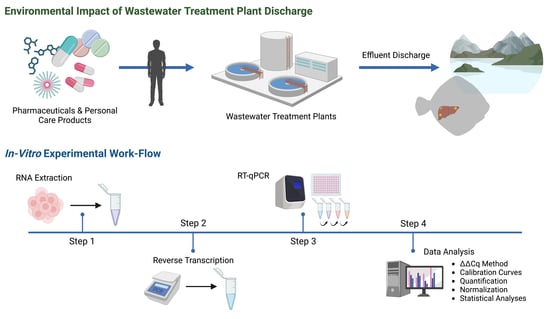
Approximately 100 mg of individual English sole liver samples were homogenized with 1 mL TRIzol® (Invitrogen, Burlington, ON, Canada) and 1 mm tungsten-carbide beads for 4 min at 30 Hz using the Retsch Mixer Mill MM400 instrument (Fisher Scientific, Ottawa, ON, Canada). Total RNA was extracted following the manufacturer’s instructions, with TURBO DNA-free kitsTM (Ambion, Austin, TX, USA) used to remove genomic DNA contamination. RNA quality was assessed using the Bio-Rad ExperionTM Automated Electrophoresis System (Version 3.20; Bio-Rad, Mississauga, ON, Canada) with an average RNA quality indicator (RQI) score ± standard error of the mean of 8.35 ± 0.18 for 136 biological samples. RNA concentrations were quantified using an Epoch 2 Microplate Spectrophotometer (BioTek, Winooski, VT, USA), with a mean 260/280 nm absorbance ratio of 2.11 ± 0.08, indicating high purity RNA. cDNA was synthesized using the Applied Biosystems High-Capacity cDNA Synthesis Kit (Waltham, MA, USA) according to the manufacturer’s instructions, and quantified using the Epoch 2 Microplate Spectrophotometer. The resulting cDNA products were diluted in double-distilled water to obtain final concentrations of 100 ng/μL prior to use in RT-qPCR reactions.

2.3. Primer Design

Gene-specific primer sets for RT-qPCR assays were designed based on highly similar phylogenetic ancestries in mitochondrial 12S and 16S sequences, assessed via BLAST (https://www.ncbi.nlm.nih.gov/, accessed on 1 November 2020), from the European plaice (Pleuronectes platessa), European flounder (Platichthys flesus), olive flounder (Paralichthys olivaceus), and the Pacific halibut (Hippoglossus stenolepis) [18]. Primers were designed using the Clustal Omega Multiple Sequence Alignment tool, and screened for optimal melting temperatures (TM) and guanine-cytosine content (GC%) via the Integrated DNA Technologies OligoAnalyzerTM Tool (www.idtdna.com/calc/analyzer, accessed on 1 January 2021). All gene-specific primer sets were PCR-amplified according to the manufacturer’s instructions using the Bio-Rad T100 Thermal Cycler (Mississauga, ON, Canada) and ABM Taq DNA Polymerase (Applied Biological Materials, Richmond, BC, Canada), and verified through Sanger Sequencing at the University of British Columbia (UBC) Sequencing and Bioinformatics Consortium (Vancouver, BC, Canada). Primer sequences, corresponding amplicon sizes, and PCR efficiencies are listed in Table 2.

2.4. Real-Time Quantitative Polymerase Chain Reaction

RT-qPCR gene amplification was conducted on the Bio-Rad CFX384TM Real-Time PCR Detection System (Bio-Rad, Mississauga, ON, Canada) using 2.5 µL of diluted cDNA template (1:80 dilution, 100 ng/µL); 0.25–0.375 µL each of the forward and reverse gene-specific primers (10 µM); 6.25 µL of the SsoFastTM EvaGreen® Supermix (Bio-Rad, Mississauga, ON, Canada), and nuclease-free water to complete the 12.5 µL reaction in a 384-well plate. The thermocycling profile included an initial denaturation step at 95 °C, for 30 s, followed by 45 amplification cycles consisting of 5 s at 95 °C for denaturation, 5 s at 55 °C for primer annealing, and 15 s at 72 °C for extension (fluorescence data was collected during the extension step). To check the specificity of the reaction and the presence of primer dimers, a dissociation protocol with a gradient from 55 °C to 95 °C was employed. Each plate included two technical replicates for each biological sample and a no-template control, as well as a positive plate control. RT-qPCR efficiency calculations were performed using seven-point standard curves of a 4-fold dilution series (1:4–1:16′384) from pooled cDNA in each assay.

2.5. Data Analysis of Gene Expression

The Livak Δ Δ Cq method was used to determine relative quantitation of target genes between exposure sites and reference genes. Reference gene stability was evaluated by calculating M-values using the Bio-Rad CFX ManagerTM Software (Mississauga, ON, Canada). Two reference genes with M-values < 1 were used to normalize the expression of target genes and compensate for differences in sample tissue amount [19].

2.6. Statistical Analyses

Statistical analyses were performed using JMP®, Ver. 16 (SAS Institute Inc., Cary, NC, USA). Normality and homogeneity of variance for each normalized gene expression dataset were tested using Shapiro-Wilk’s and Levene’s tests, respectively. Logarithmic transformation was applied to normalized gene expression data that failed normality tests. Outliers were identified and removed if individual observations were scored greater than ±3 based on studentized residuals. Two-Way ANOVA followed by a Tukey’s post-hoc analysis was used to determine statistical significance of the least square means within and between sites, sexes, and the interactions between these two variables. The significance level was set at a p-value < 0.05.

3. Results

3.1. Wild English Sole Health and Body Morphometrics

In the present study, we examined a population of mature English sole, encompassing males aged between 5 and 16 years, and females aged between 4 and 16 years [12]. Analysis of the data revealed variations in the age structure among the sampling sites (p < 0.0158) and sexes (p < 0.0108), but no interaction between the main effects was observed (F(6, 125) = 0.505, p < 0.8304) [12]. The oldest individuals were found at BIA-3, with a mean age of 11.4 years for males and 9.5 years for females, whereas the youngest individuals were observed at BIA-2, with a mean age of 7.8 years for males and 6.2 years for females (Figure 2A) [12]. Furthermore, growth rates exhibited sex-related differences, with females showing faster growth compared to males (Figure 2B) [12]. Notably, the largest and heaviest female English sole was found at the Outer Harbour (BIA-1), near the Lion’s Gate WWTP discharge site, while no such pattern was observed in males (Figure 2C,D) [12]. Additionally, a significant disparity in the levels of the gonadosomatic index (GSI) among the various collection sites was observed (p < 0.0001; Figure 2E) [12]. Female English sole from BIA-1 exhibited remarkably higher GSI levels, averaging around 13%, in comparison to all other sites where GSI levels ranged ~1.3–3.7% (p < 0.0001) [12]. Additionally, a noteworthy discovery was the consistency of GSI values among males compared to females across all sites [12]. Lastly, the hepatosomatic index (HSI) levels showed similar levels in both males and females (p < 0.4224), with the highest HSI levels being recorded at BIA-5 and the lowest at BIA-6 (p < 0.0001; Figure 2F) [12].

3.2. Gene Primer Set Design and Evaluation

A total of 14 primer sets used to amplify candidate genes of interest generated single amplicons, as demonstrated by the presence of single bands in agarose gel electrophoresis, single-peak melting curves of the PCR products, and via third-party verification through Sanger Sequencing at the UBC Sequencing and Bioinformatics Consortium (Table 2). A full list of all unsuccessful genes of interest including data related to primer design as well as detailed reasons for exclusion are provided in Table S1. Efficiency of RT-qPCR reactions ranged 90–110% with one minor exception to these criteria, the case of HSP70, which exhibited an efficiency of 112% but was deemed acceptable due to the presence of a single sharp melt peak and R2 = 0.971. The M-value for the two reference genes, ACT β and eEF1 α 1, was 0.58 with an average coefficient variance (CV) of 0.20.

3.3. RT-qPCR Gene Expression

Mean normalized expression (± standard error of the mean) values of 7–11 biological replicates per site per sex for a total of 12 genes of biological interest revealed several significant differences between sites and sexes, and in some instances, the interactive effects between these two main factors. Target genes involved in protein synthesis, which included 18S rRNA (Figure 3A) and RPS4X (Figure 3B), both exhibited significant changes in normalized gene expression. Normalized expression in 18S rRNA was found to have a main effect of site (p < 0.0001), sex (p < 0.0001), and the interaction (F(6, 119) = 3.7874, p = 0.0017). Specifically, normalized expression was significantly elevated in females at BIA-1 (3.75 ± 0.58) in comparison to some sites further from the Lion’s Gate WWTP (BIA-3 (1.23 ± 0.18), BIA-6 (0.66 ± 0.22)) and compared to both Iona WWTP sites investigated (II-NF = 0.76 ± 0.12, II-FF = 1.06 ± 0.52; Figure 3A). However, changes in 18S rRNA appeared to be sex-specific since no significant changes were observed for males between sites (p > 0.05; Figure 3A). In addition, differences in the expression of 18S rRNA were significant between sexes at one site, whereby BIA-6 females (0.66 ± 0.22) exhibited significantly lower normalized expression of 18S rRNA compared to males (1.39 ± 0.20) at this site (Figure 3A). Normalized expression in RPS4X was found to have a main effect of site (p < 0.0001) and sex (p < 0.0008), but no significant interaction was observed (F(6, 120) = 1.5506, p = 0.1676). Specifically in males, normalized expression was significantly elevated at BIA-3 (1.90 ± 0.66) in comparison to BIA-6 (0.18 ± 0.05) and to the near-field Iona WWTP site (II-NF = 0.34 ± 0.10); whereas females at BIA-6 revealed significantly lower normalized expression of RPS4X in comparison to BIA-5 (0.83 ± 0.23) and BIA-3 (1.10 ± 0.44; Figure 3B). Interestingly, at both Iona WWTP sites, males and females exhibited similar levels of RPS4X that were not significantly different from any of the Lion’s Gate WWTP sites, except for females at II-FF (0.08 ± 0.02), which differed from several sites (BIA-3 = 1.10 ± 0.44, BIA-5 = 0.83 ± 0.23; Figure 3B).
Target genes involved in xenobiotic detoxification and transcriptional regulation, which included CYP1A (Figure 4A) and HSP70 (Figure 4B), both exhibited significant changes in normalized gene expression. For CYP1A, there was no significant effect of sex (p = 0.0623) or interactive effects between sex and site (F(6, 113) = 1.9735, p = 0.0752), but site as a main effect was significant (p < 0.0001). Within females, normalized expression was significantly lower nearest to the Lion’s Gate WWTP at BIA-1 (0.08 ± 0.01) in comparison to sites further from the Lion’s Gate WWTP (BIA-2 = 0.38 ±0.05, BIA-3 = 0.43 ± 0.09, BIA-5 = 1.03 ± 0.20, BIA-6 = 1.10 ± 0.39), and compared to the near-field Iona WWTP site (II-NF = 0.44 ± 0.13; Figure 4A). However, within males, no significant differences were observed across Lion’s Gate and Iona WWTP sites, but BIA-6 (1.09 ± 0.20) exhibited higher CYP1A expression relative to II-NF (0.32 ± 0.07; Figure 4A). Normalized expression in HSP70 was found to be a main effect of site (p < 0.0001) and sex (p = 0.0258) but no significant interaction was observed (F(6, 121) = 1.1356, p = 0.3459; Figure 4B).
Target genes involved in homeostasis and metabolic function like DIO1 exhibited significant changes in normalized gene expression (Figure 5). Normalized expression in DIO1 was found to have a main effect of site (p < 0.0002), sex (p < 0.0001), and the interaction (F(6, 120) = 2.2008, p = 0.0475). Specifically, normalized expression was significantly elevated in females at BIA-1 (1.29 ± 0.26) in comparison to some sites further from the Lion’s Gate WWTP (BIA-3 (0.41 ± 0.08), BIA-5 (0.26 ± 0.05), BIA-6 (0.33 ± 0.05)), and compared to one of the Iona WWTP sites, II-NF (0.26 ± 0.05; Figure 5). However, changes in DIO1 appeared to be sex-specific since no significant changes were observed for males between sites (p > 0.05). In addition, differences in the expression of DIO1 were significant between sexes at two sites, whereby BIA-5 (0.26 ± 0.05) and II-NF (0.26 ± 0.05) females exhibited significantly lower normalized expression of DIO1 compared to males (BIA-5 = 0.96 ± 0.28, II-NF = 0.82 ± 0.14; Figure 5) at these sites.
Target genes involved in lipid and glucose metabolism included FABP1 (Figure 6A), FASN (Figure 6B), PPARδ (Figure 7A), and PPAR γ (Figure 7B), and all exhibited significant changes in normalized gene expression. For FABP1, there was no significant main effect of sex (p = 0.1509) or interactive effects between sex and site (F(6, 121) = 1.9191, p = 0.083), but site as a main effect was significant (p < 0.0034). Within females, normalized expression was significantly higher nearest to Lion’s Gate WWTP at BIA-1 (2.33 ± 0.69) in comparison to sites further from the Lion’s Gate WWTP (BIA-2 = 0.64 ± 0.96, BIA-3 = 0.66 ± 0.29, BIA-6 = 0.67 ± 0.22) and both Iona WWTP sites (II-NF = 0.78 ± 0.19, II-FF = 0.67 ± 0.39, Figure 6A). However, changes in FABP1 appeared to be sex-specific since no significant changes were observed for males between sites (p > 0.05). For FASN, there was no significant main effect of sex (p = 0.8472) or interactive effects between sex and site (F(6, 121) = 1.957, p = 0.0771), but site as a main effect was significant (p < 0.0001). Within males, normalized expression was significantly higher nearest to Lion’s Gate WWTP at BIA-1 in comparison to BIA-5 (0.8 ± 0.14) and compared to both Iona WWTP sites investigated (II-NF = 0.57 ± 0.04, II-FF = 0.64 ± 0.06, Figure 6B). Interestingly, females exhibited similar levels of FASN that were not significantly different from any of the Lion’s Gate WWTP sites, except BIA-1 (1.22 ± 0.09), which exhibited significantly higher normalized expression of FASN compared to Iona WWTP sites (II-NF = 0.60 ± 0.06, II-FF = 0.73 ± 0.11, Figure 6B). Normalized expression in PPAR δ was found to have a main effect of site (p < 0.0001), but no significant main effect of sex (p = 0.1868) and the interaction (F(6,114) = 0.4520, p = 0.8423) was observed. Within females, normalized expression was significantly higher nearest to Lion’s Gate WWTP at BIA-1 (2.78 ± 0.52) in comparison to some sites further from the primary WWTP (BIA-3 = 0.63 ± 0.14, BIA-5 = 0.68 ± 0.08, BIA-6 = 0.60 ± 0.16), and compared to both Iona WWTP sites investigated (II-NF = 0.34 ± 0.07, II-FF = 0.35 ± 0.06; Figure 7A). However, within males, the far-field Iona WWTP site (II-FF = 0.30 ± 0.03) exhibited lower PPAR δ expression relative to several sites at Lion’s Gate WWTP (BIA-1 = 1.07 ± 0.20, BIA-2 = 1.34 ± 0.22, BIA-3 = 1.00 ± 0.27), whereas the near-field Iona WWTP site (II-NF = 0.39 ± 0.08) only exhibited lower PPAR δ expression relative to BIA-2 (1.34 ± 0.22; Figure 7A). For PPAR γ , there was no significant main effect of sex (p = 0.8708), but site as a main effect (p < 0.0093) and the interactive effects between these two main factors (F(6, 121) = 3.9343, p < 0.0013) were significant. Specifically, normalized expression was significantly elevated in females at BIA-5 (0.72 ± 0.15) in comparison to the nearest site to the Lion’s Gate WWTP (BIA-1 = 0.21 ± 0.07; Figure 7B). Similarly, normalized expression was significantly elevated in males at BIA-6 (0.53 ± 0.13) in comparison to the BIA-2 (0.20 ± 0.06). Lastly, normalized expression in GLUT2 indicated no main effect of site (p = 0.0751), sex (p = 0.4418), nor an interactive effect between these two main factors (F(6, 114) = 0.4520, p = 0.8423; data not shown).
Target genes involved in reproduction included ER α (Figure 8A) and VTG (Figure 8B), and both exhibited significant changes in normalized gene expression. Normalized expression in ER α was found to have a main effect of site (p < 0.0001) and sex (p < 0.0001), but no interactive effects between these two main factors were observed (F(6,121) = 0.3278, p = 0.9213). Within females, normalized expression was significantly higher nearest to Lion’s Gate WWTP at BIA-1 (4.23 ± 1.10) in comparison to some sites further from the primary WWTP (BIA-5 = 0.36 ± 0.20, BIA-6 = 0.47 ± 0.25) and compared to the near-field Iona WWTP site (II-NF = 0.67 ± 0.37; Figure 8A). Similarly, within males, normalized expression was also significantly higher nearest to Lion’s Gate WWTP at BIA-1 (0.67 ± 0.26) in comparison to BIA-5 (0.02 ± 0.004) and compared to the near-field Iona WWTP site (II-NF = 0.03 ± 0.008; Figure 8A). Normalized expression in VTG was found to have a main effect of site (p < 0.0001), sex (p < 0.0001), and the interaction (p < 0.0014). Specifically, normalized expression was significantly elevated in females nearest to Lion’s Gate WWTP at BIA-1 (7.58 ± 1.63) in comparison to all sites further from the primary WWTP (BIA-2 = 0.83 ± 0.75, BIA-3 = 0.13 ± 0.09, BIA-5 = 0.54 ± 0.51, BIA-6 = 0.02 ± 0.01) and compared to both Iona WWTP sites investigated (II-NF = 0.15 ± 0.10, II-FF = 0.65 ± 0.52; Figure 8B). However, changes in VTG appeared to be sex-specific since no significant changes were observed for males between sites (p > 0.05). In addition, differences in the expression of VTG were significant between sexes at two sites, whereby BIA-6 (0.88 ± 0.33) and II-NF (0.30 ± 0.11) males exhibited significantly higher normalized expression of VTG compared to females (BIA-6 = 0.02 ± 0.007, II-NF = 0.15 ± 0.10; Figure 8B) at these sites.

4. Discussion

4.1. Examining the Health and Body Metrics of Resident English Sole

Several studies have successfully demonstrated the application of the GSI to improve accuracy and precision in determining sexual maturity and fertility in fish [20,21], and collectively this study demonstrates the potential of molecular biomarkers of urban contaminant exposure in wild caught English sole for use in diagnosing a broader range of adverse health effects when combined with conventional whole organism health indicators (summarized in Table S2). For instance, the heavier body weight of females at BIA-1 was likely partially attributed to the significantly higher GSI in these females, the latter indicating ovaries in an advanced stage of development and closer to a ripe ovary observed just prior to spawning. With all English sole of adult age, one would expect the reproductive status to be similar across all sites; thus, it is possible that WWTP contaminants, particularly those with estrogenic modes of action, may have caused advanced ovarian development in females residing nearest the Lions Gate WWTP effluent discharge site (BIA-1). Although no detailed studies of English sole reproductive cycles and gonad size in BC have been conducted, English sole gonad size has been shown to vary seasonally in other Pacific Northwest populations. Specifically, GSI and gonad sizes increase during the months leading up to winter spawning, at which point GSI and gonad sizes peak, and this is then followed by reduced size immediately post-spawning of spent gonads and continued gonadal recrudescence into the summer and/or early fall [22]. In the present study, we found that female English sole collected closest to the Lion’s Gate WWTP (near BIA-1) exhibited markedly higher GSI levels resembling those of pre-spawning females. However, fish collected from all other sites exhibited GSIs that resembled those of spawned-out females. This advanced ovarian development at BIA-1 may be due to estrogenic contaminants known to be present in WWTP effluent (i.e., 17 β -estradiol, estriol, estrogen 17 α -ethynyl estradiol, etc.), and all other sites were exhibiting underdeveloped ovaries. Despite the evident differences in female GSI, critical lifelong ecological patterns of English sole in the Burrard Inlet related to spawning, planktonic life stages, settlement, and foraging have remained largely unexplored. As a result, the intriguing advanced ovarian development observed in these fish could potentially be attributed to the unique reproductive ecology specific to the English sole in this region of the Burrard Inlet, coupled with the presence of estrogenic contaminants stemming from the nearby WWTP. Therefore, to avoid unfounded assumptions, further in-depth research is imperative to establish a comprehensive understanding of the reproductive ecology of English sole in the area.

4.2. Influence of Urban Pollutants on Hepatic CYP1A Gene Expression

The cytochrome P450 detoxification system is an extensively studied enzyme system that is involved in the first phase oxidation reactions of the two-phase metabolism of xenobiotics and its activity, transcript, and protein abundance has been shown to be related to sediment, whole-body, and liver tissue concentrations of various planar halogenated and non-halogenated hydrocarbons [23]; and the present study showed significant differences in the expression levels of CYP1A transcripts in female English sole between sites of interest. Indeed, several studies over the past two decades have demonstrated the induction of hepatic CYP1A enzymes or transcripts after exposure to several industrial compounds, including PCBs, PBDEs, OCPs, DDT, and PAHs, primarily through the binding and activation of the AhR pathway [24]. For instance, in a 2000 field study, results found that sediment PCBs and PAHs near the Orange County wastewater discharge (California, USA) significantly increased CYP1A protein levels in English sole, with sediment PCBs showing a dose-dependent positive correlation [25]. Similarly, high molecular weight PAHs (e.g., naphthalene, fluorene, anthracene, pyrene) have been commonly associated with petroleum operations, lumber production, WWTPs, and commercial use throughout the Burrard Inlet, and while the BIAMP aims to monitor and assess the impacts of effluents from the primary Lion’s Gate WWTP, 2017 tissue chemistry results for arsenic, lead, PCBs, OCPs (represented by total DDT and total chlordane), dioxins and furans, and the PAH metabolite, pyrene 1-glucuronide in bile, were highest in fish from BIA-5 and BIA-6 [12]. It is possible that this is due to the Port Moody and Indian Arm being subjected to a larger influx of PAHs from multiple contributors, such as Imperial Oil Ioco, Flavelle Cedar, Burrard Thermal, and active hydroelectric plants relative to the Outer Harbour, in which two sampling sites (BIA-1 and BIA-2) were set [12,13,14]. Furthermore, liver English sole CYP1A activity measured using an in vitro EROD assay was significantly higher in the eastern portions of Burrard Inlet (BIA-5, BIA-6) compared to the western Outer Harbour region (BIA-1) in 2007, 2012, and 2017 [12,13,14]. These results suggest the exposure of English sole to CYP1A-inducing chemicals (e.g., dioxins and furans, PCBs, PAHs, and 2,3,7,8-TCDF) in Metro Vancouver waters generally supports the hypothesis that sites furthest from the Lion’s Gate WWTP had higher levels of these chemicals due to local contaminant sources in the Port Moody Arm (near BIA-5) and Indian Arm (near BIA-6) [12,13,14]. Nevertheless, PCBs and PBDEs have been measured at concentrations equal to or exceeding Canadian Environmental Quality Guidelines in Metro Vancouver’s WWTP, but are also prevalent from a variety of other sources in urban areas relative to both industrial and urban wastewater discharges [12,13,14].

4.3. Exploring Sewage Effluent’s Potential Disruption of Thyroid Hormone Metabolism

Interestingly, over the past ~three decades there is mounting evidence that in teleosts, peripheral thyroxine (T4) deiodination to the biologically active triiodothyronine (T3) hormone or inactivated reverse T3 (rT3) or 3,3′-diiodothyronine (T2) via deiodinase enzymatic activity are known to be influenced by contaminants detected in wastewater effluents (e.g., plasticizers, flame retardants, surfactants, and pharmaceuticals) [26,27]. Specifically, the DIO1 enzyme is involved in both T4-outer ring deiodination (ORD) and T4-inner ring deiodination (IRD) catalytic reactions to produce active T3 and inactive rT3 hormones, respectively, whereas DIO2 and DIO3 participate solely in ORD and IRD reactions, respectively [27]. Despite, the lack of gene-specific biomarkers of contaminants that disrupt thyroid hormone metabolism, recent field-based studies in fish demonstrated the impacts on the abundance of deiodinase enzyme levels in waters downstream of WWTP discharge sites [28,29]. Considering several suspected and known EDCs with modes of action that disrupt the HPT endocrine axis of vertebrates, the findings in the present study and the presence of these contaminants in Metro Vancouver sewage effluents (i.e., alkylphenols, dioxins and furans, metals, PAHs, PBDEs, PCBs, pesticides, and PPCPs), it is likely that English sole are experiencing some disruption of the HPT axis [12,13,14].

4.4. Exploring Fatty acid Metabolism Transcripts as Promising Biomarkers of Sewage Effluent Exposure

In teleost, lipids play a major role in the storage and provision of energy through lipogenesis pathways occurring in the cytosol via FASN, a multienzyme complex which catalyzes the conversion of acetyl-CoA and malonyl-CoA to 16- and 18-carbon fatty acids (palmitic and stearic acids, respectively); while intracellular transport and metabolism of dietary lipids and exogenous lipophilic compounds are catalyzed by cytoplasmic FABP1 [30]. Nevertheless, nuclear receptors (i.e., peroxisome proliferator-activated receptors, PPARs) are key to these processes, and bind a broad range of ligands (e.g., fatty acids and fatty acid derivatives) and serve as major transcriptional regulators of enzymes involved in fatty acid metabolism and transport [30]. Together, in the present study, a trend of elevations in two enzymes and a transcriptional regulator (PPAR δ ) involved in lipid metabolism at the site most proximal to a primary WWTP effluent discharge site (BIA-1) coupled with the significantly heavier and longer females at this site provides evidence of altered metabolism in fish exposed to WWTP effluent. Whether these larger fish exhibited different levels of lipids or other metabolites or organ level alterations in metabolism was not measured in the study, but should be considered in future monitoring programs.
Although several contaminants discharged in primary and secondary WWTP effluents have been detected globally, a recent review of the two main types of lipid-regulating agents, statins, and fibrates reported that these cardiovascular drugs are the most consumed pharmaceuticals worldwide, and are routinely detected in the ng/L to µg/L range in global surface waters [31,32,33]. Although not measured in the Metro Vancouver BIAMP, it is expected that these biologically active pharmaceuticals targeting lipid metabolism in vertebrates are present to some extent proximal to Metro Vancouver’s WWTP. Furthermore, the alterations in transcripts related to lipid metabolism in the present study support this hypothesis. Even though most studies reporting significant effects of fibrates and statins on lipid metabolism are laboratory-based and variable, some WWTP effluent field-based exposures do exist, and demonstrate effects on lipids and lipid metabolites [34]. Such studies suggest the potential use of PPARs and lipid-metabolism-related transcripts as biomarkers for environmental contaminant risk assessment in fish; however, further studies are warranted.

4.5. Disruption of the Reproductive Endocrine Axis by Estrogenic Contaminant Exposure

Strong evidence of exposure to contaminants with an estrogenic mode of action was observed in this study based on male English sole VTG transcript levels either equivalent or exceeding female levels of this gene at all sites investigated. In oviparous vertebrates and some invertebrates (i.e., bivalves), the glycolipophosphoprotein VTG is the precursor of vitellin, which is used as the primary nutritional source for the developing embryo [35]. It is produced in the liver and regulated by estrogens and their receptors (ligand activated transcription factors) that mediate estrogen action, and then exported into the blood and sequestered in oocytes [35]. Although males possess the genes to produce VTG, this naturally occurs at much lower levels than in females. As a result, VTG at the transcript and protein levels has been validated as a reliable and robust biomarker of exposure to estrogenic substances [36,37,38,39]. In addition, most teleosts investigated to date exhibit sex-specific differences in ERα levels with females exhibiting higher levels than males [40]; however, this was not observed in the present study. Thus, these similar levels of ERα in males and females further support the hypothesis of the widespread presence of estrogenic contaminants in Metro Vancouver waters. Interestingly, at site BIA-1 nearest to the primary treatment Lion’s Gate WWTP, where females exhibited advanced ovarian development and elevated ER α levels, the males collected from this site exhibited the largest mean magnitude VTG transcript induction relative to males from other study sites. Previous Metro Vancouver BIAMPs conducted in 2007 and 2012 measured protein levels of VTG and reported elevated levels in males nearest to the Lion’s Gate WWTP discharge site in both years, and some induction in the Central Harbour (BIA-3), Port Moody Arm (BIA-5), and Indian Arm (BIA-6; n = 5 males per site) [13,14]. Although no true reference site was included in the present study, Johnson et al. [41] demonstrated a low frequency of detecting protein VTG in Puget Sound English sole at three non-urban sites (0–6%) compared to urban impacted sites (12–47%). Locally, in controlled laboratory-based studies on Metro Vancouver WWTP effluents, Osachoff et al. [42] reported significant induction of VTG mRNA transcript and plasma protein levels in juvenile rainbow trout (Oncorhynchus mykiss) after exposure to both Metro Vancouver WWTP influent and effluent, indicating the presence of estrogenic contaminants discharged from primary and secondary WWTPs in Metro Vancouver. Collectively, multiple years of monitoring programs suggest that English sole males are exhibiting elevated VTG, indicative of the widespread occurrence of estrogenic substances in Metro Vancouver waters, and females spawning nearest the Lion’s Gate WWTP discharge site may occur abnormally early due to advanced ovarian development. With clear evidence of organismal and population level impacts due to the disruption of the reproductive endocrine axis in fish downstream of sewage effluent discharge sites collected since the 1990s (reviewed in Marlatt et al., [43]), mitigation measures such as improved WWTP treatment processes (i.e., tertiary level) are the ideal solution.

5. Conclusions

Field studies, such as the BIAMP conducted by Metro Vancouver, provide invaluable information about point-source environmental pollution associated with WWTPs as well as other anthropogenic pollutants and stressors in urban waters. Here, we report the induction of well characterized biomarkers of reproductive endocrine axis abnormalities (i.e., elevated VTG and ER α in males and females, advanced ovarian development) that were most prevalent nearest the primary treatment Lion’s Gate WWTP, but ultimately, the gene expression analyses indicate widespread exposure to estrogenic contaminants throughout the Burrard Inlet. Routine VTG and sex steroid receptor assays are recommended for future monitoring in Burrard Inlet to track the spatial and temporal extent of WWTP effluent exposure and impacts on aquatic wildlife. Although few other genes in fish are as well characterized as VTG and ER α , several genes involved in lipid and glucose metabolism and one enzyme involved in thyroid hormone metabolism were also associated with exposure to primary WWTP effluent. We hypothesize the former is at least partly attributed to the lipid regulating pharmaceuticals known to be present near sewage discharge sites, while the latter potential thyroid hormone metabolism disruption was due to several chemicals present in WWTP and other industrial effluents (i.e., alkylphenols, dioxins and furans, metals, PAHs, PBDEs, PCBs, pesticides, and PPCPs), several of which were present in English sole tissue in the 2017 BIAMP [12]. This study demonstrates the high potential and applicability of molecular biomarkers of urban contaminant exposure in wild caught English sole to assess a wider range of adverse health effects when combined with conventional whole organism health indicators.

Supplementary Materials

The following supporting information can be downloaded at: https://www.mdpi.com/article/10.3390/toxics11080657/s1, Table S1: List of primer sequences designed to amplify genes of interest in English Sole that were unsuccessful for RT-qPCR experiments, and the reasons for exclusion; Table S2: Overview of molecular biomarker and RT-qPCR results of urban contaminant exposure in wild English sole.

Author Contributions

Conceptualization, K.P. and V.L.M.; methodology, K.P.; software, K.P.; validation, K.P. and V.L.M.; formal analysis, K.P.; investigation, V.L.M.; resources, V.L.M.; data curation, K.P.; writing—original draft preparation, K.P.; writing—review and editing, K.P. and V.L.M.; visualization, K.P.; supervision, V.L.M.; project administration, V.L.M.; funding acquisition, V.L.M. All authors have read and agreed to the published version of the manuscript.

Funding

This research was funded by NSERC Discovery (Marlatt).

Institutional Review Board Statement

All work with English soles was performed in compliance with the guidelines of the Canadian Council for Animal Care and with a permit issued by Metro Vancouver (Vancouver, BC, Canada).

Informed Consent Statement

Not applicable.

Data Availability Statement

Data available upon request.

Acknowledgments

We would like to express our sincere gratitude to Farida Bishay, Colleen Shum, and Charlene Hickling from Metro Vancouver for their invaluable contribution in providing us with the supplemental data on known points sources of contamination and comprehensive whole organism health indicators. The data, including EROD activity, external and internal abnormalities, fork length, weight, GSI, and plasma VTG, were derived from the meticulous 2007, 2012, and 2017 Burrard Inlet Ambient Monitoring Program reports conducted by Nautilus and ENKON Environmental. Their assistance and support greatly enriched our research.

Conflicts of Interest

The authors declare no conflict of interest.

References

  1. Landrigan, P.J.; Fuller, R.; Acosta, N.J.R.; Adeyi, O.; Arnold, R.; Basu, N.; Baldé, A.B.; Bertollini, R.; Bose-O’Reilly, S.; Boufford, J.I.; et al. The Lancet Commission on Pollution and Health. Lancet 2018, 391, 462–512. [Google Scholar] [CrossRef] [Green Version]
  2. Rehman, M.S.U.; Rashid, N.; Ashfaq, M.; Saif, A.; Ahmad, N.; Han, J.-I. Global Risk of Pharmaceutical Contamination from Highly Populated Developing Countries. Chemosphere 2015, 138, 1045–1055. [Google Scholar] [CrossRef]
  3. Khan, U.; Bloom, R.A.; Nicell, J.A.; Laurenson, J.P. Risks Associated with the Environmental Release of Pharmaceuticals on the U.S. Food and Drug Administration “Flush List”. Sci. Total Environ. 2017, 609, 1023–1040. [Google Scholar] [CrossRef] [PubMed]
  4. Wang, H.; Xi, H.; Xu, L.; Jin, M.; Zhao, W.; Liu, H. Ecotoxicological Effects, Environmental Fate and Risks of Pharmaceutical and Personal Care Products in the Water Environment: A Review. Sci. Total Environ. 2021, 788, 147819. [Google Scholar] [CrossRef] [PubMed]
  5. British Columbia Ministry of Environment and Climate Change Strategy. British Columbia Approved Water Quality Guidelines: Aquatic Life, Wildlife & Agriculture—Guideline Summary; Water Quality Guideline Series, WQG-20; British Columbia Ministry of Environment and Climate Change Strategy: Victoria, BC, Canada, 2021.
  6. Canadian Council of Ministers of the Environment. Canadian Council of Ministers of the Environment. Canadian water quality guidelines for the protection of aquatic life: Nonylphenol and its ethoxylates. In Canadian Environmental Quality Guidelines; Canadian Council of Ministers of the Environment: Winnipeg, MB, Canada, 1999. [Google Scholar]
  7. Sousa, J.C.G.; Ribeiro, A.R.; Barbosa, M.O.; Pereira, M.F.R.; Silva, A.M.T. A Review on Environmental Monitoring of Water Organic Pollutants Identified by EU Guidelines. J. Hazard. Mater. 2018, 344, 146–162. [Google Scholar] [CrossRef]
  8. Kidd, K.A.; Blanchfield, P.J.; Mills, K.H.; Palace, V.P.; Evans, R.E.; Lazorchak, J.M.; Flick, R.W. Collapse of a Fish Population after Exposure to a Synthetic Estrogen. Proc. Natl. Acad. Sci. USA 2007, 104, 8897–8901. [Google Scholar] [CrossRef]
  9. Matthiessen, P.; Wheeler, J.R.; Weltje, L. A Review of the Evidence for Endocrine Disrupting Effects of Current-Use Chemicals on Wildlife Populations. Crit. Rev. Toxicol. 2017, 48, 195–216. [Google Scholar] [CrossRef] [PubMed] [Green Version]
  10. Söffker, M.; Tyler, C.R. Endocrine Disrupting Chemicals and Sexual Behaviors in Fish—A Critical Review on Effects and Possible Consequences. Crit. Rev. Toxicol. 2012, 42, 653–668. [Google Scholar] [CrossRef]
  11. OECD. Detailed Review Paper on the State of the Science on Novel In Vitro and In Vivo Screening and Testing Methods and Endpoints for Evaluating Endocrine Disruptors; OECD: Paris, France, 2012. [Google Scholar] [CrossRef]
  12. ENKON Environmental Limited. Burrard Inlet Ambient Monitoring Program 2017 Fish Community and Health; ENKON Environmental Limited: Burnaby, BC, Canada, 2020. [Google Scholar]
  13. Nautilus Environmental. Metro Vancouver Ambient Burrard Inlet Monitoring Program Fish Health Survey; Nautilus Environmental: Burnaby, BC, Canada, 2009. [Google Scholar]
  14. ENKON Environmental Limited. Burrard Inlet Ambient Monitoring Program 2012 Biota Monitoring; ENKON Environmental Limited: Burnaby, BC, Canada, 2015. [Google Scholar]
  15. Lockhart, K.; Ciannelli, L.; Waldo Wakefield, W. A Comparative Analysis of Sampling Methodologies for Assessing Abundance and Distribution of Young-of-the-Year Groundfishes in Nearshore Soft-Sediment Habitats. Fish. Res. 2023, 264, 106734. [Google Scholar] [CrossRef]
  16. Levings, C.; Ong, S. Fish Communities and Life History Attributes of English Sole (Pleuronectes vetulus) in Vancouver Harbour. Mar. Environ. Res. 2004, 57, 103–120. [Google Scholar] [CrossRef]
  17. Chittaro, P.M.; Finley, R.J.; Levin, P.S. Spatial and Temporal Patterns in the Contribution of Fish from Their Nursery Habitats. Oecologia 2009, 160, 49–61. [Google Scholar] [CrossRef]
  18. Azevedo, M.; Oliveira, C.; Pardo, B.G.; Martínez, P.; Foresti, F. Phylogenetic Analysis of the Order Pleuronectiformes (Teleostei) Based on Sequences of 12S and 16S Mitochondrial Genes. Genet. Mol. Biol. 2008, 31 (Suppl. S1), 284–292. [Google Scholar] [CrossRef]
  19. Bustin, S.A.; Benes, V.; Garson, J.A.; Hellemans, J.; Huggett, J.; Kubista, M.; Mueller, R.; Nolan, T.; Pfaffl, M.W.; Shipley, G.L.; et al. The MIQE Guidelines: Minimum Information for Publication of Quantitative Real-Time PCR Experiments. Clin. Chem. 2009, 55, 611–622. [Google Scholar] [CrossRef] [PubMed] [Green Version]
  20. Flores, A.; Wiff, R.; Díaz, E. Using the Gonadosomatic Index to Estimate the Maturity Ogive: Application to Chilean Hake (Merluccius Gayi Gayi). ICES J. Mar. Sci. 2014, 72, 508–514. [Google Scholar] [CrossRef] [Green Version]
  21. Lowerre-Barbieri, S.K.; Ganias, K.; Saborido-Rey, F.; Murua, H.; Hunter, J.R. Reproductive Timing in Marine Fishes: Variability, Temporal Scales, and Methods. Mar. Coast. Fish. 2011, 3, 71–91. [Google Scholar] [CrossRef] [Green Version]
  22. Sol, S.Y.; Olson, O.P.; Lomax, D.P.; Johnson, L.L. Gonadal Development and Associated Changes in Plasma Reproductive Steroids in English sole, Pleuronectes vetulus, from Puget Sound, Washington. Fish. Bull. Natl. Ocean. Atmos. Adm. 1998, 96, 859–870. [Google Scholar]
  23. Goksøyr, A. Use of Cytochrome P450 1A (CYP1A) in Fish as a Biomarker of Aquatic Pollution. Arch. Toxicol. Suppl. 1995, 17, 80–95. [Google Scholar] [CrossRef]
  24. Oris, J.T.; Roberts, A.P. Statistical ANALYSIS of cytochrome P4501A biomarker measurements in fish. Environ. Toxicol. Chem. 2007, 26, 1742–1750. [Google Scholar] [CrossRef] [Green Version]
  25. Roy, L.A.; Armstrong, J.M.; Sakamoto, K.; Steinert, S.A.; Perkins, E.; Lomax, D.P.; Johnson, L.L.; Schlenk, D. The Relationships of Biochemical Endpoints to Histopathology and Population Metrics in Feral Flatfish Species Collected near the Municipal Wastewater Outfall of Orange County, California, USA. Environ. Toxicol. Chem. 2003, 22, 1309–1317. [Google Scholar] [CrossRef]
  26. Dang, Z.; Arena, M.; Kienzler, A. Fish Toxicity Testing for Identification of Thyroid Disrupting Chemicals. Environ. Pollut. 2021, 284, 117374. [Google Scholar] [CrossRef]
  27. Noyes, P.D.; Stapleton, H.M. PBDE Flame Retardants. Endocr. Disruptors 2014, 2, e29430. [Google Scholar] [CrossRef] [Green Version]
  28. Reinling, J.; Houde, M.; Verreault, J. Environmental Exposure to a Major Urban Wastewater Effluent: Effects on the Energy Metabolism of Northern Pike. Aquat. Toxicol. 2017, 191, 131–140. [Google Scholar] [CrossRef]
  29. Picard-Aitken, M.; Fournier, H.; Pariseau, R.; Marcogliese, D.J.; Cyr, D.G. Thyroid Disruption in Walleye (Sander vitreus) Exposed to Environmental Contaminants: Cloning and Use of Iodothyronine Deiodinases as Molecular Biomarkers. Aquat. Toxicol. 2007, 83, 200–211. [Google Scholar] [CrossRef] [PubMed]
  30. Olivares-Rubio, H.F.; Vega-López, A. Fatty Acid Metabolism in Fish Species as a Biomarker for Environmental Monitoring. Environ. Pollut. 2016, 218, 297–312. [Google Scholar] [CrossRef] [PubMed]
  31. Zhang, K.; Zhao, Y.; Fent, K. Cardiovascular Drugs and Lipid Regulating Agents in Surface Waters at Global Scale: Occurrence, Ecotoxicity and Risk Assessment. Sci. Total Environ. 2020, 729, 138770. [Google Scholar] [CrossRef] [PubMed]
  32. Miao, X.-S.; Metcalfe, C. Determination of Pharmaceuticals in Aqueous Samples Using Positive and Negative Voltage Switching Microbore Liquid Chromatography/Electrospray Ionization Tandem Mass Spectrometry. J. Mass Spectrom. 2003, 38, 27–34. [Google Scholar] [CrossRef]
  33. Miao, X.-S.; Metcalfe, C.D. Determination of Cholesterol-Lowering Statin Drugs in Aqueous Samples Using Liquid Chromatography–Electrospray Ionization Tandem Mass Spectrometry. J. Chromatogr. A 2003, 998, 133–141. [Google Scholar] [CrossRef]
  34. Simmons, D.B.D.; Miller, J.; Clarence, S.; McCallum, E.S.; Balshine, S.; Chandramouli, B.; Cosgrove, J.; Sherry, J.P. Altered Expression of Metabolites and Proteins in Wild and Caged Fish Exposed to Wastewater Effluents in Situ. Sci. Rep. 2017, 7, 17000. [Google Scholar] [CrossRef] [Green Version]
  35. Martyniuk, C.J.; Feswick, A.; Munkittrick, K.R.; Dreier, D.A.; Denslow, N.D. Twenty Years of Transcriptomics, 17alpha-Ethinylestradiol, and Fish. Gen. Comp. Endocrinol. 2020, 286, 113325. [Google Scholar] [CrossRef]
  36. Amano, H.; Uno, S.; Koyama, J.; Hiramatsu, N.; Todo, T.; Hara, A. Development of Specific Enzyme-Linked Immunosorbent Assays for Multiple Vitellogenins in Marbled Sole, Pleuronectes yokohamae. Gen. Comp. Endocrinol. 2019, 281, 67–72. [Google Scholar] [CrossRef]
  37. Barber, L.B.; Rapp, J.L.; Kandel, C.; Keefe, S.H.; Rice, J.; Westerhoff, P.; Bertolatus, D.W.; Vajda, A.M. Integrated Assessment of Wastewater Reuse, Exposure Risk, and Fish Endocrine Disruption in the Shenandoah River Watershed. Environ. Sci. Technol. 2019, 53, 3429–3440. [Google Scholar] [CrossRef] [PubMed]
  38. Tran, T.K.A.; Yu, R.M.K.; Islam, R.; Nguyen, T.H.T.; Bui, T.L.H.; Kong, R.Y.C.; O’Connor, W.A.; Leusch, F.D.L.; Andrew-Priestley, M.; MacFarlane, G.R. The Utility of Vitellogenin as a Biomarker of Estrogenic Endocrine Disrupting Chemicals in Molluscs. Environ. Pollut. 2019, 248, 1067–1078. [Google Scholar] [CrossRef] [PubMed]
  39. Zhang, Z.; Wang, J.; Gao, M.; Li, X.; Cheng, Y.; Zhang, X.; Shu, G.; Wang, W.; Ru, S. New Methods for Purification of Paralichthys olivaceus Lipovitellin and Immunoassay-Based Detection of Vitellogenin. Ecotoxicol. Environ. Saf. 2019, 180, 624–631. [Google Scholar] [CrossRef] [PubMed]
  40. Nelson, E.J.; Habibi, H.R. Estrogen Receptor Function and Regulation in Fish and Other Vertebrates. Gen. Comp. Endocrinol. 2013, 192, 15–24. [Google Scholar] [CrossRef]
  41. Johnson, L.L.; Lomax, D.P.; Myers, M.S.; Olson, O.P.; Sol, S.Y.; O’Neill, S.M.; West, J.; Collier, T.K. Xenoestrogen Exposure and Effects in English Sole (Parophrys vetulus) from Puget Sound, WA. Aquat. Toxicol. 2008, 88, 29–38. [Google Scholar] [CrossRef]
  42. Osachoff, H.L.; Osachoff, K.N.; Wickramaratne, A.E.; Gunawardane, E.K.; Venturini, F.P.; Kennedy, C.J. Altered Burst Swimming in Rainbow Trout Oncorhynchus mykiss Exposed to Natural and Synthetic Oestrogens. Fish Biol. 2014, 85, 210–227. [Google Scholar] [CrossRef]
  43. Marlatt, V.L.; Bayen, S.; Castaneda-Cortès, D.; Delbès, G.; Grigorova, P.; Langlois, V.S.; Martyniuk, C.J.; Metcalfe, C.D.; Parent, L.; Rwigemera, A.; et al. Impacts of Endocrine Disrupting Chemicals on Reproduction in Wildlife and Humans. Environ. Res. 2022, 208, 112584. [Google Scholar] [CrossRef]
Figure 1. Location of seven sampling sites for the BIAMP involved in water, sediment, and English sole collections [12,13,14]. In 2017, the BIAMP included five sites along the Burrard Inlet (BIA-1, BIA-2, BIA-3, BIA-5, BIA-6), two sites associated with Iona Island (II-NF, II-FF), and two of Metro Vancouver’s primary WWTPs, Lions Gate and Iona (in purple). The map highlights current sampling sites in red and historical sites in blue, with the omission of one site at the mouth of the Indian Arm (BIA-4) and a reference site at the North end of the Indian Arm (BIA-7).
Toxics 11 00657 g001
Figure 2. Various fish heath measures in English sole collected during the 2017 BIAMP [12] at seven sites: (A) Mean age ± one standard error of the mean; (B) Relationship between fork length and wet body weigh; (C) Fork length; (D) Wet body weight; (E) Gonadosomatic index; (F) Hepatosomatic index; and (G) Internal and external abnormalities in males and females pooled within each site. Boxplots show median values, 25th and 75th percentiles, minimum and maximum values, and outliers. A two-way ANOVA followed by a Tukey’s post-hoc test was used to determine significant differences between main effects (site and sex) and their interaction. Different superscripts indicate significant differences (p < 0.05).
Toxics 11 00657 g002
Figure 3. Hepatic gene expression levels of (A) 18S rRNA and (B) RPS4X in English sole at near- and far-field sites from the Iona and Lion’s Gate WWTP in Burrard Inlet. Boxplots show median values, 25th and 75th percentiles, minimum and maximum values, and outliers. Two-Way ANOVA with Tukey’s post-hoc analysis was used to determine significant differences between main effects (site and sex) and their interaction. Biological replicates for each site are provided. Significant site and sex effects were observed in both genes, and a significant interaction effect was observed for 18S rRNA. Different superscript letters indicate significant differences (p < 0.05).
Toxics 11 00657 g003
Figure 4. Hepatic gene expression levels of (A) CYP1A and (B) HSP70 in English sole at near- and far-field sites from the Iona and Lion’s Gate WWTP in Burrard Inlet. Boxplots show median values, 25th and 75th percentiles, minimum and maximum values, and outliers. Two-Way ANOVA with Tukey’s post-hoc analysis was used to determine significant differences between main effects (site and sex) and their interaction. Biological replicates for each site are provided. Significant site effects were observed in both genes, while HSP70 showed a significant sex effect. However, no interactive effect was found. Different superscript letters indicate significant differences (p < 0.05).
Toxics 11 00657 g004
Figure 5. Hepatic gene expression levels of DIO1 in English sole at near- and far-field sites from the Iona and Lion’s Gate WWTP in Burrard Inlet. Boxplots show median values, 25th and 75th percentiles, minimum and maximum values, and outliers. Two-Way ANOVA with Tukey’s post-hoc analysis was used to determine significant differences between main effects (site and sex) and their interaction. Biological replicates for each site are provided. A significant effect of site, sex, and an interactive effect was observed. Different superscript letters indicate significant differences (p < 0.05).
Toxics 11 00657 g005
Figure 6. Hepatic gene expression levels of (A) FABP1 and (B) FASN in English sole at near- and far-field sites from the Iona and Lion’s Gate WWTP in Burrard Inlet. Boxplots show median values, 25th and 75th percentiles, minimum and maximum values, and outliers. Two-Way ANOVA with Tukey’s post-hoc analysis was used to determine significant differences between main effects (site and sex) and their interaction. Biological replicates for each site are provided. Significant site effects were observed in both genes; however, there were no significant effects for sex or the interaction in either gene. Different superscript letters indicate significant differences (p < 0.05).
Toxics 11 00657 g006
Figure 7. Hepatic gene expression levels of (A) PPAR δ and (B) PPAR γ in English sole at near- and far-field sites from the Iona and Lion’s Gate WWTP in Burrard Inlet. Boxplots show median values, 25th and 75th percentiles, minimum and maximum values, and outliers. Two-Way ANOVA with Tukey’s post-hoc analysis was used to determine significant differences between main effects (site and sex) and their interaction. Biological replicates for each site are provided. Significant site effects were observed in both genes, with the interactive effect specifically observed in PPAR γ . However, neither gene showed a main effect of sex. Different superscript letters indicate significant differences (p < 0.05).
Toxics 11 00657 g007
Figure 8. Hepatic gene expression levels of (A) ERα and (B) VTG in English sole at near- and far-field sites from the Iona and Lion’s Gate WWTP in Burrard Inlet. Boxplots show median values, 25th and 75th percentiles, minimum and maximum values, and outliers. Two-Way ANOVA with Tukey’s post-hoc analysis was used to determine significant differences between main effects (site and sex) and their interaction. Biological replicates for each site are provided. Significant site and sex effects were observed in both genes, with the interactive effect specifically observed in VTG.
Toxics 11 00657 g008
Table 1. Sampling sites for English sole from Lions Gate and Iona primary WWTPs. Sites were designated by the BIAMP [12,13,14] and include five sites along the Burrard Inlet and two sites along Iona Island. For each site of interest, location coordinates (Universal Transverse Mercator, UTM) and brief site descriptions are provided.
SiteLocationCoordinates (UTM)Description
BIA-1Outer Harbour NorthE 483752
N 5464073
1.5 km offshore to the south of Pacific Institute
BIA-2Outer Harbour SouthE 482935
N 5460679
1.5 km north of Spanish Bank
BIA-3Inner HarbourE 493841
N 5460963
West of Loch Katrine Bank
BIA-5Port Moody ArmE 506788
N 5459875
East of Port Moody Narrows
BIA-6Indian ArmE 505549
N 5464824
Representative southern site
II-NFIona IslandE 478523
N 5453266
2–4 km north of Iona diffuser
II-FFIona IslandE 477283
N 5442000
8–10 km of south Iona diffuser
Table 2. Primer sequences designed to amplify genes of interest to examine the effects of wastewater effluents in English sole through RT-qPCR. For each target gene of interest, the National Center for Biotechnology and Information accession identifiers, forward and reverse primer sequences (5′ → 3′), annealing temperatures (Tm), amplicon sizes, efficiency of primer pairs (%E), and goodness of fit of linear regression for the relative standard curves (R2) are provided for reference genes and target genes of interest.
Target GeneAccession IDPrimer SequenceTm (°C)Amplicon SizeEfficiency (%)R2
Reference Housekeeping Genes
18S rRNAXR_004613416.1F: GGTCTGTGATGCCCTTAGATG
R: GCTTATGACCCGCGCTTAC
55.8210104.50.972
ACT β XM_035181811.1F: GACCAACTGGGATGACATGG
R: GCGTACAGGGACAGCACAGC
612041030.972
Genes of Biological Interest
CYP1AAJ310693.1F: TGTGAGGACAGGAAGCTGGA
R: GCTCCAAACAGGTCGTTGACA
588696.90.992
DIO1AB362421.1F: ACAGATGGTTGGGCCTTCAC
R: TGACTTTCCCAGCCTGAAGC
57.7196108.10.992
eEF1 α 1XM_035620145.1F: AAGATCCACATCAACATCGTG
R: CAAACTTCCACAGAGCGATG
56.722998.80.998
ER α XM_035143352.1F: GCTGAGGGATTTGAGATGGCT
R: ATGTAGTCATTGTGACCCTGGATG
56.9143106.10.96
FABP1XM_020099114.1F: GAAGGTCAAGGCGGTGGTTC
R: ACATGCGTTTGCTCGTCCTC
58.4155108.40.989
FASNXM_035162863.1F: GCAACGGCAATGACAAAGAGC
R: TTTGTCTGGTTTCCGTGCCA
57.6143107.80.952
GLUT2AY521663.1F: CCGCGCTACCTCTACATCGT
R: TGCTGCCTGTAGACGGAAGA
58.6182102.60.969
HSP70AF187726.1F: CAGTGCCCGCCTACTTCAAT
R: TTCTGACCCAACCTTCTTGTCC
57.3143111.70.971
PPAR δ XM_035158970.1F: GACCTCGCTCCACCCTTTAC
R: TCCAAGCCCGAATGTGGAAC
57.715798.50.988
PPAR γ AJ243956.2F: TGTCAGTCACGCTCTGCTGAA
R: TAGGAGATCAGGGTCCCGTCT
58.8176102.40.988
RPS4XXM_034569090.1F: GTTTGATACTGCCAACCTGTGC
R: TTGGAGAGCCTGGTAGCGAA
57.515099.70.976
VTGXM_035177204.1F: CAAGAGCCAGAGTTCACACA
R: TGCAACAGCATAAGTCTCAAC
57.3143103.60.971
Disclaimer/Publisher’s Note: The statements, opinions and data contained in all publications are solely those of the individual author(s) and contributor(s) and not of MDPI and/or the editor(s). MDPI and/or the editor(s) disclaim responsibility for any injury to people or property resulting from any ideas, methods, instructions or products referred to in the content.