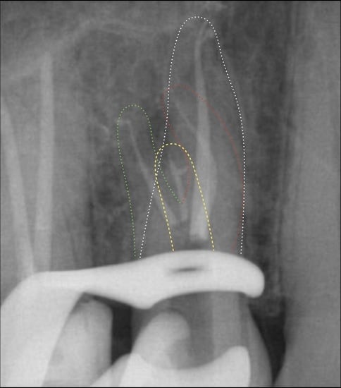
Maxillary Premolars with Four Canals: Case Series
Abstract
:1. Introduction
2. Materials and Methods
2.1. CASE 1
2.2. CASE 2
2.3. CASE 3
2.4. CASE 4
2.5. CASE 5
3. Discussion
4. Conclusions
Author Contributions
Funding
Institutional Review Board Statement
Informed Consent Statement
Data Availability Statement
Conflicts of Interest
References
- Ida, R.D.; Gutmann, J.L. Importance of anatomic variables in endodontic treatment outcomes: Case report. Endod. Dent. Traumatol. 1995, 11, 199–203. [Google Scholar] [CrossRef]
- Lin, L.M.; Pascon, E.A.; Skribner, J.; Gängler, P.; Langeland, K. Clinical, radiographic and histologic study of endodontic treatment failures. Oral Surg. Oral Med. Oral Pathol. 1991, 71, 603–611. [Google Scholar] [CrossRef] [PubMed]
- Ricucci, D.; Siqueira, J.F., Jr. Anatomic and microbiologic challenges to achieving success with endodontic treatment: A case report. J. Endod. 2008, 34, 1249–1254. [Google Scholar] [CrossRef] [PubMed]
- Carns, E.J.; Skidmore, A.E. Configurations and deviations of root canals maxillary first premolars. Oral Surg. Oral Med. Oral Pathol. 1973, 36, 880–886. [Google Scholar] [CrossRef] [PubMed]
- Pecora, J.D.; Sousa-Neto, M.D.; Saquy, P.C.; Woelfel, J.B. Root form and canal anatomy of maxillary first premolars. Braz. Dent. J. 1991, 2, 87–94. [Google Scholar]
- Atieh, M.A. Root and canal morphology of maxillary first premolars in a Saudi population. J. Contemp. Dent. Pract. 2008, 9, 46–53. [Google Scholar] [CrossRef] [PubMed] [Green Version]
- Neelakantan, P.; Subbarao, C.; Ahuia, R.; Subbarao, C.V. Root and canal morphology of Indian maxillary premolars by a modified root canal staining technique. Odontology 2011, 99, 18–21. [Google Scholar] [CrossRef] [PubMed]
- Tian, Y.Y.; Guo, B.; Zhang, R.; Yu, X.; Wang, H.; Hu, T.; Dummer, P.M.H. Root and canal morphology of maxillary first pre-molars in a Chinese subpopulation evaluated using cone-beam computed tomography. Int. Endod. J. 2012, 45, 996–1003. [Google Scholar] [CrossRef]
- Ahmad, I.A.; Alenezi, M.A. Root and Root Canal Morphology of Maxillary First Premolars: A Literature Review and Clinical Considerations. J. Endod. 2016, 42, 861–872. [Google Scholar] [CrossRef]
- De Almeida-Gomes, F.; de Sousa, B.C.; de Souza, F.D.; Dos Santos, R.A.; Maniglia-Ferreira, C. Unusual anatomy of maxillary second premolars. Eur. J. Dent. 2009, 3, 145–149. [Google Scholar] [CrossRef] [Green Version]
- De Almeida-Gomes, F.; De Sousa, B.C.; De Souza, F.D.; dos Santos, R.A.; Maniglia-Ferreira, C. Three root canals in the maxillary second premolar. Indian J. Dent. Res. 2009, 20, 241–242. [Google Scholar] [CrossRef] [PubMed]
- Barros, D.B.; Guerreiro-Tanomaru, J.M.; Tanomaru-Filho, M. Root canal treatment of three-rooted maxillary second premolars: Report of four cases. Aust. Endod. J. 2009, 35, 73–77. [Google Scholar] [CrossRef] [PubMed]
- Lea, C.; Deblinger, J.; Machado, R.; Nogueira Leal Silva, E.J.; Vansan, L.P. Maxillary premolar with 4 separate canals. J. Endod. 2014, 40, 591–593. [Google Scholar] [CrossRef] [PubMed]
- Allahem, Z.; AlYami, S. Treatment of maxillary second premolar with 4 roots. Case Rep. Dent. 2020, 2020, 8634797. [Google Scholar] [CrossRef]
- Yee, F.S.; Marlin, J.; Krakow, A.A.; Gron, P. Three-dimensional obturation of the root canal using injection-molded, thermoplasticized dental gutta-percha. J. Endod. 1977, 3, 168–174. [Google Scholar] [CrossRef]
- Messina, G. Strumentazione mista e tecnica delle conicità invertite nei canali curvi. Il Dent. Mod. 2021, 12, 70–76. [Google Scholar]
- Albuquerque, D.; Kottoor, J.; Hammo, M. Endodontic and clinical considerations in the management of variable anatomy in mandibular premolars: A literature review. Biomed Res. Int. 2014, 2014, 512574. [Google Scholar] [CrossRef]
- Kottoor, J.; Velmurugan, N.; Surendran, S. Endodontic management of a maxillary first molar with eight root canal systems evaluated using cone-beam computed tomography scanning: A case report. J. Endod. 2011, 37, 715–719. [Google Scholar] [CrossRef]
- Sberna, M.T.; Rizzo, G.; Zacchi, E.; Capparè, P.; Rubinacci, A. A preliminary study of the use of peripheral quantitative computed tomography for investigating root canal anatomy. Int. Endod. J. 2009, 42, 66–75. [Google Scholar] [CrossRef]
- Patel, S.; Brown, J.; Pimentel, T.; Kelly, R.D.; Abella, F.; Durack, C. Cone beam computed tomography in Endodontics—A review of the literature. Int. Endod. J. 2019, 52, 1138–1152. [Google Scholar] [CrossRef] [Green Version]
- Al-Fouzan, K.S. The microscopic diagnosis and treatment of a mandibular second premolar with four canals. Int. Endod. J. 2001, 34, 406–410. [Google Scholar] [CrossRef] [PubMed]
- Plotino, G.; Pameijer, C.H.; Grande, N.M.; Somma, F. Ultrasonics in endodontics: A review of the literature. J. Endod. 2007, 33, 81–95. [Google Scholar] [CrossRef] [PubMed]
- Das, S.; De Ida, A.; Das, S.; Nair, V.; Saha, N.; Chattopadhyay, S. Comparative evaluation of three different rotary instrumentation systems for removal of gutta-percha from root canal during endodontic retreatment: An in vitro study. J. Conserv. Dent. 2017, 20, 311–316. [Google Scholar] [PubMed]
- Heran, J.; Khalid, S.; Albaaj, F.; Tomson, P.L.; Camilleri, J. The single cone obturation technique with a modified warm filler. J. Dent. 2019, 89, 103181. [Google Scholar] [CrossRef] [PubMed]
- Sfeir, G.; Zogheib, C.; Patel, S.; Giraud, T.; Nagendrababu, V.; Bukiet, F. Calcium silicate- based root canal sealers: A narrative review and clinical perspectives. Materials 2021, 14, 3965. [Google Scholar] [CrossRef] [PubMed]
- Bugea, C.; Berton, F.; Rapani, A.; Di Lenarda, R.; Perinetti, G.; Pedullà, E.; Scarano, A.; Stacchi, C. In Vitro Qualitative Evaluation of Root-End Preparation Performed by Piezoelectric Instruments. Bioengineering 2022, 9, 103. [Google Scholar] [CrossRef]
- Ahmed, H.M.; Abbott, P.V. Accessory roots in maxillary molar teeth: A review and endodontic considerations. Aust. Dent. J. 2012, 57, 123–131. [Google Scholar] [CrossRef]
- Silva, E.J.N.L.; Nejaim, Y.; Silva, A.V.; Haiter-Neto, F.; Cohenca, N. Evaluation of root canal configuration of mandibular molars in a Brazilian population using cone beam computed tomography: An in vivo study. J. Endod. 2013, 39, 849–852. [Google Scholar] [CrossRef]
- Lucchese, A.; Gherlone, E.; Portelli, M.; Bertossi, D. Tooth Orthodontic Movement after Maxillofacial Surgery. Eur. J. Inflamm. 2012, 10, 227–232. [Google Scholar] [CrossRef] [Green Version]
- Ferrari Cagidiaco, E.; Carboncini, F.; Parrini, S.; Doldo, T.; Nagni, M.; Uti, N.; Ferrari, M. Functional Implant Prosthodontic Score of a one-year prospective study on three different connections for single-implant restorations. J. Osseointegr. 2018, 10, 130–135. [Google Scholar]
- Savignano, R.; Viecilli, R.F.; Oyoyo, U. Three-dimensional nonlinear prediction of tooth movement from the force system and root morphology. Angle Orthod. 2020, 90, 811–822. [Google Scholar] [CrossRef] [PubMed]
- Juodzbalys, G.; Stumbras, A.; Goyushov, S.; Duruel, O.; Tözüm, T.F. Morphological Classification of Extraction Sockets and Clinical Decision Tree for Socket Preservation/Augmentation after Tooth Extraction: A Systematic Review. J. Oral Maxillofac. Res. 2019, 10, e3. [Google Scholar] [CrossRef] [PubMed] [Green Version]


Publisher’s Note: MDPI stays neutral with regard to jurisdictional claims in published maps and institutional affiliations. |
© 2022 by the authors. Licensee MDPI, Basel, Switzerland. This article is an open access article distributed under the terms and conditions of the Creative Commons Attribution (CC BY) license (https://creativecommons.org/licenses/by/4.0/).
Share and Cite
Bugea, C.; Pontoriero, D.I.K.; Rosenberg, G.; Suardi, G.M.G.; Calabria, G.; Pedullà, E.; La Rosa, G.R.M.; Sforza, F.; Scarano, A.; Luongo, R.; et al. Maxillary Premolars with Four Canals: Case Series. Bioengineering 2022, 9, 757. https://doi.org/10.3390/bioengineering9120757
Bugea C, Pontoriero DIK, Rosenberg G, Suardi GMG, Calabria G, Pedullà E, La Rosa GRM, Sforza F, Scarano A, Luongo R, et al. Maxillary Premolars with Four Canals: Case Series. Bioengineering. 2022; 9(12):757. https://doi.org/10.3390/bioengineering9120757
Chicago/Turabian StyleBugea, Calogero, Denise Irene Karin Pontoriero, Gaia Rosenberg, Giacomo Mario Gerardo Suardi, Gianmarco Calabria, Eugenio Pedullà, Giusy Rita Maria La Rosa, Francesco Sforza, Antonio Scarano, Roberto Luongo, and et al. 2022. "Maxillary Premolars with Four Canals: Case Series" Bioengineering 9, no. 12: 757. https://doi.org/10.3390/bioengineering9120757
APA StyleBugea, C., Pontoriero, D. I. K., Rosenberg, G., Suardi, G. M. G., Calabria, G., Pedullà, E., La Rosa, G. R. M., Sforza, F., Scarano, A., Luongo, R., & Messina, G. (2022). Maxillary Premolars with Four Canals: Case Series. Bioengineering, 9(12), 757. https://doi.org/10.3390/bioengineering9120757


