1. Introduction
Gelatin is an affordable and widely accessible polymer due to its biodegradability, biocompatibility, and safety in terms of non-toxic byproducts produced upon enzymatic degradation. Gelatin contains motifs such as Arg-Gly-Asp (RGD), which exhibit cell adhesion, ensuring close contact and hence good biological activity as compared to other polymers [
1]. This adaptability of the gelatin structure has aroused interest to generate matrices viz. hydrogels, sponges, microparticles, microspheres, sheets, emulsion gels, and nano-carriers [
2]. With its good mechanical properties and capacity to provide a solid gelled texture, presently we propose to use it to develop vaginal suppositories. Gelatin is a protein that forms an interconnected, web-like matrix when dissolved in water and creates a structural network in water due to its (i) gelling and (ii) co-emulsifying properties. Not only does the gelatin give the water structure, but it can also suspend other dissolved and undissolved solids in its matrix viz. the probiotic cells. Herein, gelatin coupled with a suitable surfactant in water (aqueous phase) was used to emulsify an oil phase, into which probiotic cells were dispersed. The coupling of gelatin with water prior to the addition of the probiotic dispersed in oil helps to reduce the amount of reactive water. The latter, combined with the coating of the probiotic with oil prior to incorporation into the gelatin solution, doubly protects against germination of the probiotic during storage; the oil coat also maintains probiotic viability. Vulvovaginal candidiasis (VVC) involves inflammation of the vaginal and vulvar epithelium mucosa caused by underlying fungal infection, most commonly with
Candida albicans and
C. glabrata [
3]. Opportunistic Candida species invade the mucous membrane of the vagina, leading to exuberant mucosal inflammatory responses [
3]. Almost 50% of adult women report having experienced at least one episode of VVC when young [
4].About 1.4 million outpatient visits per year are reported for vaginal candidiasis, only in the US [
5], while the actual number of cases may be much higher, and is not known. Significant drug resistance and VVC recurrence (RVVC) is reported, with conventional antifungal treatments [
6] available as vaginal creams, ointments, ovules, and suppositories. In the case of recurrence, physicians may prescribe one to three antifungal drugs to be taken by mouth from two weeks to six months. Fluconazole is the most common drug of choice for oral treatment. Its use is, however, avoided or closely monitored in cases of women taking statins, having kidney disease, or being at high risk for arrhythmias. It is also reported to significantly increase the risk of miscarriage in pregnant women. Topical treatment with a cream or suppository has an advantage of faster onset and freedom from systemic toxicity; however, local irritation is reported with some topical products. Data suggest that recurrent vulvovaginal candidiasis occurs in 6–10% of women [
7].There is an expert consensus on topical maintenance therapy with an azole antifungal, or boric acid, at one to three times weekly, following a full-course therapy [
8].
Probiotics inhibit pathogenic Candida species and maintain and modulate microbiota profiles in the vagina [
6,
9,
10,
11]. Further, they also lower vaginal pH and downregulate inflammatory mediators such as nuclear factor-kappa B (NF-κB), and proinflammatory chemokines and cytokines such as interleukins (ILs) and tumor necrosis factor-alpha (TNF-α)) [
11,
12,
13,
14,
15]. Probiotics specifically inhibit mitogen-activated protein kinase (MAPK) and pattern recognition receptor (PRR) pathways [
16]. The pathways are responsible for the transcription of NF-κB and a range of proinflammatory cytokines, chemokines and other inflammatory mediators [
17]. Probiotics are also reported to inhibit Ikb-βα and NF-κβ inhibitor degradation, which ultimately reduces the binding of NF-κB to DNA [
18,
19].
Delivery of the probiotic through gut to alter vaginal health has previously been reported by many research groups [
20,
21]. However, the direct delivery of good microbiota to vaginal cells can restore/transplant the host’s indigenous microflora with healthy bacteria and prevent repeat pathogen onslaught. Further, low enzymatic activity in the vagina will ensure that the administered probiotics do not experience harsh conditions in contrast to those observed in the stomach, which can compromise their viability [
22]. The manipulation of vaginal microflora by direct administration has thus surfaced recently in a few studies [
23,
24]. An important consideration with respect to effective probiotic delivery, however, is that the carrier system must maintain the viability and stability of the loaded probiotic, both upon administration and during manufacture and storage. Thus, there are only limited studies on products such as suppositories incorporating whole-cell probiotics for local vaginal delivery. This, according to us, is attributed to manufacturing and storage constraints, a lack of probiotic cell germination in vaginal mucosa post-application, leakiness and limited stay, and the expulsion of products from the vaginal cavity.
Gelatin and glycerol–gelatin vaginal suppositories are extremely popular, attributed to their ease of use and soothing effect, and provide prolonged and complete release of the active load owing to their slow dispersion in mucosal secretions [
25,
26]. Further, the process involved in the manufacturing of suppositories is simple and scalable without significantly compromising probiotic viability [
27]. Stability during storage can be maintained by keeping water activity of the developed system low. When dissolved in hot water, proteins of the gelatin unravel into long strings that interlace when cooled, to create a three-dimensional structure that can trap a lot of liquid. Thus, the developed gelatin–oil-based carrier system was prepared to have low water activity to avoid probiotic multiplication and formulation spoilage during storage [
28].
The preservation of the developed system against contaminating organism is another concern with gelatin-based products, as the latter can support the growth of some organisms. Low water activity does not support the growth of such organisms, and thus there is no need to add a preservative into the suppositories. This is of significance for a probiotic product, because the incorporated preservative can affect probiotic viability [
28,
29].
In the present study, we thus endeavored to develop a suppository system with an intelligent choice of ingredients containing a self-emulsifying gel base, oil, surfactant, and a soothing agent, which keeps the incorporated probiotic viable, prevents any contamination of the product during storage, aids in the germination of the incorporated probiotic upon application, is acceptable for use and helps maintain/transplant healthy flora in the vaginal cavity for vaginal health.
Bacillus coagulans Unique IS-2, a nonpathogenic, Gram-positive, spore-forming bacteria, was loaded into the above-described oil–gel suppository base. The selected strain of bacteria has reported clinical efficacy against bacterial vaginitis [
30]. The developed suppositories were characterized for various pharmacopeial and non-pharmacopeial tests. The suitability of the suppository composition for probiotic delivery was ensured by evaluating its water activity, ex vivo bioadhesion and retention in porcine vaginal mucosa. The anti-candida potential of the probiotic-loaded suppository in terms of inhibition and filament disruption was recorded in vitro. The in vivo performance of the developed suppositories was evaluated in terms of probiotic germination, bioadhesion and retention in the rat vaginal cavity, and the anti-infective effect in comparison to a marketed product (Candid V
® gel) in VVC rat model. The developed probiotic–oil–gelatin suppositories were evaluated for stability (2–8 °C for 3 month) and in vivo 15-day safety studies.
2. Results and Discussion
As per the official guidelines, there should be uniformity in weight as well as content of the suppositories to elicit an invariable therapeutic effect. The average weight of the developed suppositories was 1.39 g (
Supplementary Figure S1). Since only two suppositories differed from the average weight by more than 5% and no suppository differed from the average weight by more than 10%, the formulated suppositories complied with the standards described in the Indian Pharmacopeia (2014) for suppositories. The total probiotic content loaded into the suppositories was found to be 23 ± 2.48 × 10
8 cfu per suppository. The content uniformity evaluation of the developed gelatin–oil suppositories indicated that the distribution of colony forming units of
Bacillus coagulans was uniform in the apex and bottom part of the suppository; however, it was considerably low in the middle part (
Table 1), indicating that the probiotic needs to be mixed properly after incorporation into the base. The cell count of the used
Bacillus coagulans powder was confirmed by the pour plate method (
n = 8) as per the procedure given in the
Supplementary Materials. The mean viable count was found to be 21.54 ± 4.51 × 10
6 cfu/mg. The cell surface hydrophobicity of
Bacillus coagulans was 30.4 ± 5.34% (
Supplementary Table S1); % Hydrophobicity index (% HPBI) is an indicator of adhesive capabilities and a value < 50% is considered low. The results indicated that
Bacillus coagulans spores were invariably less hydrophobic than their germinated counterparts. The incorporation into the gelatin–oil base thus imparted it adhesiveness until the time the spores germinated.
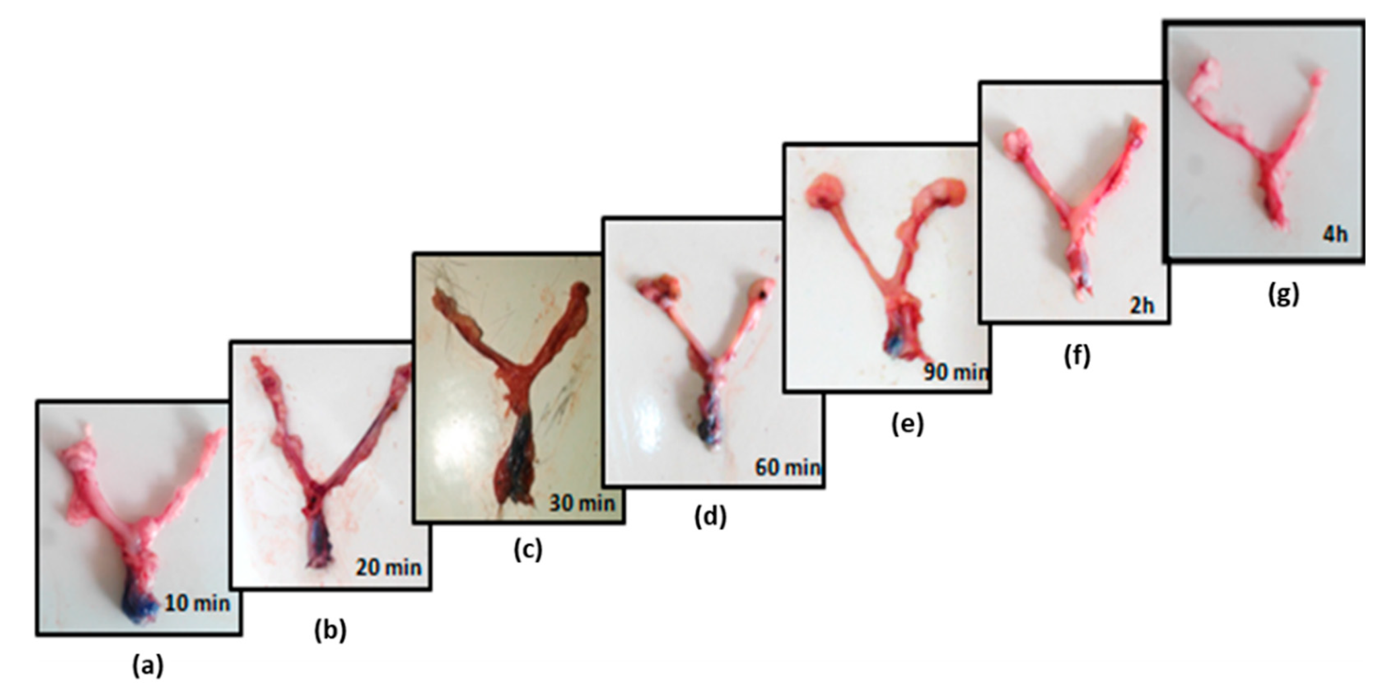
The hardness was found to be 1.43 ± 0.175 kg. Gelatin–oil suppositories, due to their elastic nature, are not too firm. It was observed that the suppositories started melting after 10 min (
Supplementary Figure S2a) of being placed in the test beaker at 37 °C, and completely melted within 2 h (
Supplementary Figure S2b). It was confirmed that the prepared suppositories had a significant capacity to swell, almost to double their initial weight (at 6 h) (
Table 2;
Figure 1). The swelling behavior of the suppositories was studied at room temperature (25 °C) and the results indicated erosion and dissolution at times after 6 h. However, upon maintaining at 37 °C, fast dissolution within 2 h was recorded. The suppositories completely disintegrated within 2 min. This complies with the Indian Pharmacopeial limits, according to which the water-soluble suppositories should dissolve within 30 min.
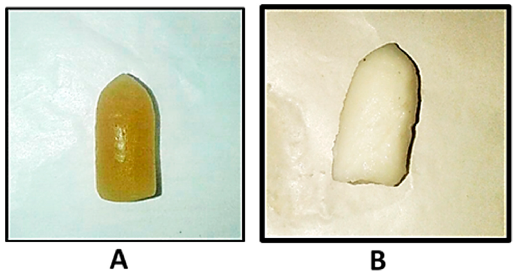
Optical images taken at 10 and 100× for blank and probiotic-loaded suppositories (
Figure 2) showed a woven/honeycomb network with air voids.
Figure 2C shows the presence of probiotic spores enmeshed as small brown clusters. Images taken at 100× depict the gel network more clearly with
Figure 2D distinctly illustrating the presence of spores within an oil globule.
Probiotic-loaded suppositories when incubated in 2 mL SVF at 37 ± 0.5 °C resulted in the complete release of probiotics within 45 min, which is well in the limits defined by I.P., 2014 (not more than 60 min) (
Figure 3).
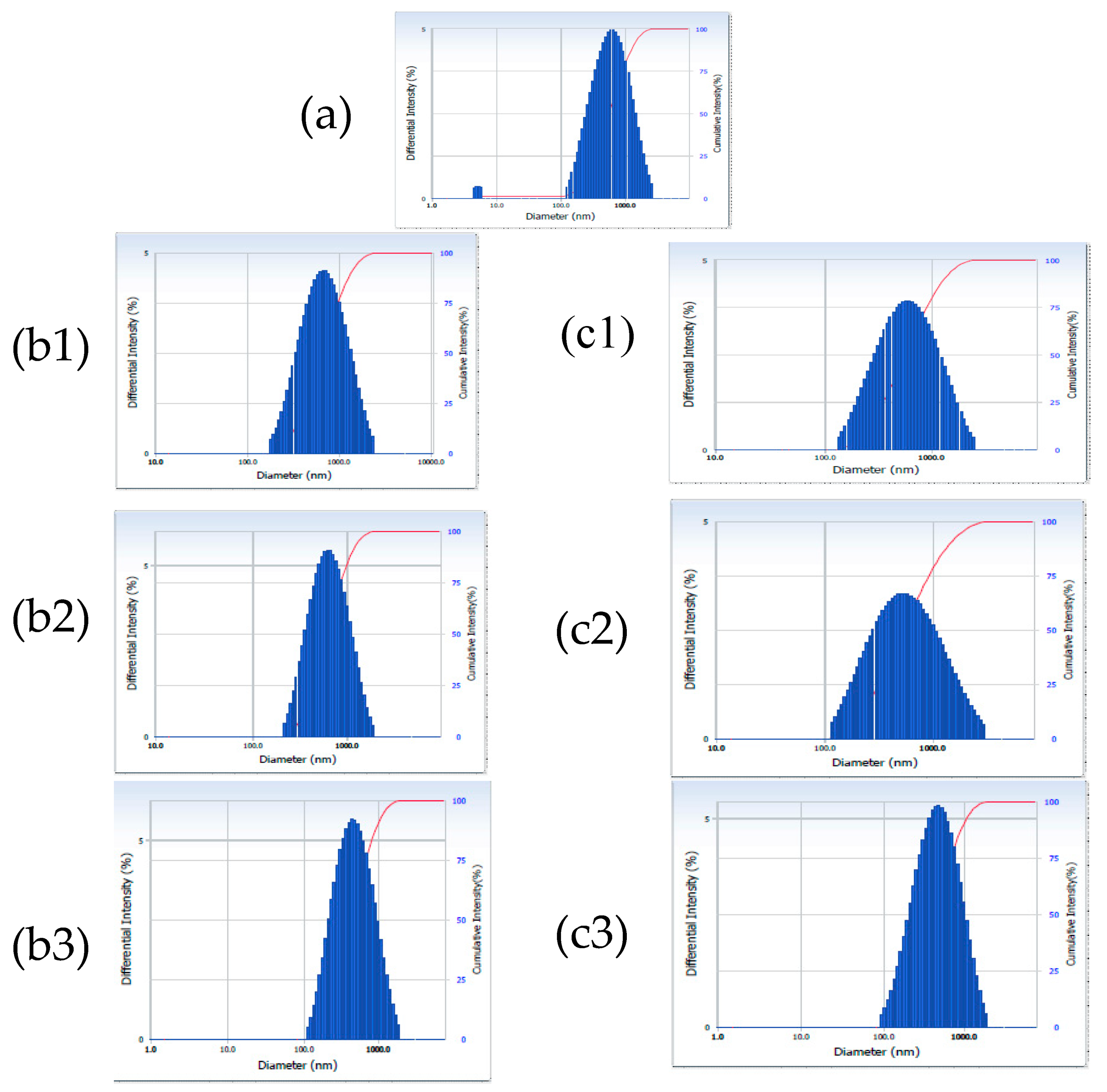
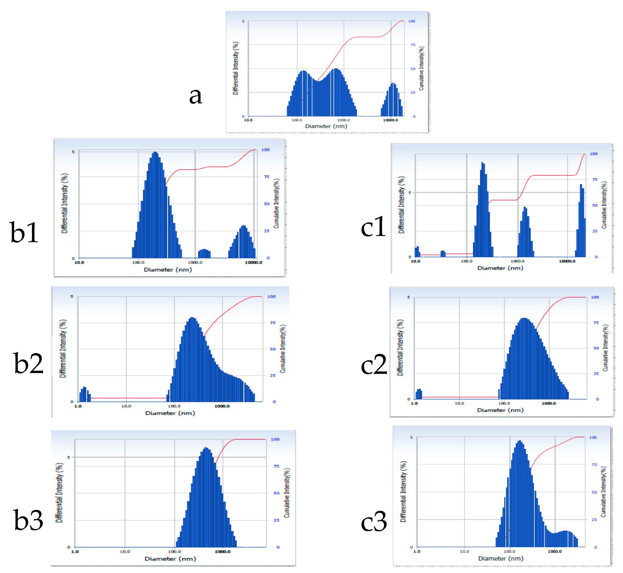
The size and size distribution of both the blank and gelatin–oil–probiotic suppositories were determined after dispersion in water. At ambient temperature, the average particle size of the blank suppository was found to be 493.1 nm. The distribution curve for the same (
Figure 4a) showed a maximum intensity at 800.0 nm and a very small peak observable at <10 nm, which probably shifted the average size to 493.1 nm. The probiotic-loaded suppository recorded an average particle size of 754 nm (
Table 3) and a trimodal distribution (
Figure 5a). The first peak in case of the probiotic-loaded suppository appeared at 160 nm and may be due to fine oil globules emulsified in the aqueous gelatin base; peak 2 at 753.2 nm may be attributed to gelatin particles; while peak 3 observed at >11,000 nm could be due to the free probiotic or its aggregates in oil globules. Gelatin is known to undergo dissolution upon extended hydration; therefore, studies were continued for 24 h post-dispersion of the suppositories in water at 25 °C (room temperature) and 37 °C (body temperature). The average particle size of the blank formulation remained stable, at both temperatures, for up to 6 h, with a slight reduction at 24 h, attributed to solubility or the erosion of gelatin particles with time. The size distribution curves for probiotic suppository suggested that with passing time the distribution pattern moved from trimodal to unimodal (
Figure 5), thus matching that of the blank suppository (
Table 3;
Figure 4). This could be due to disaggregation of the probiotic with time, attributed to its hydration.
The gelatin–oil suppositories prepared here are expected to release probiotic both by melting and dissolution. Small probiotic-loaded oil particles obtained upon the dissolution of the prepared suppositories will not only cover a larger area but will also achieve an intimate contact with the vaginal mucosa [
1]. This will help in the establishment of the probiotic on the mucosal wall.
Water activity (a
w) is defined as a measure of the energy status of the water present in a sample referred to as free, unbound, or active water. It is different from total water content, as a portion of the total water contained in a sample is at times strongly bound to specific sites of various components in a sample, and hence may not affect the preparation. Active water, on the other hand, influences the shelf life of products as it makes the preparation liable to microbial growth [
17,
18]. While temperature, pH, and several other factors may also influence the growth of an organism in a product, the rate at which they grow is often monitored by water activity. The lowest a
w at which most bacteria will grow in a product is about 0.90. The a
w for mold and yeast growth is approximately 0.61 a
w with the lower limit for the growth of mycotoxigenic mold being 0.78 a
w [
19]. The water activity of the freshly prepared suppository was 0.531 a
w, whereas during storage for two months (2–8 °C) it increased to 0.593 a
w. Although a marginal increase in water activity was recorded for the developed suppositories, the value was still lower than the reported range (0.61–0.90), favoring microbial (both bacterial and fungal) growth both of the probiotic, or the contaminating organism. Thus, it may be said that the developed gelatin–oil suppository will be non-reactive, or in other words, will keep the probiotic dormant, i.e., without germination (as confirmed in stability studies). Furthermore, suppositories per se will be endowed with a self-preservation against any harmful bacterial growth during storage and use. It may be noted that the suppository base used here was not a simple gelatin or a glycerol–gelatin base; instead, it was a gelatin base incorporating probiotic-laden small oil globules.
SEM pictures of the cross-sectioned suppository confirmed the gel matrix (
Figure 6A) interspersed with oil globules (
Figure 6B).
The probiotic spores incorporated into the developed formulation were found to maintain their viability (
p < 0.05) after storage under refrigerated conditions for three months, as shown in
Table 4. Obtaining similar values for samples with and without heat shock indicated that the spores did not undergo any germination in the formulation. This is important, as otherwise the cell population in the formulation will keep changing and the growth of vegetative cells may produce materials that foul the suppository, making it aesthetically unpleasant or unfavorable for use.
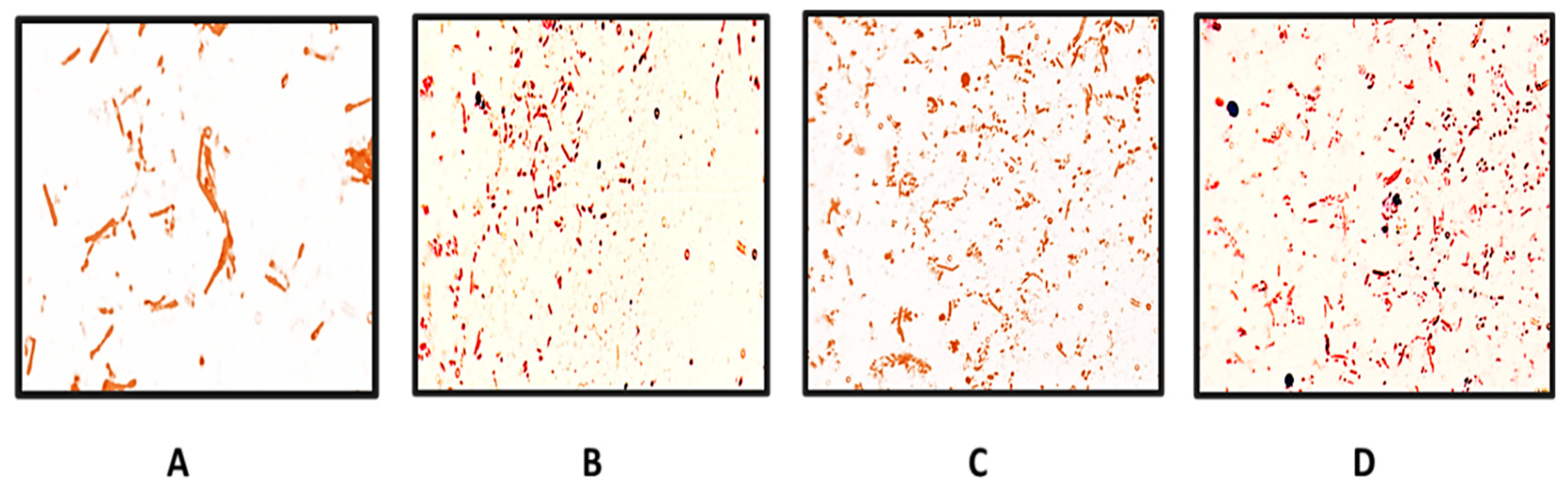
Bacillus coagulans usually exists as spores, and since the latter are resistant and stable, it was considered appropriate to incorporate and maintain them as spores in the developed formulation. However, to elicit the protective physiological effect, it is important that they grow actively upon insertion into the vaginal cavity. The in vivo germination of the probiotic following the application of the spore-loaded suppository upon insertion into the vagina was established by the swabs collected from the rat’s vagina. The presence of vegetative cells at different time intervals, i.e., 2 h, 4 h, 6 h, and 24 h, were observable on the slides and are suitably labeled in the
Figure 7. Spore germination was evidenced from 4 h onwards (
Figure 7C) in the samples and abundance was observed, with most of the spores showing germination in the 24 h sample (
Figure 7D).
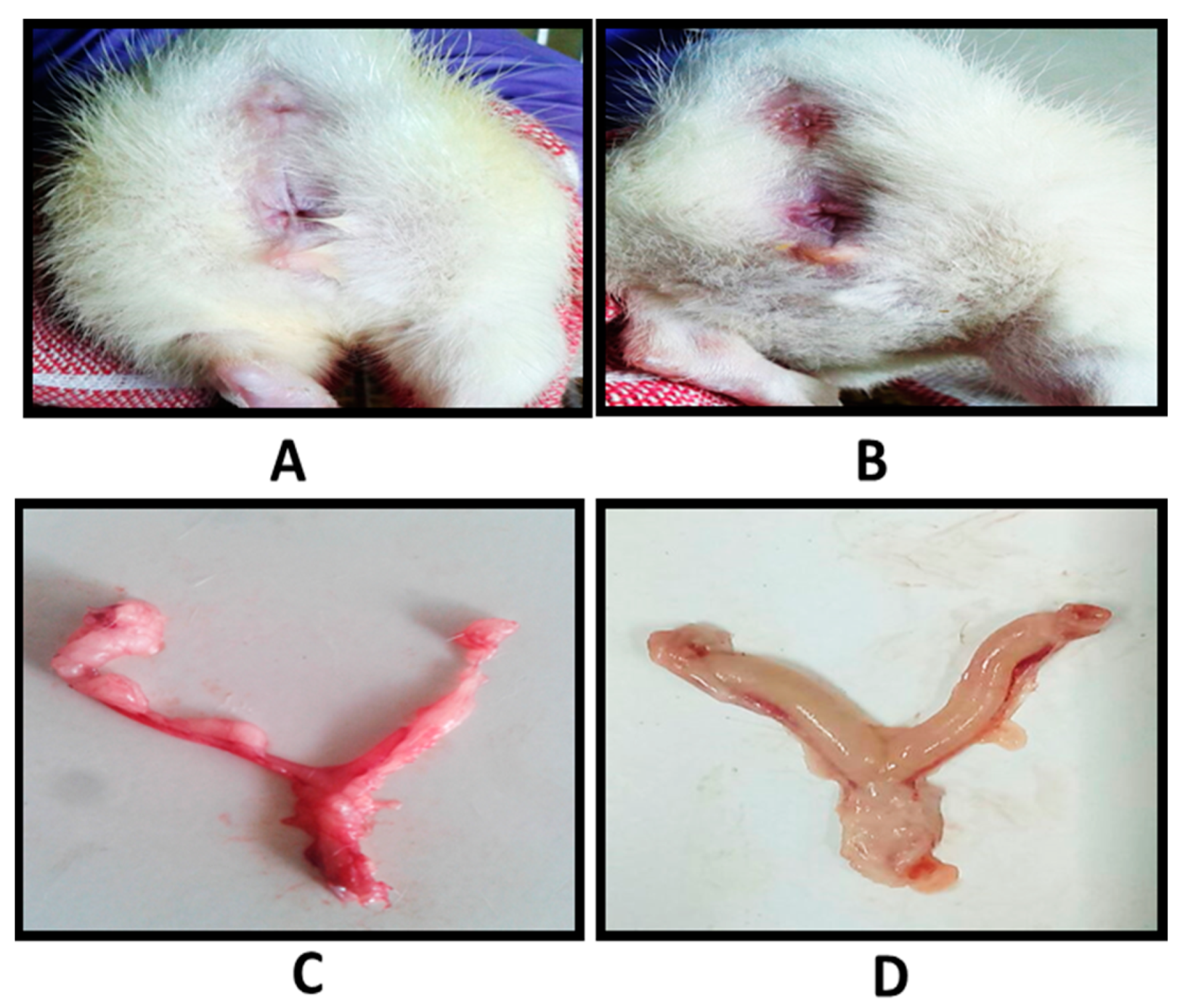
In vivo retention studies indicated that the suppository was intact and maintained its shape even at 10 min after its insertion into the vaginal cavity of the rats (
Figure 8). Complete melting of the vaginal suppository was recorded at the end of 30 min; however, no leakiness was observed through the vaginal opening of the rat (
Table 5), indicating a successful formulation. The reasons for this are:
- (1)
Swelling of gelatin suppositories helps them to snugly fit in the reproductive tract.
- (2)
Small amount of vaginal fluid is insufficient to initiate the dissolution process, such that the base melts at body temperature to a viscous gel-like consistency, as also observed in the in vitro study. Since the consistency of the melted suppository is high, it stays within the tract and does not leak out.
- (3)
Furthermore, the release of probiotic-loaded oil particles coated with viscous gel cover a larger vaginal area, with vaginal contractions further helping in a widespread distribution.
The vaginal mucosa is highly sensitive to the exposed chemicals and therapeutic agents that may result in irritation and/or inflammation and can make women more susceptible to various infections. Hence, the vaginal irritation potential of feminine care formulations and vaginally administered therapeutic agents is a significant public health concern. The pH of the developed suppository was found to be 4.3 ± 1.0. The pH is very near the normal vaginal pH, thus the suppositories are expected to be non-irritating to the vaginal mucosa. An in vivo vaginal irritation study was performed in rats to evaluate the safety of the vaginal formulations on their multilayered vaginal epithelium. The latter is a characteristic feature of mammalian vagina, too.
The results from the histopathological examination (
Figure 9) showed that the formulation was well-tolerated by the rat vagina and there were no signs of erythema or edema after 14 days of vaginal administration of the prepared suppositories. It was evident from the images that the epithelial membrane was intact, and no signs of vascular congestion/leucocyte infiltration or edema were observed in the treated groups. Further, the lower cervix, uterus and epithelial layers showed normal histology.
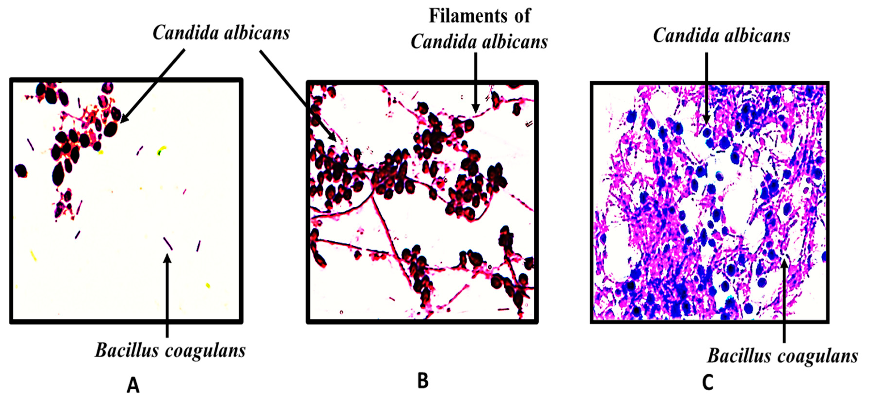
Bacillus coagulans spores are reported to form co-aggregates with
Candida albicans, and the same was also observed in the present study (
Figure 10A). The probiotic spores co-aggregated with the filaments of
Candida albicans. Visibly better co-aggregation of probiotics from suppository was observed versus free form. Only candida aggregates were observed after incubation with the blank suppository.
The in vivo efficacy of the developed formulation was evaluated in a VVC rat model. The establishment of a pseudo-estrous state was confirmed by observing epithelial cells in the vaginal lavage under an optical microscope (
Supplementary Figure S3). It was observed that well-formed, nucleated epithelial cells of the pro-estrous stage was replaced by cornified, squamous, densely packed epithelial cells after hormonal treatment. The latter signified the establishment of an estrous phase, after which standardized yeast suspension was introduced into the vagina. Once the Candida infection was confirmed in the animals, scheduled treatments (marketed formulation (Candid V
® gel), free probiotic suspension, probiotic-loaded suppositories, and blank suppositories) were initiated in the infected rats and the fungal burden was monitored every day for seven days. Macroscopic observation (
Figure 11) indicated redness of the vaginal opening and inflammation of the reproductive tract of animals infected with
Candida albicans. Correspondingly, probiotic-loaded-vaginal-suppository-treated animals showed significant improvement during the experimental period.
The probiotic-loaded suppositories did not show any inhibition by either well diffusion or the streak method. It may be concluded that the vegetative probiotic cells do not inhibit Candida albicans in vitro probably because (a) the inhibition follows mechanisms other than the release of bacteriocins/inhibitory substances in the surrounding media; (b) factors such as the immune responses of the host, changes in the immune system induced by C. albicans enzymes or metabolites present in the vaginal lumen cannot be mimicked in the in vitro tests, although they do exert a great influence on the effects demonstrated by probiotics in vivo.
Fungal burden studies (
Figure 12) indicated that the positive control (infected) group was not significantly different from the blank-suppository-treated infected group, indicating that the suppository base per se does not elicit any physiological effect. Further, in the initial phase of treatment, no significant difference (
p < 0.001) was observed between the marketed formulation and the free-probiotic-treated group, whereas the probiotic-loaded suppository group showed significantly (
p < 0.001) better results. However, on the 7th day of treatment, both the free probiotic group and probiotic-loaded suppository group showed significantly better effects than the marketed formulation. The free probiotic dispersion showed significant effects; however, it tended to leak out following application, clearly concluding that its incorporation into a suppository base, designed herein, is a successful dosage form.
Furthermore, as is expected of azole antifungals, which are fungistatic in nature, the marketed formulation containing clotrimazole did not show any significant decrease in fungal loads from day four onwards, while the decrease in fungal loads continued with both the probiotic-treated groups. The cfu/animal for the positive control group and blank suppository group did not show any significant inter-day difference, while for the free probiotic group and probiotic suppository group, a significant difference was observed. In the marketed formulation, no significant difference was observed between the 1st and 2nd day and 4th, 5th, and 6th days (
Figure 12). What is interesting to note is the fact that although
B. coagulans spores and vegetative cells did not show any activity against Candida in vitro, the effects obtained in vivo establish the usefulness of both the probiotic per se and the developed gelatin–oil suppository formulation. Thus, it may be said that the observed in vivo effects are mediated by mechanisms such as the competitive exclusion of adherence to epithelial cells by Candida, co-aggregation and auto-aggregation leading to the inhibition of Candida, and the modulation of both the immunological reactions initiated by Candida and the local immunological host response.
The in vivo translocation study indicated that only 0.007 ± 0.001% of 122 × 106 spores, administered per day for seven days, translocated to reach systemic circulation when inserted into the infected animals. This value is clearly insignificant, and it was confirmed that the presently used strain was safe for vaginal administration in infected animals. It confirms that the action of the developed probiotic-loaded vaginal suppositories remains localized.
3. Conclusions
In the present study, we developed probiotic-loaded gelatin–oil biphasic suppositories for combating vaginal fungal infections. The formulation was tested in an animal model of vulvovaginal candidiasis where it showed effects comparable to a marketed product (Candid V® gel). Unlike the available vaginal probiotic products in the market that contain either extracellular substances or bioactives obtained from probiotics, the currently presented formulation consists of whole-cell probiotics.
The probiotic-loaded vaginal gelatin suppositories described here addressed the stability and viability issues associated with the incorporation of live probiotic cells into the delivery system, not only during production but also storage. The gelatin, due to its intrinsic nature, can keep reactive water to a minimum to avoid the germination of Bacillus coagulans spores to their active vegetative form during storage.
Confirmatory proof of the germination of probiotic spores into vegetative form upon insertion of the suppository into the rat vagina was demonstrated in the study. Not only the viability, but the germination of the probiotic spores on the application site is an important consideration because the purported therapeutic effects such as the release of various enzymes, bacteriocins and metabolites (bioactives) are demonstrated by these strains in their active state.
The developed gelatin–oil suppositories were safe in terms of pH, vaginal irritation, and translocation/permeation. High patient acceptability is expected, as indicated by in the vivo retention study suggesting no leakiness.
The in vivo therapeutic effects of the probiotic-loaded suppositories were confirmed in terms of the decrease in the cfu of Candida in infected rats. The developed probiotic-loaded suppositories can also incorporate other probiotic cells, including their vegetative forms, with equal success, as they have the capability of keeping the loaded probiotic in its dormant state during storage. Furthermore, the components and equipment used in this study are cheap and widely available, which makes it an industrially viable option.


 and
and 


