Article

Microstructure and Properties of Thin-Film Submicrostructures Obtained by Rapid Thermal Treatment of Nickel Films on Silicon

by
Vasilina Lapitskaya
1,2,*,
Ruslan Trukhan
1,
Tatyana Kuznetsova
1,2,†,
Jaroslav Solovjov
3,
Sergei Chizhik
1,2,
Vladimir Pilipenko
3,
Karyna Liutsko
2,
Anastasiya Nasevich
2 and
Maksim Douhal
2
1
Laboratory of Nanoprocesses and Technology, A.V. Luikov Heat and Mass Exchange Institute of the National Academy of Sciences of Belarus, 220072 Minsk, Belarus
2
Department of Micro- and Nanotechnology, Faculty of Instrumentation, Belarusian National Technical University, 220013 Minsk, Belarus
3
JSC “INTEGRAL”—“INTEGRAL” Holding Managing Company, 121 A Kazintsa, 220108 Minsk, Belarus
*
Author to whom correspondence should be addressed.
Deceased author.
Surfaces 2024, 7(2), 196-207; https://doi.org/10.3390/surfaces7020013
Submission received: 17 February 2024 / Revised: 12 March 2024 / Accepted: 20 March 2024 / Published: 27 March 2024

Abstract

:
Nickel films of 40 nm thickness were obtained by means of magnetron sputtering on a single-crystalline silicon substrate. The films were subjected to rapid thermal treatment (RTT) for 7 s until the temperature increased from 200 to 550 °C. By means of the X-ray diffraction method, the structural-phase composition of nickel films before and after RTT was explored. The atomic force microscopy method due to direct contact with the surface under study, made it possible to accurately define the microstructure, roughness, specific surface energy and grain size of the nickel films before and after RTT, as well as to establish the relationship of these parameters with the phase composition and electrical properties of the films. Surface specific resistance was measured using the four-probe method. Based on XRD results, formation of Ni2Si and NiSi phases in the film was ascertained after RTT at 300 °C. At RTT 350–550 °C, only the NiSi phase was formed in the film. The microstructure and grain size significantly depend on the phase composition of the films. A correlation has been established between specific surface energy and resistivity with the average grain size after RTT at 350–550 °C, which is associated with the formation and constant restructuring of the crystal structure of the NiSi phase.

Graphical Abstract

1. Introduction

The good electrical and magnetic properties of nickel, as well as resistance to corrosion and oxidation, allow it to be used in electronics, optics and healthcare [1,2]. The nickel layers are used as orienting layers for growing graphene and copper films [2]. Thin nickel films are used as ohmic contacts [3] in the solar-to-thermal energy converters [1], as well as for electromagnetic radiation shielding [4].
The nickel silicide films hold a particular place in microelectronics. Low specific and contact resistance, and low silicon consumption and formation temperature [5,6,7] have opened up the possibility of using nickel silicide (Ni2Si, NiSi) in CMOS technology as self-aligned silicide contacts in the source, drain and gate regions [6,7,8,9].
Formation of nickel silicide thin films is mainly carried out by a solid-phase reaction of a nickel film with silicon. Nickel is applied by physical vapor deposition [5,6,7,9,10,11,12,13,14,15,16,17] or chemical deposition [8,18] onto a previously prepared silicon wafer. After this, the Ni/Si system is subjected to heat treatment, during which the phases Ni2Si, NiSi and NiSi2 are sequentially formed [8]. The resulting silicides differ in their formation temperatures and the value of their surface-specific resistance [5,6]. Formation of the Ni2Si phase occurs within a temperature range from 175 °C to 300 °C [5,10,14,15,17,18,19]. According to [5,9,15], the transition from the Ni2Si phase to NiSi occurs at temperatures of 275–350 °C, and at a temperature of 700 °C [6,8,14,18], the NiSi2 phase is formed. Heat treatment is carried out by steady-state annealing in a vacuum [5,7,10,11,12], in an atmosphere of argon [8] and nitrogen [9], and rapid thermal annealing (RTA) in an atmosphere of argon [16] and nitrogen [6,7,9,11,13,14,15,17,18]. RTT with lamp heating makes it possible to form nickel silicides within a time frame of 5 to 7 s [9,15,17], while steady-state annealing takes at least 30 min [5,7,8,9,10,11,12].
The formation of silicides is accompanied by changes in the surface roughness [6,11,19] due to an increase in grain size, their integration and agglomeration [6,11,14]. Grain size is one of the factors that has an impact on the resistivity of nickel silicides. The smaller the grain size in NiSi films, the higher the resistivity [14], which is associated with a large number of grain boundaries. To define the grain size, X-ray diffraction (XRD) methods [14,20,21] and electron microscopy [17,22] are used more often than probe methods [6,22].
Atomic force microscopy (AFM) is used more often to monitor changes in surface morphology, roughness [4,19,21,22,23,24,25] and changes in grain size [4,21,22,25] when studying the nickel films on silicon (after annealing). AFM images are used to describe the smoothness [4,22], homogeneity and continuity [4] of the formed films. AFM is also used to verify the process of the film agglomeration during the transition from NiSi to NiSi2 at temperatures from 500 °C when studying the structure, phase composition and resistivity [21]. In this case, attention is focused on changes in morphology [19], the formation of a needle-like [23] or columnar [25] structure and deep depressions [23,25] and an increase in roughness (which is only possible using atomic force microscopy) of the films [4,19,21]. Also, while defining the influence of doping elements on the thermal stability of NiSi films, the roughness values and film morphology identified by the AFM method make it possible to estimate either the improvement or deterioration of its stability. Establishing the relationship and correlation of roughness, specific surface energy, grain size and other properties of nickel films after rapid thermal treatment is quite a challenge now. Probe methods, due to direct contact with the surface under study, make it possible to accurately define roughness, specific surface energy and grain size, which can significantly affect the material properties.
The purpose of this work was to study the impact produced by rapid thermal treatment within a temperature range of 200 to 550 °C on the structural phase composition and properties of nickel films on silicon by means of high-precision probe methods.

2. Materials and Methods

2.1. Application and Processing of Nickel Films

Nickel films of ~40 nm thickness were deposited on silicon substrates by magnetron sputtering of a nickel target with 99.5% purity in an argon environment with 99.993% purity at a pressure of 0.35 Pa and a discharge power of 7.1 kW (the power density was about 8.15 W/cm2 at a discharge voltage of 480 V) at the SNT “Sigma” installation (StratNanoTek Invest, Minsk, Belarus). The vacuum chamber was pumped out to less than 5 × 10⁻⁴ Pa (oil-free pumping) before deposition of nickel films.
The silicon substrates represented epitaxial layers of phosphorus-doped silicon with a resistivity of 0.58–0.63 Ω × cm and a thickness of 5.3–5.8 μm, formed on p-type single-crystalline silicon substrates with 10 Ω × cm resistivity and orientation (111). Prior to nickel deposition, the substrates were treated first in an ammonia peroxide solution and then in an aqueous HF solution.
After deposition, nickel films on silicon were subjected to rapid thermal treatment in thermal balance mode using a JetFirst 100 unit (Jipelec Qualiflow Therm, Montpellier, France). The annealing chamber was pumped out twice to 1 Pa, followed by nitrogen injection to atmospheric pressure (oil-free pumping) prior to rapid thermal treatment. The reverse side of the silicon substrates was irradiated with an incoherent light flux from constant power quartz halogen lamps in a nitrogen environment for 7 s. Heating was carried out until the temperature increased from 200 to 550 °C with 50 °C increments. The temperature of the working side of the substrate was controlled by a thermocouple with an accuracy of ±0.5 °C [17].

2.2. Research Methods

The phase composition was studied by X-ray diffraction using ULTIMA IV diffractometer (Rigaku, Tokyo, Japan). A parallel beam geometry was used in copper (CuKα) radiation with a wavelength of 0.154179 nm. The thickness (t) of the films was determined by scanning electron microscopy (SEM) with an S-4800 instrument (Hitachi, Tokyo, Japan). To define the thickness of the films, a vertical shearing cut of silicon wafer with a film was made, and an SEM image was obtained (Figure 1).
The study of the structure, roughness and adhesion force of the film surface before and after rapid thermal treatment was carried out using an atomic force microscope Dimension FastScan (Bruker, Santa Barbara, CA, USA) in PeakForce QNM mode (Quantitative Nanoscale Mechanical Mapping, Bruker, Santa Barbara, CA, USA). Standard silicon cantilevers of the CSG10_SS type (TipsNano, Moscow, Russia) with a curvature radius of 3.2 nm and a cantilever stiffness of 0.26 N/m were used. The film roughness parameters Ra, Rq, Rz and grain size were determined on fields of 1 × 1 μm2. Ra is the arithmetic mean of the absolute values of the surface height deviations measured from the average plane, Rz is the average difference in height between the highest peaks and valleys relative to the average plane, and Rq is the root mean square value of the profile deviations within the base length of the profile.
Based on the surface adhesion force values [26], the specific surface energy γ was determined [27]:
γ = F ad 2 π R ,
where Fad is the surface adhesion force, N; R is the radius of curvature of the probe tip, m. The adhesion force Fad was determined from the force required to break the “AFM probe–surface” contact when the AFM probe was removed from the surface of the film under study.
The specific electrical resistivity of the films was determined using an RS-30 apparatus (KLA Tencor, Milpitas, CA, USA). The error is no more than ±5%. Calculation of the specific electrical resistivity ρ (Ω × cm) of nickel films was carried out according to the formula:
ρ = R s t ,
where RS is the surface resistance of the sample, Ω/square; t is the thickness of the nickel film on silicon after heat treatment, meter.
The rapid thermal treatment temperature was a criterion for correlation analysis [27]. The relationship between two parameters (roughness, specific surface energy, grain size and specific electrical resistivity) with respect to the third constant feature—rapid thermal treatment temperature—was found. The correlation coefficient of parameters x and y was found by the formula [27]:
r x y = x i x ¯ · y i y ¯ x i x ¯ 2 · y i y ¯ 2
where y corresponded to specific electrical resistivity at a change in rapid thermal treatment temperature, and x alternately took the values of roughness, specific surface energy and grain size.

3. Results and Discussion

XRD displayed the presence of crystalline nickel with orientation (111), (200) and (220) on the nickel film before RTT (Figure 2). At a RTT temperature of 200–250 °C, the intensity of the nickel peaks changed, but no new phases were formed. The intensity of the peak (220) slightly increased (Figure 2c) at a temperature of 250 °C. After rapid thermal treatment at 300 °C, mainly the NiSi phase is formed in the film (Figure 2c). The peak intensity of the NiSi phase corresponds to orientation at an angle of 45.5° in the film after 300 °C RTT, belonging to the Ni2Si phase, i.e., the silicide layer contains two phases. The NiSi phase in films is presented in the form of orientations (101), (002), (200), (111), (210), (112), (211), (103), (020), (013). The (111) nickel phase disappears after rapid thermal treatment at 350 °C (Figure 2d,e). A further increase in the RTT temperature leads (350–550 °C) to the formation of a NiSi phase only in the film (Figure 2e–i). The highest intensity peaks of the NiSi phase are at the (112) and (211) orientations at angles of 45.9 and 47.5°, respectively. Rapid thermal treatment at temperatures of 400–550 °C (Figure 2f–i) does not lead to virtually any changes in the phase composition of the films; there is only one NiSi phase in the composition.
The change in film thickness (Figure 1) is also explained by a change in the phase composition, as well as solid-phase interaction of nickel with silicon throughout the entire heat treatment temperature range [15]. At RTT 200–250 °C, the film thickness remains virtually unchanged, but the grain size increases (visible in the cross section, Figure 1b). At temperatures of 300–350 °C, due to the formation of the Ni2Si and NiSi phases, the structure on the cross section changes; there is no pronounced granular structure (Figure 1d,e). It should also be noted that the film thickness is uneven. After 350 °C, uniformity in film thickness is observed (Figure 1f). A comparative analysis of the ratio d/d0 (d is the thickness of the nickel film after rapid thermal treatment, and d0 is the thickness of the initial nickel film), carried out in [15], showed that at RTT temperatures of 250–300 °C, there is a complete transition of nickel to the Ni2Si phase (d/d0 = 1.47). At an RTT temperature of 350–400 °C, the formation of the NiSi phase occurs (d/d0 = 2.2). Thus, the Ni2Si–NiSi phase transition occurs in the RTT temperature range from 250 to 400 °C [15], which is in good agreement with XRD (Figure 2).
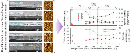
The microstructure of the film surface before and after rapid thermal treatment is significantly different (Figure 3 and Figure 4). The surface of the film before rapid thermal treatment is smooth with a granular structure and grain size—18.7 ± 4.1 nm (Figure 3a and Figure 4a). Rapid thermal treatment at 200 °C leads to an increase in grain size (Figure 3b and Figure 4b). Such an increase also leads to a rise in the height distribution over the film surface (Figure 3b). Formation of the Ni2Si and NiSi phases after rapid thermal treatment at 250 °C (Figure 3c and Figure 4c) ensues the formation of grains of various sizes from 10 nm to 40–50 nm (Figure 3c). The average grain size on this film was 23.1 nm (Figure 4c). Increasing the RTT temperature to 300–350 °C leads to the formation of a coarse-grained structure (Figure 3d and Figure 4d) with an average grain size from 24.9 nm (at 300 °C) to 27.4 nm (at 350 °C) due to the formation of the NiSi phase only. In this case, there are pores on the film’s surface (Figure 3d, shown by yellow arrows).
The formation of Ni2Si and NiSi silicides occurs by a diffusion mechanism [6,9,13,19], where the diffusion of nickel atoms into the silicon substrate is 10 times higher than that of silicon into the nickel matrix [6]. As a result, the diffusion of Ni atoms from the film surface to the substrate can lead to the formation of pores. Further elevation of the RTT temperature to 500 °C slightly changes the grain size (Figure 3f–h and Figure 4f–h), but leads to a decrease in the quantity of pores on the surface of nickel silicide films. An RTT temperature of 550 °C leads to an increase in grain size to 29.9 nm. It should be noted that in [17], by means of SEM, a larger grain size was observed in the cross section.
The film microstructure after rapid thermal treatment at 550 °C is similar to the film structure in [19], obtained by RTT in 15 s. An increase in grain size during the formation of the Ni2Si and NiSi phases was also observed for films deposited on silicon of different orientation [16,20]. The main difference between the above studies and the results in [16,19,20] is that they generally use rapid thermal treatment for 15 s and above. Thus, in [19], films were studied after rapid thermal treatment in the range of 15–300 s at temperatures of 300–500 °C, as well as after a long annealing process of 45 min at temperatures of 300–500 °C. In [20], rapid thermal treatment was carried out in 60 s at temperatures of 300–800 °C. At the same time, the grain size increased from 17.2 to 35.4 nm for films on (100)-oriented silicon wafers and from 31.2 to 44 nm for films on (110)-oriented silicon wafers [20].
The structures are obtained at the surface and not at a cross section using atomic force microscopy. The difference in grain size is due to the short time of the RTT process, which does not allow the film to be completely restructured. That is, silicon atoms only reach the surface in small quantities, and nickel atoms do not have time to completely diffuse deep into the film. Because of this, large grains comparable to the grains on the cross section in [17] are not formed on the surface. Changes in the film’s morphology can also be traced by height distribution over the surface (Figure 3). Rapid thermal treatment of nickel film leads to an expansion of height ranges and their shift towards higher values. Before rapid thermal treatment, the height distribution over the film surface ranges from 2 to 6 nm. In the presence of the Ni2Si phase in the film (Figure 3c) after rapid thermal treatment at 250 °C, the height range is from 1 to 20 nm, and at a temperature of 300 °C, with the formation of the NiSi phase (Figure 3d) it is from 2 to 22 nm. A further rise in the RTT temperature to 550 °C increases the width of range and maximum height distribution.
In the adhesion contrast (distribution maps of the adhesion force on the surface, Figure 4, blue images), the areas corresponding to pores and grain boundaries have a higher adhesion force (light or white areas in blue images). Due to the presence of pores, and unevenness of the film relief in the temperature range of 200–350 °C, the adhesion force is higher (and, accordingly, the specific surface energy, Figure 5) compared to the surface of films at 400–550 °C.
In addition to grain size, electrical properties of thin films are also affected by surface roughness. Rapid thermal treatment of nickel film leads to an increase in surface roughness (Figure 5) compared to the film roughness before rapid thermal treatment. In the temperature range of 200–250 °C, roughness steps up: Ra increases from 0.5 nm (for the initial film) to 1.6 nm (after RTT 250 °C), Rq—from 0.7 to 2.1 nm, Rz—from 1.7 to 2.9 nm. If we compare the surface roughness within the RTT temperature, then in the temperature range of 300–350 °C, the surface roughness of the films has a maximum value. Roughness Ra increases to 2.6 nm (after RTT 350 °C), Rq increases to 3.3 nm (after RTT 350 °C) and Rz increases to 5.4 nm (after RTT 300 °C). Further elevation of the RTT temperature leads to its decrease. After RTT at 350 °C, the roughness Ra decreases to 1.6 nm, Rq decreases to 2.3 nm, and Rz decreases to 3.0 nm. The roughness Rz varies independently of Ra and Rq. An increase in Rz to 5.4 nm occurs at 300 °C, where the Ni2Si phase transforms into the NiSi phase. Next, the roughness Rz gradually decreases to 2.0 nm at 500 °C and increases slightly to 3.0 nm at 550 °C. A minor increase in roughness Rz at 550 °C is caused by restructuring of the surface, during which the porosity of the film grows and the height differences increase that are visible on nanoprofiles (Figure 3d,i). The difference in roughness (Ra, Rq and Rz) between 200–350 °C and 400–550 °C is explained by the difference in surface structure and phase composition of the films. In the temperature range of 200–350 °C, surface porosity and unevenness of the relief (Figure 2 and Figure 3) are significantly higher compared to the film’s surface at 400–550 °C. Therefore, this significantly affects the values of Ra, Rq and Rz.
Rapid thermal treatment at 200 °C reduces the specific surface energy γ of the film compared to original film from 0.38 to 0.33 N/m (Figure 5). RTT at 250 °C leads to a drop in the specific surface energy from 0.33 to 0.21 N/m. Further elevation in the RTT temperature to 550 °C gradually increases the specific surface energy γ to 0.31 N/m. The increase in specific surface energy γ in the temperature range 400–550 °C is associated with an increase in grain size. A correlation has been established between the specific surface energy and grain size in the temperature range of 400–550 °C.
Resistivity ρ of the films depends significantly on the phase of the formed silicide [10,15]. RTT at temperatures of 200–250 °C (Figure 6) increases ρ by 1.8 times as compared to original nickel films (from 15 to 27–28 μΩ cm). This is due to the presence of the Ni2Si phase, a change in the microstructure and an increase in grain size [10,15].
A decrease in resistivity (up to 15 μΩ cm) at RTT temperatures of 350–400 °C to the level of the original nickel film is caused by the formation of the NiSi phase and high homogeneity over the entire thickness of the film [17]. A further increase in the RTT temperature to 550 °C increases ρ to 18 μΩ cm. This is probably due to the restructuring of the NiSi crystal structure [15], which also manifests itself in changes in the film morphology, roughness and grain size.
The specific surface energy and resistivity correlate with the average grain size (0.94 and 0.81, respectively) within an RTT temperature range of 350–550 °C, at which the film consists of the NiSi phase. This is due to further restructuring of the crystal structure of the NiSi phase and a decrease in the number of grain boundaries with increasing the grain size [14].

4. Conclusions

A study of the effect produced by rapid thermal treatment (RTT) with a temperature range from 200 to 550 °C for 7 s on the phase composition, microstructure and properties of thin nickel films deposited on a silicon wafer with a (111) orientation has been carried out. This study was carried out by applying X-ray phase analysis, scanning electron microscopy, high-precision atomic force microscopy and the four-probe method.
It has been ascertained that RTT at temperatures of 200–250 °C leads to the enlargement of nickel film grains due to the formation of the Ni2Si phase. An increase in roughness Ra, Rq and Rz and resistivity is also observed. Roughness Ra increases from 0.5 nm (for the initial film) to 1.6 nm (after RTT 250 °C), Rq—from 0.7 to 2.1 nm, Rz—from 1.7 to 2.9 nm. The specific electrical resistivity increases from 15 μΩ cm (for the initial film) to 27 μΩ cm (after RTT 250 °C). Rapid thermal treatment at 200 °C reduces the specific surface energy γ of the film compared to the original one from 0.38 to 0.33 N/m. An increase in the RTT temperature to 300 °C leads to the emergence of pores on the film surface, caused by an outflow of nickel atoms during the formation of the Ni2Si and NiSi phases. In this case, the roughness and average grain size increase, and the resistivity decreases. Elevation of the RTT temperature to 350–550 °C leads to the formation of a granular surface with a large height difference and depressions (pits) (from 3.56 nm at 450 °C to 5.1 nm at 500 °C). This is caused by the more active diffusion of nickel atoms to the substrate and the formation of the NiSi phase in the film. Increasing the RTT temperature to 550 °C gradually increases γ to 0.31 N/m. The specific surface energy γ in the temperature range 400–550 °C correlates with the grain size. The resistivity ρ of the films is determined by the silicide phase formed and the homogeneity of the structural phase composition. A correlation (correlation coefficient 0.81) of the resistivity with the average grain size after rapid thermal treatment at 350–550 °C has been established, which is associated with the formation and continuous restructuring of the crystal structure of the NiSi phase with an increasing RTT temperature. Applying high-precision atomic force microscopy made it possible to define the specific surface energy, roughness and grain size of nickel films before and after rapid thermal treatment, and to establish correlations between the obtained parameters and the electrical properties of the films. The established patterns of formation, and the obtained results and properties can be used in the technology of creating integrated electronics products with rectifying contacts.

Author Contributions

Conceptualization, V.L., T.K., S.C., V.P. and J.S.; methodology, T.K. and V.L.; software, T.K. and V.L.; validation, V.L., T.K. and J.S.; formal analysis, R.T., K.L., A.N. and M.D.; investigation, T.K., J.S. and V.L.; resources, J.S. and S.C.; data curation, V.L., T.K., S.C. and V.P.; writing—original draft preparation, V.L. and R.T.; writing—review and editing, V.L. and J.S.; visualization, V.L.; supervision, T.K., S.C. and V.P.; project administration, V.L. and T.K.; funding acquisition, S.C. All authors have read and agreed to the published version of the manuscript.

Funding

This research was partly financed by the grant of the Belarusian Republican Foundation for Fundamental Research BRFFR No. T23ME-010.

Institutional Review Board Statement

Not applicable.

Informed Consent Statement

Not applicable.

Data Availability Statement

The data presented in this study are available on request from the corresponding author. The data are not publicly available due to the ongoing character of the research.

Conflicts of Interest

The authors declare no conflicts of interest. Authors Jaroslav Solovjov and Vladimir Pilipenko was employed by the company JSC “INTEGRAL”—“INTEGRAL” Holding Managing Company. The remaining authors declare that the research was conducted in the absence of any commercial or financial relationships that could be construed as a potential conflict of interest.

References

  1. Rizwan, M.N.; Kalyar, M.A.; Bell, C.; Anwar-Ul-Haq, M.; Makhdoom, A.R. Nickel thin films grown by pulsed laser deposition: Influence of substrate and substrate temperature. Dig. J. Nanometer. Bios. 2020, 15, 1141–1151. [Google Scholar] [CrossRef]
  2. Dzhumaliev, A.S.; Nikulin, Y.V.; Filimonov, Y.A. Formation of textured Ni (200) and Ni (111) films by magnetron sputtering. Tech. Phys. 2016, 61, 924–928. [Google Scholar] [CrossRef]
  3. Kuchuk, A.V.; Gołaszewska, K.; Kladko, V.P.; Guziewicz, M.; Wzorek, M.; Kamińska, E.; Piotrowska, A. The formation mechanism of Ni-based ohmic contacts to 4H-n-SiC. Mater. Sci. Forum 2012, 717, 833–836. [Google Scholar] [CrossRef]
  4. Testov, O.A.; Komlev, A.E.; Gareev, K.G.; Khmelnitskiy, I.K.; Luchinin, V.V.; Sevost’yanov, E.N.; Testov, I.O. Providing a specified level of electromagnetic shielding with nickel thin films formed by DC magnetron sputtering. Coatings 2021, 11, 1455. [Google Scholar] [CrossRef]
  5. Mangelinck, D.; Gas, P.; Grob, A.; Pichaud, B.; Thomas, O. Formation of Ni silicide from Ni (Au) films on (111) Si. J. Appl. Phys. 1996, 79, 4078–4086. [Google Scholar] [CrossRef]
  6. Zhao, F.F.; Zheng, J.Z.; Shen, Z.X.; Osipowicz, T.; Gao, W.Z.; Chan, L.H. Thermal stability study of NiSi and NiSi2 thin films. Microelectron. Eng. 2004, 71, 104–111. [Google Scholar] [CrossRef]
  7. Okubo, K.; Tsuchiya, Y.; Nakatsuka, O.; Sakai, A.; Zaima, S.; Yasuda, Y. Influence of structural variation of Ni silicide thin films on electrical property for contact materials. Jpn. J. Appl. Phys. 2004, 43, 1896. [Google Scholar] [CrossRef]
  8. Liu, C.M.; Liu, W.L.; Hsieh, S.H.; Tsai, T.K.; Chen, W.J. Interfacial reactions of electroless nickel thin films on silicon. Appl. Surf. Sci. 2005, 243, 259–264. [Google Scholar] [CrossRef]
  9. Waidmann, S.; Kahlert, V.; Streck, C.; Press, P.; Kammler, T.; Dittmar, K.; Rinderknecht, J. Tuning nickel silicide properties using a lamp based RTA, a heat conduction based RTA or a furnace anneal. Microelectron. Eng. 2006, 83, 11–12, 2282–2286. [Google Scholar] [CrossRef]
  10. Colgan, E.G.; Mäenpää, M.; Finetti, M.; Nicolet, M.A. Electrical characteristics of thin Ni2Si, NiSi, and NiSi2 layers grown on silicon. J. Electron. Mater. 1983, 12, 413–422. [Google Scholar] [CrossRef]
  11. Tsuchiya, Y.; Tobioka, A.; Nakatsuka, O.; Ikeda, H.; Sakai, A.; Zaima, S.; Yasuda, Y. Electrical properties and solid-phase reactions in Ni/Si (100) contacts. Jpn. J. Appl. Phys. 2002, 41, 2450. [Google Scholar] [CrossRef]
  12. Bhaskaran, M.; Sriram, S.; Perova, T.S.; Ermakov, V.; Thorogood, G.J.; Short, K.T.; Holland, A.S. In situ micro-Raman analysis and X-ray diffraction of nickel silicide thin films on silicon. Micron 2009, 40, 89–93. [Google Scholar] [CrossRef]
  13. Adusumilli, P.; Seidman, D.N.; Murray, C.E. Silicide-phase evolution and platinum redistribution during silicidation of Ni0.95Pt0.05/Si(100) specimens. J. Appl. Phys. 2012, 112, 064307. [Google Scholar] [CrossRef]
  14. Utlu, G.; Artunc, N.; Selvi, S. Temperature and thickness dependence of the grain boundary scattering in the Ni–Si silicide films formed on silicon substrate at 500 °C by RTA. Mater. Chem. Phys. 2012, 132, 421–430. [Google Scholar] [CrossRef]
  15. Solovjov, J.A.; Pilipenko, V.A. Effect of Rapid Thermal Treatment Temperature on Electrophysical Properties of Nickel Films on Silicon. Doklady BGUIR 2020, 18, 81–88. [Google Scholar] [CrossRef]
  16. Pascu, R.; Romanitan, C. Phase transition of nickel silicide compounds and their electrical properties. J. Mater. Sci. Mater. Electron. 2021, 32, 16811–16823. [Google Scholar] [CrossRef]
  17. Pilipenko, V.A.; Solovjov, J.A.; Gaiduk, P.I. Nickel silicide formation with rapid thermal treatment in the heat balance mode. Dokl. Natl. Acad. Sci. Belarus 2021, 65, 111–118. (In Russian) [Google Scholar] [CrossRef]
  18. Bolisetty, S. Novel Process and Manufactur of Multi crystalline Solar Cell. 2009. Available online: http://liu.diva-portal.org/smash/get/diva2:210630/FULLTEXT01.pdf (accessed on 19 December 2023).
  19. Peter, A.P.; Meersschaut, J.; Richard, O.; Moussa, A.; Steenbergen, J.; Schaekers, M.; Adelmann, C. Phase formation and morphology of nickel silicide thin films synthesized by catalyzed chemical vapor reaction of nickel with silane. Chem. Mater. 2015, 27, 245–254. [Google Scholar] [CrossRef]
  20. Guo, X.; Yu, H.; Jiang, Y.L.; Ru, G.P.; Zhang, D.W.; Li, B.Z. Study of nickel silicide formation on Si (1 1 0) substrate. Appl. Surf. Sci. 2011, 257, 10571–10575. [Google Scholar] [CrossRef]
  21. Azimirad, R.; Kargarian, M.; Akhavan, O.; Moshfegh, A.Z. Improved thermal stability of NiSi nanolayer in Ni-Si Co-sputtered structure. Int. J. Nanosci. Nanotechnol. 2011, 7, 14–20. [Google Scholar]
  22. Bhaskaran, M.; Sriram, S.; Holland, A.S.; Evans, P.J. Characterisation of nickel silicide thin films by spectroscopy and microscopy techniques. Micron 2009, 40, 99–103. [Google Scholar] [CrossRef]
  23. Tinani, M.; Mueller, A.; Gao, Y.; Irene, E.A.; Hu, Y.Z.; Tay, S.P. In situ real-time studies of nickel silicide phase formation. J. Vac. Sci. Technol. B 2001, 19, 376–383. [Google Scholar] [CrossRef]
  24. Karabko, A.; Dragašius, E. NiSi and Ni (Pd) Si as possible interconnect and electrode materials for film bulk acoustic resonators and microelectromechanical systems. J. Vibroeng. 2013, 15, 196–203. [Google Scholar]
  25. Geetha Priyadarshini, B.; Aich, S.; Chakraborty, M. On the microstructure and interfacial properties of sputtered nickel thin film on Si (1 0 0). Bull. Mater. Sci. 2014, 37, 1265–1273. [Google Scholar] [CrossRef]
  26. Hurtado, C.; Ciampi, S. Oxidative Damage during the Operation of Si(211)-Based Triboelectric Nanogenerators. Surfaces 2023, 6, 281–290. [Google Scholar] [CrossRef]
  27. Kuznetsova, T.A.; Lapitskaya, V.A.; Chizhik, S.A.; Warcholinski, B.; Gilewicz, A. Effect of Atmosphere During Deposition on the Morphology, Mechanical Properties and Microfriction of Zr-Based Coatings. Adv. Struct. Mater. 2022, 155, 271–319. [Google Scholar]
Figure 1. SEM images of the cross section of films on silicon: (a) before rapid thermal treatment; (b) after rapid thermal treatment at 200 °C; (c) after rapid thermal treatment at 250 °C; (d) after rapid thermal treatment at 300 °C; (e) after rapid thermal treatment at 350 °C; (f) after rapid thermal treatment at 500 °C.
Surfaces 07 00013 g001
Figure 2. XRD films of nickel and silicides based on them before and after rapid thermal treatment: (a) before RTT; (b) 200 °C; (c) 250 °C; (d) 300 °C; (e) 350 °C; (f) 400 °C; (g) 450 °C; (h) 500 °C; (i) 550 °C.
Surfaces 07 00013 g002
Figure 3. Microstructure with height distribution and profiles (in a field of 1 × 1 µm2) of the film surface before (a) and after (bi) RTT: (a) 20 °C; (b) 200 °C; (c) 250 °C; (d) 300 °C; (e) 350 °C; (f) 400 °C; (g) 450 °C; (h) 500 °C; (i) 550 °C.
Surfaces 07 00013 g003aSurfaces 07 00013 g003b
Figure 4. Microstructure of the film’s surface with adhesive contrast (field 200 × 200 nm2) and histograms of grain size distribution before (a) and after RTT (bi) (a) 20 °C; (b) 200 °C; (c) 250 °C; (d) 300 °C; (e) 350 °C; (f) 400 °C; (g) 450 °C; (h) 500 °C; (i) 550 °C.
Surfaces 07 00013 g004aSurfaces 07 00013 g004b
Figure 5. Film surface roughness and specific surface energy (field 1 × 1 µm2) before and after rapid thermal treatment.
Surfaces 07 00013 g005
Figure 6. The specific resistivity and grain size of films before and after rapid thermal treatment.
Surfaces 07 00013 g006
Disclaimer/Publisher’s Note: The statements, opinions and data contained in all publications are solely those of the individual author(s) and contributor(s) and not of MDPI and/or the editor(s). MDPI and/or the editor(s) disclaim responsibility for any injury to people or property resulting from any ideas, methods, instructions or products referred to in the content.