Article

Sub-Genotyping of Acanthamoeba T4 Complex: Experience from North India

1
Department of Medical Parasitology, Postgraduate Institute of Medical Education and Research, Chandigarh 160012, India
2
Department of Microbiology, All India Institute of Medical Sciences, Bilaspur 174001, India
3
Department of Ophthalmology, Postgraduate Institute of Medical Education and Research, Chandigarh 160012, India
*
Author to whom correspondence should be addressed.
These authors contributed equally to this work.
M.S. was affiliated to 1 at the start and to 2 by the end of the study.
Parasitologia 2023, 3(1), 69-78; https://doi.org/10.3390/parasitologia3010009
Submission received: 16 December 2022 / Revised: 13 February 2023 / Accepted: 17 February 2023 / Published: 19 February 2023

Abstract

:
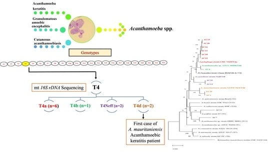
The Acanthamoeba genus comprises the free-living amoebae that are ubiquitously present as opportunistic pathogens. They cause serious human diseases—for instance, Acanthamoeba keratitis (AK), granulomatous amoebic encephalitis (GAE), cutaneous acanthamoebiasis and disseminated infections. The traditional method for classifying Acanthamoeba was based on the morphological examination of cysts. However, this method was less consistent as the morphology of cysts changes with the culture conditions. After the advent of molecular techniques, genotyping is considered an essential tool in accurately identifying Acanthamoeba at the species level and is further helpful in classification up to the sub-genotype level. The most recommended and currently used methods for Acanthamoeba genotyping are 18S and 16S rDNA gene sequencing. Based on these two genes, Acanthamoeba is classified into 23 genotypes. Out of these, it is the T4 genotype that is most commonly associated with clinical disease and isolation from environmental samples. The T4 genotype contains more than ten species within it. Differences in geographical distribution, virulence, pathogenesis and drug susceptibility profile have been observed among different genotypes. However, whether such differences exist within sub-genotypes/species under T4 is yet unknown. In the present study, 11 Acanthamoeba isolates, which were already characterized as the T4 genotype by the hypervariable region of diagnostic fragment 3 (DF3) of the 18S rDNA, were sub-genotyped using the 16S rDNA mitochondrial sequence. Nine of these were isolated from patients with AK and two from water samples. Phylogenetic analysis of these isolates attributed them to four sub-genotypes (T4a (n = 6), T4b (n = 1), T4Neff (n = 2) and T4d (n = 2)). The study highlights the potential use of 16S in the sub-genotyping of Acanthamoeba T4. The 16S rDNA sequences of two isolates, one from an Acanthamoebic keratitis (AK) patient and one environmental, were found to group with A. mauritaniensis (T4d). This group was believed to be a non-pathogenic environmental Acanthamoeba and the identification of the AK isolate may be confirmed by whole-genome sequencing.

Graphical Abstract

1. Introduction

Free-living amoebae, namely Acanthamoeba spp., Naegleria fowleri, Balamuthia mandrillaris and Sappinia diploidea, are sporadic sources of illness in both humans and animals. They are microscopic, single-celled eukaryotes that generally occur in dual morphological forms, i.e., actively dividing trophozoites and dormant but resistant cysts. However, in Naegleria fowleri, an additional flagellated form also exists. Acanthamoeba trophozoites are amoeboid in shape, often 15 to 25 µm in length and have characteristics of spine-like projection known as acanthopodia on their outer surface. These acanthopodia not only help in feeding organic particles and other microbes such as bacteria, fungi, viruses and algae but are also useful in locomotion and capturing prey. The cysts remain inert in harsh environmental conditions and revert back to trophozoite form once favorable conditions are encountered [1]. Cysts are airborne and are therefore isolated from a wide range of environmental niches. Acanthamoeba can also cause severe diseases in humans and livestock by acting as opportunistic pathogens.
The various clinical entities associated with Acanthamoeba infection include granulomatous amoebic encephalitis, cutaneous acanthamoebiasis, disseminated infection and Acanthamoeba keratitis (AK) [2,3]. Apart from AK, the remaining clinical manifestations are mostly associated with immunocompromised individuals; however, AK affects not only immunocompromised but also immunocompetent individuals. AK is a corneal infection causing severe vision-threatening disease. AK is also recognized as a progressive infection, which, if not treated early, might lead to corneal ulcers, loss of visual acuity, perforation of the cornea and ultimately loss of eyesight [4]. The maximum number of cases of AK are documented by contact lens users from developed countries, which correspond to 85–88% of AK cases [5]. Meanwhile, in developing countries, major predisposing factors include corneal injuries, followed by the use of contaminated water [6]. Recently, Acanthamoeba have been detected in the bronchoalveolar lavage fluid of immunocompetent patients with chronic respiratory disorders [7]. Acanthamoebae seem to be the most predominant and ubiquitous protozoa present in the natural environment. They have been isolated from a variety of water resources, such as domestic tap, natural, treated and bottled water; and swimming pools, sea water, dialysis units, dental treatment units, eye wash stations, dust, air, soil and sewage [8]. Their ubiquitous distribution in the environment provides them with ample opportunities for close contact with potential hosts, including humans, causing disease in both immunocompromised as well as immunocompetent individuals [1].
Genotyping is considered to be an essential tool for the accurate identification of Acanthamoeba. Acanthamoeba genotyping helps in studying their taxonomy and is useful in molecular epidemiology and further clinical studies. It also offers valuable information in studying the correlation between disease phenotype, drug susceptibility, virulence factors of different genotypes and the development of new and rapid diagnostic tests [9]. Based on 18S rDNA, Acanthamoebae are classified into different genotypes that form holophyletic clades and are distinguished from each other by 5% or greater sequence dissimilarity between isolates [10]. Out of the total 23 genotypes identified for Acanthamoeba, only a few are implicated in causing human disease. The genotypes T3, T4, T5, T6, T11 and T15 are implicated in the causation of AK, whereas genotypes T1, T3, T4, T10 and T12 are implicated in GAE [11]. Among those causing disease, it is the T4 or A. castellanii complex that contributes to the highest number of cases. The A. castellanii species complex contains more than ten species, including A. castellanii, A. polyphaga, A. lugdenensis, A. mauritaniensis, A. triangularis, A. rhysodes, A. royreba, A. divionensis, A. paradivionenesis, etc. [12]. Fuerst et al. [10], in their compilation of all available sequences of Acanthamoeba in 2014, reported that 70% (n = 1300) of them were genotype T4. Diehl et al. [13], in their recent systematic review on AK, reported that 86% of global AK infections were caused by genotype T4. The T4 genotype contains more than ten species under the A. castellanii complex, and the sub-genotyping of T4 further into species is important to understand the differences in their disease potential. Previous studies have shown that the different genotypes of Acanthamoeba differ not only in their geographical and environmental distribution, but also in their susceptibility towards common anti-acanthamoebic drugs [13,14]. It is, however, yet unknown if such differences exist among the sub-genotypes of the T4 genotype as well.
Historically, it was the cyst morphological form of Acanthamoeba that formed the basis of genotype/species identification [15]. The classification of Acanthamoebae into three morpho-groups (I, II and III) was based on the size and structure of ecto and endo cysts. Species in morpho-group I were distinguished by possessing a large cyst, in contrast to species in the other two groups, and it consists of the T7, T8 and T9 genotypes. A wrinkled ectocyst and an endocyst that could be stellate, triangular, polygonal or oval are characteristics of species in morpho-group II and it consists of T1, T3, T4 and T11. Meanwhile, the species in morpho-group III have a thin and smooth endocyst and round ectocyst, and it consists of the T2, T5, T6, T10, T12, T14 and T15 genotypes [16]. However, classification based on cyst morphology is unreliable as the characteristics vary with the culture conditions. Other conventional methods, such as immunological, physiological and biochemical assays, have also been used for species differentiation. However, numerous species exhibit similar antigenic determinants. Immunological methods such as Western blotting and immunofluorescence give ambiguous results. Different enzyme systems, such as isoenzyme electrophoresis, have also been used to compare Acanthamoeba strains. The results of this method have shown interstrain variance within species and similarities between strains of different species, despite the method’s promise to shed light on links between species. Additionally, investigations have demonstrated that when isolates are cultivated in various laboratory environments, the patterns of the enzymes alter [1]. With the advancements in molecular biology, DNA sequencing of the 18S rRNA nuclear gene has become the gold standard to identify and characterize any new Acanthamoeba strain isolated from clinical or environmental sources. Of the 18S rDNA gene, it was the diagnostic fragment 3 (DF3) of hypervariable regions that formed the basis of genotyping and epidemiological studies dividing Acanthamoeba into 23 genotypes, i.e., T1 to T23 [17,18]. However, since Acanthamoeba T4 is polyphyletic by evolution, the nuclear material of each species is actually a mix of several variants, the genes of which are present in different copy numbers. This means that it is the amplification of the predominant variant that governs the identification. While this serves well for identification up to the genotype level, these factors lead to bias in the phylogenetic signaling with 18S, thereby hindering the molecular and taxonomical classification of Acanthamoeba beyond genotypes.
With wide geographical differences in the distribution of Acanthamoeba genotypes, there is a definite need to delineate local Acanthamoeba T4 isolates. In contrast to 18S, the 16S rDNA gene serves as a better marker for the sub-genotyping of T4, given that it is mitochondrial in origin. The present study aimed to characterize the Acanthamoeba isolates belonging to genotype T4 in our culture collection to the sub-genotypic level using 16S rDNA.

2. Materials and Methods

2.1. Study Isolates

Eleven isolates of Acanthamoeba, characterized by the T4 genotype, that were processed and maintained in the Department of Medical Parasitology, Postgraduate Institute of Medical Education and Research, Chandigarh, India, were included in the study. They were isolated from clinical specimens, i.e., corneal scraping (n = 9) of Acanthamoeba keratitis patients attending the cornea clinic at the Advanced Eye Center, Postgraduate Institute of Medical Education and Research, Chandigarh, India. Additionally, water samples (n = 2) were obtained from domestic tap water. However, these water samples were not obtained from the AK patients’ residences (Table 1). All the samples were confirmed as the Acanthamoeba T4 genotype using the hypervariable region of the DF3 region of the 18S rDNA, as described previously [19]. The study was approved by the institute’s Ethics Committee (PGI/IEC/2014/90).

2.2. Maintenance of Culture and Morphological Characterization

All the Acanthamoeba isolates were cultivated in an axenic medium constituted by improvised peptone yeast dextrose medium supplemented with RNAase and NCTC109 vitamin mixture media [20]. Morphological identification of Acanthamoeba spp. was performed based on the shape of their endocysts and ectocyts. Briefly, Acanthamoeba trophozoites were grown on a 2% non-nutrient agar plate overlaid with Escherichia coli ATCC 25922 for 14 days, and, once all trophozoites converted into cysts, they were harvested and stored in phosphate-buffered saline at 4 °C, and then the wet mount was prepared from each isolate and observed under a light microscope (Olympus CX2li, Olympus Corporation, Tokyo, Japan).

2.3. DNA Extraction

DNA from axenic culture was extracted using the chloroform isoamyl alcohol method, as described previously [18]. Briefly, the aqueous lysate was extracted with an equal volume of phenol–chloroform–isoamyl alcohol in the ratio of 25:24:1 until the protein interface completely disappeared. The DNA was precipitated from the aqueous lysate with double the volume of absolute ethanol. Further, an ethanol-free DNA pellet was dissolved in 50 µL Tris EDTA buffer, pH 8.0, and kept at 37 °C for 15 min. The extracted DNA was checked for quantity by UV absorbance using a NanoDrop ND-1000 spectrophotometer (NanoDrop Technologies, Thermo Scientific, Waltham, MA, USA) and stored at −20 °C for further use.

2.4. Molecular Characterization Using 16S

The 16S rDNA PCR was performed for sub-genotypic characterization using three PCR primer sets (Fmt, Rmt and tALA) to obtain amplicons of ~1500 bp, as described previously [21]. The amplification reactions were carried out in 25 μL reaction volume and consisted of 2.5 μL of 1X buffer (Sigma, Kawasaki, Japan), 0.2 mM dNTPs (Sigma) and 10 pmol of each primer pair (Fmt and Rmt) or (tALA and Rmt), with 1.5 unit of Taq DNA polymerase (Sigma), 5 μL nuclease-free water and 2 μL DNA template. The PCR cycle profile was as follows: initial denaturation at 95 °C for 10 min, then 35 cycles at 95 °C for 1 min; annealing at 63 °C for 2 min; extension at 72 °C for 3 min and the final extension at 72 °C for 15 min to obtain a product of ~1500 bp. During each PCR run, a negative control (nuclease-free water) and appropriate positive controls were included.
Amplified PCR products were separated by 1.5% agarose electrophoresis stained with a solution of 0.5 μg/mL of ethidium bromide and visualized under UV light using an image analyzer. They were then subjected to multidirectional sequencing using nine sets of sequencing primers on an ABI 3130 automated sequencer and ABI 3130 Genetic Analyser (Applied Biosystems, Foster City, CA, USA). All the sequences were analyzed in Geospiza’s DNA sequence trace viewer software FinchTV v1.4.0 (Geospiza Inc., Denver, CO, USA). Consensus sequences for the 16S rDNA gene were created using the SeqMan software (DNASTAR Inc., Madison, WI, US). Each pair of consensus sequences was then used for the identification of the Acanthamoeba genotype and sub-genotype by matching it against the references available in a public database using BLASTn [22]. All the sequences were submitted to GenBank under accession numbers MF538583-MF538588, MF563603-MF563604 and MF563606-MF5636010 (Table 1).

2.5. Phylogenetic Analysis

The molecular phylogenetic analysis was performed for all the isolates using reference sequences of Acanthamoeba obtained from GenBank. Sequences belonging to other genotypes, T1, T3, T7-T12, from our own experience and those from reference databases, were also included for better discrimination. All the DNA sequences were aligned using multiple alignment mode in the CLUSTAL X2 software [23]. The sequence alignments were exported to the Molecular Evolutionary Genetics Analysis software version 6 (MEGA 6) [24]. The phylogenetic analysis was achieved using the neighbor-joining method with the Kimura 2 parameter model. The phylogenetic tree was reconstructed with 1000 bootstrap replications, and B. mandrillaris was used as an outgroup isolate.

3. Results

3.1. Morphological Characterization of Study Isolates

All 11 isolates from the clinical specimens and water samples showed a mixed morphology, i.e., both morpho-group II and III. The wrinkled ectocyst and stellate or polygonal endocyst was consistent with morpho-group II. The species included in this morpho-group were A. grifini (T3) A. castellanii (T4) and A. hatchetti (T11). Meanwhile, a smooth ectocyst and round endocyst was consistent with morpho-group III, and species included in this morpho-group were A. leticulata (T5) and A. culbertsoni (T10) (Figure 1).

3.2. Molecular Characterization and Phylogenetic Analysis

All 11 isolates of Acanthamoeba were identified as the T4 genotype on mitochondrial 16S rDNA sequencing and showed 94–99% homology with the available gene sequences of Acanthamoeba (Table 1). In the phylogenetic analysis, six out of 11 isolates (AC11, AC15, AC16, AC20, AC28) clustered around A. polyphaga strain CDC V029 (ATCC 50495) and were identified as the T4a sub-genotype. AC4 clustered with the Acanthamoeba Galka strain and was identified as sub-genotype T4b. AC13 and AC25 clustered along Acanthamoeba castellanii strain Neff and were identified as sub-genotype Neff under T4f. AC29 (environmental) and AC1 (Acanthamoeba keratitis patients) clustered along the Acanthamoeba mauritaniensis strain SAWE and were identified as sub-genotype T4d (Figure 2).

4. Discussion

Acanthamoebae are widely dispersed in the environment and implicated in causing keratitis, encephalitis, cutaneous and disseminated infections [1]. Since Acanthamoeba infections are associated with high mortality and considerable morbidity, it is imperative to identify the infecting organism at the earliest possible time and initiate appropriate treatments as soon as possible. Globally, the T4 genotype is the most common genotype identified in clinical and environmental samples. Genotyping of Acanthamoeba in general and the sub-genotyping of T4 in particular would help in identifying the different species of Acanthamoeba, which would provide valuable information regarding the differences in their target organs, virulence factors, pathogenesis, drug susceptibility and the development of new, more rapid diagnostics.
Traditionally, Acanthamoeba were classified into morphotypes on the basis of the morphology of the cysts. In the present study, the cysts of all 11 isolates had a dual morphological appearance resembling that of morpho-group II (which consists of genotypes T3, T4 and T11) and morpho-group III (which consists of genotypes T5 and T10). Thus, morphological classification was inconsistent in the present study. A similar observation was made earlier, where this traditional grouping was found unreliable and discordant, especially for Acanthamoeba T4, in comparison to molecular genotyping [25].
The most common sub-genotype, observed on 16S molecular analysis, in the present study, was T4a (6/11; 54.5%), followed by T4f and T4d (2/11; 18.2% each), and a single isolate of T4b (1/11; 9.1%). Our findings are similar to the observations made by Fuerst et al., where T4a was the most common sub-genotype (29.8%; 45) out of 151 global T4 isolates, followed by T4d (19.8%; 30) and T4c (15.2%; 23). Our findings are in contrast to two recent studies from South India, where T4b was reported as the predominant sub-genotype. While Rayamajhee et al. [26] reported T4b to be the predominant sub-genotype (76.9%, 10) among their 13 AK isolates, Prithviraj et al. [27] reported T4b in 50% (13/26), followed by T4d (38.4%, 10), T4a (7.6%, 2) and T4e (3.8%, 1). These differences could be due to the variations in the geographical distribution of Acanthamoeba.
In the current study, 100% of the Acanthamoeba T4 isolates were successfully sub-genotyped using the 16S gene. In our previous experience using 18S, only 22.2% (2/9) of T4 isolates could be characterized to the sub-genotype level [19]. This was in accordance with Malavin et al. [12], who, in a detailed molecular comparison of their six environmental Acanthamoeba isolates with global gene sequences, reported that the discriminatory power of 16S was greater than that of 18S for species within the T4 genotype. Fuerst et al., in their detailed analysis of the genotyping and sub-genotyping of Acanthamoeba strains using 18S, could delineate seven sub-genotypes/species (T4a to T4f, T4-Neff) [10,17] but they cautioned that subtypes under T4 were highly heterogenous and monophyletic, and recommended the redefinition of the species name [10]. Corsaro et al. [28] reported that although the tree topologies using 16S and 18S were congruent for Acanthamoeba in general, there were discrepancies within the T4 genotype. It is pertinent to note that Fuerst et al. [25], in their evaluation using 18S for >5300 Acanthamoeba sequences, could identify >3800 T4 isolates; however, only 151 of these could be sub-genotyped, having an allele frequency distribution of more than one in the database. A similar observation was made in Spain [29], Japan [30], Thailand [31], Iran [32] and another multicentric study in Europe [33], wherein 18S could not sub-genotype T4 correctly. This lower resolution of 18S in sub-genotyping T4 could be attributed to the nuclear sequences being longer (2300–3000 bp), less consistent in length and having frequent introns and fewer alignable base pairs [21,34]. Meanwhile, 16S, on the other hand, offer smaller sequences (1540 bp) with a larger proportion of alignable region. The absence of introns not only ensures the high fidelity of the sequence but also allows the use of a restriction fragment length polymorphism (RFLP) for identification as an alternative to DNA sequencing. Xuan et al. [35] reported that 16S RFLP was not inferior to 18S and 16S sequencing in identifying three isolates of the T4 genotype as A. triangularis, isolated for the first time from AK patients. Both the 16S and 18S genes have different evolutionary constraints owing to their different locations within the cell; while 16S is mitochondrial, 18S is nuclear. The 18S sequences are believed to represent the evolutionary history of the organism. Even for Acanthamoeba, it is the 18S that has been used most commonly for taxonomical and evolutionary classifications [17]. However, realizing the importance of redefining T4 up to the species level and acknowledging the inherent limitations of 18S in doing so, 16S could be used as a suitable alternative.
The 16S phylogenetic analysis in the current study yielded two interesting observations. First, two isolates (AC1, AC29) were identified as T4d, and they clustered closely with A. mauritaniensis. This is the first report of the isolation of T4d from a clinical sample (AC1) in the world, from a case of AK. The second T4d was isolated from a water sample (AC29). Our findings are in line with a recent study wherein A. mauritaniensis, earlier thought to be a non-pathogenic environmental isolate, was found to display pathogenic potential similar to the AK-causing A. castellanii strain by producing a cytopathic effect on in vitro cell lines [36]. However, since this identification is based on a single-gene analysis, confirmation may be obtained using multiple genes or whole-genome sequencing. Second, two isolates (AC13, AC25) clustered with the Neff strain of A. castellanii (T4f), with a relatively low bootstrap value of 78. Even in 16S phylogenetic trees from other studies, these two either cluster poorly with the Neff strain of A. castellanii (T4f) [12], or form a new mitochondrial subtype, T4j [28], thereby creating a possible new mitochondrial subtype. Further studies are warranted to confirm these findings, including their detailed 18S sequencing, to clarify whether they form a new nuclear subtype as well.
The study has the following limitations. Only 11 isolates of T4 were studied, and their partial sequences analyzed. Future studies could undertake an analysis of the complete sequence or the whole genome. It is also yet to be elucidated whether 16S alone could serve as a stand-alone marker for the genotyping and sub-genotyping of Acanthamoeba as its analytical sensitivity and specificity for the same has not been evaluated among Acanthamoeba and organisms closely related to Acanthamoeba. Future studies could evaluate the true potential of 16S sequencing in the unambiguous identification of species under not only genotype T4 but also other genotypes. Newer, more sensitive and robust techniques along with updated database/software could also be developed for rapid and accurate species identification. A recent study by Holmgaard et al. [37] made use of in-house 16S–18S next-generation sequencing along with the ‘Bion’ software to identify the genotypes and species of Acanthamoeba. Since gene sequencing is costly, technically demanding and takes several hours, other alternatives such as MALDI-TOF could be evaluated. MALDI-TOF has previously proven its worth as a cheap, rapid and easy method for the genotyping of Acanthamoeba by creating an in-house database that produced a concordance of 92% with 18S sequencing [38]. It is yet to be seen whether it can delineate sub-genotypes of Acanthamoeba also.

5. Conclusions

To conclude, since the different genotypes of Acanthamoeba differ in their pathogenic potential and drug susceptibility, it is important to evaluate whether such differences exist among the different subtypes/species within the same genotype, especially those under genotype T4, which is implicated in the majority of Acanthamoebic infections. We note that 16S sequencing and phylogeny could be used for the reliable differentiation of the Acanthamoeba T4 genotype into its sub-genotypes. T4a was the predominant sub-genotype among our isolates. Utilizing 16S for sub-genotyping enables greater differentiation of the T4 genotype along with the identification of newer subtypes. A pioneering case of Acanthamoebic keratitis caused by A. mauritaniensis, belonging to T4d, was reported. Although the pathogenic potential of A. mauritaniensis has been suggested, this report, if confirmed by whole-genome sequencing, would be the first documented case of AK by this species. Two isolates clustered distinctly from others and poorly with the Neff strain, possibly denoting a newer mitochondrial subtype of T4. These findings also need confirmation by whole-genome sequencing.

Author Contributions

Conceptualization: S.K. and A.G.; experiment: K.M.; data curation: K.M. and M.S.; analysis: K.M., M.S. and S.K.; draft preparation: M.S. and K.M.; revision and editing: K.M., M.S. and S.K.; study supervision: S.K. and R.S. All authors have read and agreed to the published version of the manuscript.

Funding

This work was financially supported by the Indian Council of Medical Research, grant number 5/3/3/33/2013-ECD-I.

Institutional Review Board Statement

The study was approved by the Institutional Ethics Committee (PGI/IEC/2014/90).

Informed Consent Statement

Written informed consent was obtained from all the patients involved in the study.

Data Availability Statement

All the gene sequences MF538583-MF538588, MF563603-MF563604 and MF563606-MF5636010 are available at GenBank.

Acknowledgments

We acknowledge the Indian Council of Medical Research for providing financial support for this work.

Conflicts of Interest

The authors declare no conflict of interest.

References

  1. Marciano-Cabral, F.; Cabral, G. Acanthamoeba spp. as Agents of Disease in Humans. Clin. Microbiol. Rev. 2003, 16, 273–307. [Google Scholar] [CrossRef] [PubMed] [Green Version]
  2. Khurana, S.; Sharma, M. Parasitic Keratitis—An under-Reported Entity. Trop. Parasitol. 2020, 10, 12–17. [Google Scholar] [CrossRef] [PubMed]
  3. Sharma, M.; Sudhan, S.S.; Sharma, S.; Megha, S.; Nada, R.; Khurana, S. Osteo-cutaneous acanthamoebiasis in a non-immunocompromised patient with a favorable outcome. Parasitol. Int. 2017, 66, 727–730. [Google Scholar] [CrossRef] [PubMed]
  4. Trabelsi, H.; Dendana, F.; Sellami, A.; Cheikhrouhou, F.; Neji, S.; Makni, F.; Ayadi, A. Pathogenic free-living amoebae: Epidemiology and clinical review. Pathol. Biol. 2012, 60, 399–405. [Google Scholar] [CrossRef]
  5. Schaumberg, D.A.; Snow, K.K.; Dana, M.R. The epidemic of Acanthamoeba keratitis: Where do we stand? Cornea 1998, 17, 3–10. [Google Scholar] [CrossRef]
  6. Sharma, S.; Garg, P.; Rao, G.N. Patient characteristics, diagnosis, and treatment of non-contact lens related Acanthamoeba keratitis. Br. J. Ophthalmol. 2000, 84, 1103–1108. [Google Scholar] [CrossRef] [Green Version]
  7. Saberi, R.; Nakhaei, M.; Fakhar, M.; Zarrinfar, H.; Sharifpour, A.; Hezarjaribi, H.Z. Molecular identification and genotyping of Acanthamoeba spp., in bronchoalveolar lavage fluid from immunocompetent patients with chronic respiratory disorders (CRD). Parasitol. Res. 2022, 121, 3013–3017. [Google Scholar] [CrossRef]
  8. Schuster, F.L.; Visvesvara, G.S. Free-living amoebae as opportunistic and non-opportunistic pathogens of humans and animals. Int. J. Parasitol. 2004, 34, 1001–1027. [Google Scholar] [CrossRef]
  9. Ledee, D.R.; Iovieno, A.; Miller, D.; Mandal, N.; Diaz, M.; Fell, J.; Fini, M.E.; Alfonso, E.C. Molecular Identification of T4 and T5 Genotypes in Isolates from Acanthamoeba Keratitis Patients. J. Clin. Microbiol. 2009, 47, 1458–1462. [Google Scholar] [CrossRef] [Green Version]
  10. Fuerst, P.A. Insights from the DNA databases: Approaches to the phylogenetic structure of Acanthamoeba. Exp. Parasitol. 2014, 145, S39–S45. [Google Scholar] [CrossRef]
  11. Khan, N.A.; Jarroll, E.L.; Paget, T.A. Molecular and Physiological Differentiation Between Pathogenic and Nonpathogenic Acanthamoeba. Curr. Microbiol. 2002, 45, 197–202. [Google Scholar] [CrossRef]
  12. Malavin, S.; Shmakova, L. Isolates from ancient permafrost help to elucidate species boundaries in Acanthamoeba castellanii complex (Amoebozoa: Discosea). Eur. J. Protistol. 2020, 73, 125671. [Google Scholar] [CrossRef] [PubMed]
  13. Diehl, M.L.N.; Paes, J.; Rott, M.B. Genotype distribution of Acanthamoeba in keratitis: A systematic review. Parasitol. Res. 2021, 120, 3051–3063. [Google Scholar] [CrossRef] [PubMed]
  14. Megha, K.; Sharma, M.; Sharma, C.; Gupta, A.; Sehgal, R.; Khurana, S. Evaluation of in vitro activity of five antimicrobial agents on Acanthamoeba isolates and their toxicity on human corneal epithelium. Eye 2021, 36, 1911–1917. [Google Scholar] [CrossRef]
  15. Pussard, M.; Pons, R. Morphologie de La Paroi Kystique et Taxonomie Du Genre Acanthamoeba (Protozoa, Amoebida). Protistologica 1977, 13, 557–598. [Google Scholar]
  16. Booton, G.C.; Visvesvara, G.S.; Byers, T.J.; Kelly, D.J.; Fuerst, P.A. Identification and Distribution of Acanthamoeba Species Genotypes Associated with Nonkeratitis Infections. J. Clin. Microbiol. 2005, 43, 1689–1693. [Google Scholar] [CrossRef] [PubMed] [Green Version]
  17. Fuerst, P.A.; Booton, G.C.; Crary, M. Phylogenetic Analysis and the Evolution of the 18S rRNA Gene Typing System of Acanthamoeba. J. Eukaryot. Microbiol. 2015, 62, 69–84. [Google Scholar] [CrossRef]
  18. Megha, K.; Sehgal, R.; Khurana, S. Genotyping of Acanthamoeba spp. isolated from patients with granulomatous amoebic encephalitis. Indian J. Med. Res. 2018, 148, 456–459. [Google Scholar] [CrossRef]
  19. Megha, K.; Sharma, M.; Gupta, A.; Sehgal, R.; Khurana, S. Microbiological diagnosis of Acanthamoebic keratitis: Experience from tertiary care center of North India. Diagn. Microbiol. Infect. Dis. 2021, 100, 115339. [Google Scholar] [CrossRef] [PubMed]
  20. Megha, K.; Gupta, A.; Sehgal, R.; Khurana, S. An Improvised Medium for Axenic Cultivation of Acanthamoeba spp. Indian J. Med. Microbiol. 2017, 35, 597–599. [Google Scholar] [CrossRef]
  21. Ledee, D.R.; Booton, G.C.; Awwad, M.H.; Sharma, S.; Aggarwal, R.K.; Niszl, I.A.; Markus, M.B.; Fuerst, P.A.; Byers, T.J. Advantages of Using Mitochondrial 16S rDNA Sequences to Classify Clinical Isolates of Acanthamoeba. Investig. Opthalmology Vis. Sci. 2003, 44, 1142–1149. [Google Scholar] [CrossRef] [Green Version]
  22. Johnson, M.; Zaretskaya, I.; Raytselis, Y.; Merezhuk, Y.; McGinnis, S.; Madden, T.L. NCBI BLAST: A better web interface. Nucleic Acids Res. 2008, 36, W5–W9. [Google Scholar] [CrossRef] [PubMed]
  23. Larkin, M.A.; Blackshields, G.; Brown, N.P.; Chenna, R.; McGettigan, P.A.; McWilliam, H.; Valentin, F.; Wallace, I.M.; Wilm, A.; Lopez, R.; et al. Clustal W and Clustal X version 2.0. Bioinformatics 2007, 23, 2947–2948. [Google Scholar] [CrossRef] [PubMed] [Green Version]
  24. Tamura, K.; Stecher, G.; Peterson, D.; Filipski, A.; Kumar, S. MEGA6: Molecular Evolutionary Genetics Analysis Version 6.0. Mol. Biol. Evol. 2013, 30, 2725–2729. [Google Scholar] [CrossRef] [PubMed] [Green Version]
  25. Fuerst, P.; Booton, G. Species, Sequence Types and Alleles: Dissecting Genetic Variation in Acanthamoeba. Pathogens 2020, 9, 534. [Google Scholar] [CrossRef] [PubMed]
  26. Rayamajhee, B.; Sharma, S.; Willcox, M.; Henriquez, F.L.; Rajagopal, R.N.; Shrestha, G.S.; Subedi, D.; Bagga, B.; Carnt, N. Assessment of genotypes, endosymbionts and clinical characteristics of Acanthamoeba recovered from ocular infection. BMC Infect. Dis. 2022, 22, 757. [Google Scholar] [CrossRef] [PubMed]
  27. Prithiviraj, S.R.; Rajapandian, S.G.K.; Gnanam, H.; Gunasekaran, R.; Mariappan, P.; Singh, S.S.; Prajna, L. Clinical presentations, genotypic diversity and phylogenetic analysis of Acanthamoeba species causing keratitis. J. Med. Microbiol. 2020, 69, 87–95. [Google Scholar] [CrossRef]
  28. Corsaro, D. Update on Acanthamoeba phylogeny. Parasitol. Res. 2020, 119, 3327–3338. [Google Scholar] [CrossRef]
  29. Martín-Pérez, T.; Criado-Fornelio, A.; Martínez, J.; Blanco, M.; Fuentes, I.; Pérez-Serrano, J. Isolation and molecular characterization of Acanthamoeba from patients with keratitis in Spain. Eur. J. Protistol. 2017, 61, 244–252. [Google Scholar] [CrossRef]
  30. Rahman, M.; Yagita, K.; Kobayashi, A.; Oikawa, Y.; Hussein, A.; Matsumura, T.; Tokoro, M. Genetic Characterization of Clinical Acanthamoeba Isolates from Japan using Nuclear and Mitochondrial Small Subunit Ribosomal RNA. Korean J. Parasitol. 2013, 51, 401–411. [Google Scholar] [CrossRef]
  31. Putaporntip, C.; Kuamsab, N.; Nuprasert, W.; Rojrung, R.; Pattanawong, U.; Tia, T.; Yanmanee, S.; Jongwutiwes, S. Analysis of Acanthamoeba genotypes from public freshwater sources in Thailand reveals a new genotype, T23 Acanthamoeba bangkokensis sp. nov. Sci. Rep. 2021, 11, 17290. [Google Scholar] [CrossRef] [PubMed]
  32. Spotin, A.; Moslemzadeh, H.R.; Mahami-Oskouei, M.; Ahmadpour, E.; Niyyati, M.; Hejazi, S.H.; Memari, F.; Noori, J. Phylogeography, genetic variability and structure of Acanthamoeba metapopulations in Iran inferred by 18S ribosomal RNA sequences: A systematic review and meta-analysis. Asian Pac. J. Trop. Med. 2017, 10, 855–863. [Google Scholar] [CrossRef] [PubMed]
  33. Chelkha, N.; Jardot, P.; Moussaoui, I.; Levasseur, A.; La Scola, B.; Colson, P. Core gene-based molecular detection and identification of Acanthamoeba species. Sci. Rep. 2020, 10, 1583. [Google Scholar] [CrossRef] [PubMed] [Green Version]
  34. Stothard, D.R.; Schroeder-Diedrich, J.M.; Awwad, M.H.; Gast, R.J.; Ledee, D.; Rodriguez-Zaragoza, S.; Dean, C.L.; Fuerst, P.; Byers, T.J. The Evolutionary History of the Genus Acanthamoeba and the Identification of Eight New 18S rRNA Gene Sequence Types. J. Eukaryot. Microbiol. 1998, 45, 45–54. [Google Scholar] [CrossRef] [PubMed]
  35. Xuan, Y.-H.; Chung, B.-S.; Hong, Y.-C.; Kong, H.-H.; Hahn, T.-W.; Chung, D.-I. Keratitis by Acanthamoeba triangularis: Report of Cases and Characterization of Isolates. Korean J. Parasitol. 2008, 46, 157–164. [Google Scholar] [CrossRef] [PubMed]
  36. Coronado-Velázquez, D.; Silva-Olivares, A.; Castro-Muñozledo, F.; Lares-Jiménez, L.F.; Rodríguez-Anaya, L.Z.; Shibayama, M.; Serrano-Luna, J. Acanthamoeba mauritaniensis genotype T4D: An environmental isolate displays pathogenic behavior. Parasitol. Int. 2020, 74, 102002. [Google Scholar] [CrossRef] [PubMed]
  37. Holmgaard, D.B.; Barnadas, C.; Mirbarati, S.H.; O’Brien, A.L.; Nielsen, H.V.; Stensvold, C.R. Detection and identification of acanthamoeba and other nonviral causes of infectious keratitis in corneal scrapings by real-time PCR and next-generation sequencing-based 16S–18S gene analysis. J. Clin. Microbiol. 2021, 59, e02224-20. [Google Scholar] [CrossRef]
  38. Megha, K.; Sharma, M.; Gupta, A.; Sehgal, R.; Khurana, S. Protein profiling of Acanthamoeba species using MALDI-TOF MS for specific identification of Acanthamoeba genotype. Parasitol. Res. 2018, 117, 729–736. [Google Scholar] [CrossRef]
Figure 1. Microscopic examination of Acanthamoeba cysts in wet mount showing different morpho-groups having wrinkled ectocyst and stellate/polygonal endocyst and having smooth ectocyst and round endocyst in same isolate under 40× magnification with a light microscope (Olympus CX2li, Olympus Corporation, Tokyo, Japan).
Parasitologia 03 00009 g001
Figure 2. Phylogenetic tree based on partial mitochondrial 16S rDNA sequences by the neighbor-joining method.
Parasitologia 03 00009 g002
Table 1. Comparison of nuclear 18s rDNA and mitochondrial 16S rDNA sequences of eleven isolates of Acanthamoeba with the reference sequences available in GenBank.
IDSource Origin16S rDNAGenBank Accession No.% IDIdentity with Reference GenBank Accesion No.
AC11Corneal scrapingT4aMF53858599%AF479525
AC15Corneal scrapingT4aMF53858898%AB795711
AC16Corneal scrapingT4aMF53858698%MK100243
AC20Corneal scrapingT4aMF53858499%AF479525
AC22Corneal scrapingT4aMF53858398%AB795713
AC4Corneal scrapingT4bMF56360699%AB795716
AC13Corneal scrapingT4cMF56360894%U03732
AC25Corneal scrapingT4cMF56360794%U12386
AC1Corneal scrapingT4dMF56360599%AF479510
AC28Water sampleT4aMF53858798%AB795711
AC29Water sampleT4dMF56360499%AF479510
Disclaimer/Publisher’s Note: The statements, opinions and data contained in all publications are solely those of the individual author(s) and contributor(s) and not of MDPI and/or the editor(s). MDPI and/or the editor(s) disclaim responsibility for any injury to people or property resulting from any ideas, methods, instructions or products referred to in the content.