Article

Intra-Laboratory Calibration Exercise for Quantification of Microplastic Particles in Fine-Grained Sediment Samples: Special Focus on the Influence of User Experience

by
Manuela Piccardo
1,*,
Gabriele Salvatore Priami
1,
Serena Anselmi
2,
Stanislao Bevilacqua
1 and
Monia Renzi
1
1
Dipartimento di Scienze della Vita, Università di Trieste, 34127 Trieste, Italy
2
Bioscience Research Center, 58015 Orbetello, Italy
*
Author to whom correspondence should be addressed.
Microplastics 2022, 1(3), 440-455; https://doi.org/10.3390/microplastics1030032
Submission received: 13 July 2022 / Revised: 10 August 2022 / Accepted: 11 August 2022 / Published: 15 August 2022
(This article belongs to the Topic Marine Micro & Nanoplastics)

Abstract

:
An intra-laboratory calibration to quantify microplastic in fine-grained marine sediments was performed with two objectives: (a) to determine the recovery rate of self-produced microplastics characterized by a size ranging from 220 µm to 5 mm and differing in color (pink, orange, gray, yellow, silver), shape (fragments, filaments, spheres, films), and chemical composition (polystyrene, polyethylene, polyvinyl chloride, acrylonitrile-butadiene-styrene, polypropylene, poly(methyl methacrylate)) artificially introduced into real samples; and (b) to analyze whether operator experience can be a key factor in the quality of the results. To answer this question, the same protocol was assigned to an experienced and an inexperienced operator. The results of this comparison are detailed in terms of root mean square and percent error. Possible strategies to increase the recovery rate are presented, and an ad hoc category, namely “glitter”, was created to adjust the results with respect to this unique type of microplastic usually ignored and excluded from the analysis.

Graphical Abstract

1. Introduction

Microplastics (MP) are defined as small plastic particles < 5 mm [1,2,3,4] that originate either from fragmentation of macroplastics (secondary microplastics) or directly from industrial production (primary microplastics, e.g., pellets or cosmetic beads). These particles are present in all marine matrices: in water [5,6], in sediment [7], and in biota [8,9] from the coast to nearshore areas [10]. The amount of plastic entering the seas and oceans annually has been estimated to range between 4.8 and 12.7 million tons [11]. Sediments can be considered the ultimate sink for microplastics [12], with the amount of particles being significantly higher if compared to water samples [13]. This is because even plastics that are lighter than seawater (e.g., polypropylene PP, polyethylene PE, and polystyrene PS) can experience negative buoyancy due to fouling by organisms and particle attachment, and can sink over a period of weeks to months [14,15].
Thus, although marine sediments represent an interesting environmental domain when it comes to quantifying microplastic pollution, no standardized protocols exist to date. The lack of common methods in the scientific community means that data are often difficult to compare. One strategy to develop these methods is to use calibrations within and between different laboratories.
Calibration exercises can help validate and harmonize methods, and improve data reliability. Currently, there are only two studies that address intercalibration: Tsangaris et al. (2021) [16] on biota tissues and Cadiou et al. (2020) [17] on sediment and water samples. The second study involved five European laboratories that were free to choose the protocol to be used, and worked with natural sediments (with low fines < 300 µm, without mud) decontaminated from any plastics (i.e., subjected to five extraction cycles in NaCl) and artificially enriched with seagrass fragments and mussel shells.
The calibration we performed and report here is proposed as an alternative to corroborate the data collected by Cadiou et al., 2020 [17], focusing on aspects that have not been previously studied. The study was carried out entirely in the laboratories of the University of Trieste, using real samples of marine sediments collected in the Gulf of Trieste within the project FEAMP MIT-FISH (funded by the European Maritime and Fisheries Fund, PO 2014–2020). The Gulf of Trieste is the northernmost part of the Mediterranean Sea and is characterized by a medium-fine grain size [18], a property known to hamper the extraction of plastics. Therefore, it was decided not to manipulate the sediments in any way initially to avoid variations in the physicochemical and biological components and thus maintain a high degree of realism. The set of internal standards was self-produced and consists of particles that we believe are representative of the phenomenon and heterogeneous in shape, color, composition, and size. In addition, an ad hoc category was created, namely, ‘glitter’.
The term glitter refers to an assortment of small, flat, reflective particles made of a plastic polymer and coated with metal (aluminum) to provide high reflectivity [19]. Glitter is produced by the ton each year around the globe and is used primarily in makeup and craft materials, but also in a variety of activities such as facial scrubs, furniture, toys, clothing, and accessories [20]. Plastic glitter comes in all colors and in various shapes (in precision-cut pieces of uniform size). Because commercial glitter is often made from polyethylene terephthalate films and marketed in sizes < 5 mm, it may actually be classified as microplastic [20]. To date, little attention has been paid to glitter [21], even though glitter particles can enter the environment directly or indirectly from the time of their first use (2.7–3.0 × 10⁷ of daily discharge of glitter particles from a wastewater treatment plant; [22]. Although the environmental impact is not yet known, glitter would accumulate in the environment as a pollutant. For these reasons, three different types of glitter were included in the internal standard set.
The first objective of the study was to validate the protocol we established following the guidelines of the Technical Group for the Monitoring of the Marine Litter in European Seas [2] and the current literature [23], selected specifically for the type of sediments analyzed in this study. The data obtained in this work (expressed as percent recovery) will therefore be able to support the environmental monitoring of microplastics in soft bottom marine habitats by better showing the limits of over- and underestimation of the methodology. In addition, possible strategies to increase the recovery rate are presented. The second objective of the study focused on an aspect previously highlighted by Cadiou et al., (2020) [17] but not further investigated, namely, if the operator experience may be a key factor in the quality of outcomes.

2. Materials and Methods

2.1. Laboratories and Operators Involved in the Exercise

The experiment was conducted at the University of Trieste. A total of two operators participated in the exercise: a novice (i.e., a master’s student, hereafter referred to as SO) and an experienced operator (hereafter referred to as EO). The master’s student has a bachelor’s degree in science, has completed a total of 30 h in phylogeny, botany, and zoology labs in his career, but has never applied protocols to extract microplastics from environmental matrices. In contrast, users defined as experienced have been in research for at least 4–5 years, work in ecotoxicology and marine biology laboratories, and have several years of experience in microplastic extraction.
An individual protocol was established (see Section 2.2), which was then issued to each operator, who was required to follow it slavishly. Each operator analyzed 12 sediment samples, for a total of 24 samples.

2.2. Protocols and Instructions for the Execution of the Exercise

The protocol was based on the recommendations of the Technical Group for the Monitoring of the Marine Litter in European Seas [2] and the current literature [23]. Natural sediments were predigested in hydrogen peroxide, followed by a double extraction in saturated sodium chloride solution. The protocol requires a total of 8 days, including 5 days for the actual work, in a sediment that is not excessively rich in organic matter (1.82% per unit of dry weight) and water. Otherwise, 2 additional days for the digestion and drying phases are required. This calculation does not include the time needed for chemical characterization.
The protocol was designed to be used by researchers in a closed environment (without using a vertical laminar flow hood, which is often lacking in laboratories) and with common equipment. A closed environment is a room dedicated exclusively to the extraction of plastics, at least for the time of analysis, and characterized by the absence of windows that could promote airborne contamination (alternatively, windows and doors should remain closed at all times). Figure 1 shows the flowchart of the different phases of the protocol. Further details can be found in the Supplementary Materials.

2.3. Preparation of Microplastic Particles

The following protocol aims to determine the recovery rate (%) of microplastics commonly found in the marine environment, characterized by a size between 220 µm and 5 mm and differing in color (pink, orange, gray, yellow, and silver), shape (fragments, filaments, spheres, and films) and chemical composition (polystyrene PS, polyethylene PE, polyvinyl chloride PVC, acrylonitrile-butadiene-styrene ABS, polypropylene PP, and poly(methyl methacrylate)) artificially introduced into real samples of marine sediments.
The internal standards used in this protocol were self-produced, starting from commercial items (beverage lids, industrial foils and dust, lanyards, body cleansers, food containers, and arts and crafts items), cut and/or grated, and finally sieved to obtain three different size classes: 300–500 µm; 500–1000 µm; 1000–5000 µm) (see Table 1). An ad hoc category was assigned to plastics that can be defined as “GLITTER”, characterized by very regular shapes (hexagons or rectangles) with angular edges, variable in color (silver and iridescent violet) and size (221, 954, and 2500 microns). The specific dimensions of individual particles in the sediment samples were not recorded. A representative photo of each type of internal standard is shown in Figure 2.
Each set of internal standards was prepared by the expert operator and supplied ready for use in water or on tape to speed up the process for the other operator and to ensure that an equal number and quality of particles have been added.

2.4. Sampling and Preparation of Sediments

Sediment samples were collected as part of the FEAMP MIT-FISH project. Sampling points are distributed in the Gulf of Trieste and samples were collected in February 2022 with a stainless steel grab, immediately transferred to a glass bottle, and stored at −20 °C until analysis. The samples were not pretreated or manipulated, for example, by removing plastics already present in the sediment, so as not to alter the chemical-physical and biological properties.
Particle size analysis was performed on the sediment samples used for calibration, following the method described by Romano et al., 2018, to confirm their fine grain size (<300 µm). The results show that on average 76% (±9 standard deviations, n = 2) of the particles are smaller than 300 microns.

2.5. Quality Assurance

The analysis was performed in a specific room. The room was frequently “decontaminated” by cleaning the surfaces with alcohol just before the procedure began and as needed. In addition, the room was entered only by the operator performing the extraction, who had to ensure that all necessary precautions were taken to avoid cross-contamination (wearing gloves and cotton gowns). Other precautions included pre-filtration (0.45 µm) of all solutions, covering samples as long as possible, minimizing the use of plastics, preparing blanks (at least in duplicate for each extraction slot), and washing equipment with pre-filtered distilled water (0.45 µm, hereafter referred to as “pf” water, plastic-free) immediately before use.
To test the effect of hydrogen peroxide on the integrity of the internal standards, a set of internal standards was chemically characterized by infrared before and after analysis (FT-IR).
Finally, a double and a crossed reading of the filters was performed to evaluate the accuracy of each operator and the usefulness of a double reading.

2.6. Data Processing and Statistical Analysis

Recovery %, standard deviation (SD), and coefficient of variation (CV) were used as measures of variability.
Because the recovery rate data did not meet the assumptions of normality (Shapiro–Wilk test), the Mann Whitney or Kruskal–Wallis test was used to test for differences in means among operators, polymers, size classes, colors, and types of microplastics. Analyses were performed using GraphPad prism software (GraphPad Software, San Diego, CA, USA, www.graphpad.com, accessed on 4 April 2022).
Operator filters SO were read by the operator EO to quantify and characterize the reading error of the less experienced operator. The resulting error in percent was the ratio between the difference SO-(SO-crossed) and the reference (SO-crossed).
Considering that the reading error can be of two types, namely an overestimation (if SO > SO-crossed) or an underestimation (if SO > SO-crossed), for a better understanding and comparison of the sign and magnitude of the deviation, the mean and the square root value (RMS) were calculated according to the following formula given by Cadiou et al., (2020) [17]. Specifically, the RMS error was calculated as follows:
R M S = i = 1 n y i 2 n
where, n is the number of samples; yi is the reported error in %.

3. Results

3.1. Microplastic Alterations Due to 6% Hydrogen Peroxide

A set of internal standards was analyzed by infrared spectrophotometric techniques (FT-IR) to evaluate any chemical changes due to the application of the protocol, with particular attention to the first step: the digestion of the sediments with 6% hydrogen peroxide. A set of internal standards, which were never used, served as a reference for comparison. PS films and PE spheres showed a change in spectra between 21.2% and 24.9%, all other microplastics had a match > 95.7% with microplastics not treated with peroxide. For more details, see Supplementary Materials, Table S1.

3.2. Expert Operator’s Recovery Rates (by Polymer Density, Size, Type and Color)

The expert operator’s recovery rate was 51%, with significant differences (p < 0.0001) between high (PVC) and low density polymers (Figure 3). The average recovery rate for low density polymers (PE, PS, PP, ABS) was 81%, and PVC microparticles escaped completely, while GLITTER was recovered 11% of the time (Table 2).
Always excluding the heterogeneous category GLITTER, as far as the size classes of the tested particles are concerned, the Kruskal–Wallis test showed significant differences between particles in the range 300–500 µm and those > 500 µm (p < 0.0001). In particular, an average recovery rate of 25% was found for particles between 300 and 500 µm, 40% for particles between 500 and 1000 µm, and 89% for particles > 1000 µm (Table 2).
As for the type, the sphere of PE was always recovered, and the line of PP was collected in 96% of the cases, followed by PS film (67%) and fragments of ABS, PE, and PVC (52%) (Table 2).
In the specific combination of shapes, colors, and sizes that characterized the set of internal standards used in this study, the colors of the most recovered microplastics, using the EMODnet color-class HO4 [24], are in decreasing order of orange/pink/red (75 %), black/gray (69%), yellow (33%), and silver (8%) (Table 2).

3.2.1. Effect of Additional Extraction Steps

For a subset of four samples, an additional third step of extraction in saturated sodium chloride solution was performed. This step contributed to the recovery of the particles ABS and the fragment PE, increasing their recovery rate by 13% and the overall recovery rate by 3%.

3.2.2. Effect of Double Reading

To increase the average recovery rate, each operator read its own filter twice. No statistically significant differences were found between the first and second readings (50.7% and 50.3%, respectively).

3.2.3. Influence of the Color of the Filter

To test whether filter color can help detect microplastics, half of the samples (n = 6) were filtered with a white filter (Sartorius, cellulose nitrate filter, 3 µm pore size) and the other half with a gray filter (Sartorius cellulose nitrate filter, 0.45 µm pore size) that turns completely black when wet.
The average recovery rates of white and black filters were 48% and 53%, respectively.

3.3. Experience Effect

3.3.1. Starting Operator’s Recovery Rates

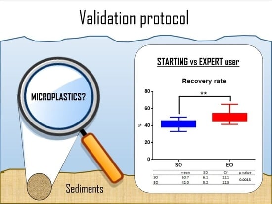
The performance of the two operators was statistically different (Mann Whitney test, p-value = 0.0002) with a recovery rate of 42% for SO versus 51% for EO (Figure 4a).
Statistically significant differences (p < 0.05) between operators were found for the smaller size class (i.e., 300–500 µm), for the polymer PE, and for the color category orange/pink/red, as shown in Figure 4b–e and in Table 2. No important differences were found for microplastic type.

3.3.2. Filter Reading Error

Further analysis was performed to better characterize the 9% difference in performance between the starting and expert operators, which could be due to: (a) errors in the extraction phase or (b) errors in reading the filters. The error in reading the filters was described using cross-reading, i.e., EO analyzed the filters from SO. The reading error corresponded to 1%. Thus, the remaining 8% could be due to sample extraction/handling errors. Specifically, SO underestimated a minimum of 1 and a maximum of 7 microparticles, with an average error of 2.8 poorly counted microparticles (Table 3). Thus, SO underestimated the value 54.3% of the time and overestimated it 45.7% of the time.
From these data, it appears that the error committed by SO had a greater impact in qualitative rather than in quantitative terms. Indeed, in terms of size, there was an overall error of −3% and an RMS of 26.6, with the largest error associated with the underestimation of the 300–500 µm size class (−71%). For the polymer type ABS _small, the error was the largest (−83%), followed by the underestimation of all three types of GLITTER (error between −50% and −67%). As for color, with the exception of the yellow particles, the colors black/gray, orange/pink/red, and silver were underestimated with errors up to −50%. The results are shown in Figure 5 and detailed in Table 4.

4. Discussion

This protocol has been based on guidance from the Marine Strategy Framework Directive Technical Subgroup [2] and the recent literature [23], as well as preliminary testing, and has attempted to adopt a common vocabulary as much as possible, as suggested by the European Marine Observation and Data Network (i.e., EMODnet Thematic Lot n°4—Chemistry, [24]).
Our findings suggest that the first step of digestion in hydrogen peroxide is necessary to avoid flocculation. Indeed, the flocs would trap the MP and prevent its proper distribution according to the density gradient generated by the brine, as well as favor the clogging of the filters (see Figure S1 in the Supplementary Materials). In this context, the studies of Konechnaya et al. (2020) [25] showed that a high content of organic material with a density similar to that of MP can worsen the conditions for the detection of microplastics. Peroxide pretreatment is recommended in the guidelines on particle size analysis of marine sediments developed by the National System for Environmental Protection (SNPA) on Italian territory [26], and has also been used by other authors such as Sanchez-Nieva et al. (2017) [27]. In the present study, the possible aggressiveness of peroxide towards MP was evaluated through a chemical analysis of MP before and after this treatment. Although there were some variations in the spectral imprint of polystyrene and polyethylene, the treatment did not condition the correct recognition of FT-IR, so we strongly recommend the use of this step in MP extraction protocols of marine sediments rich in organic material.
The settling times of 24 h used in our experiment proved to be reasonable and in agreement with those of Vermeiren, Munoz, and Ikejima (2020) [28], who worked with organic-rich and fine-grained sediments. Indeed, the granulometric analysis we performed showed a 76% fraction of particles smaller than 300 µm. Further investigations are nevertheless needed to calibrate the optimal time depending on the granulometric characteristics of the matrix.
From the results obtained in this study, it was possible to draw the following conclusions:
(i)
NaCl solution provides the best cost-benefit ratio, but it is ineffective for extracting high-density polymers
The data we collected confirm the ineffectiveness of NaCl brine (density 1.2 g/cm3) in the extraction of dense polymers such as PVC (density 1.16–1.3 g/cm3; recovery of 0%). However, we decided to use NaCl brine because it has a better cost-benefit ratio, as evidenced by its widespread use in the scientific community [17,27,29,30] and also recommended in EU guidelines. In terms of environmental monitoring, and even more so on large spatial-temporal scales, the costs associated with the use of salts would allow the buoyancy of denser polymers (e.g., ZnCl2, 125 euros/kg, density 1.6 g/cm3; NaCl 38.9 euros/kg; data from SIGMAALDRICH.COM, 4 April 2022) that might be unsustainable. The only alternative would be to implement systems for recovery and reuse of solutions, as adopted by Imhof et al. (2012) [31]. However, this would require the purchase of specialized equipment and additional time.
(ii)
The recovery rate by size, polymer density, type and color
The average recovery rate of EO was 50.7%. PVC was never recovered, and, as mentioned earlier, followed in decreasing order by lightweight polymers such as PP, PE, ABS, and PS (95.8%, 91.7%, 68.8%, and 66.7%, respectively). The least recovered colors were yellow and silver (typical of glitters 1 and 3). In the case of the yellow film, we suspect that mud contamination weighed down the plastics so that they could not float. The fragments are associated with a lower recovery rate, which is inevitably influenced by the specific shape–color–size–polymer combination of our internal standard set. Further studies with targeted experimental designs are needed to better investigate which of these factors is more relevant.
(iii)
The reasons of low recovery rate
The reasons for this low rate can be attributed to the fact that this protocol used (i) sediments with medium-fine grain size and some organic component, properties known to make microplastic extraction difficult; and (ii) a set of internal standards, including (1) dense polymers (PVC) and therefore predictably not extractable with NaCl brine; (2) plastics very similar in shape, color, and size to sand grains (see ABS _small); (3) categories not commonly used, such as, for example, GLITTER with completely unique properties. Removing these last three components from the final calculation led to an increase in the recovery rate to 87.5%, which is consistent with the literature (87.2% [32]).
(iv)
Strategies to increase the recovery rate
To further increase the recovery rate, strategies were used such as (i) dark color filters to improve the contrast between the plastic and the background, (ii) reading each filter twice, and (iii) adding a third extraction step. In the first case, a different color of filters (black and white) was used. The use of black filters increased the average recovery rate by 5%. Considering that black filters are even cheaper than white ones on the market (e.g., 1.15 Euro/filter for black and 1.58 Euro/filter for white), we recommend the use of black filters. On the other hand, reading the filters twice did not bring significant improvements and can be avoided. Finally, the usefulness of an additional step in the brine was evaluated. Overall, it contributed 3%, but extended the analysis time by 24 h and improved the recovery (13%) of only two of the seven polymers, namely ABS and PE in fragments.
(v)
The operator’s experience qualitatively affected the recovery rate
However, from a qualitative point of view, cross-reading is much more useful, especially when one or more users performing the analyses have little experience. The difference in performance between an experienced and an inexperienced operator was around 9%, of which 1% was due to reading errors and the remaining 8% was due to possible errors in sample handling, spitting, and recovery during the different phases. In particular, statistically significant differences were found in the 300–500 µm size class, i.e., the smallest, with respect to PE, which in turn has implications for the “orange/pink/red” color category, since PE is orange and pink.
(vi)
The possible reasons of error
Specifically, SO made an average error of 2.9 particles, with a minimum of 1 and a maximum of 7 per sample. As for the nature of the error in reading the filters, it is known that this error can be twofold; it can consist in overestimation or underestimation. In our particular case, the error was both in overestimation (in 45.7% of the cases) and underestimation (54.3%). The underestimation error of −50% may be due to the lack of experience and/or attention in sorting particles that, especially in the case of large surface particles such as foils, may be more encrusted and camouflaged by the silt. Figure 6 illustrates this phenomenon. The figure also shows other examples of possible confounding that justify the −83.3% from ABS (300–500 µm), which is closely related to the −18% from the category (black/gray). In this case, the error was likely due to a confusion with the sediment particles—with which they share size, irregular shape, and color-and the presence of the shells of small crabs.
(vii)
Methodological shortcomings in the extraction of GLITTER
An even more significant error is the color silver, represented here by glitters 1 and 3. The glitter category represents a unique specimen that we have chosen because, although they have the properties to be considered microplastic, they are usually ignored and excluded from the analysis. Nevertheless, their presence in sediments can be important, because glitters often contain a core of high-density polymers (such as PET or PVC). Evidence for their presence in sediments was provided by Ballent et al. (2016) [33], who found a small hexagonal glitter in sediments from Lake Ontario. In any case, the first step to scaling the problem is to recognize GLITTER as an ad hoc category and develop appropriate protocols for extraction. Our study showed that glitter is a very unfavorable category, corresponding to a recovery rate of 4.2% to 11.1% (SO and EO, respectively). Another 31.9% was found in sediments analyzed by sorting during ex post analysis. This may indicate that the use of a higher density solution would likely ensure a higher recovery rate, as suggested by Yurtsever (2019) [34] and experimentally demonstrated by Ballent et al., (2016) [32] who used a solution of polytungstate (1.5 g/cm3).
Above all, the difference between SO and EO seems to be negligible from a quantitative point of view, since the overestimation and underestimation values are quite similar (1% is therefore justified). In all cases where the correct classification of plastics is more important, experience may be a decisive factor. Contexts where the qualitative aspect may be important are, for example, those aimed at identifying the sources of MP in order to take mitigation measures. Future studies could aim to examine whether this qualitative difference is also more or less pronounced among experienced operators.
Finally, this protocol proposed solutions that are easy to disseminate, including the abandonment of the use of the vertical laminar flow hood. The flow hood undoubtedly has positive effects on reducing air pollution, but it is not widely used. Our protocol calls for the analysis to take place entirely in a room dedicated (at least for the time of the analysis) to the extraction of the plastics. Combined with other preventive measures, this resulted in an average contamination level of 1.5 fibers/lines for samples and 0.5 fragments for samples (although chemical analysis of the particles found in the controls is lacking as confirmation). Some microscopic images of the particles frequently found in the control filters can be found in the Supplemental Material (Figure S2).

5. Conclusions

The objectives of the present work were twofold: (a) to test the protocol on real, previously untreated sediment samples to obtain a recovery rate that can be used in the subsequent environmental monitoring phase; (b) to involve professionals and laypersons to evaluate whether the experience of the researcher is a key factor in the quality of the final data; and, in addition, (c) to provide useful information to the scientific community to encourage the elaboration of increasingly standardized methods.
In the first case, the low recovery rate (50%) highlighted that fine-grained, medium-rich, and microplasticized sediments (some of which are characterized by high polymer density and sediment-like color) represent conditions that significantly compromise the effectiveness of extraction protocols. Under the specific conditions we have replicated, the phenomenon of underestimation appears to be of significant importance in the context of environmental monitoring of sediments.
In the second case, the difference in performance between an experienced and an inexperienced operator was set at 9%, of which 1% was due to reading errors and the remaining 8% to possible errors in sample handling, spitting, and recovery during the different phases. In summary, the difference between SO and EO seems negligible from a quantitative point of view, but in all cases where the correct classification of plastics is more important, experience can be a decisive factor.
Our study showed that glitter is a very hostile category. Sorting during ex-post analysis suggests that a higher density solution must be used to ensure a higher recovery rate of this unique microplastic, which is usually ignored and excluded from analysis.
Finally, a protocol was proposed that uses easily accessible instruments and environments that favor rapid dissemination, with a weekly duration and with an average cost of consumables per sample (which represents a tripling in the context of monitoring) of 61 euros (through the purchase of NaCl from suppliers of chemical agents). This value decreases to six euros if you use common salt. More details can be found in the Supplementary Materials. Working in a closed room (avoiding the laminar flow hood) also ensures an acceptable level of air contamination (1.5 fibers/line and 0.5 fragments per sample).

Supplementary Materials

The following supporting information can be downloaded at: https://www.mdpi.com/article/10.3390/microplastics1030032/s1, Figure S1: A sample not digested with hydrogen peroxide exhibits flocculation, Figure S2: An example of particles found in controls; Table S1: Percentage of agreement between the spectra of 6% treated and untreated particles.

Author Contributions

Conceptualization, Formal analysis, Writing—original draft, Writing—review and editing: M.P.; Investigation: M.P., G.S.P. and S.A.; Resources, Writing—review and editing: M.R.; Funding, Writing—review and editing: S.B. All authors have read and agreed to the published version of the manuscript.

Funding

This work was carried out in the context of the MITFISH-N2K project: MITigation and monitoring of the interaction between artisanal fishing and fish fauna, protected species of aquatic avifauna, and benthic habitats in Natura 2000 sites, funded by the European Fund for Maritime Affairs and Fisheries (EMFF 2014-2020, Measure 1.40) Friuli Venezia Giulia Region.

Institutional Review Board Statement

Not applicable.

Informed Consent Statement

Not applicable.

Data Availability Statement

Not applicable.

Conflicts of Interest

The authors declare no conflict of interest.

References

  1. Hartmann, N.B.; Hüffer, T.; Thompson, R.C.; Hassellöv, M.; Verschoor, A.; Daugaard, A.E.; Rist, S.; Karlsson, T.M.; Brennholt, N.; Cole, M.; et al. Are We Speaking the Same Language? Recommendations for a Definition and Categorization Framework for Plastic Debris. Environ. Sci. Technol. 2019, 53, 1039–1047. [Google Scholar] [CrossRef] [PubMed]
  2. Bergmann, M.; Gutow, L.; Klages, M. Nanoplastics in the Aquatic Environment. Critical Review. In Marine Anthropogenic Litter; Springer: Berlin/Heidelberg, Germany, 2015; pp. 325–440. [Google Scholar]
  3. Arthur, J.B.; Bamford, H. Proceedings of the International Research Workshop on the Occurrence, Effects and Fate of Microplastic Marine Debris. In NOAA Technical Memorandum NOS-OR&R-30. 2009; p. 530. Available online: https://repository.library.noaa.gov/view/noaa/2509 (accessed on 4 April 2022).
  4. GESAMP. Guidelines for the Monitoring and Assessment of Plastic Litter in the Ocean; GESAMP: London, UK, 2019; 130p, Available online: http://www.gesamp.org/publications/guidelines-for-the-monitoring-and-assessment-of-plastic-litter-in-the-ocean (accessed on 4 April 2022).
  5. Suaria, G.; Avio, C.G.; Mineo, A.; Lattin, G.L.; Magaldi, M.G.; Belmonte, G.; Moore, C.J.; Regoli, F.; Aliani, S. The Mediterranean Plastic Soup: Synthetic polymers in Mediterranean surface waters. Sci. Rep. 2016, 6, 37551. [Google Scholar] [CrossRef] [PubMed]
  6. Baini, M.; Fossi, M.C.; Galli, M.; Caliani, I.; Campani, T.; Finoia, M.G.; Panti, C. Abundance and characterization of microplastics in the coastal waters of Tuscany (Italy): The application of the MSFD monitoring protocol in the Mediterranean Sea. Mar. Pollut. Bull. 2018, 133, 543–552. [Google Scholar] [CrossRef]
  7. Harris, P.T. The fate of microplastic in marine sedimentary environments: A review and synthesis. Mar. Pollut. Bull. 2020, 158, 111398. [Google Scholar] [CrossRef] [PubMed]
  8. Vandermeersch, G.; Van Cauwenberghe, L.; Janssen, C.R.; Marques, A.; Granby, K.; Fait, G.; Kotterman, M.J.; Diogène, J.; Bekaert, K.; Robbens, J.; et al. A critical view on microplastic quantification in aquatic organisms. Environ. Res. 2015, 143, 46–55. [Google Scholar] [CrossRef]
  9. Renzi, M.; Guerranti, C.; Blašković, A. Microplastic contents from maricultured and natural mussels. Mar. Pollut. Bull. 2018, 131, 248–251. [Google Scholar] [CrossRef]
  10. Sharma, S.; Vikas, S.; Subhankar, C. Microplastics in the Mediterranean Sea: Sources, Pollution Intensity, Sea Health, and Regulatory Policies. Front. Mar. Sci. 2021, 8, 634934. [Google Scholar] [CrossRef]
  11. Jambeck, J.R.; Geyer, R.; Wilcox, C.; Siegler, T.R.; Perryman, M.; Andrady, A.; Narayan, R.; Law, K.L. Plastic waste inputs from land into the ocean. Science 2015, 347, 768–771. [Google Scholar] [CrossRef]
  12. Woodall, L.C.; Sanchez-Vidal, A.; Canals, M.; Paterson, G.L.J.; Coppock, R.; Sleight, V.; Calafat, A.; Rogers, A.D.; Narayanaswamy, B.E.; Thompson, R.C. The deep sea is a major sink for microplastic debris. R. Soc. Open Sci. 2014, 1, 140317. Available online: http://rsos.royalsocietypublishing.org/content/1/4/140317.abstract (accessed on 4 April 2022). [CrossRef] [PubMed]
  13. Reinold, S.; Herrera, A.; Stile, N.; Saliu, F.; Hernández-González, C.; Martinez, I.; Ortega, Z.; Marrero, M.D.; Lasagni, M.; Gómez, M. An annual study on plastic accumulation in surface water and sediment cores from the coastline of Tenerife (Canary Island, Spain). Mar. Pollut. Bull. 2021, 173, 113072. [Google Scholar] [CrossRef] [PubMed]
  14. Lobelle, D.; Cunliffe, M. Early microbial biofilm formation on marine plastic debris. Mar. Pollut. Bull. 2011, 62, 197–200. [Google Scholar] [CrossRef] [PubMed]
  15. Rummel, C.D.; Jahnke, A.; Gorokhova, E.; Kühnel, D.; Schmitt-Jansen, M. Impacts of Biofilm Formation on the Fate and Potential Effects of Microplastic in the Aquatic Environment. Environ. Sci. Technol. Lett. 2017, 4, 258–267. [Google Scholar] [CrossRef]
  16. Tsangaris, C.; Panti, C.; Compa, M.; Pedà, C.; Digka, N.; Baini, M.; D’Alessandro, M.; Alomar, C.; Patsiou, D.; Giani, D.; et al. Interlaboratory comparison of microplastic extraction methods from marine biota tissues: A harmonization exercise of the Plastic Busters MPAs project. Mar. Pollut. Bull. 2021, 164, 111992. [Google Scholar] [CrossRef] [PubMed]
  17. Cadiou, J.-F.; Gerigny, O.; Koren, Š.; Zeri, C.; Kaberi, H.; Alomar, C.; Panti, C.; Fossi, M.; Adamopoulou, A.; Digka, N.; et al. Lessons learned from an intercalibration exercise on the quantification and characterisation of microplastic particles in sediment and water samples. Mar. Pollut. Bull. 2020, 154, 111097. [Google Scholar] [CrossRef] [PubMed]
  18. De Lazzari, A.; Rampazzo, G.; Pavoni, B. Geochemistry of sediments in the Northern and Central Adriatic Sea. Estuarine, Coast. Shelf Sci. 2004, 59, 429–440. [Google Scholar] [CrossRef]
  19. Green, D.S.; Jefferson, M.; Boots, B.; Stone, L. All that glitters is litter? Ecological impacts of conventional versus biodegradable glitter in a freshwater habitat. J. Hazard. Mater. 2020, 402, 124070. [Google Scholar] [CrossRef] [PubMed]
  20. Tagg, A.S.; do Sul, J.A.I. Is This Your Glitter? An Overlooked but Potentially Environmentally-Valuable Microplastic. Mar. Pollut. Bull. 2019, 146, 50–53. [Google Scholar] [CrossRef]
  21. Perosa, M.; Guerranti, C.; Renzi, M.; Bevilacqua, S. Taking the sparkle off the sparkling time. Mar. Pollut. Bull. 2021, 170, 112660. [Google Scholar] [CrossRef]
  22. Raju, S.; Carbery, M.; Kuttykattil, A.; Senthirajah, K.; Lundmark, A.; Rogers, Z.; Scb, S.; Evans, G.; Palanisami, T. Improved methodology to determine the fate and transport of microplastics in a secondary wastewater treatment plant. Water Res. 2020, 173, 115549. [Google Scholar] [CrossRef]
  23. Enders, K.; Lenz, R.; Sul, J.A.I.D.; Tagg, A.S.; Labrenz, M. When every particle matters: A QuEChERS approach to extract microplastics from environmental samples. MethodsX 2020, 7, 100784. [Google Scholar] [CrossRef] [PubMed]
  24. Galgani, F.; Giorgetti, A.; Vinci, M.; Le Moigne, M.; Moncoiffe, G.; Brosich, A.; Molina, E.; Lipizer, M.; Holdsworth, N.; Schlitzer, R.; et al. Proposal for Gathering and Managing Data Sets on Marine Micro-Litter on a European Scale; EMODnet: Oostende, Belgium, 2019. [Google Scholar]
  25. Konechnaya, O.; Lüchtrath, S.; Dsikowitzky, L.; Schwarzbauer, J. Optimized Microplastic Analysis Based on Size Fractionation, Density Separation and μ-FTIR. Water Sci. Technol. 2020, 81, 834–844. [Google Scholar] [CrossRef]
  26. Romano, E.; Ausili, A.; Bergamin, L.; Celia Magno, M.; Pierfranceschi, G.; Venti, F. Analisi Granulometriche Dei Sedimenti Marini; Linee Guida SNPA 18/2018; ISPRA: Roma, Italy, 2018. [Google Scholar]
  27. Sánchez-Nieva, J.; Perales, J.A.; González-Leal, J.M.; Rojo-Nieto, E. A new analytical technique for the extraction and quantification of microplastics in marine sediments focused on easy implementation and repeatability. Anal. Methods 2017, 9, 6371–6378. [Google Scholar] [CrossRef]
  28. Vermeiren, P.; Muñoz, C.; Ikejima, K. Microplastic identification and quantification from organic rich sediments: A validated laboratory protocol. Environ. Pollut. 2020, 262, 114298. [Google Scholar] [CrossRef]
  29. Reed, S.; Clark, M.; Thompson, R.; Hughes, K.A. Microplastics in marine sediments near Rothera Research Station, Antarctica. Mar. Pollut. Bull. 2018, 133, 460–463. [Google Scholar] [CrossRef]
  30. Vianello, A.; Boldrin, A.; Guerriero, P.; Moschino, V.; Rella, R.; Sturaro, A.; Da Ros, L. Microplastic particles in sediments of Lagoon of Venice, Italy: First observations on occurrence, spatial patterns and identification. Estuar. Coast. Shelf Sci. 2013, 130, 54–61. [Google Scholar] [CrossRef]
  31. Imhof, H.K.; Schmid, J.; Niessner, R.; Ivleva, N.P.; Laforsch, C. A novel, highly efficient method for the separation and quantification of plastic particles in sediments of aquatic environments. Limnol. Oceanogr. Methods 2012, 10, 524–537. [Google Scholar] [CrossRef]
  32. Scherer, C.; Weber, A.; Stock, F.; Vurusic, S.; Egerci, H.; Kochleus, C.; Arendt, N.; Foeldi, C.; Dierkes, G.; Wagner, M.; et al. Comparative Assessment of Microplastics in Water and Sediment of a Large European River Sci. Total Environ. 2020, 738, 139866. [Google Scholar] [CrossRef]
  33. Ballent, A.; Corcoran, P.L.; Madden, O.; Helm, P.A.; Longstaffe, F.J. Sources and sinks of microplastics in Canadian Lake Ontario nearshore, tributary and beach sediments. Mar. Pollut. Bull. 2016, 110, 383–395. [Google Scholar] [CrossRef] [PubMed]
  34. Yurtsever, M. Tiny, Shiny, and Colorful Microplastics: Are Regular Glitters a Significant Source of Microplastics? Mar. Pollut. Bull. 2019, 146, 678–682. [Google Scholar] [CrossRef]
Figure 1. Schematic representation of the different phases of the protocol. RT = room temperature.
Microplastics 01 00032 g001
Figure 2. Representative photo of each type of internal standard: from (ae) particles in the 1000–5000 µm size range; (f,g) particles between 500–1000 µm; (h,i) particles between 300–500 µm; from (jl) glitter.
Microplastics 01 00032 g002aMicroplastics 01 00032 g002b
Figure 3. Recovery rate (%) by polymer type. Mean value of low density category (in black) was compared to PVC (high-density polymer) and GLITTER (mixed composition). **** means they are statistically different from each other (p-value < 0.0001, Mann Whitney test).
Microplastics 01 00032 g003
Figure 4. Comparison of the performances of SO and EO, given as mean values (%) (a) and by size class (b), polymer type (c), color (d), and microplastic type (e). * and ** indicate that they are statistically different from each other (p-value < 0.05 and 0.001, respectively, Mann Whitney test).
Microplastics 01 00032 g004
Figure 5. Difference in (a) size, (b) polymer type, (c) color and (d) microplastic type in comparison to the reference (SO-crossed) in % and RMS errors.
Microplastics 01 00032 g005
Figure 6. Example of possible confusion: letter A shows how ABS particles (A2) can be confused with the shells of small crabs (A1); letter B shows how film particles (B2) can be camouflaged by silt (B1).
Microplastics 01 00032 g006
Table 1. Detailed description of the internal standards used in the protocol. The size class, shape, chemical composition, color, commercial origin, number of added particles by type, total added plastics for each size class, and finally the amount to be added in each sediment sample (24 plastics) are given. The vocabulary proposed by Galgani et al., 2020 [24] was used for the description of the particles. The polymer composition of GLITTER category refers to the outer layer.
Size ClassesTypeShapePolymerColorPrecursorItems AddedTotal per Size ClassTotal per Beaker
300–500 µmfragmentsbroken edgesPVCyellowindustrial foil2424
fragmentsbroken edgesABSgrayindustrial dust2
500–1000 µmfragmentsbroken edgesPVCyellowindustrial foil24
fragmentsangular edgesPEorangebeverage lids2
1000–5000 µmfragmentsangular edgesPEorangebeverage lids210
fragmentsbroken edgesABSgrayindustrial dust2
filaments/PPorangelanyards2
filmsbroken edgesPSyellowfood container2
spherespheruloidPEpinkbody cleanser2
GLITTER/exagonalPMMAsilverhair gel for kids26
exagonalPMMApinkpurpurine2
rectangularPEsilverpurpurine2
Table 2. Recovery rate (%) of expert (EO) and starting operator (SO) expressed as mean values, standard deviation (SD), and coefficient of variation (CV). Data are reported by size category, polymer type, color, and type of microplastic. The color categories are those proposed by Galgani et al., 2020 [24] for the construction of a common vocabulary in the management of data sets on marine micro-litter on a European scale. Comparison of the performances of SO and EO expressed as p-value.
Recovery Rate (%)
SO EO p Value
meanSDCVmeanSDCV
GLOBAL42.05.212.350.76.112.10.0016
300–500 µum4.29.7233.525.015.160.30.0124
500–1000 µm22.916.772.939.616.742.20.2303
1000–5000 µm87.511.413.089.29.010.10.8378
glitter4.214.0336.411.121.1189.70.1887
PVC8.319.0228.40.00.0-0.1092
PE72.217.924.891.713.314.50.0224
ABS47.947.799.668.832.347.00.1184
PS75.039.953.266.738.958.40.6399
PP87.522.625.895.814.415.10.5901
black/gray47.947.799.668.832.347.00.1184
yellow41.729.570.733.319.558.40.3856
orange/pink/red54.618.333.568.117.425.60.0477
silver4.214.1338.88.319.0228.40.6662
fragment38.244.0115.252.144.885.90.0644
line87.522.625.895.814.415.10.5901
sphere100.00.00.0100.00.00.0>0.9999
film75.039.953.266.738.958.40.6399
Table 3. Mean, standard deviation, minimum, and maximum number of particles not registered by the starting operator. OVER and UNDER refer to the sum of overestimated and underestimated particles, respectively.
# of Particles Not Registered by SO
mean2.9
SD2.5
min1.0
max7.0
OVER16.0
UNDER19.0
Table 4. Root mean square (RMS) and percentage error divided for size, color, microplastic type and polymer type. PVC_s = PVC 300–500 µm; PVC_b = PVC 500–1000 µm; ABS_s = ABS 300–500 µm; ABS_b = ABS 1000–5000 µm; PE_s = PE 500–1000 µm; PE_b = PET 1000–5000 µm.
SIZECOLORTYPE
300–500500–10001000–5000TOTAL Black/GrayYellowOrange/Pink/RedSilverTOTALFragmentLINESPHEREFILMTOTAL
RMS20.63.21.626.6 5.24.64.114.416.51.03.01.30.016.9
error (%)−71.411.15.5−2.6 −17.915.8−14.3−50.0−11.6−3.510.54.30.0−2.4
POLYMER TYPE
PVC_sABS_sPVC_bPE_sPE_bABS_bPPPSGLITTER 1GLITTER 2GLLITTER 3TOTAL
RMS 0.024.1nd5.82.10.03.80.014.419.214.437.7
error (%) 0.0−83.3nd−20.07.30.013.30.0−50.0−66.7−50.0−2.5
Publisher’s Note: MDPI stays neutral with regard to jurisdictional claims in published maps and institutional affiliations.