Non-invasive Early Detection of Cancers (Closed)

A topical collection in Cancers (ISSN 2072-6694). This collection belongs to the section "Cancer Causes, Screening and Diagnosis".

Viewed by 17989

Editor


E-Mail Website
Collection Editor
Department of Maxillofacial Diagnostic and Surgical Science, Field of Oral and Maxillofacial Rehabilitation, Graduate School of Medical and Dental Sciences, Kagoshima University, 8-35-1, Sakuragaoka, Kagoshima 890-8544, Japan
Interests: oral cancer; metastasis; oral hypofunction; cell-free DNA; DNA methylation; miRNA; circulating tumor cell (CTC); liquid biopsy
Special Issues, Collections and Topics in MDPI journals

Topical Collection Information

Dear Colleagues,

The early detection of cancer is currently the most effective way to improve treatment success and prognosis. Early detection usually means the detection of cancers at a very early stage, before the early cancer signs or symptoms arise in mass population screening. Detecting cancer in mass screening is commonly performed in certain cancer types (breast, cervical, lung, and gastric cancer). However, there are problems with mass screening (detection rate, sensitivity, specificity and screening rate, cost). It is also a big issue that while cancer screening is usually useful when it comes to common cancers, there is no screening test for rare cancers. In this regard, the development of novel detection methods with high accuracy for cancer (especially for rare types of cancer) is expected.

A non-invasive detection method for cancer has recently been reported based on the cancer cell biology, which works by detecting cancer cell metabolites including cell-free DNA, microRNA, and circulating tumor cells (CTCs). This Topical Collection will highlight non-invasive early detection methods (screening test, device) for various types of cancers for improving prognosis.

Prof. Dr. Tsuyoshi Sugiura
Collection Editor

Manuscript Submission Information

Manuscripts should be submitted online at www.mdpi.com by registering and logging in to this website. Once you are registered, click here to go to the submission form. Manuscripts can be submitted until the deadline. All submissions that pass pre-check are peer-reviewed. Accepted papers will be published continuously in the journal (as soon as accepted) and will be listed together on the collection website. Research articles, review articles as well as communications are invited. For planned papers, a title and short abstract (about 100 words) can be sent to the Editorial Office for announcement on this website.

Submitted manuscripts should not have been published previously, nor be under consideration for publication elsewhere (except conference proceedings papers). All manuscripts are thoroughly refereed through a single-blind peer-review process. A guide for authors and other relevant information for submission of manuscripts is available on the Instructions for Authors page. Cancers is an international peer-reviewed open access semimonthly journal published by MDPI.

Please visit the Instructions for Authors page before submitting a manuscript. The Article Processing Charge (APC) for publication in this open access journal is 2900 CHF (Swiss Francs). Submitted papers should be well formatted and use good English. Authors may use MDPI's English editing service prior to publication or during author revisions.

Published Papers (6 papers)

2023

Jump to: 2022, 2021

12 pages, 1459 KiB  
Article
Sniffer Dogs Diagnose Lung Cancer by Recognition of Exhaled Gases: Using Breathing Target Samples to Train Dogs Has a Higher Diagnostic Rate Than Using Lung Cancer Tissue Samples or Urine Samples
by Shih-Feng Liu, Hung-I Lu, Wei-Lien Chi, Guan-Heng Liu and Ho-Chang Kuo
Cancers 2023, 15(4), 1234; https://doi.org/10.3390/cancers15041234 - 15 Feb 2023
Cited by 3 | Viewed by 2013
Abstract
Introduction: Sniffer dogs can diagnose lung cancer. However, the diagnostic yields of different samples and training methods for lung cancer remain undetermined. Objective: Six dogs were trained in three stages with the aim of improving the diagnostic yield of lung cancer by comparing [...] Read more.
Introduction: Sniffer dogs can diagnose lung cancer. However, the diagnostic yields of different samples and training methods for lung cancer remain undetermined. Objective: Six dogs were trained in three stages with the aim of improving the diagnostic yield of lung cancer by comparing training methods and specimens. Methods: The pathological tissues of 53 lung cancer patients and 6 non-lung cancer patients in the Department of Thoracic Surgery of Kaohsiung Chang Gung Hospital were collected, and the exhaled breath samples and urine samples were collected. Urine and exhaled breath samples were also collected from 20 healthy individuals. The specimens were sent to the Veterinary Department of Pingtung University of Science and Technology. Results: The dogs had a very low response rate to urine target samples in the first and second stages of training. The experimental results at the second stage of training found that after lung cancer tissue training, dogs were less likely to recognize lung cancer and healthy controls than through breath target training: the response rate to exhaled breathing target samples was about 8–55%; for urine target samples, it was only about 5–30%. When using exhaled air samples for training, the diagnosis rate of these dogs in lung cancer patients was 71.3% to 97.6% (mean 83.9%), while the false positive rate of lung cancer in the healthy group was 0.5% to 27.6% (mean 7.6%). Compared with using breathing target samples for training, the diagnosis rate of dogs trained with lung cancer tissue lung cancer was significantly lower (p < 0.05). The sensitivity and specificity of lung cancer tissue training (50.4% and 50.1%) were lower than the exhaled breath target training (91.7% and 85.1%). There is no difference in lung cancer diagnostic rate by sniff dogs among lung cancer histological types, location, and staging. Conclusion: Training dogs using breathing target samples to train dogs then to recognize exhaled samples had a higher diagnostic rate than training using lung cancer tissue samples or urine samples. Dogs had a very low response rate to urine samples in our study. Six canines were trained on lung cancer tissues and breathing target samples of lung cancer patients, then the diagnostic rate of the recognition of exhaled breath of lung cancer and non-lung cancer patients were compared. When using exhaled air samples for training, the diagnosis rate of these dogs in lung cancer patients was 71.3% to 97.6% (mean 83.9%), while the false positive rate of lung cancer in the healthy group was 0.5% to 27.6% (mean 7.6%). There was a significant difference in the average diagnosis rate of individual dog and overall dogs between the lung cancer group and the healthy group (p < 0.05). When using lung cancer tissue samples for training, lung cancer diagnosis rate of these dogs among lung cancer patients was only 15.5% to 40.9% (mean 27.7%). Compared with using breathing target samples for training, the diagnosis rate of dogs trained with lung cancer tissue lung cancer was significantly lower (p < 0.05). The sensitivity and specificity of lung cancer tissue training (50.4% and 50.1%) were lower than the exhaled breath target training (91.7% and 85.1%). The diagnostic rate of lung cancer by sniffer dogs has nothing to do with the current stage of lung cancer, pathologic type, and the location of tumor mass. Even in stage IA lung cancer, well-trained dogs can have a diagnostic rate of 100%. Using sniffer dogs to screen early lung cancer may have good clinical and economic benefits. Full article
Show Figures

Figure 1

11 pages, 1669 KiB  
Article
Tumour Cell Seeding to Lymph Nodes from In Situ Colorectal Cancer
by Maria Teresa Rodrigo-Calvo, Karmele Saez de Gordoa, Sandra Lopez-Prades, Ivan Archilla, Alba Diaz, Mario Berrios, Jordi Camps, Eva Musulen and Miriam Cuatrecasas
Cancers 2023, 15(3), 842; https://doi.org/10.3390/cancers15030842 - 30 Jan 2023
Cited by 3 | Viewed by 2217
Abstract
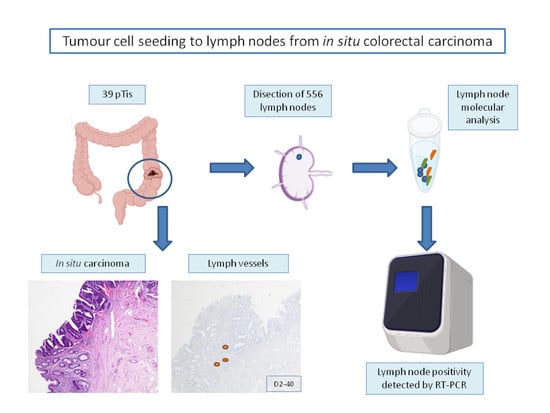
Lymph node (LN) metastasis is an important prognostic factor in colorectal cancer (CRC). We aimed to demonstrate the presence of lymphatic vessels (LV) in the mucosa of in-situ (pTis) CRC, and of detectable tumour burden in regional LNs. This is an observational retrospective [...] Read more.
Lymph node (LN) metastasis is an important prognostic factor in colorectal cancer (CRC). We aimed to demonstrate the presence of lymphatic vessels (LV) in the mucosa of in-situ (pTis) CRC, and of detectable tumour burden in regional LNs. This is an observational retrospective study of 39 surgically resected in situ CRCs. The number of LVs was evaluated in both pTis and normal mucosa using D2-40 immunostains. All LNs were assessed with both H&E and the One Step Nucleic Acid Amplification (OSNA) assay, and the results were correlated with clinicopathological features. D2-40 immunohistochemisty revealed LVs in the lamina propria of all pTis CRC (100%), being absent in normal mucosa. A median of 16 LNs were freshly dissected per patient, and all cases were pN0 with H&E. Molecular LN analysis with OSNA revealed the presence of low amounts of tumour burden in 11/39 (28%) cases (range 400 to 4270 CK19 mRNA copies/µL), which had no clinical consequences. This study demonstrates the presence of LVs in the lamina propria in 100% of pTis CRC, as well as the presence of low amounts of tumour burden in regional LNs, only detected by molecular methods. Given the prognostic value of LN tumour burden, its molecular quantification may help a patient’s clinical management. Full article
Show Figures

Graphical abstract

11 pages, 1600 KiB  
Article
Al[18F]F-NOTA-Octreotide Is Comparable to [68Ga]Ga-DOTA-TATE for PET/CT Imaging of Neuroendocrine Tumours in the Latin-American Population
by Arlette Haeger, Cristian Soza-Ried, Vasko Kramer, Ana Hurtado de Mendoza, Elisabeth Eppard, Noémie Emmanuel, Johanna Wettlin, Horacio Amaral and René Fernández
Cancers 2023, 15(2), 439; https://doi.org/10.3390/cancers15020439 - 10 Jan 2023
Cited by 5 | Viewed by 2201
Abstract
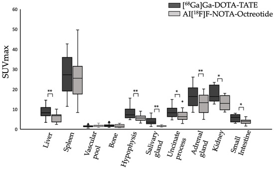
PET imaging of neuroendocrine tumours (NET) is well established for staging and therapy follow-up. The short half-life, increasing costs, and regulatory issues significantly limit the availability of approved imaging agents, such as [68Ga]Ga-DOTA-TATE. Al[18F]F-NOTA-Octreotide provides a similar biodistribution and [...] Read more.
PET imaging of neuroendocrine tumours (NET) is well established for staging and therapy follow-up. The short half-life, increasing costs, and regulatory issues significantly limit the availability of approved imaging agents, such as [68Ga]Ga-DOTA-TATE. Al[18F]F-NOTA-Octreotide provides a similar biodistribution and tumour uptake, can be produced on a large scale and may improve access to precision imaging. Here we prospectively compared the clinical utility of [68Ga]Ga-DOTA-TATE and Al[18F]F-NOTA-Octreotide in the Latin-American population. Our results showed that in patients with stage IV NETs [68Ga]Ga-DOTA-TATE presents higher physiological uptake than Al[18F]F-NOTA-Octreotide in the liver, hypophysis, salivary glands, adrenal glands (all p < 0.001), pancreatic uncinated process, kidneys, and small intestine (all p < 0.05). Nevertheless, despite the lower background uptake of Al[18F]F-NOTA-Octreotide, comparative analysis of tumour-to-liver (TLR) and tumour-to-spleen (TSR) showed no statistically significant difference for lesions in the liver, bone, lymph nodes, and other tissues. Only three discordant lesions in highly-metastases livers were detected by [68Ga]Ga-DOTA-TATE but not by Al[18F]F-NOTA-Octreotide and only one discordant lesion was detected by Al[18F]F-NOTA-Octreotide but not by [68Ga]Ga-DOTA-TATE. Non-inferiority analysis showed that Al[18F]F-NOTA-Octreotide is comparable to [68Ga]Ga-DOTA-TATE. Hence, our results demonstrate that Al[18F]F-NOTA-Octreotide provided excellent image quality, visualized NET lesions with high sensitivity and represents a highly promising, clinical alternative to [68Ga]Ga-DOTA-TATE. Full article
Show Figures

Figure 1

2022

Jump to: 2023, 2021

14 pages, 1044 KiB  
Article
A Novel Blood-Based microRNA Diagnostic Model with High Accuracy for Multi-Cancer Early Detection
by Andrew Zhang and Hai Hu
Cancers 2022, 14(6), 1450; https://doi.org/10.3390/cancers14061450 - 11 Mar 2022
Cited by 10 | Viewed by 4104
Abstract
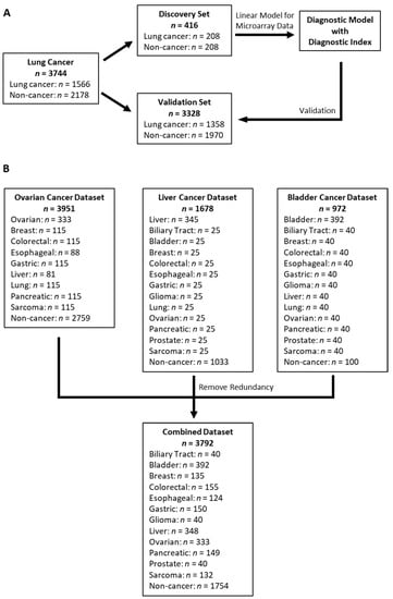
Early detection is critical to reduce cancer deaths as treating early stage cancers is more likely to be successful. However, patients with early stage diseases are often asymptomatic and thus less likely to be diagnosed. Here, we utilized four microarray datasets with a [...] Read more.
Early detection is critical to reduce cancer deaths as treating early stage cancers is more likely to be successful. However, patients with early stage diseases are often asymptomatic and thus less likely to be diagnosed. Here, we utilized four microarray datasets with a standardized platform to investigate comprehensive microRNA expression profiles from 7536 serum samples. A 4-miRNA diagnostic model was developed from the lung cancer training set (n = 416, 208 lung cancer patients and 208 non-cancer participants). The model showed 99% sensitivity and specificity in the lung cancer validation set (n = 3328, 1358 cancer patients and 1970 non-cancer participants); and the sensitivity remained to be >99% for patients with stage 1 disease. When applied to the additional combined dataset of 3792 participants including 2038 cancer patients across 12 different cancer types and 1754 independent non-cancer controls, the model demonstrated high sensitivities ranging from 83.2 to 100% for biliary tract, bladder, colorectal, esophageal, gastric, glioma, liver, pancreatic, and prostate cancers, and showed reasonable sensitivities of 68.2 and 72.0% for ovarian cancer and sarcoma, respectively, while maintaining 99.3% specificity. Our study provided a proof-of-concept data in demonstrating that the 4-miRNA model has the potential to be developed into a simple, inexpensive and noninvasive blood test for early detection of multiple cancers with high accuracy. Full article
Show Figures

Figure 1

2021

Jump to: 2023, 2022

10 pages, 1094 KiB  
Article
Contrast-Enhanced Mammographic Features of In Situ and Invasive Ductal Carcinoma Manifesting Microcalcifications Only: Help to Predict Underestimation?
by Yun-Chung Cheung, Kueian Chen, Chi-Chang Yu, Shir-Hwa Ueng, Chia-Wei Li and Shin-Cheh Chen
Cancers 2021, 13(17), 4371; https://doi.org/10.3390/cancers13174371 - 30 Aug 2021
Cited by 16 | Viewed by 2940
Abstract
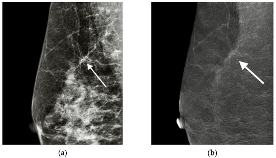
Background: The contrast-enhanced mammographic features of ductal carcinoma in situ (DCIS) and invasive ductal carcinoma (IDC) manifesting microcalcifications only on mammograms were evaluated to determine whether they could predict IDC underestimation. Methods: We reviewed patients who underwent mammography-guided biopsy on suspicious breast microcalcifications [...] Read more.
Background: The contrast-enhanced mammographic features of ductal carcinoma in situ (DCIS) and invasive ductal carcinoma (IDC) manifesting microcalcifications only on mammograms were evaluated to determine whether they could predict IDC underestimation. Methods: We reviewed patients who underwent mammography-guided biopsy on suspicious breast microcalcifications only and received contrast-enhanced spectral mammography (CESM) within 2 weeks before the biopsy. Those patients who were proven to have cancers (DCIS or IDC) by biopsy and subsequently had surgical treatment in our hospital were included for analysis. The presence or absence, size, morphology and texture of enhancement on contrast-enhanced spectral mammography were reviewed by consensus of two radiologists. Results: A total of 49 patients were included for analysis. Forty patients (81.6%) showed enhancement, including 18 (45%) DCIS and 22 (55%) IDC patients. All nine unenhanced cancers were pure DCIS. Pure DCIS showed 72.22% nonmass enhancement and 83.33% pure ground glass enhancement. IDC showed more mass (72.2% vs. 27.8%) and solid enhancements (83.33% vs. 16.67%). The cancer and texture of enhancement were significantly different between pure DCIS and IDC, with moderate diagnostic performance for the former (p-value < 0.01, AUC = 0.66, sensitivity = 93%, specificity = 39%) and the latter (p-value < 0.01, AUC = 0.74, sensitivity = 65%, specificity = 83%). Otherwise, pure DCIS showed a significant difference in enhanced texture compared with upgraded IDC and IDC (p = 0.0226 and 0.0018, respectively). Conclusions: Nonmass and pure ground glass enhancements were closely related to pure DCIS, and cases showing mass and unpurified solid enhancements should be suspected as IDC. Unenhanced DCIS with microcalcifications only has a low DCIS upgrade rate. The CESM-enhanced features could feasibly predict IDC underestimation. Full article
Show Figures

Figure 1

13 pages, 1747 KiB  
Systematic Review
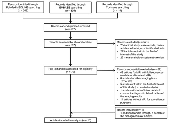
Meta-Analysis of the Accuracy of Abbreviated Magnetic Resonance Imaging for Hepatocellular Carcinoma Surveillance: Non-Contrast versus Hepatobiliary Phase-Abbreviated Magnetic Resonance Imaging
by Dong Hwan Kim, Sang Hyun Choi, Ju Hyun Shim, So Yeon Kim, Seung Soo Lee, Jae Ho Byun and Joon-Il Choi
Cancers 2021, 13(12), 2975; https://doi.org/10.3390/cancers13122975 - 14 Jun 2021
Cited by 21 | Viewed by 2631
Abstract
We aimed to determine the performance of surveillance abbreviated magnetic resonance imaging (AMRI) for detecting hepatocellular carcinoma (HCC), and to compare the performance of surveillance AMRI according to different protocols. Original research studies reporting the performance of surveillance AMRI for the detection of [...] Read more.
We aimed to determine the performance of surveillance abbreviated magnetic resonance imaging (AMRI) for detecting hepatocellular carcinoma (HCC), and to compare the performance of surveillance AMRI according to different protocols. Original research studies reporting the performance of surveillance AMRI for the detection of HCC were identified in MEDLINE, EMBASE, and Cochrane databases. The pooled sensitivity and specificity of surveillance AMRI were calculated using a hierarchical model. The pooled sensitivity and specificity of contrast-enhanced hepatobiliary phase (HBP)-AMRI and non-contrast (NC)-AMRI were calculated and compared using bivariate meta-regression. Ten studies, including 1547 patients, reported the accuracy of surveillance AMRI. The pooled sensitivity and specificity of surveillance AMRI for detecting any-stage HCC were 86% (95% confidence interval (CI), 80–90%; I2 = 0%) and 96% (95% CI, 93–98%; I2 = 80.5%), respectively. HBP-AMRI showed a significantly higher sensitivity for detecting HCC than NC-AMRI (87% vs. 82%), but significantly lower specificity (93% vs. 98%) (p = 0.03). Study quality and MRI magnet field strength were factors significantly associated with study heterogeneity (p ≤ 0.01). In conclusion, surveillance AMRI showed good overall diagnostic performance for detecting HCC. HBP-AMRI had significantly higher sensitivity for detecting HCC than NC-AMRI, but lower specificity. Full article
Show Figures

Figure 1

Back to TopTop