Extracellular Vesicles and Nucleic Acids in Health and Disease

Editors


E-Mail Website
Collection Editor
Department of Chemistry, Biology and Biotechnology, University of Perugia, Via del Giochetto, 06123 Perugia, Italy
Interests: extracellular vesicles; lipidomics; phospholipid metabolism; cell signaling
Special Issues, Collections and Topics in MDPI journals

E-Mail Website
Collection Editor
Department of Chemistry, Biology and Biotechnology, University of Perugia, Via del Giochetto, 06123 Perugia, Italy
Interests: extracellular vesicles; autophagy; lysosomes; senescence; aging; lipidomics
Special Issues, Collections and Topics in MDPI journals

Topical Collection Information

Dear Colleagues,

The term extracellular vesicles (EVs) is used to identify nanosized vesicles released by almost all cell types. EVs are present in every body fluid and their composition mirrors the type and the physio-pathological status of the originating cells. Due to these features and their targeting properties by recipient cells, EVs have been recently considered promising diagnostic and therapeutic tools. EVs contain different types of nucleic acids. The first studies demonstrated that EVs contain mRNAs that are translated and functionally active in target cells. This result led to the discovery that the horizontal transfer of genetic information via EVs represents one of the cell-to-cell communication mechanisms. Further, other non-coding RNA involved in protein synthesis or in regulatory functions, including tRNA, miRNA, siRNA, vtRNA, mtRNA, lncRNA, Y RNA, piRNA and snoRNA, have been found to be associated with EVs. Additionally, EVs isolated from cell culture media and biofluids carry DNA molecules, including mitochondrial DNA, single-stranded DNA and double-stranded DNA fragments.

In this Special Issue, authors are invited to submit original research and reviews on the nucleic acid content of EVs isolated from different sources and/or under different physio-pathological conditions, their biological roles, and their potential to be used as a diagnostic and therapeutic tool.

Dr. Sandra Buratta
Dr. Lorena Urbanelli
Collection Editors

Manuscript Submission Information

Manuscripts should be submitted online at www.mdpi.com by registering and logging in to this website. Once you are registered, click here to go to the submission form. Manuscripts can be submitted until the deadline. All submissions that pass pre-check are peer-reviewed. Accepted papers will be published continuously in the journal (as soon as accepted) and will be listed together on the collection website. Research articles, review articles as well as short communications are invited. For planned papers, a title and short abstract (about 100 words) can be sent to the Editorial Office for announcement on this website.

Submitted manuscripts should not have been published previously, nor be under consideration for publication elsewhere (except conference proceedings papers). All manuscripts are thoroughly refereed through a single-blind peer-review process. A guide for authors and other relevant information for submission of manuscripts is available on the Instructions for Authors page. Cells is an international peer-reviewed open access semimonthly journal published by MDPI.

Please visit the Instructions for Authors page before submitting a manuscript. The Article Processing Charge (APC) for publication in this open access journal is 2700 CHF (Swiss Francs). Submitted papers should be well formatted and use good English. Authors may use MDPI's English editing service prior to publication or during author revisions.

Keywords

  • extracellular vesicles
  • exosomes
  • microvesicles
  • nucleic acid delivery
  • cell-free nucleic acids
  • circulating RNAs
  • vesicular RNAs
  • nucleic acid biomarkers
  • liquid biopsy

Published Papers (6 papers)

2024

Jump to: 2023

19 pages, 2458 KiB  
Article
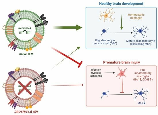
All but Small: miRNAs from Wharton’s Jelly-Mesenchymal Stromal Cell Small Extracellular Vesicles Rescue Premature White Matter Injury after Intranasal Administration
by Vera Tscherrig, Marel Steinfort, Valérie Haesler, Daniel Surbek, Andreina Schoeberlein and Marianne Simone Joerger-Messerli
Cells 2024, 13(6), 543; https://doi.org/10.3390/cells13060543 - 19 Mar 2024
Cited by 1 | Viewed by 1480
Abstract
White matter injury (WMI) is a common neurological issue in premature-born neonates, often causing long-term disabilities. We recently demonstrated a key beneficial role of Wharton’s jelly mesenchymal stromal cell-derived small extracellular vesicles (WJ-MSC-sEVs) microRNAs (miRNAs) in WMI-related processes in vitro. Here, we studied [...] Read more.
White matter injury (WMI) is a common neurological issue in premature-born neonates, often causing long-term disabilities. We recently demonstrated a key beneficial role of Wharton’s jelly mesenchymal stromal cell-derived small extracellular vesicles (WJ-MSC-sEVs) microRNAs (miRNAs) in WMI-related processes in vitro. Here, we studied the functions of WJ-MSC-sEV miRNAs in vivo using a preclinical rat model of premature WMI. Premature WMI was induced in rat pups through inflammation and hypoxia-ischemia. Small EVs were purified from the culture supernatant of human WJ-MSCs. The capacity of WJ-MSC-sEV-derived miRNAs to decrease microglia activation and promote oligodendrocyte maturation was evaluated by knocking down (k.d) DROSHA in WJ-MSCs, releasing sEVs containing significantly less mature miRNAs. Wharton’s jelly MSC-sEVs intranasally administrated 24 h upon injury reached the brain within 1 h, remained detectable for at least 24 h, significantly reduced microglial activation, and promoted oligodendrocyte maturation. The DROSHA k.d in WJ-MSCs lowered the therapeutic capabilities of sEVs in experimental premature WMI. Our results strongly indicate the relevance of miRNAs in the therapeutic abilities of WJ-MSC-sEVs in premature WMI in vivo, opening the path to clinical application. Full article
Show Figures

Graphical abstract

24 pages, 3616 KiB  
Article
Effects of Eribulin on the RNA Content of Extracellular Vesicles Released by Metastatic Breast Cancer Cells
by Matteo Giulietti, Francesco Piva, Monia Cecati, Serena Maggio, Michele Guescini, Tiziana Saladino, Laura Scortichini, Sonia Crocetti, Miriam Caramanti, Nicola Battelli and Emanuela Romagnoli
Cells 2024, 13(6), 479; https://doi.org/10.3390/cells13060479 - 8 Mar 2024
Viewed by 1721
Abstract
Extracellular vesicles (EVs) are small lipid particles secreted by almost all human cells into the extracellular space. They perform the essential function of cell-to-cell communication, and their role in promoting breast cancer progression has been well demonstrated. It is known that EVs released [...] Read more.
Extracellular vesicles (EVs) are small lipid particles secreted by almost all human cells into the extracellular space. They perform the essential function of cell-to-cell communication, and their role in promoting breast cancer progression has been well demonstrated. It is known that EVs released by triple-negative and highly aggressive MDA-MB-231 breast cancer cells treated with paclitaxel, a microtubule-targeting agent (MTA), promoted chemoresistance in EV-recipient cells. Here, we studied the RNA content of EVs produced by the same MDA-MB-231 breast cancer cells treated with another MTA, eribulin mesylate. In particular, we analyzed the expression of different RNA species, including mRNAs, lncRNAs, miRNAs, snoRNAs, piRNAs and tRNA fragments by RNA-seq. Then, we performed differential expression analysis, weighted gene co-expression network analysis (WGCNA), functional enrichment analysis, and miRNA-target identification. Our findings demonstrate the possible involvement of EVs from eribulin-treated cells in the spread of chemoresistance, prompting the design of strategies that selectively target tumor EVs. Full article
Show Figures

Figure 1

28 pages, 1581 KiB  
Review
Extracellular Vesicles, Circulating Tumor Cells, and Immune Checkpoint Inhibitors: Hints and Promises
by Sara Bandini, Paola Ulivi and Tania Rossi
Cells 2024, 13(4), 337; https://doi.org/10.3390/cells13040337 - 13 Feb 2024
Cited by 4 | Viewed by 2102
Abstract
Immune checkpoint inhibitor (ICI) therapy has revolutionized the treatment of cancer, in particular lung cancer, while the introduction of predictive biomarkers from liquid biopsies has emerged as a promising tool to achieve an effective and personalized therapy response. Important progress has also been [...] Read more.
Immune checkpoint inhibitor (ICI) therapy has revolutionized the treatment of cancer, in particular lung cancer, while the introduction of predictive biomarkers from liquid biopsies has emerged as a promising tool to achieve an effective and personalized therapy response. Important progress has also been made in the molecular characterization of extracellular vesicles (EVs) and circulating tumor cells (CTCs), highlighting their tremendous potential in modulating the tumor microenvironment, acting on immunomodulatory pathways, and setting up the pre-metastatic niche. Surface antigens on EVs and CTCs have proved to be particularly useful in the case of the characterization of potential immune escape mechanisms through the expression of immunosuppressive ligands or the transport of cargos that may mitigate the antitumor immune function. On the other hand, novel approaches, to increase the expression of immunostimulatory molecules or cargo contents that can enhance the immune response, offer premium options in combinatorial clinical strategies for precision immunotherapy. In this review, we discuss recent advances in the identification of immune checkpoints using EVs and CTCs, their potential applications as predictive biomarkers for ICI therapy, and their prospective use as innovative clinical tools, considering that CTCs have already been approved by the Food and Drug Administration (FDA) for clinical use, but providing good reasons to intensify the research on both. Full article
Show Figures

Graphical abstract

19 pages, 4129 KiB  
Article
Small RNA Changes in Plasma Have Potential for Early Diagnosis of Alzheimer’s Disease before Symptom Onset
by Joanna Palade, Eric Alsop, Amanda Courtright-Lim, Michael Hsieh, Timothy G. Whitsett, Douglas Galasko and Kendall Van Keuren-Jensen
Cells 2024, 13(3), 207; https://doi.org/10.3390/cells13030207 - 23 Jan 2024
Viewed by 2287
Abstract
Alzheimer’s disease (AD), due to its multifactorial nature and complex etiology, poses challenges for research, diagnosis, and treatment, and impacts millions worldwide. To address the need for minimally invasive, repeatable measures that aid in AD diagnosis and progression monitoring, studies leveraging RNAs associated [...] Read more.
Alzheimer’s disease (AD), due to its multifactorial nature and complex etiology, poses challenges for research, diagnosis, and treatment, and impacts millions worldwide. To address the need for minimally invasive, repeatable measures that aid in AD diagnosis and progression monitoring, studies leveraging RNAs associated with extracellular vesicles (EVs) in human biofluids have revealed AD-associated changes. However, the validation of AD biomarkers has suffered from the collection of samples from differing points in the disease time course or a lack of confirmed AD diagnoses. Here, we integrate clinical diagnosis and postmortem pathology data to form more accurate experimental groups and use small RNA sequencing to show that EVs from plasma can serve as a potential source of RNAs that reflect disease-related changes. Importantly, we demonstrated that these changes are identifiable in the EVs of preclinical patients, years before symptom manifestation, and that machine learning models based on differentially expressed RNAs can help predict disease conversion or progression. This research offers critical insight into early disease biomarkers and underscores the significance of accounting for disease progression and pathology in human AD studies. Full article
Show Figures

Figure 1

2023

Jump to: 2024

24 pages, 912 KiB  
Review
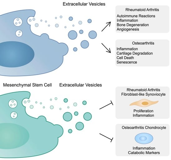
The Role of Extracellular Vesicles in the Pathogenesis and Treatment of Rheumatoid Arthritis and Osteoarthritis
by Estera Bakinowska, Kajetan Kiełbowski and Andrzej Pawlik
Cells 2023, 12(23), 2716; https://doi.org/10.3390/cells12232716 - 27 Nov 2023
Cited by 8 | Viewed by 2355
Abstract
Cells can communicate with each other through extracellular vesicles (EVs), which are membrane-bound structures that transport proteins, lipids and nucleic acids. These structures have been found to mediate cellular differentiation and proliferation apoptosis, as well as inflammatory responses and senescence, among others. The [...] Read more.
Cells can communicate with each other through extracellular vesicles (EVs), which are membrane-bound structures that transport proteins, lipids and nucleic acids. These structures have been found to mediate cellular differentiation and proliferation apoptosis, as well as inflammatory responses and senescence, among others. The cargo of these vesicles may include immunomodulatory molecules, which can then contribute to the pathogenesis of various diseases. By contrast, EVs secreted by mesenchymal stem cells (MSCs) have shown important immunosuppressive and regenerative properties. Moreover, EVs can be modified and used as drug carriers to precisely deliver therapeutic agents. In this review, we aim to summarize the current evidence on the roles of EVs in the progression and treatment of rheumatoid arthritis (RA) and osteoarthritis (OA), which are important and prevalent joint diseases with a significant global burden. Full article
Show Figures

Graphical abstract

15 pages, 8609 KiB  
Article
Oral Delivery of mRNA Vaccine by Plant-Derived Extracellular Vesicle Carriers
by Margherita A. C. Pomatto, Chiara Gai, Federica Negro, Lucia Massari, Maria Chiara Deregibus, Francesco Giuseppe De Rosa and Giovanni Camussi
Cells 2023, 12(14), 1826; https://doi.org/10.3390/cells12141826 - 11 Jul 2023
Cited by 8 | Viewed by 3596
Abstract
mRNA-based vaccines were effective in contrasting SARS-CoV-2 infection. However, they presented several limitations of storage and supply chain, and their parenteral administration elicited a limited mucosal IgA immune response. Extracellular vesicles (EVs) have been recognized as a mechanism of cell-to-cell communication well-preserved in [...] Read more.
mRNA-based vaccines were effective in contrasting SARS-CoV-2 infection. However, they presented several limitations of storage and supply chain, and their parenteral administration elicited a limited mucosal IgA immune response. Extracellular vesicles (EVs) have been recognized as a mechanism of cell-to-cell communication well-preserved in all life kingdoms, including plants. Their membrane confers protection from enzyme degradation to encapsulated nucleic acids favoring their transfer between cells. In the present study, EVs derived from the juice of an edible plant (Citrus sinensis) (oEVs) were investigated as carriers of an orally administered mRNA vaccine coding for the S1 protein subunit of SARS-CoV-2 with gastro-resistant oral capsule formulation. The mRNA loaded into oEVs was protected and was stable at room temperature for one year after lyophilization and encapsulation. Rats immunized via gavage administration developed a humoral immune response with the production of specific IgM, IgG, and IgA, which represent the first mucosal barrier in the adaptive immune response. The vaccination also triggered the generation of blocking antibodies and specific lymphocyte activation. In conclusion, the formulation of lyophilized mRNA-containing oEVs represents an efficient delivery strategy for oral vaccines due to their stability at room temperature, optimal mucosal absorption, and the ability to trigger an immune response. Full article
Show Figures

Graphical abstract

Back to TopTop