Cellular and Molecular Mechanisms of Atherosclerosis

A topical collection in Cells (ISSN 2073-4409). This collection belongs to the section "Cells of the Cardiovascular System".

Viewed by 37401

Editors


E-Mail Website
Guest Editor
Division of Biomedical Sciences, School of Medicine, University of California, Riverside, CA, USA
Interests: ahterosclerosis; inflammation; NF-kB; nulcear receptor; endocrine disrupting chemicals
Vascular Biology Program, Department of Surgery, Boston Children’s Hospital, Harvard Medical School, Boston, MA 02115, USA
Interests: atherosclerosis; angiogenesis; diabetes; lymphatic disorders; obesity; vascular inflammation; hypertension; stroke; cancer

Topical Collection Information

Dear Colleagues,

Atherosclerotic cardiovascular disease (CVD) is still the leading cause of mortality and morbidity worldwide despite advanced diagnosis and treatments. Atherosclerosis is a chronic inflammatory disease characterized by the accumulation of lipids and inflammatory molecules in the subendothelium of the artery, leading to restricted blood flow and ultimately myocardial infarction and stroke. Atherosclerosis is a complex chronic disease involving the interaction of genetic and environmental factors over multiple years. A multitude of treatments have been developed to halt or reverse the progression of this disease, including lifestyle changes, interventional procedures such as angioplasty and stent placement, and the prescription of medications to lower blood cholesterol levels, prevent clot formation, and reduce blood pressure. While these treatments have undoubtedly improved outcomes, there is more room for improvement. As a result, new classes of therapeutics are being developed to address the shortcomings of existing treatments.  At the same time, inquiries into the basic biological mechanisms of atherosclerosis continue at a rapid pace.

In this Special Issue of Cells, we invite you to contribute original research articles, reviews, or shorter perspective articles on all aspects related to the theme of “Atherosclerosis”. Expert articles describing the mechanistic, functional, cellular, biochemical, or general aspects of atherosclerosis are highly welcome.

Dr. Changcheng Zhou
Dr. Hong Chen
Guest Editors

Manuscript Submission Information

Manuscripts should be submitted online at www.mdpi.com by registering and logging in to this website. Once you are registered, click here to go to the submission form. Manuscripts can be submitted until the deadline. All submissions that pass pre-check are peer-reviewed. Accepted papers will be published continuously in the journal (as soon as accepted) and will be listed together on the collection website. Research articles, review articles as well as short communications are invited. For planned papers, a title and short abstract (about 100 words) can be sent to the Editorial Office for announcement on this website.

Submitted manuscripts should not have been published previously, nor be under consideration for publication elsewhere (except conference proceedings papers). All manuscripts are thoroughly refereed through a single-blind peer-review process. A guide for authors and other relevant information for submission of manuscripts is available on the Instructions for Authors page. Cells is an international peer-reviewed open access semimonthly journal published by MDPI.

Please visit the Instructions for Authors page before submitting a manuscript. The Article Processing Charge (APC) for publication in this open access journal is 2700 CHF (Swiss Francs). Submitted papers should be well formatted and use good English. Authors may use MDPI's English editing service prior to publication or during author revisions.

Keywords

  • signaling pathways
  • macrophage function
  • endothelial cell function and endothelial–mesenchymal transition in atherosclerosis
  • smooth muscle cell function and smooth muscle transdifferentiation in atherosclerosis
  • lipid homeostasis
  • mechanotransduction
  • system biology
  • environmental exposure
  • novel biomarkers
  • microbiota metabolism
  • in vitro and in vivo models
  • atherosclerosis calcification
  • small noncoding rnas and long noncoding rnas
  • covid-19
  • bioimaging
  • translational medicine
  • sex differences in atherosclerosis
  • new therapeutics

Published Papers (12 papers)

2023

Jump to: 2022

14 pages, 2926 KiB  
Article
Role of the Autism Risk Gene Shank3 in the Development of Atherosclerosis: Insights from Big Data and Mechanistic Analyses
by Hsiu-Wen Chang, Ming-Jen Hsu, Li-Nien Chien, Nai-Fang Chi, Meng-Chieh Yu, Hsiu-Chen Chen, Yuan-Feng Lin and Chaur-Jong Hu
Cells 2023, 12(21), 2546; https://doi.org/10.3390/cells12212546 - 30 Oct 2023
Viewed by 2323
Abstract
Increased medical attention is needed as the prevalence of autism spectrum disorder (ASD) rises. Both cardiovascular disorder (CVD) and hyperlipidemia are closely associated with adult ASD. Shank3 plays a key genetic role in ASD. We hypothesized that Shank3 contributes to CVD development in [...] Read more.
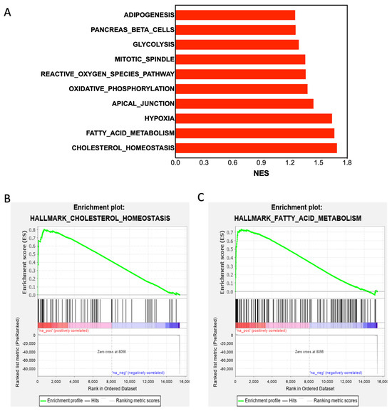
Increased medical attention is needed as the prevalence of autism spectrum disorder (ASD) rises. Both cardiovascular disorder (CVD) and hyperlipidemia are closely associated with adult ASD. Shank3 plays a key genetic role in ASD. We hypothesized that Shank3 contributes to CVD development in young adults with ASD. In this study, we investigated whether Shank3 facilitates the development of atherosclerosis. Using Gene Set Enrichment Analysis software (Version No.: GSEA-4.0.3), we analyzed the data obtained from Shank3 knockout mice (Gene Expression Omnibus database), a human population-based study cohort (from Taiwan’s National Health Insurance Research Database), and a Shank3 knockdown cellular model. Shank3 knockout upregulated the expression of genes of cholesterol homeostasis and fatty acid metabolism but downregulated the expression of genes associated with inflammatory responses. Individuals with autism had higher risks of hyperlipidemia (adjusted hazard ratio (aHR): 1.39; p < 0.001), major adverse cardiac events (aHR: 2.67; p < 0.001), and stroke (aHR: 3.55; p < 0.001) than age- and sex-matched individuals without autism did. Shank3 downregulation suppressed tumor necrosis factor-α-induced fatty acid synthase expression; vascular cell adhesion molecule 1 expression; and downstream signaling pathways involving p38, Jun N-terminal kinase, and nuclear factor-κB. Thus, Shank3 may influence the development of early-onset atherosclerosis and CVD in ASD. Furthermore, regulating Shank3 expression may reduce inflammation-related disorders, such as atherosclerosis, by inhibiting tumor necrosis factor-alpha-mediated inflammatory cascades. Full article
Show Figures

Figure 1

2022

Jump to: 2023

18 pages, 3750 KiB  
Article
Bioinformatic Analysis Revealed the Essential Regulatory Genes and Pathways of Early and Advanced Atherosclerotic Plaque in Humans
by Luling He, Andrea Palos-Jasso, Yao Yi, Manman Qin, Liang Qiu, Xiaofeng Yang, Yifeng Zhang and Jun Yu
Cells 2022, 11(24), 3976; https://doi.org/10.3390/cells11243976 - 8 Dec 2022
Cited by 2 | Viewed by 2105
Abstract
Atherosclerosis (AS) is a lipid-induced, chronic inflammatory, autoimmune disease affecting multiple arteries. Although much effort has been put into AS research in the past decades, it is still the leading cause of death worldwide. The complex genetic network regulation underlying the pathogenesis of [...] Read more.
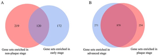
Atherosclerosis (AS) is a lipid-induced, chronic inflammatory, autoimmune disease affecting multiple arteries. Although much effort has been put into AS research in the past decades, it is still the leading cause of death worldwide. The complex genetic network regulation underlying the pathogenesis of AS still needs further investigation to provide effective targeted therapy for AS. We performed a bioinformatic microarray data analysis at different atherosclerotic plaque stages from the Gene Expression Omnibus database with accession numbers GSE43292 and GSE28829. Using gene set enrichment analysis, we further confirmed the immune-related pathways that play an important role in the development of AS. We are reporting, for the first time, that the metabolism of the three branched-chain amino acids (BCAAs; leucine, isoleucine, and valine) and short-chain fatty acids (SCFA; propanoate, and butanoate) are involved in the progression of AS using microarray data of atherosclerotic plaque tissue. Immune and muscle system-related pathways were further confirmed as highly regulated pathways during the development of AS using gene expression pattern analysis. Furthermore, we also identified four modules mainly involved in histone modification, immune-related processes, macroautophagy, and B cell activation with modular differential connectivity in the dataset of GSE43292, and three modules related to immune-related processes, B cell activation, and nuclear division in the dataset of GSE28829 also display modular differential connectivity based on the weighted gene co-expression network analysis. Finally, we identified eight key genes related to the pathways of immune and muscle system function as potential therapeutic biomarkers to distinguish patients with early or advanced stages in AS, and two of the eight genes were validated using the gene expression dataset from gene-deficient mice. The results of the current study will improve our understanding of the molecular mechanisms in the progression of AS. The key genes and pathways identified could be potential biomarkers or new drug targets for AS management. Full article
Show Figures

Figure 1

11 pages, 2000 KiB  
Article
Plasma Cell-Free DNA as a Novel Biomarker for the Diagnosis and Monitoring of Atherosclerosis
by Benheng Qian, Kexin Li, Xiaoying Lou, Ye Guo, Yidong Wang, Lianpin Wu and Donghong Zhang
Cells 2022, 11(20), 3248; https://doi.org/10.3390/cells11203248 - 16 Oct 2022
Cited by 3 | Viewed by 2203
Abstract
Atherosclerosis (AS) is the leading cause of cardiovascular diseases (CVDs) with a high rate of mortality worldwide. Plasma cell-free DNA (cfDNA), mainly originating from apoptosis, necrosis, and active secretion, has been recognized as a promising biomarker for the diagnosis and prognosis of multiple [...] Read more.
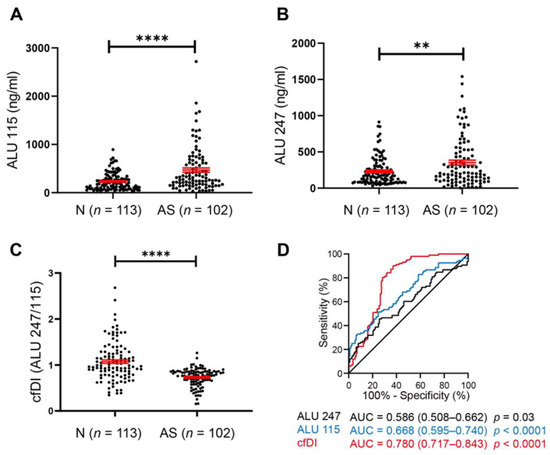
Atherosclerosis (AS) is the leading cause of cardiovascular diseases (CVDs) with a high rate of mortality worldwide. Plasma cell-free DNA (cfDNA), mainly originating from apoptosis, necrosis, and active secretion, has been recognized as a promising biomarker for the diagnosis and prognosis of multiple cancers, whereas there are no reports about cfDNA in CVDs. Here, we found an increased quantity and decreased integrity of cfDNA (cfDI) in the serum from AS patients compared with normal controls. Moreover, the reduced cfDI is inversely correlated with serum LDL levels, carotid plaque size, and carotid plaque thickness in the progression of AS. Consistently, in vivo experiments confirmed that the release and cleavage of cfDNA were increased concomitantly with the development and progression of AS in ApoE/ mice. Our study sheds light on the potential of cfDNA and cfDI as molecular biomarkers for detecting and monitoring AS. Full article
Show Figures

Figure 1

11 pages, 1555 KiB  
Article
Retinoic Acid Receptor Alpha (RARα) in Macrophages Protects from Diet-Induced Atherosclerosis in Mice
by Fathima N. Cassim Bawa, Raja Gopoju, Yanyong Xu, Shuwei Hu, Yingdong Zhu, Shaoru Chen, Kavita Jadhav and Yanqiao Zhang
Cells 2022, 11(20), 3186; https://doi.org/10.3390/cells11203186 - 11 Oct 2022
Cited by 11 | Viewed by 2384
Abstract
Retinoic acid signaling plays an important role in regulating lipid metabolism and inflammation. However, the role of retinoic acid receptor alpha (RARα) in atherosclerosis remains to be determined. In the current study, we investigated the role of macrophage RARα in the development of [...] Read more.
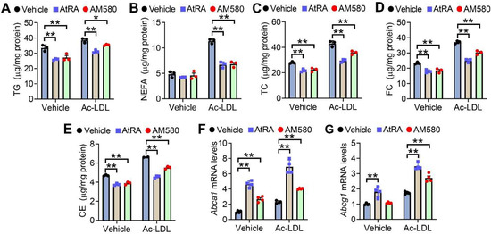
Retinoic acid signaling plays an important role in regulating lipid metabolism and inflammation. However, the role of retinoic acid receptor alpha (RARα) in atherosclerosis remains to be determined. In the current study, we investigated the role of macrophage RARα in the development of atherosclerosis. Macrophages isolated from myeloid-specific Rarα-/- (RarαMac-/-) mice showed increased lipid accumulation and inflammation and reduced cholesterol efflux compared to Rarαfl/fl (control) mice. All-trans retinoic acid (AtRA) induced ATP-binding cassette subfamily A member 1 (Abca1) and Abcg1 expression and cholesterol efflux in both RarαMac-/- mice and Rarαfl/fl mice. In Ldlr-/- mice, myeloid ablation of RARα significantly reduced macrophage Abca1 and Abcg1 expression and cholesterol efflux, induced inflammatory genes, and aggravated Western diet-induced atherosclerosis. Our data demonstrate that macrophage RARα protects against atherosclerosis, likely via inducing cholesterol efflux and inhibiting inflammation. Full article
Show Figures

Figure 1

16 pages, 2256 KiB  
Article
Influence of Shear Stress, Inflammation and BRD4 Inhibition on Human Endothelial Cells: A Holistic Proteomic Approach
by Johannes Jarausch, Lisa Neuenroth, Reiner Andag, Andreas Leha, Andreas Fischer, Abdul R. Asif, Christof Lenz and Abass Eidizadeh
Cells 2022, 11(19), 3086; https://doi.org/10.3390/cells11193086 - 30 Sep 2022
Cited by 3 | Viewed by 2259
Abstract
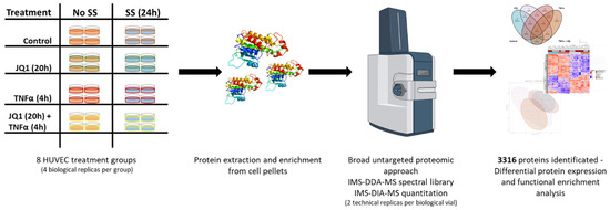
Atherosclerosis is an important risk factor in the development of cardiovascular diseases. In addition to increased plasma lipid concentrations, irregular/oscillatory shear stress and inflammatory processes trigger atherosclerosis. Inhibitors of the transcription modulatory bromo- and extra-terminal domain (BET) protein family (BETi) could offer a [...] Read more.
Atherosclerosis is an important risk factor in the development of cardiovascular diseases. In addition to increased plasma lipid concentrations, irregular/oscillatory shear stress and inflammatory processes trigger atherosclerosis. Inhibitors of the transcription modulatory bromo- and extra-terminal domain (BET) protein family (BETi) could offer a possible therapeutic approach due to their epigenetic mechanism and anti-inflammatory properties. In this study, the influence of laminar shear stress, inflammation and BETi treatment on human endothelial cells was investigated using global protein expression profiling by ion mobility separation-enhanced data independent acquisition mass spectrometry (IMS-DIA-MS). For this purpose, primary human umbilical cord derived vascular endothelial cells were treated with TNFα to mimic inflammation and exposed to laminar shear stress in the presence or absence of the BRD4 inhibitor JQ1. IMS-DIA-MS detected over 4037 proteins expressed in endothelial cells. Inflammation, shear stress and BETi led to pronounced changes in protein expression patterns with JQ1 having the greatest effect. To our knowledge, this is the first proteomics study on primary endothelial cells, which provides an extensive database for the effects of shear stress, inflammation and BETi on the endothelial proteome. Full article
Show Figures

Figure 1

8 pages, 733 KiB  
Review
Endothelial-to-Mesenchymal Transition in Atherosclerosis: Friend or Foe?
by Sarin Gole, Svyatoslav Tkachenko, Tarek Masannat, Richard A. Baylis and Olga A. Cherepanova
Cells 2022, 11(19), 2946; https://doi.org/10.3390/cells11192946 - 21 Sep 2022
Cited by 9 | Viewed by 3042
Abstract
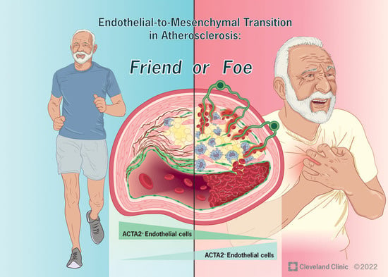
Despite many decades of research, complications of atherosclerosis resulting from the rupture or erosion of unstable plaques remain the leading cause of death worldwide. Advances in cellular lineage tracing techniques have allowed researchers to begin investigating the role of individual cell types in [...] Read more.
Despite many decades of research, complications of atherosclerosis resulting from the rupture or erosion of unstable plaques remain the leading cause of death worldwide. Advances in cellular lineage tracing techniques have allowed researchers to begin investigating the role of individual cell types in the key processes regulating plaque stability, including maintenance of the fibrous cap, a protective collagen-rich structure that underlies the endothelium. This structure was previously thought to be entirely derived from smooth muscle cells (SMC), which migrated from the vessel wall. However, recent lineage tracing studies have identified endothelial cells (EC) as an essential component of this protective barrier through an endothelial-to-mesenchymal transition (EndoMT), a process that has previously been implicated in pulmonary, cardiac, and kidney fibrosis. Although the presence of EndoMT in atherosclerotic plaques has been shown by several laboratories using EC-lineage tracing mouse models, whether EndoMT is detrimental (i.e., worsening disease progression) or beneficial (i.e., an athero-protective response that prevents plaque instability) remains uncertain as there are data to support both possibilities, which will be further discussed in this review. Full article
Show Figures

Graphical abstract

19 pages, 3349 KiB  
Article
Whole-Transcriptome Sequencing Analyses of Nuclear Antixoxidant-1 in Endothelial Cells: Role in Inflammation and Atherosclerosis
by Varadarajan Sudhahar, Yang Shi, Jack H. Kaplan, Masuko Ushio-Fukai and Tohru Fukai
Cells 2022, 11(18), 2919; https://doi.org/10.3390/cells11182919 - 18 Sep 2022
Cited by 5 | Viewed by 3098
Abstract
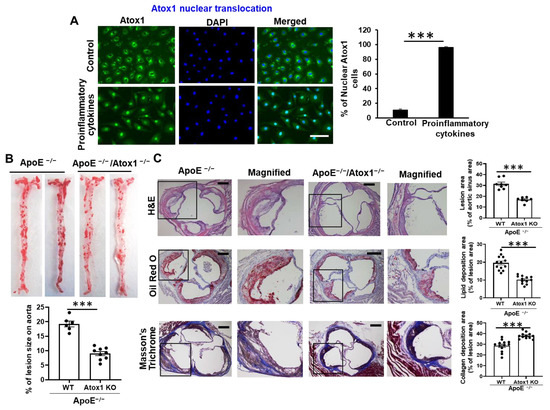
Inflammation, oxidative stress, and copper (Cu) play an important role in cardiovascular disease, including atherosclerosis. We previously reported that cytosolic Cu chaperone antioxidant-1 (Atox1) translocates to the nucleus in response to inflammatory cytokines or exogenous Cu and that Atox1 is localized at the [...] Read more.
Inflammation, oxidative stress, and copper (Cu) play an important role in cardiovascular disease, including atherosclerosis. We previously reported that cytosolic Cu chaperone antioxidant-1 (Atox1) translocates to the nucleus in response to inflammatory cytokines or exogenous Cu and that Atox1 is localized at the nucleus in the endothelium of inflamed atherosclerotic aorta. However, the roles of nuclear Atox1 and their function are poorly understood. Here we showed that Atox1 deficiency in ApoE−/− mice with a Western diet exhibited a significant reduction of atherosclerotic lesion formation. In vitro, adenovirus-mediated overexpression of nuclear-targeted Atox1 (Ad-Atox1-NLS) in cultured human endothelial cells (ECs) increased monocyte adhesion and reactive oxygen species (ROS) production compared to control cells (Ad-null). To address the underlying mechanisms, we performed genome-wide mapping of Atox1-regulated targets in ECs, using an unbiased systemic approach integrating sequencing data. Combination of ChIP-Seq and RNA-Seq analyses in ECs transfected with Ad-Atox1-NLS or Ad-null identified 1387 differentially expressed genes (DEG). Motif enrichment assay and KEGG pathway enrichment analysis revealed that 248 differentially expressed genes, including inflammatory and angiogenic genes, were regulated by Atox1-NLS, which was then confirmed by real-time qPCR. Among these genes, functional analysis of inflammatory responses identified CD137, CSF1, and IL5RA as new nuclear Atox1-targeted inflammatory genes, while CD137 is also a key regulator of Atox1-NLS-induced ROS production. These findings uncover new nuclear Atox1 downstream targets involved in inflammation and ROS production and provide insights into the nuclear Atox1 as a potential therapeutic target for the treatment of inflammatory diseases such as atherosclerosis. Full article
Show Figures

Figure 1

15 pages, 3508 KiB  
Article
Myeloid TM6SF2 Deficiency Inhibits Atherosclerosis
by Wenzhen Zhu, Wenying Liang, Haocheng Lu, Lin Chang, Jifeng Zhang, Y. Eugene Chen and Yanhong Guo
Cells 2022, 11(18), 2877; https://doi.org/10.3390/cells11182877 - 15 Sep 2022
Cited by 5 | Viewed by 2414
Abstract
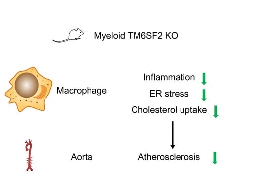
Genetic variants in transmembrane 6 superfamily member 2 (TM6SF2), such as E167K, are associated with atherosclerotic cardiovascular disease (ASCVD). Chronic inflammation and lipid-laden macrophage foam cell formation are the central pathogeneses in the development of atherosclerosis. This study was undertaken to [...] Read more.
Genetic variants in transmembrane 6 superfamily member 2 (TM6SF2), such as E167K, are associated with atherosclerotic cardiovascular disease (ASCVD). Chronic inflammation and lipid-laden macrophage foam cell formation are the central pathogeneses in the development of atherosclerosis. This study was undertaken to illustrate the biological function of TM6SF2 in macrophages and its role during atherosclerosis development. We generated myeloid cell-specific Tm6sf2 knockout mice on ApoE-deficient background (LysM Cre+/Tm6sf2fl/fl/ApoE−/−, TM6 mKO) with littermate LysM Cre−/Tm6sf2fl/fl/ApoE−/− (Control) mice as controls. Mice were fed a Western diet for 12 weeks to induce atherosclerosis. Myeloid Tm6sf2 deficiency inhibited atherosclerosis and decreased foam cells in the plaques without changing the plasma lipid profile. RNA sequencing of bone marrow-derived macrophages (BMDMs) from TM6 mKO mice demonstrated the downregulation of genes associated with inflammation, cholesterol uptake, and endoplasmic reticulum (ER) stress. TM6SF2 was upregulated by oxidized low-density lipoprotein (oxLDL) in macrophages. Silencing TM6SF2 in THP-1-derived macrophages and Tm6sf2 deficiency in BMDMs reduced inflammatory responses and ER stress and attenuated cholesterol uptake and foam cell formation, while the overexpression of TM6SF2 showed opposite effects. In conclusion, myeloid TM6SF2 deficiency inhibits atherosclerosis development and is a potential therapeutic target for the treatment of atherogenesis. Full article
Show Figures

Graphical abstract

27 pages, 12605 KiB  
Article
Cigarette Smoke and Morphine Promote Treg Plasticity to Th17 via Enhancing Trained Immunity
by Ying Shao, Fatma Saaoud, William Cornwell, Keman Xu, Aaron Kirchhoff, Yifan Lu, Xiaohua Jiang, Hong Wang, Thomas J. Rogers and Xiaofeng Yang
Cells 2022, 11(18), 2810; https://doi.org/10.3390/cells11182810 - 8 Sep 2022
Cited by 9 | Viewed by 3169
Abstract
CD4+ regulatory T cells (Tregs) respond to environmental cues to permit or suppress inflammation, and atherosclerosis weakens Treg suppression and promotes plasticity. However, the effects of smoking plus morphine (SM + M) on Treg plasticity remain unknown. To determine whether SM + [...] Read more.
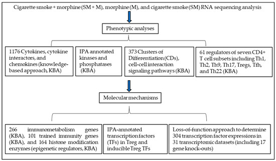
CD4+ regulatory T cells (Tregs) respond to environmental cues to permit or suppress inflammation, and atherosclerosis weakens Treg suppression and promotes plasticity. However, the effects of smoking plus morphine (SM + M) on Treg plasticity remain unknown. To determine whether SM + M promotes Treg plasticity to T helper 17 (Th17) cells, we analyzed the RNA sequencing data from SM, M, and SM + M treated Tregs and performed knowledge-based and IPA analysis. We demonstrated that (1) SM + M, M, and SM upregulated the transcripts of cytokines, chemokines, and clusters of differentiation (CDs) and modulated the transcripts of kinases and phosphatases in Tregs; (2) SM + M, M, and SM upregulated the transcripts of immunometabolism genes, trained immunity genes, and histone modification enzymes; (3) SM + M increased the transcripts of Th17 transcription factor (TF) RORC and Tfh factor CXCR5 in Tregs; M increased the transcripts of T helper cell 1 (Th1) TF RUNX3 and Th1-Th9 receptor CXCR3; and SM inhibited Treg TGIF1 transcript; (4) six genes upregulated in SM + M Tregs were matched with the top-ranked Th17 pathogenic genes; and 57, 39 genes upregulated in SM + M Tregs were matched with groups II and group III Th17 pathogenic genes, respectively; (5) SM + M upregulated the transcripts of 70 IPA-TFs, 11 iTregs-specific TFs, and 4 iTregs-Th17 shared TFs; and (6) SM + M, M, and SM downregulated Treg suppression TF Rel (c-Rel); and 35 SM + M downregulated genes were overlapped with Rel−/− Treg downregulated genes. These results provide novel insights on the roles of SM + M in reprogramming Treg transcriptomes and Treg plasticity to Th17 cells and novel targets for future therapeutic interventions involving immunosuppression in atherosclerotic cardiovascular diseases, autoimmune diseases, transplantation, and cancers. Full article
Show Figures

Figure 1

15 pages, 3787 KiB  
Article
HHcy Induces Pyroptosis and Atherosclerosis via the Lipid Raft-Mediated NOX-ROS-NLRP3 Inflammasome Pathway in apoE−/− Mice
by Sijun Liu, Jun Tao, Fengqi Duan, Huangjing Li and Hongmei Tan
Cells 2022, 11(15), 2438; https://doi.org/10.3390/cells11152438 - 6 Aug 2022
Cited by 21 | Viewed by 3239
Abstract
Lipid rafts play important roles in signal transduction, particularly in responses to inflammatory processes. The current study aimed to identify whether lipid raft-mediated inflammation contributes to hyperhomocysteinemia (HHcy)-accelerated atherosclerosis (AS), and to investigate the underlying mechanisms. THP-1-derived macrophages were used for in vitro [...] Read more.
Lipid rafts play important roles in signal transduction, particularly in responses to inflammatory processes. The current study aimed to identify whether lipid raft-mediated inflammation contributes to hyperhomocysteinemia (HHcy)-accelerated atherosclerosis (AS), and to investigate the underlying mechanisms. THP-1-derived macrophages were used for in vitro experiments. ApoE−/− mice were fed a high-fat diet for 12 weeks to establish an AS model, and a high-fat plus high-methionine diet was used to induce HHcy. We found that homocysteine (Hcy) increased the expression of p22phox and p67phox and promoted their recruitment into lipid rafts (indicating the assembly of the NOX complex), thereby increasing ROS generation and NOX activity, NLRP3 inflammasome activation, and pyroptosis. Mechanistically, Hcy activated the NOX-ROS-NLRP3 inflammasome pathway and induced pyroptosis by increasing the expression of acid sphingomyelinase (ASM) to promote the formation of lipid raft clustering. Importantly, lipid raft-mediated pyroptosis was confirmed in HHcy mice, and HHcy-promoted macrophage recruitment in atherosclerotic lesions and HHcy-aggravated AS were blocked by the lipid raft disruptor methyl-β-cyclodextrin. The study findings indicate that Hcy promotes lipid raft clustering via the upregulation of ASM, which mediates the assembly of the NOX complex, causing an increase in ROS generation, NLRP3 inflammasome activation, and pyroptosis, and contributes to HHcy-induced AS. Full article
Show Figures

Figure 1

15 pages, 805 KiB  
Review
The Role of Endothelial-to-Mesenchymal Transition in Cardiovascular Disease
by Qianman Peng, Dan Shan, Kui Cui, Kathryn Li, Bo Zhu, Hao Wu, Beibei Wang, Scott Wong, Vikram Norton, Yunzhou Dong, Yao Wei Lu, Changcheng Zhou and Hong Chen
Cells 2022, 11(11), 1834; https://doi.org/10.3390/cells11111834 - 3 Jun 2022
Cited by 21 | Viewed by 5996
Abstract
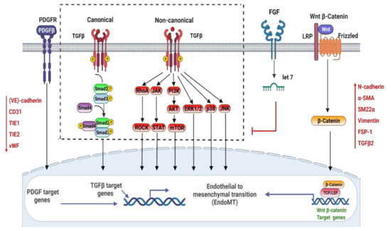
Endothelial-to-mesenchymal transition (EndoMT) is the process of endothelial cells progressively losing endothelial-specific markers and gaining mesenchymal phenotypes. In the normal physiological condition, EndoMT plays a fundamental role in forming the cardiac valves of the developing heart. However, EndoMT contributes to the development of [...] Read more.
Endothelial-to-mesenchymal transition (EndoMT) is the process of endothelial cells progressively losing endothelial-specific markers and gaining mesenchymal phenotypes. In the normal physiological condition, EndoMT plays a fundamental role in forming the cardiac valves of the developing heart. However, EndoMT contributes to the development of various cardiovascular diseases (CVD), such as atherosclerosis, valve diseases, fibrosis, and pulmonary arterial hypertension (PAH). Therefore, a deeper understanding of the cellular and molecular mechanisms underlying EndoMT in CVD should provide urgently needed insights into reversing this condition. This review summarizes a 30-year span of relevant literature, delineating the EndoMT process in particular, key signaling pathways, and the underlying regulatory networks involved in CVD. Full article
Show Figures

Figure 1

16 pages, 2874 KiB  
Article
Poloxamer 407 Induces Hypertriglyceridemia but Decreases Atherosclerosis in Ldlr−/− Mice
by Xueying Peng, Zeqin Lian, Xiao-Yuan Dai Perrard, Yunjie Xiao, Jing Ni, Veronica O’Brien, Henry Dong, Henry J. Pownall, Christie M. Ballantyne and Huaizhu Wu
Cells 2022, 11(11), 1795; https://doi.org/10.3390/cells11111795 - 30 May 2022
Cited by 5 | Viewed by 3187
Abstract
Background: Hypertriglyceridemia (HTG) increases the risk for atherosclerotic cardiovascular disease, but underlying mechanisms are incompletely understood. Circulating monocytes play an important role in atherogenesis by infiltrating arterial walls, where they differentiate into macrophages. We tested the hypothesis that HTG is mechanistically linked to [...] Read more.
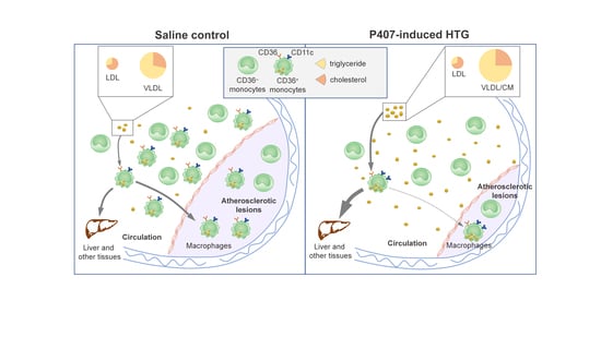
Background: Hypertriglyceridemia (HTG) increases the risk for atherosclerotic cardiovascular disease, but underlying mechanisms are incompletely understood. Circulating monocytes play an important role in atherogenesis by infiltrating arterial walls, where they differentiate into macrophages. We tested the hypothesis that HTG is mechanistically linked to atherogenesis by altering the monocyte phenotype and infiltration into atherosclerotic lesions in a model of diet-induced atherogenesis in Ldlr−/− mice. Methods: HTG was induced in male Ldlr−/− mice, fed a Western, high-fat high-cholesterol diet, by daily injection of poloxamer 407 (P407), a lipoprotein lipase inhibitor, for seven weeks. Atherosclerosis, monocyte phenotypes, and monocyte migration into atherosclerotic lesions were determined by well-validated methods. Results: Compared with the saline control, P407 injection in Ldlr−/− mice rapidly induced profound and persistent HTG, modestly elevated plasma cholesterol levels, and increased levels of triglyceride and cholesterol carried in very-low-density lipoprotein and low-density lipoprotein. Unexpectedly, mice receiving P407 versus saline control showed less atherosclerosis. Following induction of HTG by P407, CD36+ (also CD11c+), but not CD36 (CD11c), monocytes showed early increases in lipid accumulation, but the number of CD36+ (not CD36) monocytes was dramatically decreased afterwards in the circulation until the end of the test. Concurrently, CD36+ (CD11c+) monocyte migration into atherosclerotic lesions was also reduced in mice receiving P407 versus controls. Conclusions: P407 induced severe HTG, but reduced atherosclerosis, in Ldlr−/− mice, possibly because of profound reductions of circulating CD36+ (CD11c+) monocytes, leading to decreased monocyte migration into atherosclerotic lesions. Full article
Show Figures

Graphical abstract

Back to TopTop