Immunity, Inflammation, Oxidative Stress and Cancer

A topical collection in Cells (ISSN 2073-4409). This collection belongs to the section "Cellular Immunology".

Editor


E-Mail Website
Collection Editor
Laboratory for Oxidative Stress (LabOS), Rudjer Boskovic Institute, Bijenička 54, HR-10000 Zagreb, Croatia
Interests: oxidative stress; growth regulation; cancer; lipid peroxidation; 4-hydroxynonenal (HNE)
Special Issues, Collections and Topics in MDPI journals

Topical Collection Information

Dear Colleagues,

The ongoing pandemics force science to reconsider many fundamental aspects of major diseases, which were mostly considered to be non-transmissible, chronic and associated with stress and aging until the onset of COVID-19. When going to the molecular biomedicine level of these diseases, many researchers found oxidative stress either as their crucial pathogenic factor or as an important epiphenomenon that might help define novel biomarkers and/or adjuvant therapies according to the modern concepts of integrative biomedicine. Nowadays, these diseases are often denoted as “comorbidities” in respect to COVID-19; however, cancer was and is still of particular interest not only because of its severity and socio-medical relevance but also because it is a constant enigma for the immune system, which we assume acts as our crucial defense mechanism, due to the simplified perception of COVID-19. Similarly, since the 1970s, scientists have also assumed immunology will eventually provide effective solutions to cure cancer, while the ongoing pandemics demand reconsidering crucial pathophysiological aspects of immunology, notably of immunity and of inflammation in general (cytokine storm, trained immunity, etc.) and in respect to cancer in particular (the most fearsome, immunosuppressive “comorbidity”).  

Therefore, the aim of this Topical Collection (TC) is to collect comprehensive reviews and original research papers that provide new findings on the complex relationship between cancer and the immune system, focusing on inflammation, cancer development and therapies in particular. The onset of oxidative stress and endogenous and exogenous pro- and anti-oxidants will be of particular interest for the scope of this SI that aims to provide a better understanding of immunity, inflammation and cancer, thus offering new ideas and concepts for better diagnostics and treatment protocols.

Prof. Dr. Neven Zarkovic
Collection Editor

Manuscript Submission Information

Manuscripts should be submitted online at www.mdpi.com by registering and logging in to this website. Once you are registered, click here to go to the submission form. Manuscripts can be submitted until the deadline. All submissions that pass pre-check are peer-reviewed. Accepted papers will be published continuously in the journal (as soon as accepted) and will be listed together on the collection website. Research articles, review articles as well as short communications are invited. For planned papers, a title and short abstract (about 100 words) can be sent to the Editorial Office for announcement on this website.

Submitted manuscripts should not have been published previously, nor be under consideration for publication elsewhere (except conference proceedings papers). All manuscripts are thoroughly refereed through a single-blind peer-review process. A guide for authors and other relevant information for submission of manuscripts is available on the Instructions for Authors page. Cells is an international peer-reviewed open access semimonthly journal published by MDPI.

Please visit the Instructions for Authors page before submitting a manuscript. The Article Processing Charge (APC) for publication in this open access journal is 2700 CHF (Swiss Francs). Submitted papers should be well formatted and use good English. Authors may use MDPI's English editing service prior to publication or during author revisions.

Keywords

  • cancer
  • immune system
  • oxidative stress
  • inflammation
  • immunity

Published Papers (2 papers)

2022

21 pages, 2304 KiB  
Review
IRF8: Mechanism of Action and Health Implications
by Hannah R. Moorman, Yazmin Reategui, Dakota B. Poschel and Kebin Liu
Cells 2022, 11(17), 2630; https://doi.org/10.3390/cells11172630 - 24 Aug 2022
Cited by 15 | Viewed by 6208
Abstract
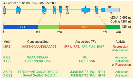
Interferon regulatory factor 8 (IRF8) is a transcription factor of the IRF protein family. IRF8 was originally identified as an essentialfactor for myeloid cell lineage commitment and differentiation. Deletion of Irf8 leads to massive accumulation of CD11b+Gr1+ immature myeloid cells [...] Read more.
Interferon regulatory factor 8 (IRF8) is a transcription factor of the IRF protein family. IRF8 was originally identified as an essentialfactor for myeloid cell lineage commitment and differentiation. Deletion of Irf8 leads to massive accumulation of CD11b+Gr1+ immature myeloid cells (IMCs), particularly the CD11b+Ly6Chi/+Ly6G polymorphonuclear myeloid-derived suppressor cell-like cells (PMN-MDSCs). Under pathological conditions such as cancer, Irf8 is silenced by its promoter DNA hypermethylation, resulting in accumulation of PMN-MDSCs and CD11b+ Ly6G+Ly6Clo monocytic MDSCs (M-MDSCs) in mice. IRF8 is often silenced in MDSCs in human cancer patients. MDSCs are heterogeneous populations of immune suppressive cells that suppress T and NK cell activity to promote tumor immune evasion and produce growth factors to exert direct tumor-promoting activity. Emerging experimental data reveals that IRF8 is also expressed in non-hematopoietic cells. Epithelial cell-expressed IRF8 regulates apoptosis and represses Osteopontin (OPN). Human tumor cells may use the IRF8 promoter DNA methylation as a mechanism to repress IRF8 expression to advance cancer through acquiring apoptosis resistance and OPN up-regulation. Elevated OPN engages CD44 to suppress T cell activation and promote tumor cell stemness to advance cancer. IRF8 thus is a transcription factor that regulates both the immune and non-immune components in human health and diseases. Full article
Show Figures

Figure 1

19 pages, 15357 KiB  
Article
Post-mortem Findings of Inflammatory Cells and the Association of 4-Hydroxynonenal with Systemic Vascular and Oxidative Stress in Lethal COVID-19
by Neven Zarkovic, Antonia Jakovcevic, Ana Mataic, Morana Jaganjac, Tea Vukovic, Georg Waeg and Kamelija Zarkovic
Cells 2022, 11(3), 444; https://doi.org/10.3390/cells11030444 - 27 Jan 2022
Cited by 17 | Viewed by 4345
Abstract
A recent comparison of clinical and inflammatory parameters, together with biomarkers of oxidative stress, in patients who died from aggressive COVID-19 and survivors suggested that the lipid peroxidation product 4-hydroxynonenal (4-HNE) might be detrimental in lethal SARS-CoV-2 infection. The current study further explores [...] Read more.
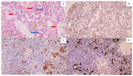
A recent comparison of clinical and inflammatory parameters, together with biomarkers of oxidative stress, in patients who died from aggressive COVID-19 and survivors suggested that the lipid peroxidation product 4-hydroxynonenal (4-HNE) might be detrimental in lethal SARS-CoV-2 infection. The current study further explores the involvement of inflammatory cells, systemic vascular stress, and 4-HNE in lethal COVID-19 using specific immunohistochemical analyses of the inflammatory cells within the vital organs obtained by autopsy of nine patients who died from aggressive SAR-CoV-2 infection. Besides 4-HNE, myeloperoxidase (MPO) and mitochondrial superoxide dismutase (SOD2) were analyzed alongside standard leukocyte biomarkers (CDs). All the immunohistochemical slides were simultaneously prepared for each analyzed biomarker. The results revealed abundant 4-HNE in the vital organs, but the primary origin of 4-HNE was sepsis-like vascular stress, not an oxidative burst of the inflammatory cells. In particular, inflammatory cells were often negative for 4-HNE, while blood vessels were always very strongly immunopositive, as was edematous tissue even in the absence of inflammatory cells. The most affected organs were the lungs with diffuse alveolar damage and the brain with edema and reactive astrocytes, whereas despite acute tubular necrosis, 4-HNE was not abundant in the kidneys, which had prominent SOD2. Although SOD2 in most cases gave strong immunohistochemical positivity similar to 4-HNE, unlike 4-HNE, it was always limited to the cells, as was MPO. Due to their differential expressions in blood vessels, inflammatory cells, and the kidneys, we think that SOD2 could, together with 4-HNE, be a potential link between a malfunctioning immune system, oxidative stress, and vascular stress in lethal COVID-19. Full article
Show Figures

Figure 1

Back to TopTop