jcm-logo

Journal Browser

Journal Browser

Pelvic Instability: Currents Insights (Closed)

A topical collection in Journal of Clinical Medicine (ISSN 2077-0383). This collection belongs to the section "Orthopedics".

Viewed by 45040

Editor


E-Mail Website
Guest Editor
Academic Department of Trauma & Orthopaedic Surgery, School of Medicine, University of Leeds, Leeds, UK
Interests: pelvic instability; pelvic reconstruction; non-union; bone regeneration; post fracture fixation complications
Special Issues, Collections and Topics in MDPI journals

Topical Collection Information

Dear Colleagues,

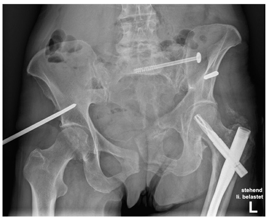
Pelvic instability is a clinical entity usually seen in the female patient population secondary to post-partum ligamentous laxity and hypermobility syndrome. In males, instability can occur due to degenerative conditions but also due to poorly stabilized pelvic fractures that continue to have a degree of mechanical instability.

Chronic pain and functional restriction, amongst others, represent the hallmark of the syndrome. Non-operative treatment modalities may provide temporary suppression of symptoms, but the ultimate treatment is the surgical reconstruction of the pelvic ring in the form of fusion. Management of this condition involves a variety of clinicians and specialties, as well as a combined interdisciplinary approach.

Despite the extensive clinical interest and the availability of several related publications, there is still a lack of consensus regarding the incidence, clinical manifestations, pathophysiology, diagnostics, treatment algorithms, and the final outcome of surgical reconstruction.

We invite both clinicians and scientists to contribute their work in this Special Issue focusing on this important clinical entity with the ultimate aim to advance further our understanding and the results of surgical treatment.

Prof. Dr. Peter V. Giannoudis
Guest Editor

Manuscript Submission Information

Manuscripts should be submitted online at www.mdpi.com by registering and logging in to this website. Once you are registered, click here to go to the submission form. Manuscripts can be submitted until the deadline. All submissions that pass pre-check are peer-reviewed. Accepted papers will be published continuously in the journal (as soon as accepted) and will be listed together on the collection website. Research articles, review articles as well as short communications are invited. For planned papers, a title and short abstract (about 100 words) can be sent to the Editorial Office for announcement on this website.

Submitted manuscripts should not have been published previously, nor be under consideration for publication elsewhere (except conference proceedings papers). All manuscripts are thoroughly refereed through a single-blind peer-review process. A guide for authors and other relevant information for submission of manuscripts is available on the Instructions for Authors page. Journal of Clinical Medicine is an international peer-reviewed open access semimonthly journal published by MDPI.

Please visit the Instructions for Authors page before submitting a manuscript. The Article Processing Charge (APC) for publication in this open access journal is 2600 CHF (Swiss Francs). Submitted papers should be well formatted and use good English. Authors may use MDPI's English editing service prior to publication or during author revisions.

Keywords

  • Post-partum
  • Pelvic instability
  • Hypermobility
  • Pubis symphysis dysfunction
  • Pelvic fusion
  • Sacroiliac joint fusion

Published Papers (11 papers)

2022

Jump to: 2021, 2020

9 pages, 2833 KiB  
Article
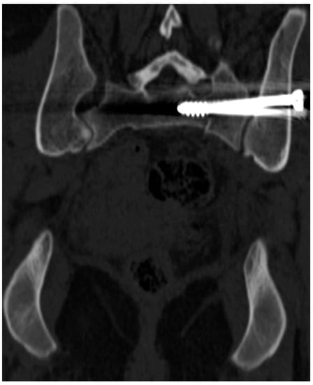
Differences in Accuracy and Radiation Dose in Placement of Iliosacral Screws: Comparison between 3D and 2D Fluoroscopy
by Michał Kułakowski, Paweł Reichert, Karol Elster, Jarosław Witkowski, Paweł Ślęczka, Piotr Morasiewicz, Łukasz Oleksy and Aleksandra Królikowska
J. Clin. Med. 2022, 11(6), 1466; https://doi.org/10.3390/jcm11061466 - 8 Mar 2022
Cited by 6 | Viewed by 2070
Abstract
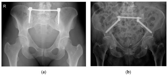
Percutaneous iliosacral screw fixation is a widely accepted method of stabilizing the posterior pelvic ring. Recently developed tools such as 3D-navigated fluoroscopy and computed navigation seem to prevent a surgeon from conducting screw misplacement. The study aimed to comparatively assess the introduction of [...] Read more.
Percutaneous iliosacral screw fixation is a widely accepted method of stabilizing the posterior pelvic ring. Recently developed tools such as 3D-navigated fluoroscopy and computed navigation seem to prevent a surgeon from conducting screw misplacement. The study aimed to comparatively assess the introduction of sacroiliac screw placement using 2D and 3D fluoroscopy in terms of accuracy and radiation exposure. Iliosacral screws were introduced in 37 patients using 2D (group N1) and in 36 patients using 3D fluoroscopy (group N2) techniques. Overall, 61 and 56 screws were introduced in groups N1 and N2, respectively. Screw placement accuracy was assessed using postoperative computed tomography and Smith’s scale. Intraoperative radiation exposure was also assessed. No differences were noted between groups in terms of screw positioning accuracy and radiation dose. Both 2D and 3D fluoroscopy provide good visualization for safely placing percutaneous iliosacral joint screws. Using 3D fluoroscopy-based navigation in comparison with 2D fluoroscopy is not advantageous. Full article
Show Figures

Figure 1

2021

Jump to: 2022, 2020

14 pages, 2886 KiB  
Article
Are Sarcopenia and Myosteatosis in Elderly Patients with Pelvic Ring Injury Related to Mortality, Physical Functioning and Quality of Life?
by Hester Banierink, Julia J. C. Bombach, Kaj ten Duis, Frank F. A. IJpma, Erik Heineman, Sven H. van Helden, Robert J. Nijveldt, Alain R. Viddeleer and Inge H. F. Reininga
J. Clin. Med. 2021, 10(21), 4874; https://doi.org/10.3390/jcm10214874 - 22 Oct 2021
Cited by 1 | Viewed by 1869
Abstract
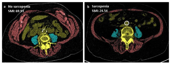
The purpose of this study was to evaluate the prevalence of sarcopenia and/or myosteatosis in elderly patients with pelvic ring injuries and their influence on mortality, patient-perceived physical functioning and quality of life (QoL). A multicenter retrospective cohort study was conducted including elderly [...] Read more.
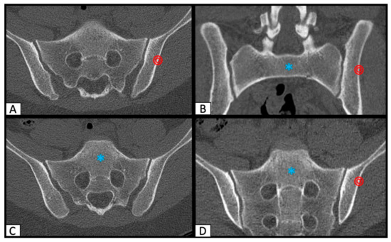
The purpose of this study was to evaluate the prevalence of sarcopenia and/or myosteatosis in elderly patients with pelvic ring injuries and their influence on mortality, patient-perceived physical functioning and quality of life (QoL). A multicenter retrospective cohort study was conducted including elderly patients aged ≥ 65 treated for a pelvic ring injury. Cross-sectional computed tomography (CT) muscle measurements were obtained to determine the presence of sarcopenia and/or myosteatosis. Kaplan–Meier analysis was used for survival analysis, and Cox proportional hazards regression analysis was used to determine risk factors for mortality. Patient-reported outcome measures for physical functioning (SMFA) and QoL (EQ-5D) were used. Multivariable linear regression analyses were used to determine the effect of sarcopenia and myosteatosis on patient-perceived physical functioning and QoL. Data to determine sarcopenia and myosteatosis were available for 199 patients, with a mean follow-up of 2.4 ± 2.2 years: 66 patients (33%) were diagnosed with sarcopenia and 65 (32%) with myosteatosis, while 30 of them (15%) had both. Mortality rates in patients at 1 and 3 years without sarcopenia and myosteatosis were 13% and 21%, compared to 11% and 36% in patients with sarcopenia, 17% and 31% in patients with myosteatosis and 27% and 43% in patients with both. Higher age at the time of injury and a higher Charlson Comorbidity Index (CCI) were independent risk factors for mortality. Patient-reported mental and emotional problems were significantly increased in patients with sarcopenia. Full article
Show Figures

Figure 1

11 pages, 967 KiB  
Review
Peripartum Pubic Symphysis Diastasis—Practical Guidelines
by Artur Stolarczyk, Piotr Stępiński, Łukasz Sasinowski, Tomasz Czarnocki, Michał Dębiński and Bartosz Maciąg
J. Clin. Med. 2021, 10(11), 2443; https://doi.org/10.3390/jcm10112443 - 31 May 2021
Cited by 12 | Viewed by 8915
Abstract
Optimal development of a fetus is made possible due to a lot of adaptive changes in the woman’s body. Some of the most important modifications occur in the musculoskeletal system. At the time of childbirth, natural widening of the pubic symphysis and the [...] Read more.
Optimal development of a fetus is made possible due to a lot of adaptive changes in the woman’s body. Some of the most important modifications occur in the musculoskeletal system. At the time of childbirth, natural widening of the pubic symphysis and the sacroiliac joints occur. Those changes are often reversible after childbirth. Peripartum pubic symphysis separation is a relatively rare disease and there is no homogeneous approach to treatment. The paper presents the current standards of diagnosis and treatment of pubic diastasis based on orthopedic and gynecological indications. Full article
Show Figures

Figure 1

16 pages, 21405 KiB  
Article
Biomechanical Comparison of Five Fixation Techniques for Unstable Fragility Fractures of the Pelvic Ring
by Moritz F. Lodde, J. Christoph Katthagen, Clemens O. Schopper, Ivan Zderic, Geoff Richards, Boyko Gueorguiev, Michael J. Raschke and René Hartensuer
J. Clin. Med. 2021, 10(11), 2326; https://doi.org/10.3390/jcm10112326 - 26 May 2021
Cited by 24 | Viewed by 4098
Abstract
Background: Incidence of pelvic ring fractures has increased over the past four decades, especially after low-impact trauma—classified as fragility fractures of the pelvis (FFP). To date, there is a lack of biomechanical evidence for the superiority of one existing fixation technique over another. [...] Read more.
Background: Incidence of pelvic ring fractures has increased over the past four decades, especially after low-impact trauma—classified as fragility fractures of the pelvis (FFP). To date, there is a lack of biomechanical evidence for the superiority of one existing fixation technique over another. An FFP type IIc was simulated in 50 artificial pelvises, assigned to 5 study groups: Sacroiliac (SI) screw, SI screw plus supra-acetabular external fixator, SI screw plus plate, SI screw plus retrograde transpubic screw, or S1/S2 ala–ilium screws. The specimens were tested under progressively increasing cyclic loading. Axial stiffness and cycles to failure were analysed. Displacement at the fracture sites was evaluated, having been continuously captured via motion tracking. Results: Fixation with SI screw plus plate and SI screw plus retrograde transpubic screw led to higher stability than the other tested techniques. The S1/S2 ala–ilium screws were more stable than the SI screw or the SI screw plus external fixator. Conclusions: In cases with displaced fractures, open reduction and plate fixation provides the highest stability, whereas in cases where minimally invasive techniques are applicable, a retrograde transpubic screw or S1/S2 ala–ilium screws can be considered as successful alternative treatment options. Full article
Show Figures

Figure 1

14 pages, 3298 KiB  
Article
Pelvic Floor Morbidity Following Vaginal Delivery versus Cesarean Delivery: Systematic Review and Meta-Analysis
by Juan A. Barca, Coral Bravo, Maria P. Pintado-Recarte, Ángel Asúnsolo, Ignacio Cueto-Hernández, Javier Ruiz-Labarta, Julia Buján, Miguel A. Ortega and Juan A. De León-Luis
J. Clin. Med. 2021, 10(8), 1652; https://doi.org/10.3390/jcm10081652 - 13 Apr 2021
Cited by 20 | Viewed by 3888
Abstract
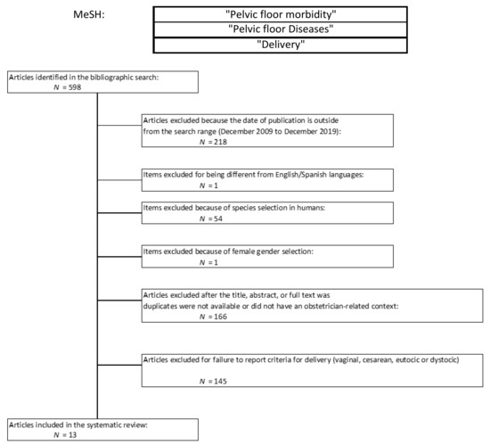
Objective: To compare pelvic floor disorders between vaginal delivery (VD) and cesarean delivery (CD). Methods: For this study, a PUBMED database search was used, utilizing a combination of relevant medical subjects’ headings (MeSH) terms, with the following keywords: “Pelvic floor disorders” or “Pelvic [...] Read more.
Objective: To compare pelvic floor disorders between vaginal delivery (VD) and cesarean delivery (CD). Methods: For this study, a PUBMED database search was used, utilizing a combination of relevant medical subjects’ headings (MeSH) terms, with the following keywords: “Pelvic floor disorders” or “Pelvic floor morbidity” and “Delivery”. Search limits were articles in English or Spanish, about women, published from December 2009 to December 2019. The STATA 16 package was used for meta-analysis and data heterogeneity assessment. Results: Thirteen studies meeting eligibility criteria were identified comprising 1,597,303 participants. Abstract: Pelvic floor morbidity prevalence was Urinary Incontinence (UI) 27.9% (5411 patients in 7 studies with reported cases), Pelvic Organ Prolapse (POP) 14.2% (6019 patients in 8 studies with reported cases), and Anal Incontinence (AI) 0.4% (1,589,740 patients in 5 studies with reported cases). Our meta-analyses revealed significantly higher rates of all three morbidities and overall morbidity in the VD versus CD group: UI OR = 2.17, 95% CI 1.64–2.87, p for heterogeneity ≤ 0.0001, I2 = 84%; POP OR = 3.28, 95% CI 1.91–5.63, p for heterogenicity ≤ 0.043, I2 = 63%; AI OR = 1.53, 95% CI 1.32–1.77; p for heterogeneity ≤ 0.291, I2 = 20%; and overall morbidity (OR = 2.17, 95% CI 1.64–2.87; p for heterogeneity ≤ 0.0001, I2 = 84%). Conclusion: Vaginal delivery is directly related to the appearance of pelvic floor disorders, mainly UI, POP, and AI. The risk of POP should be taken into higher consideration after vaginal delivery and postpartum follow-up should be performed, to identify and/or treat it at the earliest stages. Full article
Show Figures

Figure 1

6 pages, 2374 KiB  
Article
Iliac Bone Corridors to Host the Transiliac Internal Fixator—An Experimental CT Based Analysis
by Paul Schmitz, Maximilian Kerschbaum, Philipp Lamby, Siegmund Lang, Volker Alt and Michael Worlicek
J. Clin. Med. 2021, 10(7), 1500; https://doi.org/10.3390/jcm10071500 - 4 Apr 2021
Cited by 1 | Viewed by 2473
Abstract
Background: The transiliac internal fixator (TIFI) is a novel minimally invasive surgical procedure to stabilize posterior pelvic ring fractures. Two bone corridors with different lengths, widths, and angulations are suitable to host screws in the posterior iliac wing. While the length and the [...] Read more.
Background: The transiliac internal fixator (TIFI) is a novel minimally invasive surgical procedure to stabilize posterior pelvic ring fractures. Two bone corridors with different lengths, widths, and angulations are suitable to host screws in the posterior iliac wing. While the length and the width have been described previously, the angulation has not been determined yet. Methods: We created a computer tomography-based 3D-model of 40 patients (20 women, 20 men). The possible bone corridors to host the ilium screws for the TIFIcc (cranio-caudal) and the TIFIdv (dorso-ventral) procedure were identified. After reaching the optimal position, the angles in relation to the sagittal and axial plane were measured. The anterior pelvic plane was chosen as the reference plane. Results: The mean angle of the TIFIcc screws related to the axial plane was 63.4° (±1.8°) and to the sagittal plane was 12.3° (±1.5°). The mean angle of the TIFIdv screws related to the axial plane was 16.1° (±1.2°) and to the sagittal plane was 20.1° (±2.0°). In each group, a high constancy was apparent irrespective of the age or physical dimension of the patient, although a significant gender-dependent difference was observed”. Conclusions: Due to a high inter-individual constancy in length, width, and angulation, bone corridors in the posterior iliac wing are reliable to host screws for posterior pelvic ring fixation irrespective of each individual patient’s anatomy. Full article
Show Figures

Figure 1

8 pages, 216 KiB  
Article
Can CRP Levels Predict Infection in Presumptive Aseptic Long Bone Non-Unions? A Prospective Cohort Study
by Theodoros H. Tosounidis, Colin Holton, Vasileios P. Giannoudis, Nikolaos K. Kanakaris, Robert M. West and Peter V. Giannoudis
J. Clin. Med. 2021, 10(3), 425; https://doi.org/10.3390/jcm10030425 - 22 Jan 2021
Cited by 7 | Viewed by 1928
Abstract
Nonunion remains a major complication of the management of long bone fractures. The primary aim of the present study was to investigate whether raised levels of C-reactive protein (CRP) and white blood cell count (WBC), in the absence of clinical signs, are correlated [...] Read more.
Nonunion remains a major complication of the management of long bone fractures. The primary aim of the present study was to investigate whether raised levels of C-reactive protein (CRP) and white blood cell count (WBC), in the absence of clinical signs, are correlated with positive intraoperative tissue cultures in presumptive aseptic long-bone nonunions. Infection was classified as positive if any significant growth of microorganisms was observed from bone/tissue samples sent from the theater at the time of revision surgery. Preoperatively all patients were investigated with full blood count, white blood count differential as well as C-reactive protein (CRP). A total of 105 consecutive patients (59 males) were included in the study, with an average age of 46.76 years (range 16–92 years) at the time of nonunion diagnosis. The vast majority were femoral (56) and tibial (37) nonunions. The median time from the index surgical procedure to the time of nonunion diagnosis was 10 months (range 9 months to 10 years). Positive cultures revealed a mixed growth of microorganisms, with coagulase-negative Staphylococcus (56.4%) being the most prevalent microorganism, followed by Staphylococcusaureus (20.5%). Pseudomonas, Methicillin-Resistant Staphylococcus aureus (MRSA), coliforms and micrococcus were present in the remainder of the cases (23.1%). Overall, the risk of infection with normal CRP levels (<10 mg/L) was 21/80 = 0.26. Elevated CRP levels (≥10 mg/L) increased the risk of infection to 0.72. The relative risk given a positive CRP test was RR = 0.72/0.26 = 2.74. Overall, the WBC count was found to be an unreliable marker to predict infection. Solid union was achieved in all cases after an average of 6.5 months (3–24 months) from revision surgery. In patients with presumed aseptic long bone nonunion and normal CRP levels, the risk of underlying low-grade indolent infection can be as high as 26%. Patients should be made aware of this finding, which can complicate their treatment course and outcomes. Full article
8 pages, 9148 KiB  
Article
Two-Dimensional Visualization of the Three-Dimensional Planned Sacroiliac Screw Corridor with the Slice Fusion Method
by Maximilian Kerschbaum, Siegmund Lang, Florian Baumann, Volker Alt and Michael Worlicek
J. Clin. Med. 2021, 10(2), 184; https://doi.org/10.3390/jcm10020184 - 6 Jan 2021
Cited by 2 | Viewed by 2468
Abstract
Insertion of sacro-iliac (SI) screws for stabilization of the posterior pelvic ring without intraoperative navigation or three-dimensional imaging can be challenging. The aim of this study was to develop a simple method to visualize the ideal SI screw corridor, on lateral two-dimensional images, [...] Read more.
Insertion of sacro-iliac (SI) screws for stabilization of the posterior pelvic ring without intraoperative navigation or three-dimensional imaging can be challenging. The aim of this study was to develop a simple method to visualize the ideal SI screw corridor, on lateral two-dimensional images, corresponding to the lateral fluoroscopic view, used intraoperatively while screw insertion, to prevent neurovascular injury. We used multiplanar reconstructions of pre- and postoperative computed tomography scans (CT) to determine the position of the SI corridor. Then, we processed the dataset into a lateral two-dimensional slice fusion image (SFI) matching head and tip of the screw. Comparison of the preoperative SFI planning and the screw position in the postoperative SFI showed reproducible results. In conclusion, the slice fusion method is a simple technique for translation of three-dimensional planned SI screw positioning into a two-dimensional strict lateral fluoroscopic-like view. Full article
Show Figures

Figure 1

2020

Jump to: 2022, 2021

26 pages, 6150 KiB  
Review
Pelvic Girdle Pain, Hypermobility Spectrum Disorder and Hypermobility-Type Ehlers-Danlos Syndrome: A Narrative Literature Review
by Ahmed Ali, Paul Andrzejowski, Nikolaos K. Kanakaris and Peter V. Giannoudis
J. Clin. Med. 2020, 9(12), 3992; https://doi.org/10.3390/jcm9123992 - 9 Dec 2020
Cited by 11 | Viewed by 9654
Abstract
Pelvic girdle pain (PGP) refers specifically to musculoskeletal pain localised to the pelvic ring and can be present at its anterior and/or posterior aspects. Causes such as trauma, infection and pregnancy have been well-established, while patients with hypermobile joints are at greater risk [...] Read more.
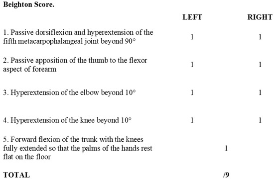
Pelvic girdle pain (PGP) refers specifically to musculoskeletal pain localised to the pelvic ring and can be present at its anterior and/or posterior aspects. Causes such as trauma, infection and pregnancy have been well-established, while patients with hypermobile joints are at greater risk of developing PGP. Research exploring this association is limited and of varying quality. In the present study we report on the incidence, pathophysiology, diagnostic and treatment modalities for PGP in patients suffering from Hypermobility Spectrum Disorder (HSD) and Hypermobility-Type Ehlers-Danlos Syndrome (hEDS). Recommendations are made for clinical practice by elaborating on screening, diagnosis and management of such patients to provide a holistic approach to their care. It appears that this cohort of patients are at greater risk particularly of mental health issues. Moreover over, they may require a multidisciplinary approach for their management. Ongoing research is still required to expand our understanding of the relationship between PGP, HSD and hEDS by appropriately diagnosing patients using the latest updated terminologies and by conducting randomised control trials to compare outcomes of interventions using standardised patient reported outcome measures. Full article
Show Figures

Figure 1

13 pages, 487 KiB  
Article
The Rolf Method of Structural Integration and Pelvic Floor Muscle Facilitation: Preliminary Results of a Randomized, Interventional Study
by Martyna Kasper-Jędrzejewska, Grzegorz Jędrzejewski, Lucyna Ptaszkowska, Kuba Ptaszkowski, Robert Schleip and Tomasz Halski
J. Clin. Med. 2020, 9(12), 3981; https://doi.org/10.3390/jcm9123981 - 9 Dec 2020
Cited by 2 | Viewed by 3908
Abstract
The management of pelvic floor dysfunctions might need to be based on a comprehensive neuro-musculoskeletal therapy such as The Rolf Method of Structural Integration (SI). The aim of the study was to evaluate the pelvic floor muscle (PFM) after the tenth session of [...] Read more.
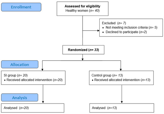
The management of pelvic floor dysfunctions might need to be based on a comprehensive neuro-musculoskeletal therapy such as The Rolf Method of Structural Integration (SI). The aim of the study was to evaluate the pelvic floor muscle (PFM) after the tenth session of SI by using surface electromyography (sEMG). This was a randomized, interventional study. Thirty-three healthy women were randomly assigned to the experimental (SI) or control group. The outcome measures included PFM bioelectrical activity, assessed using sEMG and endovaginal probes. An intervention in the SI group included 60 min of SI once a week, and teaching on how to contract and relax PFMs; in the control group, only the teaching was carried out. In the SI group, a significant difference was found between the PFM sEMG activity during “pre-baseline rest” (p < 0.014) and that during “rest after tonic contraction” (p = 0.021) in the supine position, as were significant increases in “phasic contraction” in the standing position (p = 0.014). In the intergroup comparison, higher PFM sEMG activity after the intervention “phasic contraction” (p = 0.037) and “pre-baseline rest” (p = 0.028) was observed in the SI group. The SI intervention significantly changes some functional bioelectrical activity of PFMs, providing a basis for further research on a new approach to PFM facilitation, particularly in clinical populations. Full article
Show Figures

Figure 1

2 pages, 162 KiB  
Editorial
Instability of the Pelvic Ring: A Special Clinical Entity?
by Peter V Giannoudis
J. Clin. Med. 2020, 9(6), 1985; https://doi.org/10.3390/jcm9061985 - 25 Jun 2020
Cited by 3 | Viewed by 1841
Abstract
The presence of the pathological movement of pubic symphysis under normal activities characterises a syndrome know as anterior pelvic ring instability [...] Full article
Back to TopTop