Coating Deposition and Surface Functionalization of Implants for Biomedical Applications

Editor


E-Mail Website
Collection Editor
Commonwealth Scientific and Industrial Research Organisation (CSIRO), Melbourne, Australia
Interests: composite materials; functional materials; cementitious composites; additive manufacturing
Special Issues, Collections and Topics in MDPI journals

Topical Collection Information

Dear Colleagues,

The human body is able to promote spontaneous healing phenomena to face the adverse consequences of diseases, aging processes or traumatic events. However such natural reactions are not always sufficient to recover extensive functional losses and, in that case, medical or surgical interventions are required. For this reason, the demand for new biomaterials to support or restore the role of damaged tissues is a major clinical and socioeconomic need. The deposition of a proper coating or the chemicophysical treatment of the surface may boost the performance of implant devices, conveying site-specific properties to the substrate material. The apposition of glass-based glazes which resemble the original enamel of tooth, the chemical modification of titanium to activate the bone-bonding ability or the deposition of calcium-phosphate layers on metal substrates to elicit the surface development of hydroxyapatite are just a few examples of the new approaches to improve the behaviour of medical grafts by means of biocoatings and surface functionalization methods.

Dr. Antonella Sola
Collection Editor

Manuscript Submission Information

Manuscripts for the topical collection can be submitted online at www.mdpi.com by registering and logging in to this website. Once you are registered, click here to go to the submission form. All papers will be peer-reviewed. Accepted papers will be published continuously in the journal (as soon as accepted) and will be listed together on this website. The topical collection considers regular research articles, short communications and review articles. A guide for authors and other relevant information for submission of manuscripts is available on the Instructions for Authors page.

Please visit the Instructions for Authors page before submitting a manuscript. The article processing charge (APC) for publication in this open access journal is 2700 CHF (Swiss Francs). English correction and/or formatting fees will be charged in certain cases for those articles accepted for publication that require extensive additional formatting and/or English corrections. For further details see here.

Keywords

  • biocoatings
  • functionalization
  • surface treatments
  • coating deposition methods
  • implant materials

Related Special Issue

Published Papers (5 papers)

2012

1054 KiB  
Review
Biomimetic Strategies for Bone Repair and Regeneration
by Maria G. Raucci, Vincenzo Guarino and Luigi Ambrosio
J. Funct. Biomater. 2012, 3(3), 688-705; https://doi.org/10.3390/jfb3030688 - 20 Sep 2012
Cited by 36 | Viewed by 8110
Abstract
The osseointegration rate of implants is related to their composition and surface roughness. Implant roughness favors both bone anchoring and biomechanical stability. Osteoconductive calcium phosphate (Ca-P) coatings promote bone healing and apposition, leading to the rapid biological fixation of implants. It has been [...] Read more.
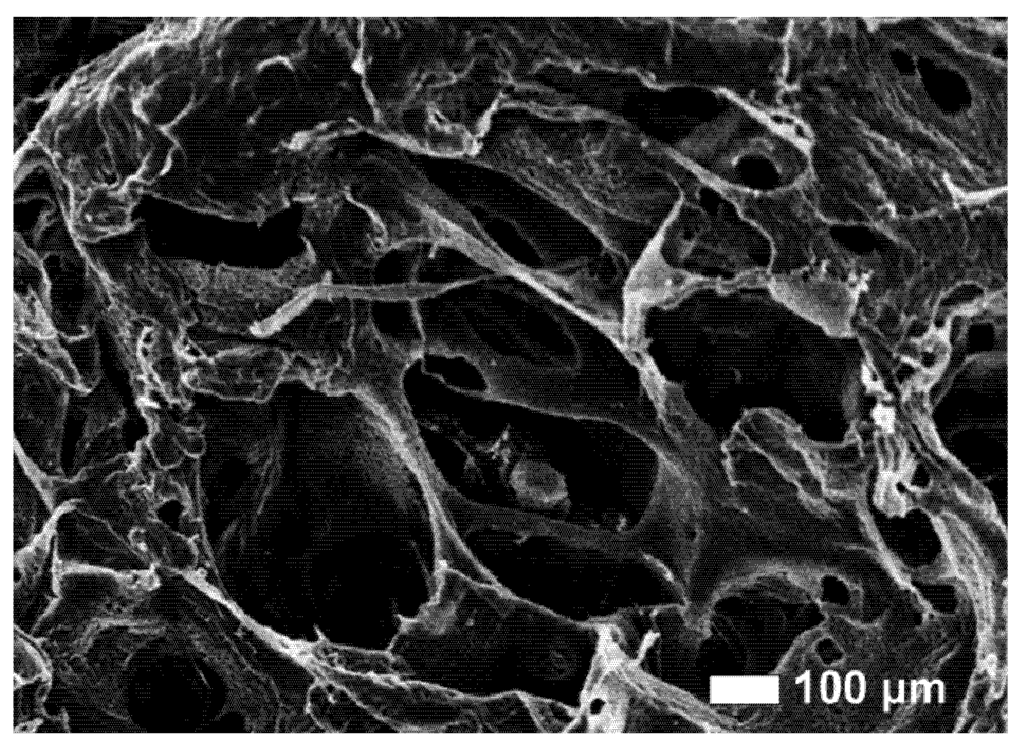
The osseointegration rate of implants is related to their composition and surface roughness. Implant roughness favors both bone anchoring and biomechanical stability. Osteoconductive calcium phosphate (Ca-P) coatings promote bone healing and apposition, leading to the rapid biological fixation of implants. It has been clearly shown in many publications that Ca-P coating accelerates bone formation around the implant. This review discusses two main routes for the manufacturing of polymer-based osteoconductive scaffolds for tissue engineering, namely the incorporation of bioceramic particles in the scaffold and the coating of a scaffold with a thin layer of apatite through a biomimetic process. Full article
Show Figures

Figure 1

2584 KiB  
Article
Cell Growth on Different Types of Ultrananocrystalline Diamond Thin Films
by Bing Shi, Qiaoling Jin, Liaohai Chen, Amina S. Woods, Albert J. Schultz and Orlando Auciello
J. Funct. Biomater. 2012, 3(3), 588-600; https://doi.org/10.3390/jfb3030588 - 16 Aug 2012
Cited by 15 | Viewed by 7491
Abstract
Unique functional materials provide a platform as scaffolds for cell/tissue regeneration. Investigation of cell-materials’ chemical and biological interactions will enable the application of more functional materials in the area of bioengineering, which provides a pathway to the novel treatment for patients who suffer [...] Read more.
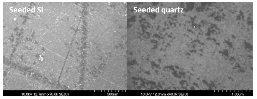
Unique functional materials provide a platform as scaffolds for cell/tissue regeneration. Investigation of cell-materials’ chemical and biological interactions will enable the application of more functional materials in the area of bioengineering, which provides a pathway to the novel treatment for patients who suffer from tissue/organ damage and face the limitation of donation sources. Many studies have been made into tissue/organ regeneration. Development of new substrate materials as platforms for cell/tissue regeneration is a key research area. Studies discussed in this paper focus on the investigation of novel ultrananocrystalline diamond (UNCD) films as substrate/scaffold materials for developmental biology. Specially designed quartz dishes have been coated with different types of UNCD films and cells were subsequently seeded on those films. Results showed the cells’ growth on UNCD-coated culture dishes are similar to cell culture dishes with little retardation, indicating that UNCD films have no or little inhibition on cell proliferation and are potentially appealing as substrate/scaffold materials. The mechanisms of cell adhesion on UNCD surfaces are proposed based on the experimental results. The comparisons of cell cultures on diamond-powder-seeded culture dishes and on UNCD-coated dishes with matrix-assisted laser desorption/ionization—time-of-flight mass spectroscopy (MALDI-TOF MS) and X-ray photoelectron spectroscopy (XPS) analyses provided valuable data to support the mechanisms proposed to explain the adhesion and proliferation of cells on the surface of the UNCD platform. Full article
Show Figures

Figure 1

635 KiB  
Article
A Method for Quantitative Determination of Biofilm Viability
by Ken Welch, Yanling Cai and Maria Strømme
J. Funct. Biomater. 2012, 3(2), 418-431; https://doi.org/10.3390/jfb3020418 - 1 Jun 2012
Cited by 73 | Viewed by 12519
Abstract
In this study we present a scheme for quantitative determination of biofilm viability offering significant improvement over existing methods with metabolic assays. Existing metabolic assays for quantifying viable bacteria in biofilms usually utilize calibration curves derived from planktonic bacteria, which can introduce large [...] Read more.
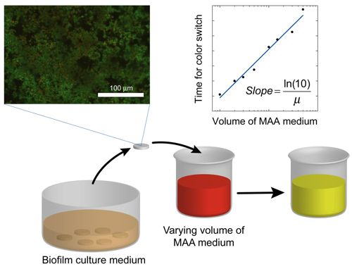
In this study we present a scheme for quantitative determination of biofilm viability offering significant improvement over existing methods with metabolic assays. Existing metabolic assays for quantifying viable bacteria in biofilms usually utilize calibration curves derived from planktonic bacteria, which can introduce large errors due to significant differences in the metabolic and/or growth rates of biofilm bacteria in the assay media compared to their planktonic counterparts. In the presented method we derive the specific growth rate of Streptococcus mutans bacteria biofilm from a series of metabolic assays using the pH indicator phenol red, and show that this information could be used to more accurately quantify the relative number of viable bacteria in a biofilm. We found that the specific growth rate of S. mutans in biofilm mode of growth was 0.70 h−1, compared to 1.09 h−1 in planktonic growth. This method should be applicable to other bacteria types, as well as other metabolic assays, and, for example, to quantify the effect of antibacterial treatments or the performance of bactericidal implant surfaces. Full article
Show Figures

Graphical abstract

965 KiB  
Article
Characterization of Porous TiO2 Surfaces Formed on 316L Stainless Steel by Plasma Electrolytic Oxidation for Stent Applications
by Zhiguang Huan, Lidy E. Fratila-Apachitei, Iulian Apachitei and Jurek Duszczyk
J. Funct. Biomater. 2012, 3(2), 349-360; https://doi.org/10.3390/jfb3020349 - 11 May 2012
Cited by 16 | Viewed by 8965
Abstract
In this study, a porous oxide layer was formed on the surface of 316L stainless steel (SS) by combining Ti magnetron sputtering and plasma electrolytic oxidation (PEO) with the aim to produce a polymer-free drug carrier for drug eluting stent (DES) applications. The [...] Read more.
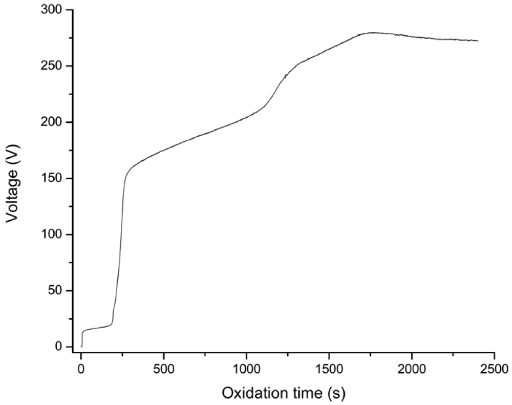
In this study, a porous oxide layer was formed on the surface of 316L stainless steel (SS) by combining Ti magnetron sputtering and plasma electrolytic oxidation (PEO) with the aim to produce a polymer-free drug carrier for drug eluting stent (DES) applications. The oxidation was performed galvanostatically in Na3PO4 electrolyte. The surface porosity, average pore size and roughness varied with PEO treatment duration, and under optimum conditions, the surface showed a porosity of 7.43%, an average pore size of 0.44 µm and a roughness (Ra) of 0.34 µm. The EDS analyses revealed that the porous layer consisted of Ti, O and P. The cross-sectional morphology evidenced a double-layer structure, with a porous titania surface and an un-oxidized dense Ti film towards the interface with 316L SS. After the PEO treatment, wettability and surface free energy increased significantly. The results of the present study confirm the feasibility of forming a porous TiO2 layer on stainless steel by combining sputtering technology and PEO. Further, the resultant porous oxide layer has the potential to be used as a drug carrier for DES, thus avoiding the complications associated with the polymer based carriers. Full article
Show Figures

Figure 1

902 KiB  
Article
Hemocompatibility of Inorganic Physical Vapor Deposition (PVD) Coatings on Thermoplastic Polyurethane Polymers
by Juergen M. Lackner, Wolfgang Waldhauser, Paul Hartmann, Franz Bruckert, Marianne Weidenhaupt, Roman Major, Marek Sanak, Martin Wiesinger and Daniel Heim
J. Funct. Biomater. 2012, 3(2), 283-297; https://doi.org/10.3390/jfb3020283 - 17 Apr 2012
Cited by 15 | Viewed by 8241
Abstract
Biocompatibility improvements for blood contacting materials are of increasing interest for implanted devices and interventional tools. The current study focuses on inorganic (titanium, titanium nitride, titanium oxide) as well as diamond-like carbon (DLC) coating materials on polymer surfaces (thermoplastic polyurethane), deposited by magnetron [...] Read more.
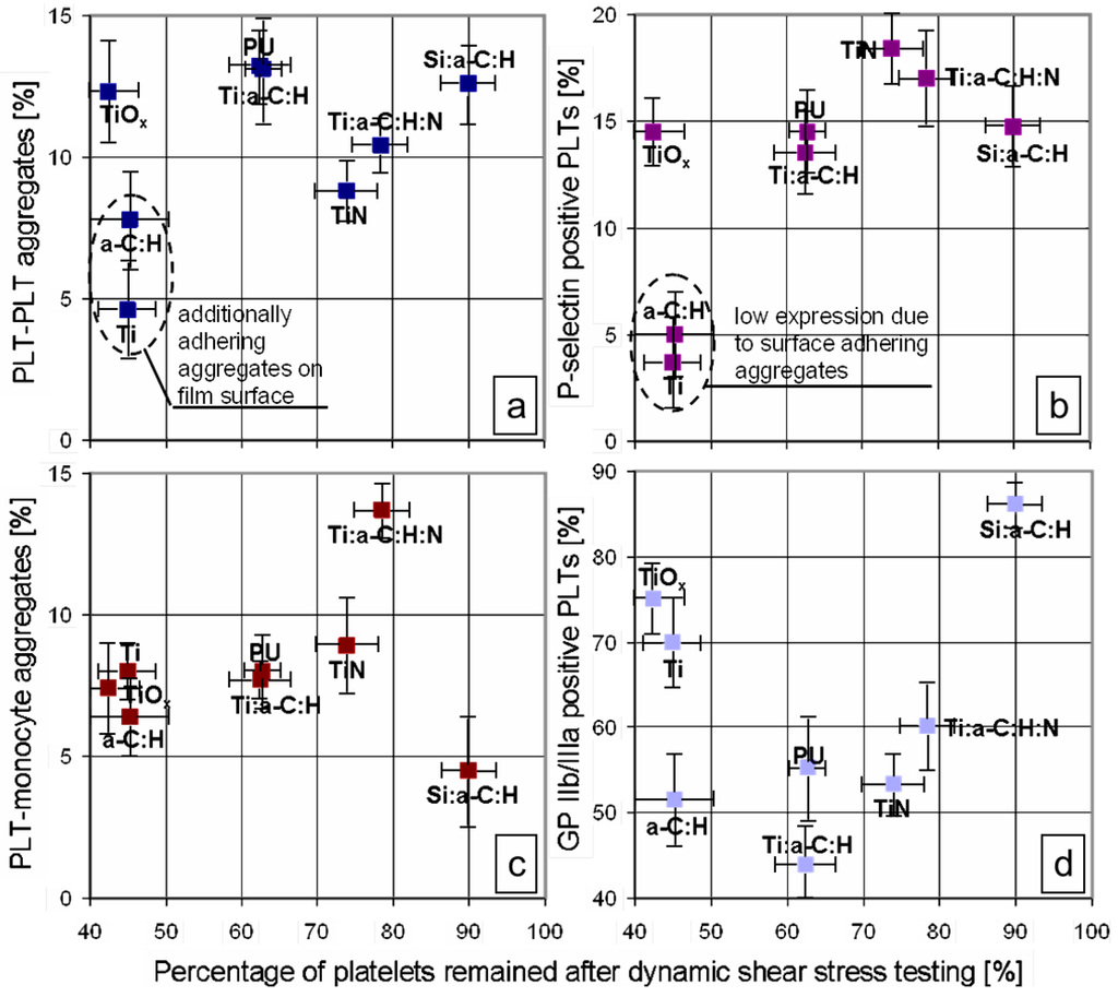
Biocompatibility improvements for blood contacting materials are of increasing interest for implanted devices and interventional tools. The current study focuses on inorganic (titanium, titanium nitride, titanium oxide) as well as diamond-like carbon (DLC) coating materials on polymer surfaces (thermoplastic polyurethane), deposited by magnetron sputtering und pulsed laser deposition at room temperature. DLC was used pure (a-C:H) as well as doped with silicon, titanium, and nitrogen + titanium (a-C:H:Si, a-C:H:Ti, a-C:H:N:Ti). In-vitro testing of the hemocompatibility requires mandatory dynamic test conditions to simulate in-vivo conditions, e.g., realized by a cone-and-plate analyzer. In such tests, titanium- and nitrogen-doped DLC and titanium nitride were found to be optimally anti-thrombotic and better than state-of-the-art polyurethane polymers. This is mainly due to the low tendency to platelet microparticle formation, a high content of remaining platelets in the whole blood after testing and low concentration of platelet activation and aggregation markers. Comparing this result to shear-flow induced cell motility tests with e.g., Dictostelium discoideum cell model organism reveals similar tendencies for the investigated materials. Full article
Show Figures

Figure 1

Back to TopTop