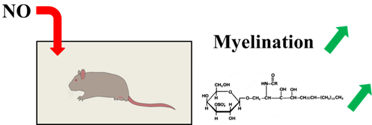
Impact of Inhaled Nitric Oxide on the Sulfatide Profile of Neonatal Rat Brain Studied by TOF-SIMS Imaging
Abstract
:1. Introduction
2. Results and Discussion
2.1. Imaging Data
2.2. Semi-Quantitative Approach
3. Experimental Section
3.1. Animals
3.2. Sample Preparation
3.3. TOF-SIMS Imaging Acquisition
3.4. TOF-SIMS Imaging Processing
4. Conclusions
Supplementary Information
ijms-15-05233-s001.pdfAcknowledgments
Conflicts of Interest
- Author ContributionsH.K. and H.P. performed all experiments and data treatments. Writing was done by H.K., D.T., O.B. and A.B., and management and submission tasks were done by D.T., O.B. and A.B.
References
- Larroque, B.; Ancel, P.Y.; Marchand-Martin, L.; Cambonie, G.; Fresson, J.; Pierrat, V.; Roze, J.C.; Marpeau, L.; Thiriez, G.; Alberge, C.; et al. Special care and school difficulties in 8-year-old very preterm children: The epipage cohort study. PLoS One 2010, 6, e21361. [Google Scholar]
- Woodward, L.J.; Clark, C.A.C.; Bora, S.; Inder, T.E. Neonatal white matter abnormalities an important predictor of neurocognitive outcome for very preterm children. PLoS One 2012, 7, e51879. [Google Scholar]
- Woodward, L.J.; Moor, S.; Hood, K.M.; Champion, P.R.; Foster-Cohen, S.; Inder, T.E.; Austin, N.C. Very preterm children show impairments across multiple neurodevelopmental domains by age 4 years. Arch. Dis. Child-Fetal Neonatal Ed 2009, 94, F339–F344. [Google Scholar]
- Volpe, J.J. Brain injury in premature infants: A complex amalgam of destructive and developmental disturbances. Lancet Neurol 2009, 8, 110–124. [Google Scholar]
- Zhang, Y.P.; Huang, Q.L.; Zhao, C.M.; Tang, J.L.; Wang, Y.L. GM1 improves neurofascin 155 association with lipid rafts and prevents rat brain myelin injury after hypoxia-ischemia. Braz. J. Med. Biol. Res 2011, 28, 9741–9754. [Google Scholar]
- Inder, T.; Neil, J.; Kroenke, C.; Dieni, S.; Yoder, B.; Rees, S. Investigation of cerebral development and injury in the prematurely born primate by magnetic resonance imaging and histopathology. Dev. Neurosci 2005, 27, 100–111. [Google Scholar]
- Khwaja, O.; Volpe, J.J. Pathogenesis of cerebral white matter injury of prematurity. Arch. Dis. Child-Fetal 2008, 93, F153–F161. [Google Scholar]
- Baumann, N.; Pham-Dinh, D. Biology of oligodendrocyte and myelin in the mammalian central nervous system. Physiol. Rev 2001, 81, 871–927. [Google Scholar]
- Siegel, J.S.; Wayne Albers, R.; Brady, S.; Price, D. Basic Neurochemistry, Molecular, Cellular and Medical Aspects; Elsevier Academic Press: Burlington, MA, USA, 2006. [Google Scholar]
- Zöller, I.; Meixner, M.; Hartmann, D.; Bussow, H.; Meyer, R.; Gieselmann, V.; Eckhardt, M. Absence of 2-hydroxylated sphingolipids is compatible with normal neural development but causes late-onset axon and myelin sheath degeneration. J. Neurosci 2008, 28, 9741–9754. [Google Scholar]
- Olivier, P.; Loron, G.; Fontaine, R.H.; Pansiot, J.; Dalous, J.; Thi, H.P.; Charriaut-Marlangue, C.; Thomas, J.L.; Mercier, J.C.; Gressens, P.; et al. Nitric Oxide Plays a Key Role in Myelination in the Developing Brain. J. Neuropathol. Exp. Neurol 2010, 69, 828–837. [Google Scholar]
- Olivier, P.; Baud, O.; Bouslama, M.; Evrard, P.; Gressens, P.; Verney, C. Moderate growth restriction: Deleterious and protective effects on white matter damage. Neurobiol. Dis 2007, 26, 253–263. [Google Scholar]
- Dean, J.M.; Farrag, D.; Zahkouk, S.A.M.; El Zawahry, E.Y.I.; Hagberg, H.; Kjellmer, I.; Mallard, C. Cerebellar white matter injury following systemic endotoxemia in preterm fetal sheep. Neuroscience 2009, 160, 606–615. [Google Scholar]
- Setou, M. Imaging Mass Spectrometry; Springer: Tokyo, Japan, 2010. [Google Scholar]
- Rubakhin, S.S.; Sweedler, J.V. Mass Spectrometry Imaging, Principles and Protocols; Humana Press: New York, NY, USA, 2010. [Google Scholar]
- Wu, C.; Dill, A.L.; Eberlin, L.S.; Cooks, R.G.; Ifa, D.R. Mass spectrometry imaging under ambient conditions. Mass Spectrom. Rev 2013, 32, 218–243. [Google Scholar]
- Gode, D.; Volmer, D.A. Lipid imaging by mass spectrometry. Analyst 2013, 138, 1289–1315. [Google Scholar]
- Brunelle, A.; Touboul, D.; Laprévote, O. Recent advances in biological tissue Imaging with time-of-flight secondary ion mass spectrometry: Polyatomic ion sources, sample preparation, and applications. Curr. Pharm. Des 2007, 13, 3335–3343. [Google Scholar]
- Cazares, L.H.; Troyer, D.A.; Wang, B.; Drake, R.R.; Semmes, O.J. MALDI tissue imaging: From biomarker discovery to clinical applications. Anal. Bioanal. Chem 2011, 401, 17–27. [Google Scholar]
- Bakry, R.; Rainer, M.; Huck, C.W.; Bonn, G.K. Protein profiling for cancer biomarker discovery using matrix-assisted laser desorption/ionization time-of-flight mass spectrometry and infrared imaging: A review. Anal. Chim. Acta 2011, 690, 26–34. [Google Scholar]
- Touboul, D.; Brunelle, A.; Halgand, F.; de La Porte, S.; Laprévote, O. Lipid imaging by gold cluster time-of-flight secondary ion mass spectrometry: Application to Duchenne muscular dystrophy. J. Lipid Res 2005, 46, 1388–1395. [Google Scholar]
- Tahallah, N.; Brunelle, A.; de La Porte, S.; Laprévote, O. Lipid mapping in human dystrophic muscle by cluster-time-of-flight secondary ion mass spectrometry imaging. J. Lipid Res 2008, 49, 438–454. [Google Scholar]
- Gemoll, T.; Roblick, U.J.; Habermann, J.K. MALDI mass spectrometry imaging in oncology. Mol. Med. Rep 2011, 4, 1045–1051. [Google Scholar]
- Guerquin-Kern, J.L.; Wu, T.D.; Quintana, C.; Croisy, A. Progress in analytical imaging of the cell by dynamic secondary ion mass spectrometry (SIMS microscopy). Biochim. Biophys. Acta 2005, 1724, 228–238. [Google Scholar]
- Passarelli, M.K.; Winograd, N. Lipid imaging with time-of-flight secondary ion mass spectrometry (ToF-SIMS). Biochim. Biophys. Acta 2011, 1811, 976–990. [Google Scholar]
- Touboul, D.; Laprévote, O.; Brunelle, A. Micrometric molecular histology of lipids by mass spectrometry imaging. Curr. Opin. Chem. Biol 2011, 15, 725–732. [Google Scholar]
- Touboul, D.; Halgand, F.; Brunelle, A.; Kersting, R.; Tallarek, E.; Hagenhoff, B.; Laprévote, O. Tissue molecular ion imaging by gold cluster ion bombardment. Anal. Chem 2004, 76, 1550–1559. [Google Scholar]
- Sjövall, P.; Lausmaa, J.; Johansson, B. Mass spectrometric imaging of lipids in brain tissue. Anal. Chem 2004, 76, 4271–4278. [Google Scholar]
- Touboul, D.; Brunelle, A.; Germain, D.P.; Laprévote, O. A new imaging technique as a diagnostic tool: Mass spectrometry. Presse Med 2007, 36, S82–S87. [Google Scholar]
- Debois, D.; Bralet, M.P.; le Naour, F.; Brunelle, A.; Laprévote, O. In situ lipidomic analysis of nonalcoholic fatty liver by cluster TOF-SIMS imaging. Anal. Chem 2009, 81, 2823–2831. [Google Scholar]
- Seyer, A.; Cantiello, M.; Bertrand-Michel, J.; Roques, V.; Nauze, M.; Bezirard, V.; Collet, X.; Touboul, D.; Brunelle, A.; Comera, C. Lipidomic and spatio-temporal imaging of fat by mass spectrometry in mice duodenum during lipid digestion. PLoS One 2013, 8, e58224. [Google Scholar]
- Calabrese, V.; Cornelius, C.; Rizzarelli, E.; Owen, J.B.; Dinkova-Kostova, A.T.; Butterfield, D.A. Nitric oxide in cell survival: A janus molecule. Antioxid Redox Sign 2009, 11, 2717–2739. [Google Scholar]
- Charriaut-Marlangue, C.; Bonnin, P.; Gharib, A.; Leger, P.L.; Villapol, S.; Pocard, M.; Gressens, P.; Renolleau, S.; Baud, O. Inhaled nitric oxide reduces brain damage by collateral recruitment in a neonatal stroke model. Stroke 2012, 43, 3078–3084. [Google Scholar]
- Pansiot, J.; Loron, G.; Olivier, P.; Fontaine, R.; Charriaut-Marlangue, C.; Mercier, J.C.; Gressens, P.; Baud, O. Neuroprotective effect of inhaled nitric oxide on excitotoxic-induced brain damage in neonatal rat. PLoS One 2010, 5, e10916. [Google Scholar]
- Terpolilli, N.A.; Kim, S.W.; Thal, S.C.; Kataoka, H.; Zeisig, V.; Nitzsche, B.; Klaesner, B.; Zhu, C.L.; Schwarzmaier, S.; Meissner, L.; et al. Inhalation of nitric oxide prevents ischemic brain damage in experimental stroke by selective dilatation of collateral arterioles. Circ. Res 2012, 110, 727–738. [Google Scholar]
- El Ghazi, F.; Desfeux, A.; Brasse-Lagnel, C.; Roux, C.; Lesueur, C.; Mazur, D.; Remy-Jouet, I.; Richard, V.; Jégou, S.; Laudenbach, V.; et al. NO-dependent protective effect of VEGF against excitotoxicity on layer VI of the developing cerebral cortex. Neurobiol. Dis 2012, 45, 871–886. [Google Scholar]
- O’Brien, J.S.; Sampson, E.L. Fatty acid and fatty aldehyde composition of the major brain lipids in normal human gray matter, white matter, and myelin. J. Lipid Res 1965, 6, 545–551. [Google Scholar]
- Ramsey, R.B.; Scott, T.; Banik, N.L. Fatty acid composition of myelin isolated from the brain of a patient with cellular deficiency of co-enzyme forms of vitamin B12. J. Neurol. Sci 1977, 34, 221–232. [Google Scholar]
- Marbois, B.N.; Faull, K.F.; Fluharty, A.L.; Raval-Fernandes, S.; Rome, L.H. Analysis of sulfatide from rat cerebellum and multiple sclerosis white matter by negative ion electrospray mass spectrometry. Biochim. Biophys. Acta 2010, 1484, 59–70. [Google Scholar]
- Pernber, Z.; Richter, K.; Mansson, J.E.; Nygren, H. Sulfatide with different fatty acids has unique distributions in cerebellum as imaged by Time-Of-Flight Secondary Ion Mass Spectrometry (TOF-SIMS). Biochim. Biophys. Acta 2007, 1771, 202–209. [Google Scholar]
- Han, X.; Holtzman, D.M.; McKeel, D.W., Jr.; Kelley, J.; Morris, J.C. Substantial sulfatide deficiency and ceramide elevation in very early Alzheimer’s disease: Potential role in disease pathogenesis. J. Neurochem 2002, 82, 809–818. [Google Scholar]
- Taguchi, R.; Hayakawa, J.; Takeuchi, Y.; Ishida, M. Two-dimensional analysis of phospholipids by capillary liquid chromatography/electrospray ionization mass spectrometry. J. Mass Spectrom 2000, 35, 953–966. [Google Scholar]
- Ageta, H.; Asai, S.; Sugiura, Y.; Goto-Inoue, N.; Zaima, N.; Setou, M. Layer-specific sulfatide localization in rat hippocampus middle molecular layer is revealed by nanoparticle-assisted laser desorption/ionization imaging mass spectrometry. Med. Mol. Morphol 2009, 42, 16–23. [Google Scholar]
- Yuki, D.; Sugiura, Y.; Zaima, N.; Akatsu, H.; Hashizume, Y.; Yamamoto, T.; Fujiwara, M.; Sugiyama, K.; Setou, M. Hydroxylated and non-hydroxylated sulfatide are distinctly distributed in the human cerebral cortex. Neuroscience 2011, 193, 44–53. [Google Scholar]
- Bich, C.; Touboul, D.; Brunelle, A. Study of experimental variability in TOF-SIMS mass spectrometry imaging of biological samples. Int. J. Mass Spectrom 2013, 337, 43–49. [Google Scholar]
- Börner, K.; Malmberg, P.; Mansson, J.E.; Nygren, H. Molecular imaging of lipids in cells and tissues. Int. J. Mass Spectrom 2007, 260, 128–136. [Google Scholar]
- Richter, K.; Nygren, H.; Malmberg, P.; Hagenhoff, B. Localization of fatty acids with selective chain length by imaging time-of-flight secondary ion mass spectrometry. Microsc. Res. Tech 2007, 70, 640–647. [Google Scholar]
- Benabdellah, F.; Seyer, A.; Quinton, L.; Touboul, D.; Brunelle, A.; Laprévote, O. Mass spectrometry imaging of rat brain sections: Nanomolar sensitivity with MALDI versus nanometer resolution by TOF-SIMS. Anal. Bioanal. Chem 2010, 396, 151–162. [Google Scholar]
- Malmberg, P.; Jennische, E.; Nilsson, D.; Nygren, H. High-resolution, imaging TOF-SIMS: Novel applications in medical research. Anal. Bioanal. Chem 2011, 399, 2711–2718. [Google Scholar]


| Species | Abbreviations | Formula | Calculated m/z | Experimental m/z | Error (ppm) |
|---|---|---|---|---|---|
| (3′-Sulf)Galβ-Cer(d18:1/16:0) | ST C16 | C40H76SNO11 | 778.51 | 778.52 | 13 |
| (3′-Sulf)Galβ-Cer(d18:1/2-OH-16:0) | ST C16-OH | C40H76SNO12 | 794.51 | 794.53 | 25 |
| (3′-Sulf)Galβ-Cer(d18:1/18:0) | ST C18 | C42H80SNO11 | 806.54 | 806.54 | 0 |
| (3′-Sulf)Galβ-Cer(d18:1/2-OH-18:0) | ST C18-OH | C42H80SNO12 | 822.54 | 822.55 | 12 |
| (3′-Sulf)Galβ-Cer(d18:1/20:0) | ST C20 | C44H84SNO11 | 834.57 | 834.55 | −24 |
| (3′-Sulf)Galβ-Cer(d18:1/2-OH-20:0) | ST C20-OH | C44H84SNO12 | 850.57 | 850.56 | −12 |
| (3′-Sulf)Galβ-Cer(d18:1/22:0) | ST C22 | C46H88SNO11 | 862.61 | 862.60 | −12 |
| (3′-Sulf)Galβ-Cer(d18:1/2-OH-22:0) | ST C22-OH | C46H88SNO12 | 878.60 | 878.59 | −11 |
| (3′-Sulf)Galβ-Cer(d18:1/24:0) | ST C24 | C48H92SNO11 | 890.64 | 890.62 | −22 |
| (3′-Sulf)Galβ-Cer(d18:1/2-OH-24:0) | ST C24-OH | C46H92SNO12 | 906.63 | 906.61 | −22 |
© 2014 by the authors; licensee MDPI, Basel, Switzerland This article is an open access article distributed under the terms and conditions of the Creative Commons Attribution license (http://creativecommons.org/licenses/by/3.0/).
Share and Cite
Kadar, H.; Pham, H.; Touboul, D.; Brunelle, A.; Baud, O. Impact of Inhaled Nitric Oxide on the Sulfatide Profile of Neonatal Rat Brain Studied by TOF-SIMS Imaging. Int. J. Mol. Sci. 2014, 15, 5233-5245. https://doi.org/10.3390/ijms15045233
Kadar H, Pham H, Touboul D, Brunelle A, Baud O. Impact of Inhaled Nitric Oxide on the Sulfatide Profile of Neonatal Rat Brain Studied by TOF-SIMS Imaging. International Journal of Molecular Sciences. 2014; 15(4):5233-5245. https://doi.org/10.3390/ijms15045233
Chicago/Turabian StyleKadar, Hanane, Hoa Pham, David Touboul, Alain Brunelle, and Olivier Baud. 2014. "Impact of Inhaled Nitric Oxide on the Sulfatide Profile of Neonatal Rat Brain Studied by TOF-SIMS Imaging" International Journal of Molecular Sciences 15, no. 4: 5233-5245. https://doi.org/10.3390/ijms15045233
APA StyleKadar, H., Pham, H., Touboul, D., Brunelle, A., & Baud, O. (2014). Impact of Inhaled Nitric Oxide on the Sulfatide Profile of Neonatal Rat Brain Studied by TOF-SIMS Imaging. International Journal of Molecular Sciences, 15(4), 5233-5245. https://doi.org/10.3390/ijms15045233


