Next Issue
Volume 19, February
Previous Issue
Volume 18, December
 
 
ijms-logo

Journal Browser

Journal Browser

Int. J. Mol. Sci., Volume 19, Issue 1 (January 2018) – 316 articles

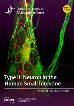
Cover Story (view full-size image): The enteric nervous system consists of more neurons than the spinal cord. These neurons generate different reflex arcs located completely within the gut wall. Our basic knowledge on different neuron types is derived from studies in the guinea pig gut. To identify the equivalent human enteric neuron populations, morpho–chemical analysis is a powerful method. Type III neurons have long, slender, branched dendrites. Beyond their immunoreactivity for neurofilaments (green) and calbindin (red), their unique chemical code needs to be deciphered. View this paper
  • Issues are regarded as officially published after their release is announced to the table of contents alert mailing list.
  • You may sign up for e-mail alerts to receive table of contents of newly released issues.
  • PDF is the official format for papers published in both, html and pdf forms. To view the papers in pdf format, click on the "PDF Full-text" link, and use the free Adobe Reader to open them.
Order results
Result details
Section
Select all
Export citation of selected articles as:
17 pages, 2608 KiB  
Article
Comparative Genomics of the Balsaminaceae Sister Genera Hydrocera triflora and Impatiens pinfanensis
by Zhi-Zhong Li, Josphat K. Saina, Andrew W. Gichira, Cornelius M. Kyalo, Qing-Feng Wang and Jin-Ming Chen
Int. J. Mol. Sci. 2018, 19(1), 319; https://doi.org/10.3390/ijms19010319 - 23 Jan 2018
Cited by 20 | Viewed by 7572
Abstract
The family Balsaminaceae, which consists of the economically important genus Impatiens and the monotypic genus Hydrocera, lacks a reported or published complete chloroplast genome sequence. Therefore, chloroplast genome sequences of the two sister genera are significant to give insight into the phylogenetic [...] Read more.
The family Balsaminaceae, which consists of the economically important genus Impatiens and the monotypic genus Hydrocera, lacks a reported or published complete chloroplast genome sequence. Therefore, chloroplast genome sequences of the two sister genera are significant to give insight into the phylogenetic position and understanding the evolution of the Balsaminaceae family among the Ericales. In this study, complete chloroplast (cp) genomes of Impatiens pinfanensis and Hydrocera triflora were characterized and assembled using a high-throughput sequencing method. The complete cp genomes were found to possess the typical quadripartite structure of land plants chloroplast genomes with double-stranded molecules of 154,189 bp (Impatiens pinfanensis) and 152,238 bp (Hydrocera triflora) in length. A total of 115 unique genes were identified in both genomes, of which 80 are protein-coding genes, 31 are distinct transfer RNA (tRNA) and four distinct ribosomal RNA (rRNA). Thirty codons, of which 29 had A/T ending codons, revealed relative synonymous codon usage values of >1, whereas those with G/C ending codons displayed values of <1. The simple sequence repeats comprise mostly the mononucleotide repeats A/T in all examined cp genomes. Phylogenetic analysis based on 51 common protein-coding genes indicated that the Balsaminaceae family formed a lineage with Ebenaceae together with all the other Ericales. Full article
(This article belongs to the Special Issue Chloroplast)
Show Figures

Figure 1

15 pages, 4391 KiB  
Article
Affinity Purification and Comparative Biosensor Analysis of Citrulline-Peptide-Specific Antibodies in Rheumatoid Arthritis
by Eszter Szarka, Petra Aradi, Krisztina Huber, Judit Pozsgay, Lili Végh, Anna Magyar, Gergő Gyulai, György Nagy, Bernadette Rojkovich, Éva Kiss, Ferenc Hudecz and Gabriella Sármay
Int. J. Mol. Sci. 2018, 19(1), 326; https://doi.org/10.3390/ijms19010326 - 22 Jan 2018
Cited by 12 | Viewed by 6459
Abstract
Background: In rheumatoid arthritis (RA), anti-citrullinated protein/peptide antibodies (ACPAs) are responsible for disease onset and progression, however, our knowledge is limited on ligand binding affinities of autoantibodies with different citrulline-peptide specificity. Methods: Citrulline-peptide-specific ACPA IgGs were affinity purified and tested by ELISA. Binding [...] Read more.
Background: In rheumatoid arthritis (RA), anti-citrullinated protein/peptide antibodies (ACPAs) are responsible for disease onset and progression, however, our knowledge is limited on ligand binding affinities of autoantibodies with different citrulline-peptide specificity. Methods: Citrulline-peptide-specific ACPA IgGs were affinity purified and tested by ELISA. Binding affinities of ACPA IgGs and serum antibodies were compared by surface plasmon resonance (SPR) analysis. Bifunctional nanoparticles harboring a multi-epitope citrulline-peptide and a complement-activating peptide were used to induce selective depletion of ACPA-producing B cells. Results: KD values of affinity-purified ACPA IgGs varied between 10−6 and 10−8 M and inversely correlated with disease activity. Based on their cross-reaction with citrulline-peptides, we designed a novel multi-epitope peptide, containing Cit-Gly and Ala-Cit motifs in two–two copies, separated with a short, neutral spacer. This peptide detected antibodies in RA sera with 66% sensitivity and 98% specificity in ELISA and was recognized by 90% of RA sera, while none of the healthy samples in SPR. When coupled to nanoparticles, the multi-epitope peptide specifically targeted and depleted ACPA-producing B cells ex vivo. Conclusions: The unique multi-epitope peptide designed based on ACPA cross-reactivity might be suitable to develop better diagnostics and novel therapies for RA. Full article
(This article belongs to the Special Issue Musculoskeletal Diseases Therapy)
Show Figures

Graphical abstract

40 pages, 2232 KiB  
Review
Heat Shock Proteins and Autophagy Pathways in Neuroprotection: From Molecular Bases to Pharmacological Interventions
by Botond Penke, Ferenc Bogár, Tim Crul, Miklós Sántha, Melinda E. Tóth and László Vígh
Int. J. Mol. Sci. 2018, 19(1), 325; https://doi.org/10.3390/ijms19010325 - 22 Jan 2018
Cited by 72 | Viewed by 14145
Abstract
Neurodegenerative diseases (NDDs) such as Alzheimer’s disease, Parkinson’s disease and Huntington’s disease (HD), amyotrophic lateral sclerosis, and prion diseases are all characterized by the accumulation of protein aggregates (amyloids) into inclusions and/or plaques. The ubiquitous presence of amyloids in NDDs suggests the involvement [...] Read more.
Neurodegenerative diseases (NDDs) such as Alzheimer’s disease, Parkinson’s disease and Huntington’s disease (HD), amyotrophic lateral sclerosis, and prion diseases are all characterized by the accumulation of protein aggregates (amyloids) into inclusions and/or plaques. The ubiquitous presence of amyloids in NDDs suggests the involvement of disturbed protein homeostasis (proteostasis) in the underlying pathomechanisms. This review summarizes specific mechanisms that maintain proteostasis, including molecular chaperons, the ubiquitin-proteasome system (UPS), endoplasmic reticulum associated degradation (ERAD), and different autophagic pathways (chaperon mediated-, micro-, and macro-autophagy). The role of heat shock proteins (Hsps) in cellular quality control and degradation of pathogenic proteins is reviewed. Finally, putative therapeutic strategies for efficient removal of cytotoxic proteins from neurons and design of new therapeutic targets against the progression of NDDs are discussed. Full article
(This article belongs to the Special Issue Neuroprotective Strategies 2022)
Show Figures

Figure 1

11 pages, 1265 KiB  
Article
Overall Survival of Ovarian Cancer Patients Is Determined by Expression of Galectins-8 and -9
by Heiko Schulz, Christina Kuhn, Simone Hofmann, Doris Mayr, Sven Mahner, Udo Jeschke and Elisa Schmoeckel
Int. J. Mol. Sci. 2018, 19(1), 323; https://doi.org/10.3390/ijms19010323 - 22 Jan 2018
Cited by 25 | Viewed by 5576
Abstract
The evaluation of new prognostic factors that can be targeted in ovarian cancer diagnosis and therapy is of the utmost importance. Galectins are a family of carbohydrate binding proteins with various implications in cancer biology. In this study, the presence of galectin (Gal)-8 [...] Read more.
The evaluation of new prognostic factors that can be targeted in ovarian cancer diagnosis and therapy is of the utmost importance. Galectins are a family of carbohydrate binding proteins with various implications in cancer biology. In this study, the presence of galectin (Gal)-8 and -9 was investigated in 156 ovarian cancer samples using immunohistochemistry (IHC). Staining was evaluated using semi-quantitative immunoreactivity (IR) scores and correlated to clinical and pathological data. Different types of galectin expression were compared with respect to disease-free survival (DFS) and overall survival (OS). Gal-8 served as a new positive prognostic factor for the OS and DFS of ovarian cancer patients. Gal-9 expression determined the DFS and OS of ovarian cancer patients in two opposing ways—moderate Gal-9 expression was correlated with a reduced outcome as compared to Gal-9 negative cases, while patients with high Gal-9 expression showed the best outcome. Full article
(This article belongs to the Special Issue Galectins in Cancer and Translational Medicine)
Show Figures

Graphical abstract

17 pages, 1609 KiB  
Review
Role of 3-Hydroxy Fatty Acid-Induced Hepatic Lipotoxicity in Acute Fatty Liver of Pregnancy
by Sathish Kumar Natarajan and Jamal A. Ibdah
Int. J. Mol. Sci. 2018, 19(1), 322; https://doi.org/10.3390/ijms19010322 - 22 Jan 2018
Cited by 36 | Viewed by 13972
Abstract
Acute fatty liver of pregnancy (AFLP), a catastrophic illness for both the mother and the unborn offspring, develops in the last trimester of pregnancy with significant maternal and perinatal mortality. AFLP is also recognized as an obstetric and medical emergency. Maternal AFLP is [...] Read more.
Acute fatty liver of pregnancy (AFLP), a catastrophic illness for both the mother and the unborn offspring, develops in the last trimester of pregnancy with significant maternal and perinatal mortality. AFLP is also recognized as an obstetric and medical emergency. Maternal AFLP is highly associated with a fetal homozygous mutation (1528G>C) in the gene that encodes for mitochondrial long-chain hydroxy acyl-CoA dehydrogenase (LCHAD). The mutation in LCHAD results in the accumulation of 3-hydroxy fatty acids, such as 3-hydroxy myristic acid, 3-hydroxy palmitic acid and 3-hydroxy dicarboxylic acid in the placenta, which are then shunted to the maternal circulation leading to the development of acute liver injury observed in patients with AFLP. In this review, we will discuss the mechanistic role of increased 3-hydroxy fatty acid in causing lipotoxicity to the liver and in inducing oxidative stress, mitochondrial dysfunction and hepatocyte lipoapoptosis. Further, we also review the role of 3-hydroxy fatty acids in causing placental damage, pancreatic islet β-cell glucolipotoxicity, brain damage, and retinal epithelial cells lipoapoptosis in patients with LCHAD deficiency. Full article
(This article belongs to the Special Issue Hepatotoxicity: Molecular Mechanisms and Pathophysiology)
Show Figures

Graphical abstract

10 pages, 2951 KiB  
Communication
Identification of a Rare Germline Heterozygous Deletion Involving the Polycistronic miR-17–92 Cluster in Two First-Degree Relatives from a BRCA 1/2 Negative Chilean Family with Familial Breast Cancer: Possible Functional Implications
by Tomás De Mayo, Annemarie Ziegler, Sebastián Morales and Lilian Jara
Int. J. Mol. Sci. 2018, 19(1), 321; https://doi.org/10.3390/ijms19010321 - 22 Jan 2018
Cited by 4 | Viewed by 5624
Abstract
Micro-RNAs (miRNAs) have emerged as novel gene expression regulators. Recent evidence strongly suggests a role for miRNAs in a large variety of cancer-related pathways. Different studies have shown that 18.7 to 37% of all human miRNA genes are clustered. miR-17–92 polycistronic cluster overexpression [...] Read more.
Micro-RNAs (miRNAs) have emerged as novel gene expression regulators. Recent evidence strongly suggests a role for miRNAs in a large variety of cancer-related pathways. Different studies have shown that 18.7 to 37% of all human miRNA genes are clustered. miR-17–92 polycistronic cluster overexpression is associated with human hematolymphoid and solid malignancies including breast cancer (BC). Here, we report the identification of rs770419845, a rare 6 bp deletion located within the polycistronic miR-17–92 cluster, in two first-degree relatives from a Chilean family with familial BC and negative for point mutations in BRCA 1/2 genes. The deletion was identified by Sanger sequencing when 99 BRCA1/2 mutation-negative BC cases with a strong family history were initially screened. In silico analysis predicts that rs770419845 affects the secondary structure and stability of the pre-miR-17–pre-miR-18 region and the entire 17–92 cluster. The deletion was screened in 458 high-risk BRCA1/2-negative Chilean families and 480 controls. rs770419845 was not detected in any control but identified in a single family with two cases of BC and other cancers. Both BC cases, the mother and her daughter, carried the deletion. Based on bioinformatic analyses, the location of the deletion and its low frequency, we presume rs770419845 may be a pathogenic variant. Functional studies are needed to support this hypothesis. Full article
(This article belongs to the Special Issue Advances in Molecular Oncology)
Show Figures

Graphical abstract

10 pages, 1607 KiB  
Review
Hereditary Fibrinogen Aα-Chain Amyloidosis in Asia: Clinical and Molecular Characteristics
by Masahide Yazaki, Tsuneaki Yoshinaga, Yoshiki Sekijima, Fuyuki Kametani and Nobuo Okumura
Int. J. Mol. Sci. 2018, 19(1), 320; https://doi.org/10.3390/ijms19010320 - 22 Jan 2018
Cited by 11 | Viewed by 5873
Abstract
Hereditary fibrinogen Aα-chain amyloidosis (Aα-chain amyloidosis) is a type of autosomal dominant systemic amyloidosis caused by mutations in fibrinogen Aα-chain gene (FGA). Patients with Aα-chain amyloidosis have been mainly reported in Western countries but have been rarely reported in [...] Read more.
Hereditary fibrinogen Aα-chain amyloidosis (Aα-chain amyloidosis) is a type of autosomal dominant systemic amyloidosis caused by mutations in fibrinogen Aα-chain gene (FGA). Patients with Aα-chain amyloidosis have been mainly reported in Western countries but have been rarely reported in Asia, with only five patients with Aα-chain amyloidosis being reported in Korea, China, and Japan. Clinically, the most prominent manifestation in Asian patients with Aα-chain amyloidosis is progressive nephropathy caused by excessive amyloid deposition in the glomeruli, which is similar to that observed in patients with Aα-chain amyloidosis in Western countries. In molecular features in Asian Aα-chain amyloidosis, the most common variant, E526V, was found in only one Chinese kindred, and other four kindred each had a different variant, which have not been identified in other countries. These variants are located in the C-terminal region (amino acid residues 517–555) of mature Aα-chain, which was similar to that observed in patients with Aα-chain amyloidosis in other countries. The precise number of Asian patients with Aα-chain amyloidosis is unclear. However, patients with Aα-chain amyloidosis do exist in Asian countries, and the majority of these patients may be diagnosed with other types of systemic amyloidosis. Full article
(This article belongs to the Special Issue Genetic Basis of Fibrinogen Disorders)
Show Figures

Figure 1

27 pages, 2924 KiB  
Review
Microglia and Aging: The Role of the TREM2–DAP12 and CX3CL1-CX3CR1 Axes
by Carmen Mecca, Ileana Giambanco, Rosario Donato and Cataldo Arcuri
Int. J. Mol. Sci. 2018, 19(1), 318; https://doi.org/10.3390/ijms19010318 - 22 Jan 2018
Cited by 175 | Viewed by 18694
Abstract
Depending on the species, microglial cells represent 5–20% of glial cells in the adult brain. As the innate immune effector of the brain, microglia are involved in several functions: regulation of inflammation, synaptic connectivity, programmed cell death, wiring and circuitry formation, phagocytosis of [...] Read more.
Depending on the species, microglial cells represent 5–20% of glial cells in the adult brain. As the innate immune effector of the brain, microglia are involved in several functions: regulation of inflammation, synaptic connectivity, programmed cell death, wiring and circuitry formation, phagocytosis of cell debris, and synaptic pruning and sculpting of postnatal neural circuits. Moreover, microglia contribute to some neurodevelopmental disorders such as Nasu-Hakola disease (NHD), and to aged-associated neurodegenerative diseases, such as Alzheimer’s disease (AD), Parkinson’s disease (PD), and others. There is evidence that human and rodent microglia may become senescent. This event determines alterations in the microglia activation status, associated with a chronic inflammation phenotype and with the loss of neuroprotective functions that lead to a greater susceptibility to the neurodegenerative diseases of aging. In the central nervous system (CNS), Triggering Receptor Expressed on Myeloid Cells 2-DNAX activation protein 12 (TREM2-DAP12) is a signaling complex expressed exclusively in microglia. As a microglial surface receptor, TREM2 interacts with DAP12 to initiate signal transduction pathways that promote microglial cell activation, phagocytosis, and microglial cell survival. Defective TREM2-DAP12 functions play a central role in the pathogenesis of several diseases. The CX3CL1 (fractalkine)-CX3CR1 signaling represents the most important communication channel between neurons and microglia. The expression of CX3CL1 in neurons and of its receptor CX3CR1 in microglia determines a specific interaction, playing fundamental roles in the regulation of the maturation and function of these cells. Here, we review the role of the TREM2-DAP12 and CX3CL1-CX3CR1 axes in aged microglia and the involvement of these pathways in physiological CNS aging and in age-associated neurodegenerative diseases. Full article
(This article belongs to the Special Issue Microglia in Aging and Neurodegenerative Disease)
Show Figures

Figure 1

18 pages, 958 KiB  
Review
Human MHC-II with Shared Epitope Motifs Are Optimal Epstein-Barr Virus Glycoprotein 42 Ligands—Relation to Rheumatoid Arthritis
by Nicole Trier, Jose Izarzugaza, Anna Chailyan, Paolo Marcatili and Gunnar Houen
Int. J. Mol. Sci. 2018, 19(1), 317; https://doi.org/10.3390/ijms19010317 - 21 Jan 2018
Cited by 29 | Viewed by 10727
Abstract
Rheumatoid arthritis (RA) is a chronic systemic autoimmune disorder of unknown etiology, which is characterized by inflammation in the synovium and joint damage. Although the pathogenesis of RA remains to be determined, a combination of environmental (e.g., viral infections) and genetic factors influence [...] Read more.
Rheumatoid arthritis (RA) is a chronic systemic autoimmune disorder of unknown etiology, which is characterized by inflammation in the synovium and joint damage. Although the pathogenesis of RA remains to be determined, a combination of environmental (e.g., viral infections) and genetic factors influence disease onset. Especially genetic factors play a vital role in the onset of disease, as the heritability of RA is 50–60%, with the human leukocyte antigen (HLA) alleles accounting for at least 30% of the overall genetic risk. Some HLA-DR alleles encode a conserved sequence of amino acids, referred to as the shared epitope (SE) structure. By analyzing the structure of a HLA-DR molecule in complex with Epstein-Barr virus (EBV), the SE motif is suggested to play a vital role in the interaction of MHC II with the viral glycoprotein (gp) 42, an essential entry factor for EBV. EBV has been repeatedly linked to RA by several lines of evidence and, based on several findings, we suggest that EBV is able to induce the onset of RA in predisposed SE-positive individuals, by promoting entry of B-cells through direct contact between SE and gp42 in the entry complex. Full article
(This article belongs to the Special Issue Research of Pathogenesis and Novel Therapeutics in Arthritis)
Show Figures

Figure 1

14 pages, 2707 KiB  
Article
Effects of Exogenous Melatonin on Methyl Viologen-Mediated Oxidative Stress in Apple Leaf
by Zhiwei Wei, Tengteng Gao, Bowen Liang, Qi Zhao, Fengwang Ma and Chao Li
Int. J. Mol. Sci. 2018, 19(1), 316; https://doi.org/10.3390/ijms19010316 - 21 Jan 2018
Cited by 47 | Viewed by 6609
Abstract
Oxidative stress is a major source of damage of plants exposed to adverse environments. We examined the effect of exogenous melatonin (MT) in limiting of oxidative stress caused by methyl viologen (MV; paraquatin) in apple leaves (Malus domestica Borkh.). When detached leaves [...] Read more.
Oxidative stress is a major source of damage of plants exposed to adverse environments. We examined the effect of exogenous melatonin (MT) in limiting of oxidative stress caused by methyl viologen (MV; paraquatin) in apple leaves (Malus domestica Borkh.). When detached leaves were pre-treated with melatonin, their level of stress tolerance increased. Under MV treatment, melatonin effectively alleviated the decrease in chlorophyll concentrations and maximum potential Photosystem II efficiency while also mitigating membrane damage and lipid peroxidation when compared with control leaves that were sprayed only with water prior to the stress experiment. The melatonin-treated leaves also showed higher activities and transcripts of antioxidant enzymes superoxide dismutase, peroxidase, and catalase. In addition, the expression of genes for those enzymes was upregulated. Melatonin-synthesis genes MdTDC1, MdT5H4, MdAANAT2, and MdASMT1 were also upregulated under oxidative stress in leaves but that expression was suppressed in response to 1 mM melatonin pretreatment during the MV treatments. Therefore, we conclude that exogenous melatonin mitigates the detrimental effects of oxidative stress, perhaps by slowing the decline in chlorophyll concentrations, moderating membrane damage and lipid peroxidation, increasing the activities of antioxidant enzymes, and changing the expression of genes for melatonin synthesis. Full article
(This article belongs to the Section Molecular Plant Sciences)
Show Figures

Figure 1

20 pages, 1463 KiB  
Review
Endothelium-Dependent Hyperpolarization (EDH) in Hypertension: The Role of Endothelial Ion Channels
by Kenichi Goto, Toshio Ohtsubo and Takanari Kitazono
Int. J. Mol. Sci. 2018, 19(1), 315; https://doi.org/10.3390/ijms19010315 - 21 Jan 2018
Cited by 63 | Viewed by 15006
Abstract
Upon stimulation with agonists and shear stress, the vascular endothelium of different vessels selectively releases several vasodilator factors such as nitric oxide and prostacyclin. In addition, vascular endothelial cells of many vessels regulate the contractility of the vascular smooth muscle cells through the [...] Read more.
Upon stimulation with agonists and shear stress, the vascular endothelium of different vessels selectively releases several vasodilator factors such as nitric oxide and prostacyclin. In addition, vascular endothelial cells of many vessels regulate the contractility of the vascular smooth muscle cells through the generation of endothelium-dependent hyperpolarization (EDH). There is a general consensus that the opening of small- and intermediate-conductance Ca2+-activated K+ channels (SKCa and IKCa) is the initial mechanistic step for the generation of EDH. In animal models and humans, EDH and EDH-mediated relaxations are impaired during hypertension, and anti-hypertensive treatments restore such impairments. However, the underlying mechanisms of reduced EDH and its improvement by lowering blood pressure are poorly understood. Emerging evidence suggests that alterations of endothelial ion channels such as SKCa channels, inward rectifier K+ channels, Ca2+-activated Cl channels, and transient receptor potential vanilloid type 4 channels contribute to the impaired EDH during hypertension. In this review, we attempt to summarize the accumulating evidence regarding the pathophysiological role of endothelial ion channels, focusing on their relationship with EDH during hypertension. Full article
(This article belongs to the Special Issue Ion Transporters and Channels in Physiology and Pathophysiology)
Show Figures

Figure 1

15 pages, 517 KiB  
Review
Deregulation of Frizzled Receptors in Hepatocellular Carcinoma
by Kristy Kwan-Shuen Chan and Regina Cheuk-Lam Lo
Int. J. Mol. Sci. 2018, 19(1), 313; https://doi.org/10.3390/ijms19010313 - 21 Jan 2018
Cited by 17 | Viewed by 8472
Abstract
G protein-coupled receptors (GPCRs) have a substantial role in tumorigenesis and are described as a “cancer driver”. Aberrant expression or activation of GPCRs leads to the deregulation of downstream signaling pathways, thereby promoting cancer progression. In hepatocellular carcinoma (HCC), the Wnt signaling pathway [...] Read more.
G protein-coupled receptors (GPCRs) have a substantial role in tumorigenesis and are described as a “cancer driver”. Aberrant expression or activation of GPCRs leads to the deregulation of downstream signaling pathways, thereby promoting cancer progression. In hepatocellular carcinoma (HCC), the Wnt signaling pathway is frequently activated and it is associated with an aggressive HCC phenotype. Frizzled (FZD) receptors, a family member of GPCRs, are known to mediate Wnt signaling. Accumulating findings have revealed the deregulation of FZD receptors in HCC and their functional roles have been implicated in HCC progression. Given the important role of FZD receptors in HCC, we summarize here the expression pattern of FZD receptors in HCC and their corresponding functional roles during HCC progression. We also further review and highlight the potential targeting of FZD receptors as an alternative therapeutic strategy in HCC. Full article
(This article belongs to the Special Issue Cancer-Driver G Protein-Coupled Receptors as Therapeutic Targets)
Show Figures

Figure 1

13 pages, 5291 KiB  
Article
In Vitro Antitumor Activity of Aloperine on Human Thyroid Cancer Cells through Caspase-Dependent Apoptosis
by Ying-Ray Lee, Shu-Hsin Chen, Ching-Yen Lin, Wen-Ying Chao, Yun-Ping Lim, Hui-I Yu and Chieh-Hsiang Lu
Int. J. Mol. Sci. 2018, 19(1), 312; https://doi.org/10.3390/ijms19010312 - 21 Jan 2018
Cited by 40 | Viewed by 6162
Abstract
The global incidence of thyroid cancer, one of the most common endocrine malignancies, is especially high among women. Although most patients with thyroid cancers exhibit a good prognosis with standard treatment, there are no effective therapies for patients with anaplastic thyroid cancers or [...] Read more.
The global incidence of thyroid cancer, one of the most common endocrine malignancies, is especially high among women. Although most patients with thyroid cancers exhibit a good prognosis with standard treatment, there are no effective therapies for patients with anaplastic thyroid cancers or cancers that have reached an advanced or recurrent level. Therefore, it is important to develop highly effective compounds for treating such patients. Aloperine, a natural compound isolated from Sophora alopecuroides, has been reported to possess antioxidant, anti-inflammatory, anti-neuronal injury, anti-renal injury, antitumor, anti-allergic, and antiviral properties. In this study, we show that aloperine can inhibit cell growth in human anaplastic thyroid cancers and multidrug-resistant papillary thyroid cancers. Moreover, it could suppress in vitro tumorigenesis and promote cellular apoptosis. Further analysis demonstrated the involvement of caspase-dependent apoptosis, including intrinsic and/or extrinsic pathways, in aloperine-induced cellular apoptosis. However, cell cycle regulation was not detected with aloperine treatment. This study suggests the potential therapeutic use of aloperine in human anaplastic thyroid cancers and multidrug-resistant papillary thyroid cancers. Full article
(This article belongs to the Collection Programmed Cell Death and Apoptosis)
Show Figures

Figure 1

26 pages, 3155 KiB  
Review
Chondrosarcoma: A Rare Misfortune in Aging Human Cartilage? The Role of Stem and Progenitor Cells in Proliferation, Malignant Degeneration and Therapeutic Resistance
by Karen A. Boehme, Sabine B. Schleicher, Frank Traub and Bernd Rolauffs
Int. J. Mol. Sci. 2018, 19(1), 311; https://doi.org/10.3390/ijms19010311 - 21 Jan 2018
Cited by 52 | Viewed by 10514
Abstract
Unlike other malignant bone tumors including osteosarcomas and Ewing sarcomas with a peak incidence in adolescents and young adults, conventional and dedifferentiated chondrosarcomas mainly affect people in the 4th to 7th decade of life. To date, the cell type of chondrosarcoma origin is [...] Read more.
Unlike other malignant bone tumors including osteosarcomas and Ewing sarcomas with a peak incidence in adolescents and young adults, conventional and dedifferentiated chondrosarcomas mainly affect people in the 4th to 7th decade of life. To date, the cell type of chondrosarcoma origin is not clearly defined. However, it seems that mesenchymal stem and progenitor cells (MSPC) in the bone marrow facing a pro-proliferative as well as predominantly chondrogenic differentiation milieu, as is implicated in early stage osteoarthritis (OA) at that age, are the source of chondrosarcoma genesis. But how can MSPC become malignant? Indeed, only one person in 1,000,000 will develop a chondrosarcoma, whereas the incidence of OA is a thousandfold higher. This means a rare coincidence of factors allowing escape from senescence and apoptosis together with induction of angiogenesis and migration is needed to generate a chondrosarcoma. At early stages, chondrosarcomas are still assumed to be an intermediate type of tumor which rarely metastasizes. Unfortunately, advanced stages show a pronounced resistance both against chemo- and radiation-therapy and frequently metastasize. In this review, we elucidate signaling pathways involved in the genesis and therapeutic resistance of chondrosarcomas with a focus on MSPC compared to signaling in articular cartilage (AC). Full article
(This article belongs to the Special Issue Current Advances in Soft Tissue and Bone Sarcoma)
Show Figures

Graphical abstract

15 pages, 3362 KiB  
Article
Abscisic Acid (ABA ) Promotes the Induction and Maintenance of Pear (Pyrus pyrifolia White Pear Group) Flower Bud Endodormancy
by Jianzhao Li, Ying Xu, Qingfeng Niu, Lufang He, Yuanwen Teng and Songling Bai
Int. J. Mol. Sci. 2018, 19(1), 310; https://doi.org/10.3390/ijms19010310 - 20 Jan 2018
Cited by 87 | Viewed by 8745
Abstract
Dormancy is an adaptive mechanism that allows temperate deciduous plants to survive unfavorable winter conditions. In the present work, we investigated the possible function of abscisic acid (ABA) on the endodormancy process in pear. The ABA content increased during pear flower bud endodormancy [...] Read more.
Dormancy is an adaptive mechanism that allows temperate deciduous plants to survive unfavorable winter conditions. In the present work, we investigated the possible function of abscisic acid (ABA) on the endodormancy process in pear. The ABA content increased during pear flower bud endodormancy establishment and decreased towards endodormancy release. In total, 39 putative genes related to ABA metabolism and signal transductions were identified from pear genome. During the para- to endodormancy transition, PpNCED-2 and PpNCED-3 had high expression levels, while PpCYP707As expression levels were low. However, during endodormancy, the expression of PpCYP707A-3 sharply increased with increasing cold accumulation. At the same time, the ABA content of pear buds declined, and the percentage of bud breaks rapidly increased. On the other hand, the expression levels of PpPYLs, PpPP2Cs, PpSnRK2s, and PpABI4/ABI5s were also changed during the pear flower bud dormancy cycle. Furthermore, exogenous ABA application to para-dormant buds significantly reduced the bud breaks and accelerated the transition to endodormancy. During the whole treatment time, the expression level of PpPP2C-12 decreased to a greater extent in ABA-treated buds than in control. However, the expression levels of PpSnRK2-1, PpSnRK2-4, and PpABI5-1 were higher in ABA-treated buds. Our results indicated that PpCYP707A-3 and PpNCEDs play pivotal roles on the regulation of endodormancy release, while ABA signal transduction pathway also appears to be involved in the process. The present work provided the basic information about the function of ABA-related genes during pear flower bud dormancy process. Full article
Show Figures

Graphical abstract

14 pages, 2887 KiB  
Article
Enhancing Flower Color through Simultaneous Expression of the B-peru and mPAP1 Transcription Factors under Control of a Flower-Specific Promoter
by Da-Hye Kim, Sangkyu Park, Jong-Yeol Lee, Sun-Hwa Ha and Sun-Hyung Lim
Int. J. Mol. Sci. 2018, 19(1), 309; https://doi.org/10.3390/ijms19010309 - 20 Jan 2018
Cited by 26 | Viewed by 7526
Abstract
Flower color is a main target for flower breeding. A transgenic approach for flower color modification requires a transgene and a flower-specific promoter. Here, we expressed the B-peru gene encoding a basic helix loop helix (bHLH) transcription factor (TF) together with the mPAP1 [...] Read more.
Flower color is a main target for flower breeding. A transgenic approach for flower color modification requires a transgene and a flower-specific promoter. Here, we expressed the B-peru gene encoding a basic helix loop helix (bHLH) transcription factor (TF) together with the mPAP1 gene encoding an R2R3 MYB TF to enhance flower color in tobacco (Nicotiana tabacum L.), using the tobacco anthocyanidin synthase (ANS) promoter (PANS) to drive flower-specific expression. The transgenic tobacco plants grew normally and produced either dark pink (PANSBP_DP) or dark red (PANSBP_DR) flowers. Quantitative real time polymerase chain reaction (qPCR) revealed that the expression of five structural genes in the flavonoid biosynthetic pathway increased significantly in both PANSBP_DP and PANSBP_DR lines, compared with the non-transformed (NT) control. Interestingly, the expression of two regulatory genes constituting the active MYB-bHLH-WD40 repeat (WDR) (MBW) complex decreased significantly in the PANSBP_DR plants but not in the PANSBP_DP plants. Total flavonol and anthocyanin abundance correlated with flower color, with an increase of 1.6–43.2 fold in the PANSBP_DP plants and 2.0–124.2 fold in the PANSBP_DR plants. Our results indicate that combinatorial expression of B-peru and mPAP1 genes under control of the ANS promoter can be a useful strategy for intensifying flower color without growth retardation. Full article
(This article belongs to the Section Molecular Plant Sciences)
Show Figures

Graphical abstract

22 pages, 1238 KiB  
Review
Post-Transcriptional Regulation of Anti-Apoptotic BCL2 Family Members
by Jia Cui and William J. Placzek
Int. J. Mol. Sci. 2018, 19(1), 308; https://doi.org/10.3390/ijms19010308 - 20 Jan 2018
Cited by 111 | Viewed by 10114
Abstract
Anti-apoptotic B cell lymphoma 2 (BCL2) family members (BCL2, MCL1, BCLxL, BCLW, and BFL1) are key players in the regulation of intrinsic apoptosis. Dysregulation of these proteins not only impairs normal development, but also contributes to tumor progression and resistance to various anti-cancer [...] Read more.
Anti-apoptotic B cell lymphoma 2 (BCL2) family members (BCL2, MCL1, BCLxL, BCLW, and BFL1) are key players in the regulation of intrinsic apoptosis. Dysregulation of these proteins not only impairs normal development, but also contributes to tumor progression and resistance to various anti-cancer therapies. Therefore, cells maintain strict control over the expression of anti-apoptotic BCL2 family members using multiple mechanisms. Over the past two decades, the importance of post-transcriptional regulation of mRNA in controlling gene expression and its impact on normal homeostasis and disease have begun to be appreciated. In this review, we discuss the RNA binding proteins (RBPs) and microRNAs (miRNAs) that mediate post-transcriptional regulation of the anti-apoptotic BCL2 family members. We describe their roles and impact on alternative splicing, mRNA turnover, and mRNA subcellular localization. We also point out the importance of future studies in characterizing the crosstalk between RBPs and miRNAs in regulating anti-apoptotic BCL2 family member expression and ultimately apoptosis. Full article
(This article belongs to the Collection Programmed Cell Death and Apoptosis)
Show Figures

Figure 1

12 pages, 628 KiB  
Article
Circulating Plasma Levels of miR-20b, miR-29b and miR-155 as Predictors of Bevacizumab Efficacy in Patients with Metastatic Colorectal Cancer
by Paola Ulivi, Matteo Canale, Alessandro Passardi, Giorgia Marisi, Martina Valgiusti, Giovanni Luca Frassineti, Daniele Calistri, Dino Amadori and Emanuela Scarpi
Int. J. Mol. Sci. 2018, 19(1), 307; https://doi.org/10.3390/ijms19010307 - 20 Jan 2018
Cited by 63 | Viewed by 5842
Abstract
Targeting angiogenesis in the treatment of colorectal cancer (CRC) is a common strategy, for which potential predictive biomarkers have been studied. miRNAs are small non-coding RNAs involved in several processes including the angiogenic pathway. They are very stable in biological fluids, which turns [...] Read more.
Targeting angiogenesis in the treatment of colorectal cancer (CRC) is a common strategy, for which potential predictive biomarkers have been studied. miRNAs are small non-coding RNAs involved in several processes including the angiogenic pathway. They are very stable in biological fluids, which turns them into potential circulating biomarkers. In this study, we considered a case series of patients with metastatic (m) CRC treated with a bevacizumab (B)-based treatment, enrolled in the prospective multicentric Italian Trial in Advanced Colorectal Cancer (ITACa). We then analyzed a panel of circulating miRNAs in relation to the patient outcome. In multivariate analysis, circulating basal levels of hsa-miR-20b-5p, hsa-miR-29b-3p and hsa-miR-155-5p resulted in being significantly associated with progression-free survival (PFS) (p = 0.027, p = 0.034 and p = 0.039, respectively) and overall survival (OS) (p = 0.044, p = 0.024 and p = 0.032, respectively). We also observed that an increase in hsa-miR-155-5p at the first clinical evaluation was significantly associated with shorter PFS (HR 3.03 (95% CI 1.06–9.09), p = 0.040) and OS (HR 3.45 (95% CI 1.18–10.00), p = 0.024), with PFS and OS of 9.5 (95% CI 6.8–18.7) and 15.9 (95% CI 8.4–not reached), respectively, in patients with an increase ≥30% of hsa-miR-155-5p and 22.3 (95% CI 10.2–25.5) and 42.9 (24.8–not reached) months, respectively, in patients without such increase. In conclusion, our results highlight the potential usefulness of circulating basal levels of hsa-miR-20b-5p, hsa-miR-29b-3p and hsa-miR-155-5p in predicting the outcome of patients with mCRC treated with B. In addition, the variation of circulating hsa-miR-155-5p could also be indicative of the patient survival. Full article
(This article belongs to the Special Issue MicroRNA as Biomarkers in Cancer Diagnostics and Therapy)
Show Figures

Graphical abstract

14 pages, 4348 KiB  
Article
Compatibility with Panax notoginseng and Rehmannia glutinosa Alleviates the Hepatotoxicity and Nephrotoxicity of Tripterygium wilfordii via Modulating the Pharmacokinetics of Triptolide
by Qichun Zhang, Yiqun Li, Mengzhu Liu, Jinao Duan, Xueping Zhou and Huaxu Zhu
Int. J. Mol. Sci. 2018, 19(1), 305; https://doi.org/10.3390/ijms19010305 - 19 Jan 2018
Cited by 22 | Viewed by 6440
Abstract
Tripterygium wilfordii (TW) and the representative active component triptolide show positive therapeutic effect on the autoimmune disorders and simultaneously ineluctable hepatotoxicity and nephrotoxicity. Combinational application of Panax notoginseng (PN) and Rehmannia glutinosa (RG) weakens the toxicity of TW according the clinical application of [...] Read more.
Tripterygium wilfordii (TW) and the representative active component triptolide show positive therapeutic effect on the autoimmune disorders and simultaneously ineluctable hepatotoxicity and nephrotoxicity. Combinational application of Panax notoginseng (PN) and Rehmannia glutinosa (RG) weakens the toxicity of TW according the clinical application of traditional Chinese medicine. This article was aimed at the mechanism of decreasing toxicity of TW by the combinational application of PN and RG. Biochemical and pathohistological analysis were utilized to assess the toxicity on liver and kidney in rats administrated with TW, TW-PN, TW-RG and TW-PN-RG for 3 and 7 days. Meanwhile, the pharmacokinetics profiling of triptolide and wilforlide A was determined based on the plasma concentration analyzed by ultra-high performance liquid chromatography-tandem mass spectrometry (UPLC-MS/MS). TW-induced alkaline phosphatase (ALP), the marker for liver injury, was enhanced from 22.83 ± 1.29 to 40.73 ± 1.42 King’s unit/100 mL (p < 0.01) at day 7. TW-PN-RG decreased the serum ALP of TW-treated rats at 30.15 ± 1.27 King’s unit/100 mL (p < 0.01). For nephrotoxicity, TW pronouncedly elevated serum creatinine (SCr) in rats from 20.33 ± 1.77 to 49.82 ± 2.35 μmol/L (p < 0.01). However, rats treated with TW-PN-RG showed lower SCr at 30.48 ± 1.98 μmol/L (p < 0.01). Moreover, TW-PN-RG significantly decreased the TW-induced elevation of total bilirubin (T-BIL), alanine amino transferase (ALT), aspartate amino transferase (AST), blood urea nitrogen (Bun), and reversed the TW-resulted pathohistological characteristics of liver and kidney. The delayed time to reach Cmax (Tmax) and reduced maximum concentration (Cmax) and area under plasma concentration-time curve (AUC) of triptolide and wilforlide A were explored in rats with combinational formulas. Synergism of PN and RG obviously prolonged the half-life (t1/2) and apparent volume of distribution (Vd), but exerted no action on the clearance rate. The compatibility of TW, PN and RG influences intracorporal process of both triptolide and wilforlide A on the steps of absorption and tissue distribution contributing to less toxicity of TW on liver and kidney. Full article
(This article belongs to the Special Issue Traditional Medicine – Unraveling Its Molecular Mechanism)
Show Figures

Graphical abstract

1 pages, 132 KiB  
Erratum
Erratum: Lydia Visser et al. Characterization of the Microenvironment of Nodular Lymphocyte Predominant Hodgkin Lymphoma, Int. J. Mol. Sci. 2016, 17, 2127
by Ahmad Sattarzadeh, Lydia Visser, Bea Rutgers, Arjan Diepstra and Anke Van den Berg
Int. J. Mol. Sci. 2018, 19(1), 304; https://doi.org/10.3390/ijms19010304 - 19 Jan 2018
Cited by 1 | Viewed by 3356
Abstract
The authors regret to have made a mistake in publishing this paper [1] with an incorrect author list [...]
Full article
(This article belongs to the Special Issue Tumor Microenvironment and Metabolism)
76 pages, 1336 KiB  
Editorial
Acknowledgement to Reviewers of the International Journal of Molecular Sciences in 2017
by International Journal of Molecular Sciences Editorial Office
Int. J. Mol. Sci. 2018, 19(1), 303; https://doi.org/10.3390/ijms19010303 - 19 Jan 2018
Viewed by 16677
Abstract
Peer review is an essential part in the publication process, ensuring that the International Journal of Molecular Sciences maintains high quality standards for its published papers [...]
Full article
15 pages, 3436 KiB  
Article
The Role of Sugar Transporter Genes during Early Infection by Root-Knot Nematodes
by Dan Zhao, Yang You, Haiyan Fan, Xiaofeng Zhu, Yuanyuan Wang, Yuxi Duan, Yuanhu Xuan and Lijie Chen
Int. J. Mol. Sci. 2018, 19(1), 302; https://doi.org/10.3390/ijms19010302 - 19 Jan 2018
Cited by 36 | Viewed by 7071
Abstract
Although pathogens such as nematodes are known to hijack nutrients from host plants, the mechanisms whereby nematodes obtain sugars from plants remain largely unknown. To determine the effects of nematode infection on host plant sugar allocation, soluble sugar (fructose, glucose, sucrose) content was [...] Read more.
Although pathogens such as nematodes are known to hijack nutrients from host plants, the mechanisms whereby nematodes obtain sugars from plants remain largely unknown. To determine the effects of nematode infection on host plant sugar allocation, soluble sugar (fructose, glucose, sucrose) content was investigated using high-performance liquid chromatography with refractive index detection and was found to increase significantly in tomato (Solanum lycopersicum, Sl) leaves and roots during early infection by root-knot nematodes (RKNs). To further analyze whether sugar transporters played a role in this process, the expression levels of sucrose transporter (SUT/SUC), Sugars Will Eventually be Exported Transporter (SWEET), tonoplast monosaccharide transporter (TMT), and vacuolar glucose transporter (VGT) gene family members were examined by qRT-PCR analysis after RKN infection. The results showed that three SlSUTs, 17 SlSWEETs, three SlTMTs, and SlVGT1 were upregulated in the leaves, whereas three SlSUTs, 17 SlSWEETs, two SlTMTs, and SlVGT1 were induced in the roots. To determine the function of the sugar transporters in the RKN infection process, we examined post-infection responses in the Atsuc2 mutant and pAtSUC2-GUS lines. β-glucuronidase expression was strongly induced at the infection sites, and RKN development was significantly arrested in the Atsuc2 mutant. Taken together, our analyses provide useful information for understanding the sugar transporter responses during early infection by RKNs in tomato. Full article
(This article belongs to the Special Issue Plant Defense Genes Against Biotic Stresses)
Show Figures

Graphical abstract

19 pages, 2609 KiB  
Article
B Type and Complex A/B Type Epicatechin Trimers Isolated from Litchi pericarp Aqueous Extract Show High Antioxidant and Anticancer Activity
by Yihui Gong, Fang Fang, Xin Zhang, Bin Liu, Honghui Luo, Zhen Li, Xuelian Zhang, Zhaoqi Zhang and Xuequn Pang
Int. J. Mol. Sci. 2018, 19(1), 301; https://doi.org/10.3390/ijms19010301 - 19 Jan 2018
Cited by 36 | Viewed by 6254
Abstract
Litchi (Litchi chinensis Sonn.) fruit is known for its rich source of phenolics. Litchi pericarp contains high levels of epicatechin that may form oligomers of various lengths. Except for several A or B type epicatechin dimers, other soluble oligomers have rarely been [...] Read more.
Litchi (Litchi chinensis Sonn.) fruit is known for its rich source of phenolics. Litchi pericarp contains high levels of epicatechin that may form oligomers of various lengths. Except for several A or B type epicatechin dimers, other soluble oligomers have rarely been identified in the pericarp. Here, bioassay-guided column fractionation was applied to isolate bioactive phenolics from aqueous pericarp extract. A fraction (S3) was obtained by two rounds of Sephadex LH-20 column chromatography, and showed higher antioxidant activity and inhibition on the proliferation of human lung cancer cells (A549) than Litchi anthocyanins. S3 was further separated to isolate fractions P1–P4, which all showed higher antioxidant activity than vitamin C. P3 showed 32.9% inhibition on A549 cells at 30 μg/mL, higher than other fractions and cis-Dichlorodiamineplatinum (DDP, 0.5 μg/mL), but not as high as the combination of the four fractions. Using HPLC-Q-TOF-MS/MS, one B-type and complex A/B type epicatechin trimers were identified in P3; another B-type and two A/B-type trimers were identified in P4. P1 and P2, containing epicatechin and proanthocyanidin B2, respectively, showed no cell inhibition at 30 μg/mL. It is the first time that the two B type trimers of epicatechins (Litchitannin B1 and B2), have been found in Litchi species. The identified proanthocyanidins were detected in the pericarp of the young fruit, and the levels of the compounds decreased as the fruit developed, correlating to the decreasing patterns of the expression of LcLAR and LcANR, two key genes in the catechin biosynthesis pathway. Full article
Show Figures

Figure 1

12 pages, 3613 KiB  
Article
Changes in the Expression of AQP4 and AQP9 in the Hippocampus Following Eclampsia-Like Seizure
by Xinjia Han, Qian Huang, Lei Liu, Xiaoyan Sha, Bihui Hu and Huishu Liu
Int. J. Mol. Sci. 2018, 19(1), 300; https://doi.org/10.3390/ijms19010300 - 19 Jan 2018
Cited by 14 | Viewed by 5077
Abstract
Eclampsia is a hypertensive disorder of pregnancy that is defined by the new onset of grand mal seizures on the basis of pre-eclampsia. Until now, the mechanisms underlying eclampsia were poorly understood. Brain edema is considered a leading cause of eclamptic seizures; aquaporins [...] Read more.
Eclampsia is a hypertensive disorder of pregnancy that is defined by the new onset of grand mal seizures on the basis of pre-eclampsia. Until now, the mechanisms underlying eclampsia were poorly understood. Brain edema is considered a leading cause of eclamptic seizures; aquaporins (AQP4 and AQP9), the glial water channel proteins mainly expressed in the nervous system, play an important role in brain edema. We studied AQP4 and AQP9 expression in the hippocampus of pre-eclamptic and eclamptic rats in order to explore the molecular mechanisms involved in brain edema. Using our previous animal models, we found several neuronal deaths in the hippocampal CA1 and CA3 regions after pre-eclampsia and that eclampsia induced more neuronal deaths in both areas by Nissl staining. In the current study, RT-PCR and Western blotting data showed significant upregulation of AQP4 and AQP9 mRNA and protein levels after eclamptic seizures in comparison to pre-eclampsia and at the same time AQP4 and AQP9 immunoreactivity also increased after eclampsia. These findings showed that eclamptic seizures induced cell death and that upregulation of AQP4 and AQP9 may play an important role in this pathophysiological process. Full article
(This article belongs to the Special Issue Aquaporins: Water Channels Essential for Living Organisms)
Show Figures

Figure 1

25 pages, 1499 KiB  
Review
“Vessels in the Storm”: Searching for Prognostic and Predictive Angiogenic Factors in Colorectal Cancer
by Adriano Angelucci, Simona Delle Monache, Alessio Cortellini, Monica Di Padova and Corrado Ficorella
Int. J. Mol. Sci. 2018, 19(1), 299; https://doi.org/10.3390/ijms19010299 - 19 Jan 2018
Cited by 29 | Viewed by 5349
Abstract
High expectations are placed upon anti-angiogenic compounds for metastatic colorectal cancer (mCRC), the first malignancy for which such type of treatment has been approved. Indeed, clinical trials have confirmed that targeting the formation of new vessels can improve in many cases clinical outcomes [...] Read more.
High expectations are placed upon anti-angiogenic compounds for metastatic colorectal cancer (mCRC), the first malignancy for which such type of treatment has been approved. Indeed, clinical trials have confirmed that targeting the formation of new vessels can improve in many cases clinical outcomes of mCRC patients. However, current anti-angiogenic drugs are far from obtaining the desirable or expected curative results. Many are the factors probably involved in such disappointing results, but particular attention is currently focused on the validation of biomarkers able to improve the direction of treatment protocols. Because clinical studies have clearly demonstrated that serum or tissue concentration of some angiogenic factors is associated with the evolution of the disease of mCRC patients, they are currently explored as potential biomarkers of prognosis and of tumor response to therapy. However, the complex biology underlying CRC -induced angiogenesis is a hurdle in finding rapid solutions. The aim of this review was to explore molecular mechanisms that determine the formation of tumor-associated vessels during CRC progression, and to discuss the potential role of angiogenic factors as diagnostic, prognostic and predictive biomarkers in CRC. Full article
Show Figures

Graphical abstract

18 pages, 17197 KiB  
Article
Cyclin B3 Deficiency Impairs Germline Stem Cell Maintenance and Its Overexpression Delays Cystoblast Differentiation in Drosophila Ovary
by Dongsheng Chen, Lijuan Zhou, Fuling Sun, Mingzhong Sun and Xiaoqian Tao
Int. J. Mol. Sci. 2018, 19(1), 298; https://doi.org/10.3390/ijms19010298 - 19 Jan 2018
Cited by 6 | Viewed by 6660
Abstract
It is well known that cyclin B3 (cycB3) plays a key role in the control of cell cycle progression. However, whether cycB3 is involved in stem cell fate determination remains unknown. The Drosophila ovary provides an exclusive model for studying the [...] Read more.
It is well known that cyclin B3 (cycB3) plays a key role in the control of cell cycle progression. However, whether cycB3 is involved in stem cell fate determination remains unknown. The Drosophila ovary provides an exclusive model for studying the intrinsic and extrinsic factors that modulate the fate of germline stem cells (GSCs). Here, using this model, we show that Drosophila cycB3 plays a new role in controlling the fate of germline stem cells (GSC). Results from cycB3 genetic analyses demonstrate that cycB3 is intrinsically required for GSC maintenance. Results from green fluorescent protein (GFP)-transgene reporter assays show that cycB3 is not involved in Dad-mediated regulation of Bmp signaling, or required for dpp-induced bam transcriptional silencing. Double mutants of bam and cycB3 phenocopied bam single mutants, suggesting that cycB3 functions in a bam-dependent manner in GSCs. Deficiency of cycB3 fails to cause apoptosis in GSCs or influence cystoblast (CB) differentiation into oocytes. Furthermore, overexpression of cycB3 dramatically increases the CB number in Drosophila ovaries, suggesting that an excess of cycB3 function delays CB differentiation. Given that the cycB3 gene is evolutionarily conserved, from insects to humans, cycB3 may also be involved in controlling the fate of GSCs in humans. Full article
(This article belongs to the Section Biochemistry)
Show Figures

Graphical abstract

11 pages, 1659 KiB  
Article
The Prognostic Significance of Histone Demethylase UTX in Esophageal Squamous Cell Carcinoma
by Shau-Hsuan Li, Hung-I Lu, Wan-Ting Huang, Wan-Yu Tien, Ya-Chun Lan, Wei-Che Lin, Hsin-Ting Tsai and Chang-Han Chen
Int. J. Mol. Sci. 2018, 19(1), 297; https://doi.org/10.3390/ijms19010297 - 19 Jan 2018
Cited by 17 | Viewed by 4632
Abstract
The dysregulation of the ubiquitously transcribed TPR gene on the X chromosome (UTX) has been reported to be involved in the oncogenesis of several types of cancers. However, the expression and significance of UTX in esophageal squamous cell carcinoma (ESCC) remains [...] Read more.
The dysregulation of the ubiquitously transcribed TPR gene on the X chromosome (UTX) has been reported to be involved in the oncogenesis of several types of cancers. However, the expression and significance of UTX in esophageal squamous cell carcinoma (ESCC) remains largely undetermined. Immunohistochemistry was performed in 106 ESCC patients, and correlated with clinicopathological features and survival. The functional role of UTX in ESCC cells was determined by UTX-mediated siRNA. Univariate analyses showed that high UTX expression was associated with superior overall survival (OS, p = 0.011) and disease-free survival (DFS, p = 0.01). UTX overexpression was an independent prognosticator in multivariate analysis for OS (p = 0.013, hazard ratio = 1.996) and DFS (p = 0.009, hazard ratio = 1.972). The 5-year OS rates were 39% and 61% in patients with low expression and high expression of UTX, respectively. Inhibition of endogenous UTX in ESCC cells increased cell viability and BrdU incorporation, and decreased the expression of epithelial marker E-cadherin. Immunohistochemically, UTX expression was also positively correlated with E-cadherin expression. High UTX expression is independently associated with a better prognosis in patients with ESCC and downregulation of UTX increases ESCC cell growth and decreases E-cadherin expression. Our results suggest that UTX may be a novel therapeutic target for patients with ESCC. Full article
Show Figures

Figure 1

11 pages, 1829 KiB  
Communication
Preservation Method and Phosphate Buffered Saline Washing Affect the Acute Myeloid Leukemia Proteome
by Rebecca Wangen, Elise Aasebø, Andrea Trentani, Stein-Ove Døskeland, Øystein Bruserud, Frode Selheim and Maria Hernandez-Valladares
Int. J. Mol. Sci. 2018, 19(1), 296; https://doi.org/10.3390/ijms19010296 - 19 Jan 2018
Cited by 5 | Viewed by 7617
Abstract
Acute myeloid leukemia (AML) primary cells can be isolated from peripheral blood, suspended with media containing bovine serum and cryoprotectant, and stored in liquid nitrogen before being processed for proteomic analysis by mass spectrometry (MS). The presence of bovine serum and human blood [...] Read more.
Acute myeloid leukemia (AML) primary cells can be isolated from peripheral blood, suspended with media containing bovine serum and cryoprotectant, and stored in liquid nitrogen before being processed for proteomic analysis by mass spectrometry (MS). The presence of bovine serum and human blood proteins in AML samples can hamper the identifications of proteins, and thereby reduce the proteome coverage of the study. Herein, we have established the effect of phosphate buffered saline (PBS) washing on AML patient samples stored in media. Although PBS washes effectively removed serum and blood contaminants, the saline wash resulted in cell burst and remarkable protein material loss. We also compared different methods to preserve the AML proteome from THP-1 and Molm-13 cell lines before MS analysis: (1) stored in media containing bovine serum and dimethyl sulfoxide (DMSO); (2) stored as dried cell pellets; and (3) stored as cell lysates in 4% sodium dodecyl sulfate (SDS). MS analysis of differently preserved AML cell samples shows that preservation with DMSO produce a high number of fragile cells that will burst during freezing and thawing. Our studies encourage the use of alternative preservation methods for future MS analysis of the AML proteome. Full article
Show Figures

Figure 1

9 pages, 232 KiB  
Article
Differential Association of Cx37 and Cx40 Genetic Variants in Atrial Fibrillation with and without Underlying Structural Heart Disease
by Sebastian Carballo, Anna Pfenniger, David Carballo, Nicolas Garin, Richard W James, François Mach, Dipen Shah and Brenda R Kwak
Int. J. Mol. Sci. 2018, 19(1), 295; https://doi.org/10.3390/ijms19010295 - 19 Jan 2018
Cited by 14 | Viewed by 4225
Abstract
Atrial fibrillation (AF) appears in the presence or absence of structural heart disease. The majority of foci causing AF are located near the ostia of pulmonary veins (PVs), where cardiomyocytes and vascular smooth muscle cells interdigitate. Connexins (Cx) form gap junction channels and [...] Read more.
Atrial fibrillation (AF) appears in the presence or absence of structural heart disease. The majority of foci causing AF are located near the ostia of pulmonary veins (PVs), where cardiomyocytes and vascular smooth muscle cells interdigitate. Connexins (Cx) form gap junction channels and participate in action potential propagation. Genetic variants in genes encoding Cx40 and Cx37 affect their expression or function and may contribute to PV arrhythmogenicity. DNA was obtained from 196 patients with drug-resistant, symptomatic AF with and without structural heart disease, who were referred for percutaneous catheter ablation. Eighty-nine controls were matched for age, gender, hypertension, and BMI. Genotyping of the Cx40 −44G > A, Cx40 +71A > G, Cx40 −26A > G, and Cx37 1019C > T polymorphisms was performed. The promoter A Cx40 polymorphisms (−44G > A and +71A > G) showed no association with non-structural or structural AF. Distribution of the Cx40 promoter B polymorphism (−26A > G) was different in structural AF when compared to controls (p = 0.03). There was no significant difference with non-structural AF (p = 0.50). The distribution of the Cx37 1019C > T polymorphism was different in non-structural AF (p = 0.03) but not in structural AF (p = 0.08) when compared to controls. Our study describes for the first time an association of drug-resistant non-structural heart disease AF with the Cx37 1019C > T gene polymorphism. We also confirmed the association of the Cx40 − 26G > A polymorphism in patients with AF and structural disease. Full article
(This article belongs to the Special Issue Interplay of Connexins and Pannexins in Tissue Function and Disease)
Show Figures

Graphical abstract

21 pages, 673 KiB  
Review
Telomeres: Implications for Cancer Development
by Aina Bernal and Laura Tusell
Int. J. Mol. Sci. 2018, 19(1), 294; https://doi.org/10.3390/ijms19010294 - 19 Jan 2018
Cited by 61 | Viewed by 10076
Abstract
Telomeres facilitate the protection of natural ends of chromosomes from constitutive exposure to the DNA damage response (DDR). This is most likely achieved by a lariat structure that hides the linear telomeric DNA through protein-protein and protein-DNA interactions. The telomere shortening associated with [...] Read more.
Telomeres facilitate the protection of natural ends of chromosomes from constitutive exposure to the DNA damage response (DDR). This is most likely achieved by a lariat structure that hides the linear telomeric DNA through protein-protein and protein-DNA interactions. The telomere shortening associated with DNA replication in the absence of a compensatory mechanism culminates in unmasked telomeres. Then, the subsequent activation of the DDR will define the fate of cells according to the functionality of cell cycle checkpoints. Dysfunctional telomeres can suppress cancer development by engaging replicative senescence or apoptotic pathways, but they can also promote tumour initiation. Studies in telomere dynamics and karyotype analysis underpin telomere crisis as a key event driving genomic instability. Significant attainment of telomerase or alternative lengthening of telomeres (ALT)-pathway to maintain telomere length may be permissive and required for clonal evolution of genomically-unstable cells during progression to malignancy. We summarise current knowledge of the role of telomeres in the maintenance of chromosomal stability and carcinogenesis. Full article
(This article belongs to the Special Issue Role of Telomeres and Telomerase in Cancer and Aging)
Show Figures

Graphical abstract

Previous Issue
Next Issue
Back to TopTop