Article

Boron-Implanted Silicon Substrates for Physical Adsorption of DNA Origami

1
Micron School of Materials Science & Engineering, Boise State University, Boise, ID 83725, USA
2
Department of Electrical & Computer Engineering, Boise State University, Boise, ID 83725, USA
3
Micron Technology, Inc., 8000 South Federal Way, Boise, ID 83707-0006, USA
*
Author to whom correspondence should be addressed.
Int. J. Mol. Sci. 2018, 19(9), 2513; https://doi.org/10.3390/ijms19092513
Submission received: 23 July 2018 / Accepted: 23 August 2018 / Published: 24 August 2018
(This article belongs to the Special Issue Nucleic Acid Nanotechnology)

Abstract

:
DNA nanostructures routinely self-assemble with sub-10 nm feature sizes. This capability has created industry interest in using DNA as a lithographic mask, yet with few exceptions, solution-based deposition of DNA nanostructures has remained primarily academic to date. En route to controlled adsorption of DNA patterns onto manufactured substrates, deposition and placement of DNA origami has been demonstrated on chemically functionalized silicon substrates. While compelling, chemical functionalization adds fabrication complexity that limits mask efficiency and hence industry adoption. As an alternative, we developed an ion implantation process that tailors the surface potential of silicon substrates to facilitate adsorption of DNA nanostructures without the need for chemical functionalization. Industry standard 300 mm silicon wafers were processed, and we showed controlled adsorption of DNA origami onto boron-implanted silicon patterns; selective to a surrounding silicon oxide matrix. The hydrophilic substrate achieves very high surface selectivity by exploiting pH-dependent protonation of silanol-groups on silicon dioxide (SiO2), across a range of solution pH values and magnesium chloride (MgCl2) buffer concentrations.

Graphical Abstract

1. Introduction

The semiconductor industry has followed Moore’s observation that the number of components per integrated circuit would increase exponentially with time [1,2]. This trend has been reinforced by decades of top-down scaling of photolithography. Today, 193 nm immersion lithography prints up to ~1014 features in a single exposure, at spatial pitches down to 80 nm [3]. Below the diffraction limit of light, 40 and 20 nm pitches are routine with self-aligned double patterning (SADP) [4] and quadruple patterning (SAQP) [5]. While extreme ultraviolet lithography is projected to extend direct-print lithography to a 32 nm pitch [6], SADP is still required to extend it below 20 nm [7]. In response to the escalating cost of photolithography [8,9,10], directed self-assembly (DSA) of block copolymers (BCP) have been explored [11,12,13,14,15,16]. While compelling, critical challenges have gated adoption of DSA-BCP technology as a volume manufacturing technology. First, its line edge roughness and critical dimension uniformity are too high when compared to traditional lithography. In addition, macro-molecular defects are very difficult to characterize in real-time during manufacturing. To overcome these challenges, programmable molecules such as DNA are starting to be explored as an alternative to BCP for patterning sub-lithographic features [17,18,19,20]. With a theoretical feature resolution of ~3 nm [21,22], and the ability to incorporate programmable optical defect metrology [23], DSA of DNA origami [24] or bricks [25] offers potential for sub-10 nm patterning [26,27,28].
Similar to block copolymers, DNA selectively adsorbs onto surfaces with favorable thermodynamic interactions. Adsorption is directed by pre-patterning a substrate with regions, or boundaries, that chemically differentiate favorable and unfavorable binding sites. For example, Sarveswaran et al. adsorbed DNA origami onto a positively charged self-assembled monolayer (SAM) that was pre-patterned on a silicon substrate [29]. Binding and non-binding sites were differentiated using aminopropyltrimethoxysilane (APTES) and a native oxide, respectively. In contrast, Gopinath et al. attached DNA origami onto a pre-patterned silicon dioxide substrate with negatively charged functional groups [30]. The binding and non-binding sites were differentiated using silanol and hexamethyldisilazane (HMDS) [30].
In general, surface differentiation via SAM layers is not desirable because it increases the complexity of the system and becomes a source of additional defects [31]. An alternative to SAM functionalization is to differentiate the surface using pre-patterned materials [32,33]. While DNA routinely adsorbs onto negatively charged mica surfaces [24,34], the binding strength of the DNA is non-uniform on the surface because of heterogeneous ionic exchange [35]. As an alternative to mica, Kershner et al. physically adsorbed DNA origami onto electron-beam patterned and topographically isolated diamond-like carbon sites [32]. Although their approach produced elevated surface densities, diamond-like carbon is sensitive to its processing conditions and not always amenable to DNA adsorption [30] Instead of growing a layer of material onto a substrate surface, altering the silicon surface property is a simpler approach to promote DNA origami adsorption.
To bring directed self-assembly of DNA closer to semiconductor manufacturing, we developed a boron-implantation process to tailor the surface potential of silicon substrates for physical adsorption of DNA origami. Physical adsorption of the origami was studied as a function of the deposition buffer pH, as well as the MgCl2 concentration. Origami adsorption was characterized as a function of pH on: (I) A boron-implanted silicon substrate, and (II) a thermally grown silicon dioxide (SiO2) substrate. Industry standard 300 mm wafers were also patterned in a semiconductor manufacturing facility with boron-implanted silicon features separated by a SiO2 matrix. Selective adsorption of DNA origami was then demonstrated on the implanted silicon surface with no deposition on SiO2.

2. Results

DNA origami triangles were modified from a previously reported study [24], as described in Supplement S1, and custom boron-implanted and SiO2 p-type silicon wafers were prepared by Micron Technology, as described in Supplement S2. The boron-implanted silicon substrate was characterized using X-ray Photoelectron Spectroscopy (XPS), Secondary Ion Mass Spectroscopy (SIMS), Transmission Electron Microscopy (TEM), and spectroscopic ellipsometry. According to XPS analysis of the as-received wafer, a 2–3 nm boron-rich oxide layer had formed within the substrate surface during the annealing process. As seen in Figure 1a, SIMS analysis of the as-received wafer showed that the boron concentration was ~14.6 atomic percent (at. %) at the surface, well beyond the <1 at. % solubility limit (~1 × 10²⁰ per cm3) [36,37,38], and that the majority of the implanted boron was within ~7 nm of the surface. Figure 1b shows a cross-sectional TEM image of the as-received boron-implanted silicon substrate prepared by focused-ion beam (FIB) sectioning. The image reveals two layers above the bulk crystalline silicon, and the combined thickness of these layers agrees with the boron-rich region of the SIMS depth profile, which is colored to match the layers observed in the TEM image. Fast-Fourier Transforms (FFTs) of the layers in the TEM image revealed that the boron-rich surface layers were both amorphous. The outer layer appears inhomogeneous in the TEM cross-section images, possibly revealing boron precipitates within the boron-rich amorphous oxide [38,39]. Given the very high concentration of implanted boron, a boron-silicon phase is expected to have formed at the interface of the amorphous layer and the crystalline silicon [40], and this layer is known to be resistant to etching in hydrofluoric acid (HF), although the etch rates depend strongly on the etchant and boron concentration [40,41]. After cleaning the substrate with Piranha and 1:100 hydrofluoric acid (Piranha + HF, Supplement S1), the boron concentration at the substrate surface was determined to be ~2.5 at. %, which still exceeds the solubility limit of boron in silicon and represents the boron-rich amorphous layer. The resulting boron-rich layer was uniformly distributed over a ~1 cm × 1 cm area of the substrate surface, based on XPS. For characterization details, see Supplement S3. The boron-implantation greatly reduced the hydrophobicity of the substrate surface, as shown in Supplement S4. Similarly, a boron and phosphorus co-doped silicon surface is also hydrophilic and is known to possess negative potential [42]. Thus, we expect that the boron-implanted silicon substrate also possesses a surface potential.
DNA origami triangles were deposited onto freshly cleaned boron-implanted silicon substrates with a deposition buffer using the procedure described in Supplement S1. As seen in Figure 2a, uniform, high density deposition of DNA origami triangles was observed. The average surface density (ρave) of DNA origami triangles was 90 ± 6/µm2. While the average surface density was statistically comparable to that of naturally occurring mica, the adsorption uniformity was greater (see Supplement S5). The DNA origami were incubated with the substrate for ~24 h since this incubation time was observed to give the highest surface density, as shown in Supplement S6. For longer deposition times, the density decreased by ~20%, likely due to changes in the surface chemical state. Since the time needed to reach monolayer DNA origami deposition depends on the origami concentration, shorter deposition times are expected for higher DNA origami concentrations during incubation. For the DNA nanostructure counting protocol, see Supplement S7.
The concentration of divalent magnesium ions is known to strongly affect the properties of DNA nanostructures, especially deposition onto substrates. DNA origami triangles were deposited onto boron-implanted silicon substrates to study how the MgCl2 concentration ([MgCl2]) in the deposition buffer impacted the average surface density. The [MgCl2] was varied from 0–35 mM and the deposition buffer pH was held constant at 6.6. As shown in Figure 2, DNA origami did not adhere at 0 mM and the average surface density increased from 0 to 90 ± 6 per µm2 as the [MgCl2] increased from 0 to 35 mM. Similar to deposition on mica, the deposition on the boron-implanted Si wafers shows a strong dependence on the [MgCl2].
Physical adsorption of DNA origami was studied for a fixed [MgCl2] of 35 mM in a deposition buffer pH range between 5.8 and 8.3 on the boron-implanted silicon substrate and a thermally grown SiO2 substrate (Figure 3). The average surface density range of 39 to 49 nanostructures per µm2 was observed on the boron-implanted silicon surface. In comparison, the surface density was significantly lower for the thermally grown SiO2 substrate, showing nearly zero adsorption at pH values between 5.8 and 7.2, and ~40 nanostructures per µm2 at a pH value of 8.3. For consistency, the thermally grown SiO2 substrate was also cleaned with Piranha + HF, which was not expected to completely remove the 100 nm thick oxide. The surface density seen in Figure 3b is lower compared to Figure 2a due to different incubation periods of ~1 h and ~24 h, respectively. For an expanded DNA origami adsorption dataset, see the atomic force microscopy (AFM) images in Supplement S8. The relationship between the DNA origami adsorption and the thickness of the boron-rich amorphous oxide on the boron-implanted silicon substrates is shown in Supplement S9. These data indicate that selective adsorption of DNA origami should be possible on a substrate with lithographically defined boron-implanted silicon features that are separated by a SiO2 matrix.
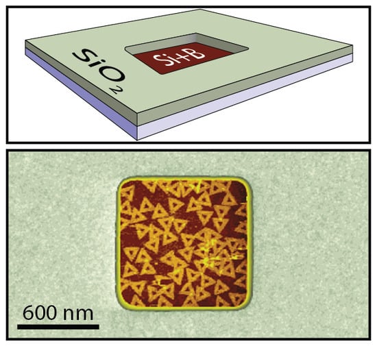
To test this hypothesis and demonstrate selective adsorption, DNA origami triangles were then deposited onto a patterned substrate with boron-implanted silicon features, including 1 µm × 1 µm and 5 µm × 5 µm in size wells, separated by a 100 nm thick, thermally grown SiO2 matrix. An optical image of the patterned wafer is shown in Supplement S2. The substrates were cleaned with Piranha + HF, and then DNA origami was deposited from a deposition buffer with an optimized pH of 6.6 and [MgCl2] of 35 mM. As shown in Figure 4, selective adsorption of DNA origami was achieved on boron-implanted silicon features but not on the SiO2 matrix. Unlike Gopinath et al. [30], spatial homogeneity was observed in the corners, edges, and interior of the patterned features, reflecting bulk adsorption from the solution, rather than 2D surface diffusion-limited coverage.

3. Discussion

Physical adsorption of charged nanostructures in an electrolyte on silicon substrates is extremely complicated and includes pH-dependent specific ion effects on the structure and density of water and ions at the surface [43,44]. However, all pH values studied here are above the isoelectric point for SiO2, and the results are consistent with behavior described by Derjaguin, Landau, Verwey, and Overbeek (DLVO) theory [45,46], where deposition, or the lack thereof, is due to a net electrostatic attraction or repulsion between the substrate and the DNA origami nanostructures. Briefly, in the DNA origami buffer solution, an electric double layer forms around both the DNA origami, and at the boron-silicon surface. The thicknesses of both double layers depend on the electrostatic screening of ions in solution, particularly divalent Mg, and can include complex contributions from multiple forces [46]. In simple terms, this electric double layer creates repulsive electrostatic interactions between the origami and surface. Increased electrostatic screening from increasing cation concentrations allows the repulsive barrier to be overcome, and structures become bound through van der Waals interactions. However, at our Mg concentrations, the DNA origami themselves exhibit minimal aggregation; but for Mg concentrations over ~20 mM at pH 6.6, the electrostatic repulsion between the DNA origami and the boron-implanted silicon surface is sufficiently reduced to allow adsorption. In contrast, the electric double layer of the thermal oxide, and thus, its repulsive electric potential barrier, is strongly dependent on the pH-dependent surface concentration of silanol-groups and adsorption of hydrated Mg ions [44]. Based on the work of Ong et al. [47], we calculated that ~20% of the silanol-groups at the surface would be deprotonated between pH values of 5.8 to 7.2. In comparison, at a pH value of 8.3, ~50% of the silanol-groups were deprotonated, giving rise to a more negative surface charge (see Supplement S10 for the derivation), increasing adsorption of hydrated Mg ions, and reducing the barrier for DNA origami adsorption. This is consistent with the pH-dependent deposition results, which are summarized in Figure 5.

4. Materials and Methods

4.1. DNA Origami Synthesis

The DNA origami triangle design was adopted/adapted from a previously reported study [24]. The design was modified to include 6 fluorescent 5’ FAM dyes (Integrated DNA Technologies, Skokie, IL, USA) to facilitate sample purification. DNA origami triangles were self-assembled from single-stranded scaffolds (Bayou Biolabs, Metairie, LA, USA), sourced from the M13mp18 bacteriophage, and staple strands (Integrated DNA Technologies, Skokie, IL, USA). The DNA scaffolds and corresponding staples were mixed in a 1:10 molar ratio, in a 1× tris-acetate-ethylenediaminetetraacetic acid (EDTA) buffer (Fisher Scientific, Hampton, NH, USA), with a pH of 8.3, and a [MgCl2] of 12.5 mM. The mixture was annealed at 70 °C for 20 min and then cooled to 20 °C at a rate of 0.6 °C/min. Well-formed nanostructures were purified using rate-zonal centrifugation as described in Supplement S1 [48,49]. After purification, the solution was normalized to a 5 nM concentration and stored in 5 µL aliquots at −80 °C to minimize sample degradation.

4.2. Substrate Cleaning

Silicon substrates were sonicated in deionized water, followed by 100% acetone (KMG, Fort Worth, TX, USA), and then 100% isopropanol (KMG, Fort Worth, TX, USA). The substrates were then sequentially soaked in Piranha to remove the organic contaminants and then 1:100 HF (Fisher Scientific, Hampton, NH, USA), to remove the surface oxide. For additional details, see Supplement S1.

4.3. DNA Origami Deposition

First, 20 µL of a deposition buffer was added to a 5 µL DNA origami solution with a 5 nM concentration. Once combined, the solution was gently pipette mixed. Cleaned silicon substrates were individually placed inside a petri dish (Fisher Scientific, Hampton, NH, USA) on top of a general-purpose lab wipe that was soaked in a deposition buffer so as to combat evaporation and subsequent change in the concentration of the deposited DNA solution. Then 25 µL of the DNA origami mixture was deposited onto the silicon substrates within 40 min of their cleaning, but before the native oxide grew back (Supplement S11). After deposition, the petri dish was sealed to minimize evaporation during incubation and sample transport between labs. For the MgCl2 concentration screening experiments, boron-implanted silicon samples were incubated at room temperature for ~24 h. For the pH screening experiments and the DNA origami deposition onto patterned substrates with boron-implanted silicon features, samples were incubated at room temperature for ~1 h. The DNA origami surface density on Piranha + HF cleaned boron-implanted silicon substrates, as a function of incubation time, is shown in Supplement S6. After incubation, excess DNA origami was removed by gravity-assisted rinsing, see Supplement S1.

4.4. Determining Surface Density

With the exception of boron-implanted silicon features surrounded by a raised SiO2 matrix, three high-resolution 5 µm × 5 µm images were captured in fluid AFM (Dimension FastScan, Bruker, Santa Barbara, CA, USA). Each image was divided into twenty-five 1 µm × 1 µm squares, and the number of DNA origami triangles in each square was manually counted. The average number of DNA nanostructures per 1 µm × 1 µm square is indicated by the average surface density.

5. Conclusions

Physical adsorption of DNA nanostructures was demonstrated on boron-implanted silicon substrates. While the average surface density was statistically comparable to natural mica, the adsorption uniformity was greater. The surface density dramatically increased from 0–91 nanostructures per µm2 as the salt concentration of the deposition buffer increased from 0–35 mM. Adsorption was also pH independent on the boron-implanted silicon surfaces, for the range we tested. The adsorption contrast between the boron-implanted silicon surface and SiO2 was optimized in a deposition buffer pH range of 5.8–7.2 at a [MgCl2] of 35 mM. In support of prior experiments, deprotonated silanol-groups at elevated pH in the presence of hydrated Mg ions promoted extensive binding between DNA origami and the oxide surfaces. Surface electrostatics provided by boron implantation enabled DNA adsorption, but further research is necessary to fully understand the electrochemical interactions controlling DNA origami deposition. Recommended next steps include further characterization of the boron-silicon surface structure, exploring how adsorption varies as a function of the feature size, and tuning the binding strength through mono and divalent cation concentrations to enable formation of ordered DNA origami arrays within the boron-implanted silicon wells [50,51,52].

Supplementary Materials

Supplementary materials can be found at https://www.mdpi.com/1422-0067/19/9/2513/s1.

Author Contributions

Conceptualization—S.S., W.L.H.; Methodology—S.T., J.F.-E.; Formal Analysis—S.T., S.K., J.F.-E., E.S.; Investigation—S.T., S.K., J.F.-E., E.S.; Resources—S.S.; Data Curation—S.T., S.K., J.F.-E., E.S.; Writing-Original Draft Preparation—S.T.; Writing-Review & Editing—S.T., S.K., J.E.P., L.C.G., E.G., W.K., S.S., W.L.H.; Visualization—S.T., E.G., W.L.H.; Supervision—S.S., E.G., W.L.H.; Project Administration—W.L.H.; Funding Acquisition—E.G., W.K., S.S., W.L.H.

Funding

This research was funded in part by the National Science Foundation (CMMI 1344915 and ECCS 1807809), the Semiconductor Research Corporation, and the National Institutes of Health (NIH K25GM093233 from the National Institute of General Medical Sciences).

Acknowledgments

(A) the Process Development team at Micron Technology for fabricating the patterned substrate; (B) Shu Qin at Micron Technology for developing the boron implant process; (C) Daniel Kelly for helping clean the wafers at Boise State; (D) William Bill Knowlton and Mike Hurley at Boise State for useful discussions; (E) Ashwin Gopinath and Paul Wilhelm Karl Rothemund at Caltech for technical insight, (F) Yaqiao Wu and the Center for Advanced Energy Studies-Microscopy and Characterization Suite (CAES-MaCS) in Idaho Falls for the FIB sample preparation and TEM imaging, and (G) the Surface Science Laboratory at Boise State for AFM sample characterization.

Conflicts of Interest

The authors declare no conflicts of interest.

Abbreviations

DNAdeoxyribonucleic acid
SiO2silicon dioxide
MgCl2magnesium chloride
[MgCl2]magnesium chloride concentration
SADPself-aligned double patterning
SAQPself-aligned quadruple patterning
DSAdirected self-assembly
BCPblock copolymers
DSA-BCPblock copolymer directed self-assembly
APTESaminopropyltrimethoxysilane
HMDShexamethyldisilazane
SAMself-assembled monolayer
XPSX-ray Photoelectron Spectroscopy
SIMSSecondary Ion Mass Spectroscopy
TEMTransmission Electron Microscopy
FIBfocused-ion beam
HFhydrofluoric acid
FFTFast Fourier Transform
HClhydrochloric acid
AFMatomic force microscopy
EDTAethylenediaminetetraacetic acid
FAMfluorescein amidite

References

  1. Moore, G.E. Cramming more components onto integrated circuits. Electronics 1965, 38, 114–117. [Google Scholar] [CrossRef]
  2. Moore, G.E. Progress in digital integrated electronics. Int. Electron Devices Meet. 1975, 21, 11–13, reprinted in IEEE Solid-State Circuits Soc. Newsl. 2006, 11, 36–37. [Google Scholar] [CrossRef]
  3. Gallatin, G. Nanofabrication: A perspective from litho practitioners. In Proceedings of the SRC/IBM/ONR Workshop on Bio-Nanofabrication and Materials, San Jose, CA, USA; 16–17 November 2016. [Google Scholar]
  4. Xu, K.; Souriau, L.; Hellin, D.; Versluijs, J.; Wong, P.; Vangoidsenhoven, D.; Vandenbroeck, N.; Dekkers, H.; Shi, X.; Albert, J.; et al. Key contributors for improvement of line width roughness, line edge roughness, and critical dimension uniformity: 15 nm half-pitch patterning with extreme ultraviolet and self-aligned double patterning. J. Micro Nanolithogr. MEMS MOEMS 2013, 12, 041302. [Google Scholar] [CrossRef]
  5. Kodama, C.; Ichikawa, H.; Nakayama, K.; Nakajima, F.; Nojima, S.; Kotani, T.; Ihara, T.; Takahashi, A. Self-Aligned Double and Quadruple Patterning Aware Grid Routing Methods. IEEE Trans. Comput. Des. Integr. Circuits Syst. 2015, 34, 753–765. [Google Scholar] [CrossRef]
  6. Wu, B.; Kumar, A. Extreme Ultraviolet Lithography; McGraw-Hill: New York, NY, USA, 2009; pp. 12–34. ISBN 9780071549189. [Google Scholar]
  7. Mimotogi, S. Extension of patterning technologies down to sub-10nm half pitch. Proc. SPIE 2013, 8685, 868503–868509. [Google Scholar]
  8. SemiSynBio Consortium and Roadmap Development. Available online: https://www.src.org/program/grc/semisynbio/semisynbio-consortium-roadmap/ (accessed on 28 July 2017).
  9. International Technology Roadmap for Semiconductors 2.0 2015 Edition. Available online: http://www.itrs2.net/itrs-reports.html (accessed on 20 October 2017).
  10. Quader, K.N. Flash Memory at a Cross-Road: Challenges and Opportunities. In Proceedings of the 2012 4th IEEE International Memory Workshop, Milan, Italy, 20 March 2012; IEEE: Piscataway, NJ, USA, 2012; pp. 1–4. [Google Scholar]
  11. Edwards, E.W.; Müller, M.; Stoykovich, M.P.; Solak, H.H.; de Pablo, J.J.; Nealey, P.F. Dimensions and Shapes of Block Copolymer Domains Assembled on Lithographically Defined Chemically Patterned Substrates. Macromolecules 2007, 40, 90–96. [Google Scholar] [CrossRef]
  12. Seeman, N.C. DNA in a material world. Nature 2003, 421, 427–431. [Google Scholar] [CrossRef] [PubMed]
  13. Deng, Z.; Mao, C. Molecular Lithography with DNA Nanostructures. Angew. Chem. Int. Ed. 2004, 43, 4068–4070. [Google Scholar] [CrossRef] [PubMed]
  14. He, Y.; Ye, T.; Ribbe, A.E.; Mao, C. DNA-Templated Fabrication of Two-Dimensional Metallic Nanostructures by Thermal Evaporation Coating. J. Am. Chem. Soc. 2011, 133, 1742–1744. [Google Scholar] [CrossRef] [PubMed]
  15. Ma, X.; Huh, J.; Park, W.; Lee, L.P.; Kwon, Y.J.; Sim, S.J. Gold nanocrystals with DNA-directed morphologies. Nat. Commun. 2016, 7, 12873. [Google Scholar] [CrossRef] [PubMed] [Green Version]
  16. Bates, F.S.; Fredrickson, G.H. Block Copolymer Thermodynamics: Theory and Experiment. Annu. Rev. Phys. Chem. 1990, 41, 525–557. [Google Scholar] [CrossRef] [PubMed]
  17. Diagne, C.T.; Brun, C.; Gasparutto, D.; Baillin, X.; Tiron, R. DNA Origami Mask for Sub-Ten-Nanometer Lithography. ACS Nano 2016, 10, 6458–6463. [Google Scholar] [CrossRef] [PubMed]
  18. Surwade, S.P.; Zhou, F.; Wei, B.; Sun, W.; Powell, A.; O’Donnell, C.; Yin, P.; Liu, H. Nanoscale Growth and Patterning of Inorganic Oxides Using DNA Nanostructure Templates. J. Am. Chem. Soc. 2013, 135, 6778–6781. [Google Scholar] [CrossRef] [PubMed]
  19. Shen, B.; Linko, V.; Tapio, K.; Kostiainen, M.A.; Toppari, J.J. Custom-shaped metal nanostructures based on DNA origami silhouettes. Nanoscale 2015, 7, 11267–11272. [Google Scholar] [CrossRef] [PubMed] [Green Version]
  20. Zhou, F.; Sun, W.; Ricardo, K.B.; Wang, D.; Shen, J.; Yin, P.; Liu, H. Programmably Shaped Carbon Nanostructure from Shape-Conserving Carbonization of DNA. ACS Nano 2016, 10, 3069–3077. [Google Scholar] [CrossRef] [PubMed]
  21. Ke, Y.; Ong, L.L.; Shih, W.M.; Yin, P. Three-Dimensional Structures Self-Assembled from DNA Bricks. Science. 2012, 338, 1177–1183. [Google Scholar] [CrossRef] [PubMed] [Green Version]
  22. Mandelkern, M.; Elias, J.G.; Eden, D.; Crothers, D.M. The dimensions of DNA in solution. J. Mol. Biol. 1981, 152, 153–161. [Google Scholar] [CrossRef]
  23. Green, C.M.; Schutt, K.; Morris, N.; Zadegan, R.M.; Hughes, W.L.; Kuang, W.; Graugnard, E. Metrology of DNA arrays by super-resolution microscopy. Nanoscale 2017, 9, 10205–10211. [Google Scholar] [CrossRef] [PubMed]
  24. Rothemund, P.W.K. Folding DNA to create nanoscale shapes and patterns. Nature 2006, 440, 297–302. [Google Scholar] [CrossRef] [PubMed] [Green Version]
  25. Scheible, M.B.; Ong, L.L.; Woehrstein, J.B.; Jungmann, R.; Yin, P.; Simmel, F.C. A Compact DNA Cube with Side Length 10 nm. Small 2015, 11, 5200–5205. [Google Scholar] [CrossRef] [PubMed] [Green Version]
  26. Jin, Z.; Sun, W.; Ke, Y.; Shih, C.-J.; Paulus, G.L.C.; Hua Wang, Q.; Mu, B.; Yin, P.; Strano, M.S. Metallized DNA nanolithography for encoding and transferring spatial information for graphene patterning. Nat. Commun. 2013, 4, 1663. [Google Scholar] [CrossRef] [PubMed] [Green Version]
  27. Surwade, S.P.; Zhao, S.; Liu, H. Molecular Lithography through DNA-Mediated Etching and Masking of SiO2. J. Am. Chem. Soc. 2011, 133, 11868–11871. [Google Scholar] [CrossRef] [PubMed]
  28. Zhou, F.; Michael, B.; Surwade, S.P.; Ricardo, K.B.; Zhao, S.; Liu, H. Mechanistic Study of the Nanoscale Negative-Tone Pattern Transfer from DNA Nanostructures to SiO2. Chem. Mater. 2015, 27, 1692–1698. [Google Scholar] [CrossRef]
  29. Sarveswaran, K.; Gao, B.; Kim, K.N.; Bernstein, G.H.; Lieberman, M. Adhesion of DNA nanostructures and DNA origami to lithographically patterned self-assembled monolayers on Si[100]. In Alternative Lithographic Technologies II; Herr, D.J.C., Ed.; International Society for Optics and Photonics: Bellingham, WA, USA, 2010; p. 76370M. [Google Scholar]
  30. Gopinath, A.; Rothemund, P.W.K. Optimized Assembly and Covalent Coupling of Single-Molecule DNA Origami Nanoarrays. ACS Nano 2014, 8, 12030–12040. [Google Scholar] [CrossRef] [PubMed]
  31. Love, J.C.; Estroff, L.A.; Kriebel, J.K.; Nuzzo, R.G.; Whitesides, G.M. Self-Assembled Monolayers of Thiolates on Metals as a Form of Nanotechnology. Chem. Rev. 2005, 105, 1103–1170. [Google Scholar] [CrossRef] [PubMed]
  32. Kershner, R.J.; Bozano, L.D.; Micheel, C.M.; Hung, A.M.; Fornof, A.R.; Cha, J.N.; Rettner, C.T.; Bersani, M.; Frommer, J.; Rothemund, P.W.K.; et al. Placement and orientation of individual DNA shapes on lithographically patterned surfaces. Nat. Nanotechnol. 2009, 4, 557–561. [Google Scholar] [CrossRef] [PubMed] [Green Version]
  33. Tanaka, S.; Taniguchi, M.; Kawai, T. Selective Adsorption of DNA onto SiO2 Surface in SiO2/SiH Pattern. Jpn. J. Appl. Phys. 2004, 43, 7346–7349. [Google Scholar] [CrossRef]
  34. Amodio, A.; Adedeji, A.F.; Castronovo, M.; Franco, E.; Ricci, F. pH-Controlled Assembly of DNA Tiles. J. Am. Chem. Soc. 2016, 138, 12735–12738. [Google Scholar] [CrossRef] [PubMed]
  35. Billingsley, D.J.; Lee, A.J.; Johansson, N.A.B.; Walton, A.; Stanger, L.; Crampton, N.; Bonass, W.A.; Thomson, N.H. Patchiness of ion-exchanged mica revealed by DNA binding dynamics at short length scales. Nanotechnology 2014, 25, 025704. [Google Scholar] [CrossRef] [PubMed]
  36. Whittle, K.M.; Vick, G.L. Control of Boron Diffusion from a Pyrolitic Borosilicate Glass Source. J. Electrochem. Soc. 1969, 116, 645–648. [Google Scholar] [CrossRef]
  37. Sadigh, B.; Lenosky, T.J.; Caturla, M.-J.; Quong, A.A.; Benedict, L.X.; Diaz de la Rubia, T.; Giles, M.M.; Foad, M.; Spataru, C.D.; Louie, S.G. Large enhancement of boron solubility in silicon due to biaxial stress. Appl. Phys. Lett. 2002, 80, 4738–4740. [Google Scholar] [CrossRef]
  38. Vick, G.L.; Whittle, K.M. Solid Solubility and Diffusion Coefficients of Boron in Silicon. J. Electrochem. Soc. 1969, 116, 1142–1144. [Google Scholar] [CrossRef]
  39. Brown, D.M.; Kennicott, P.R. Glass Source Diffusion in Si and SiO2. J. Electrochem. Soc. 1971, 118, 293–300. [Google Scholar] [CrossRef]
  40. Busen, K.M.; FitzGibbons, W.A.; Tsang, W.K. Ellipsometric Investigations of Boron-Rich Layers on Silicon. J. Electrochem. Soc. 1968, 115, 291–294. [Google Scholar] [CrossRef]
  41. Tenney, A.S.; Ghezzo, M. Etch Rates of Doped Oxides in Solutions of Buffered HF. J. Electrochem. Soc. 1973, 120, 1091–1095. [Google Scholar] [CrossRef]
  42. Fujii, M.; Sugimoto, H.; Hasegawa, M.; Imakita, K. Silicon nanocrystals with high boron and phosphorus concentration hydrophilic shell—Raman scattering and X-ray photoelectron spectroscopic studies. J. Appl. Phys. 2014, 115, 084301. [Google Scholar] [CrossRef]
  43. Zhang, X.G. Electrochemistry of Silicon and Its Oxide; Kluwer Academic/Plenum Publishers: New York, NY, USA, 2001; pp. 45–89. ISBN 0-306-46541-8. [Google Scholar]
  44. DeWalt-Kerian, E.L.; Kim, S.; Azam, M.S.; Zeng, H.; Liu, Q.; Gibbs, J.M. pH-Dependent Inversion of Hofmeister Trends in the Water Structure of the Electrical Double Layer. J. Phys. Chem. Lett. 2017, 8, 2855–2861. [Google Scholar] [CrossRef] [PubMed]
  45. Besra, L.; Liu, M. A review on fundamentals and applications of electrophoretic deposition (EPD). Prog. Mater. Sci. 2007, 52, 1–61. [Google Scholar] [CrossRef]
  46. Boström, M.; Deniz, V.; Franks, G.V.; Ninham, B.W. Extended DLVO theory: Electrostatic and non-electrostatic forces in oxide suspensions. Adv. Colloid Interface Sci. 2006, 123, 5–15. [Google Scholar] [CrossRef] [PubMed]
  47. Ong, S.; Zhao, X.; Eisenthal, K.B. Polarization of water molecules at a charged interface: Second harmonic studies of the silica/water interface. Chem. Phys. Lett. 1992, 191, 327–335. [Google Scholar] [CrossRef]
  48. Zhirnov, V.; Zadegan, R.M.; Sandhu, G.S.; Church, G.M.; Hughes, W.L. Nucleic acid memory. Nat. Mater. 2016, 15, 366–370. [Google Scholar] [CrossRef] [PubMed]
  49. Lin, C.; Perrault, S.D.; Kwak, M.; Graf, F.; Shih, W.M. Purification of DNA-origami nanostructures by rate-zonal centrifugation. Nucleic Acids Res. 2013, 41, e40. [Google Scholar] [CrossRef] [PubMed]
  50. Aghebat Rafat, A.; Pirzer, T.; Scheible, M.B.; Kostina, A.; Simmel, F.C. Surface-assisted large-scale ordering of DNA origami tiles. Angew. Chem. Int. Ed. Engl. 2014, 53, 7665–7668. [Google Scholar] [CrossRef] [PubMed]
  51. Kocabey, S.; Kempter, S.; List, J.; Xing, Y.; Bae, W.; Schiffels, D.; Shih, W.M.; Simmel, F.C.; Liedl, T. Membrane-Assisted Growth of DNA Origami Nanostructure Arrays. ACS Nano 2015, 9, 3530–3539. [Google Scholar] [CrossRef] [PubMed]
  52. Woo, S.; Rothemund, P.W.K. Self-assembly of two-dimensional DNA origami lattices using cation-controlled surface diffusion. Nat. Commun. 2014, 5, 4889. [Google Scholar] [CrossRef] [PubMed] [Green Version]
Figure 1. The Secondary Ion Mass Spectroscopy (SIMS) and cross-sectional Transmission Electron Microscopy (TEM) analyses of the boron-implanted silicon substrate revealed: (a) The boron concentration depth profile, and (b) structure of the substrate. The boron concentration at the surface of the substrate was ~14.6 atomic percent, and the majority of the boron was found within ~7 nm from the surface. The red, green, and blue colors on the plot (a) and the line in (b) correspond to the boron-rich amorphous oxide layer, the boron-rich amorphous layer, and the bulk silicon, respectively. The cross-sectional TEM image and the Fast-Fourier Transforms (FFTs) of the image showed that both outer layers were amorphous, and that the bulk was crystalline. The platinum protective layer was deposited onto the substrate surface prior to fabrication of the TEM specimen using focused-ion beam (FIB).
Ijms 19 02513 g001
Figure 2. Atomic force microscopy (AFM) images of DNA origami triangles adsorbed onto boron-implanted silicon substrates. Prior to the DNA origami triangle deposition, the samples were cleaned with Piranha + hydrofluoric acid (HF). For all conditions, the deposition buffers were 10 mM bis-tris hydrochloric acid (HCl) with a pH of 6.6 and the deposition incubation time was ~24 h. The MgCl2 concentration ([MgCl2]) for (a) was 35 mM. The average surface density increased from 0 to 90 ± 6/μm2 as the [MgCl2] increased from 0 to 35 mM. Scale bars are 500 nm.
Ijms 19 02513 g002
Figure 3. AFM images of DNA origami triangles adsorbed onto boron-implanted silicon substrates (ad) and thermally grown silicon dioxide (SiO2) substrates (eh) as a function of deposition buffer pH. The substrates were cleaned with Piranha + HF. For the pH values below 8.3, the deposition buffer was 10 mM bis-tris HCl with a [MgCl2] of 35 mM. For the pH value of 8.3, 1× tris-acetate- ethylenediaminetetraacetic acid (EDTA) with a [MgCl2] of 35 mM was selected to stay within the buffer range. For all conditions, the deposition incubation time was ~1 h. Scale bars are 500 nm.
Ijms 19 02513 g003
Figure 4. Schematic of a boron-implanted silicon feature surrounded by 100 nm thick SiO2 (a). AFM height images of lithographically fabricated 1 μm × 1 μm (b) and 5 μm × 5 μm (c) boron-implanted silicon features surrounded with SiO2. The z-axis color scale was adjusted to show nanometer-scale contrast both within the well and on SiO2 (100 nm above the well). The deposition buffer was 10 mM bis-tris HCl, with a pH of 6.6, and a [MgCl2] of 35 mM. DNA origami triangles adsorbed with a surface density of 65/μm2 in (b) and 33 ± 5/μm2 in (c) on the boron-implanted silicon surfaces, while no DNA origami adsorbed onto the SiO2 surfaces. The rectangular insert is a 2× magnified image of the boron-implanted silicon surface shown in (c). For both samples, the deposition incubation time was ~1 h. Scale bars are 600 nm for (b) and 2 μm for (c).
Ijms 19 02513 g004
Figure 5. DNA origami surface density on boron-implanted silicon substrates oxide (green circle) and thermally grown SiO2 substrates (red triangle) as a function of deposition buffer pH. Representative data points for the AFM images in Figure 3 are highlighted with red circles, and the theoretical population fraction of deprotonated silanol-groups on the oxide surfaces is depicted with a green dotted line.
Ijms 19 02513 g005