Article

Anticancer Effect of Citrus hystrix DC. Leaf Extract and Its Bioactive Constituents Citronellol and, Citronellal on the Triple Negative Breast Cancer MDA-MB-231 Cell Line

1
Cellular and Molecular Immunology Research Unit, Faculty of Allied Health Sciences, Naresuan University, Phitsanulok 65000, Thailand
2
Department of Chemistry, Faculty of Science and Center of Excellence for Innovation in Chemistry, Naresuan University, Phitsanulok 65000, Thailand
*
Author to whom correspondence should be addressed.
Pharmaceuticals 2020, 13(12), 476; https://doi.org/10.3390/ph13120476
Submission received: 3 November 2020 / Revised: 8 December 2020 / Accepted: 14 December 2020 / Published: 18 December 2020
(This article belongs to the Special Issue Anticancer Compounds in Medicinal Plants)

Abstract

:
Triple negative breast cancer is one of the most aggressive breast cancer type with abilities of early metastasis and chemoresistance. The tropical plant Citrus hystrix DC. has been reported to promote many biological activities including anticancer. However, the effect of C. hystrix against triple negative breast cancer has not yet been identified. This study aimed to evaluate the anticancer properties of C. hystrix leaf extract and its bioactive constituents citronellol and citronellal against the triple negative breast cancer MDA-MB-231 cell line. C. hystrix leaves were powdered and sequentially macerated. The in vitro anticancer effects of C. hystrix leaf extracts, and its bioactive constituents (citronellol and citronellal) were evaluated against MDA-MB-231 cell line using cytotoxic MTT assay, cell proliferation, wound scratch migration, colony formation, cell cycle, apoptosis assay, Hoechst staining, RT-qPCR, and Western blot analysis. Results showed that crude hexane extract, citronellol, and citronellal significantly reduced cell proliferation, colony formation, and cell migration by inducing cell cycle arrest, while also inducing apoptosis in MDA-MB-231 cells through inhibition of anti-apoptotic Bcl-2 expression, leading to activation of the caspase-3-dependent pathway. This study is the first report to demonstrate the effect of C. hystrix, citronellol, and citronellal against triple negative breast cancer MDA-MB-231 cells.

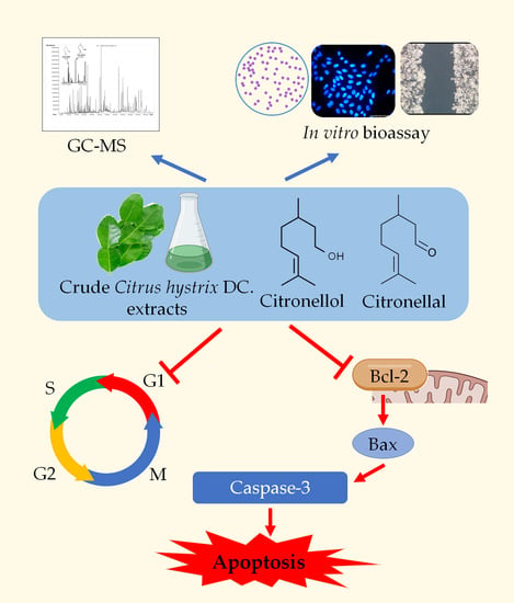
Graphical Abstract

1. Introduction

Triple negative breast cancer (TNBC) is a sub-type of breast cancer. TNBC is characterized by tumors that do not express estrogen receptor (ER) or progesterone receptor (PR), with immunohistochemical (IHC) staining present at less than 1% repartitioning of these receptors in the tissue and less than 10% for human epidermal receptor 2 (Her-2) [1,2]. TNBC accounts for 15–20% of invasive breast cancers in women [3] and presents a higher frequency of recurrence and lower overall survival rate compared to non-TNBC patients [4]. It is one of the most aggressive types of breast cancer due to its early metastasis and resistance to chemotherapies such as anthracyclines, taxanes, and many others [5]. Several new anticancer plant-derived molecules have been put through clinical trials [6] and some such as cabazitaxel and homoharringtonine have been approved by the FDA for use in treatment resistance [7,8]. Plant materials are now becoming increasingly recognized as a substantial source of anticancer agents and urgently require assessment for future development.
Citrus hystrix DC., also known as kaffir lime, is a tropical plant belonging to the Rutaceae family. The tree can grow up to six meters high and has double pointy oval shaped leaf, white fragrant flowers, and green to yellow ellipsoid fruits with irregular bumpy skin. Many compounds have been identified in the fruit including phenolics, flavonoids, terpenoids, alkaloids, coumarins, glycosides, saponins, tannins, hydrocarbons, and fatty acids [9,10,11]. In Thailand, C. hystrix is believed to have cancer-prevention and anti-inflammation properties and it is used as a remedy in traditional medicine [12]. C. hystrix has also been reported to exert potency against many types of cancers. It induced selective cytotoxicity against brain and cervical cancer cells [13], induced cell cycle arrest in lymphoma cells [14], and also suppressed cell migration and induced cell shrinkage in human pancreatic cancer cells [15]. Citronellol and citronellal are two acyclic monoterpenoids that have been identified in C. hystrix. They have been reported to exert many varied biological activities including anti-inflammatory and anti-microbial activities [16]. Citronellal exhibited toxicity against MCF-7 breast cancer cells [17] and reduced proliferation by inducing apoptosis in Huh7 hepatocellular carcinoma [18], while citronellol induced necroptosis of lung cancer [19]. Both molecules have also been reported to inhibit the action of multidrug resistance P-glycoprotein, which ameliorates the efficacity of drug treatments [20]. However, the effect of C. hystrix and its bioactive compounds citronellol and citronellal have not yet been observed on triple negative breast cancer. Here, the anticancer effects of C. hystrix crude extracts, citronellol, and citronellal were evaluated against the triple negative breast cancer MDA-MB-231 cell line in terms of cell proliferation, migration, cell cycle arrest and apoptosis. Findings revealed that C. hystrix leaf extract and its active compounds citronellol and citronellal strongly inhibited tumor cell growth and triggered apoptosis via over-expression of cleaved caspase-3 and Bax, while down-regulating the anti-apoptotic Bcl-2.

2. Results

2.1. Extraction Yields of C. hystrix Leaf Powder

Maceration C. hystrix leaf powder with hexane solvent resulted in a sticky dark green extract with yield of 1.1% (w/w dried powder). The following sub-sequential extractions culminated in a similar consistency of extracts with yield of 1.8% obtained using ethyl acetate and 7.6% using 95% ethanol.

2.2. Identification of Volatile Components in Crude Hexane Extract by GC-MS

Analysis of the total ionic chromatogram by GC-MS identified 45 volatile compounds from crude hexane extract. Relative amounts (%) of the compositions were calculated by peak-area normalization (Table 1). Most of the identified compounds of crude C. hystrix hexane extract were terpenoids (19.84%). The extract contained oxygenated monoterpenes (3.99%), hydrocarbon monoterpenes (2.19%), oxygenated sesquiterpenes (7.67%), and hydrocarbon sesquiterpenes (5.99%) as major compounds. The identification of two oxygenated monoterpenes, citronellal and citronellol, were found in leaf extract and then confirmed with authentic standards which they were observed at retention time (RT) at 10.75 min and 12.85 min, respectively (Figure S1). Other constituents comprised long-chain hydrocarbons, phytosterols, fatty acids, fatty alcohols, and vitamins.

2.3. Cytotoxicity of Crude Extracts, Citronellol, and Citronellal

After 24 h of treatment, half maximal inhibitory concentration (IC50) values were obtained using a dose-response inhibition curve. C. hystrix crude extracts, citronellol, and citronellal reduced cell viability of MDA-MB-231 cells with IC50 of 317.63 ± 2.00 µg/mL for crude hexane extract (Figure 1a), 547.10 ± 0.90 µg/mL for crude ethyl acetate extract (Figure 1b) and IC50 > 1000 µg/mL for crude ethanolic extract (Figure 1c). For citronellol, IC50 values were 1.16 ± 0.10 nM (Figure 1d) and 1.41 ± 0.03 nM for citronellal (Figure 1e). The cytotoxicity of crude hexane extract, citronellol, and citronellal on normal cells, human monocyte-derived macrophages, were performed as the experimental model (Figure S2).

2.4. Effect of Crude Hexane, Citronellol, and Citronellal on Cell Proliferation

The cytotoxicity of crude hexane extract, citronellol and citronellal on cell proliferation rate of MDA-MB-231 cells was performed by the MTT assay. For the non-treated group, cell viability value doubled at 48 h, indicating that the cells significantly duplicated their population. By contrast, results showed that all treatment groups significantly reduced proliferation of MDA-MB-231 cells in both a dose- and time-dependent manner when compared to the non-treated group at each specific incubation time of 24 h and 48 h (Figure 2).

2.5. Crude Hexane, Citronellol, and Citronellal on Inhibited MDA-MB-231 Cell Migration

Cell migration is one of the important processes in cancer metastasis. The effects of crude hexane extract, citronellol, and citronellal on cell migration were evaluated by in vitro wound scratch migration assay. In the control group, MDA-MB-231 cells migrated to completely close the wound areas at 24 h (Figure 3). However, in the treatment groups, crude hexane extract, citronellol, and citronellal significantly reduced cell migration into wound areas at 6 h, 12 h, and 24 h in both a dose- and time-dependent manner.

2.6. C. hystrix Hexane Extract, Citronellol, and Citronellal Reduced Number of Colonies Forming in MDA-MB-231 Cells

The colony forming assay was used to evaluate cell survival by studying cell growth from a single cell to form colonies in response to toxic substances. Here, colony formations of MDA-MB-231 cells under treatments by crude hexane extract, citronellol, citronellal, and doxorubicin were observed. In the treatment groups, crude hexane extract, citronellol, and citronellal significantly reduced colony formation of MDA-MB-231 cells in a dose-dependent manner, while only a few colonies were formed under treatment of doxorubicin 0.5 µM (Figure 4).

2.7. Crude Hexane, Citronellol, and Citronellal Induced Cell Cycle Arrest in MDA-MB-231 Cell

Cell cycle arrest is a mechanism that cells use to prevent cell cycle progression when they encounter a toxic substance, DNA damage during DNA replication. In the treatment groups, crude hexane extract significantly induced cell cycle arrest at the G0/G1 phase when compared to the non-treated group. However, in the citronellol- and citronellal-treated groups, the cells were significantly arrested at the G2/M phase, while doxorubicin significantly induced cell cycle arrest at the G2/M phase (Figure 5).

2.8. Crude Hexane, Citronellol, and Citronellal Induced Apoptosis in MDA-MB-231 Cells

Crude hexane extract, citronellol, and citronellal reduced cell proliferation and migration, while also decreasing the number of colony formations of MDA-MB-231 cells. When cells encounter toxic substances, they undergo a repair mechanism that reduces their active state. However, if the damage cannot be repaired, cell death or apoptosis results. Hoechst 33342 staining was used to confirm morphological change during apoptosis. The non-treated group showed normal nuclear cell structure; however, in the treatment groups, some condensed blue fluorescence appeared in cell nuclei indicating chromatin condensation and/or DNA fragmentation in cells treated by crude hexane extract 200 µg/mL (Figure 6b), citronellol (Figure 6c), citronellal (Figure 6d), and doxorubicin 0.5 µM (Figure 6e). Furthermore, apoptotic cells under treatments of crude hexane extract, citronellol, and citronellal for 24 h were observed using the MuseTM Cell Analyser. Results revealed that a dose of 200 µg/mL crude hexane extract induced apoptosis in MDA-MB-231 cells at up to 11.02 ± 0.53% (Figure 6g) when compared to the control (Figure 6f). Similarly, citronellol at 1 nM induced cell apoptosis of 14.30 ± 0.89% (Figure 6h) with 13.92 ± 0.31% for citronellal at 1 nM (Figure 6i), while doxorubicin at 0.5 µM induced cell apoptosis of 19.02 ± 0.81% (Figure 6j).

2.9. Crude Hexane, Citronellol, and Citronellal Modulated Apoptosis-Related Proteins Gene Expression in MDA-MB-231 Cells

The expression level of apoptosis-related genes was performed using RT-qPCR to better understand cell apoptosis. Cells were treated 24 h before mRNA extraction. Results showed that the expression level of the Bax gene significantly increased in the treatment of crude hexane extract 200 µg/mL, citronellol 1 nM, citronellal 1 nM and doxorubicin 0.5 µM (Figure 7a). On the other hand, expression level of the Bcl-2 gene was markedly downregulated in each treated group compared to the control (Figure 7b).

2.10. Crude Hexane, Citronellol, and Citronellal Induced Apoptosis and DNA Fragmentation in the Cells by Inhibiting the Anti-Apoptotic Bcl-2 Protein and Activating Caspase Dependent Apoptotic Pathway

To further confirm the results, another experiment was conducted to measure the protein expression of both Bax and Bcl-2 proteins using Western blot analysis. Surprisingly, the expression of Bcl-2 protein significantly decreased after treatment by crude hexane extract 200 µg/mL, citronellol 1 nM, citronellal 1 nM, and doxorubicin 0.5 µM compared to the control group (Figure 8a), while Bax protein expression was upregulated in the same treated groups (Figure 8b). In response to the treatments, cells upregulated Bax protein expression and downregulated Bcl-2 protein expression. Alteration of Bax/Bcl-2 expression may lead to the activation of caspase-3 protein, which is an executioner in apoptosis caspase-dependent activation. To confirm this hypothesis, pro-caspase-3 and cleaved-caspase-3 protein levels were measured by Western blot analysis. Results demonstrated a significant decrease in intensities of pro-caspase-3 in the treatment groups compared to the control (Figure 8c), with significantly increase cleaved-caspase-3 protein intensity (Figure 8d).

3. Discussion

C. hystrix has been reported to possess many biological activities. In this experiment, we extracted C. hystrix leaf by the sequential maceration method using lower polar solvent to higher polar solvent (hexane, ethyl acetate, and 95% ethanol). Higher yield was obtained from the ethanolic extract compared to crude hexane and ethyl acetate extract. The MTT assay showed that crude C. hystrix hexane extract had higher efficiency in reducing MDA-MB-231 cell viability compared to the two other crude extracts, while the two pure compounds showed similar toxicity on MDA-MB-231 cell viability. Moreover, TLC fingerprints of crude C. hystrix hexane extract showed more trace of compounds compared to other two extracts with the trace of citronellol and citronellal could be identified at similar retention factor (Rf) (Figure S4). A previous study reported that crude ethyl acetate and crude ethanolic extract showed a higher effect against HeLa cells compared to crude hexane extract [21]. Doxorubicin is an anticancer agent that is widely used in cancer treatments. It acts as an anti-topoisomerase 2, which prevents DNA from replication [22]. We used doxorubicin as a drug control in all experiments.
Crude C. hystrix hexane extract was selected for chemical constituent analysis by GC-MS. Results indicated that terpenoids comprised the most compounds identified from C. hystrix leaf extract, while citronellal (0.67%) and citronellol (1.42%) of C. hystrix leaf crude hexane extract from the GC chromatogram appeared at retention times of 10.75 min and 12.85 min, respectively.
We then tested the effect of crude hexane extract, citronellol, and citronellal on the proliferation of MDA-MB-231 breast cancer cells. Results showed that crude hexane extract significantly reduced cell growth rate compared to the non-treated group. Anti-proliferative effects of C. hystrix were also reported in previous studies on leukemic cell lines [23] and on HeLa cervical cancer cells [21], while the anti-proliferative effect on MDA-MB-231 cells was also significant under treatments of both citronellol and citronellal. A previous study reported that citronellal reduced proliferation in Huh7 hepatocellular carcinoma cells [18], while citronellol inhibited non-small cell lung carcinoma A-549 cells [24]. Further experiments were performed to support the results of pre-existing anti-proliferative effects of both crude hexane extract and the two pure compounds. Clonogenic assay was performed to mimic cancer growth from single mutated cells to form cancer cell colonies or tumor mass. In the control group, MDA-MB-231 cells grew and formed cell colonies within 15 days of incubation. However, in the treatment groups, crude hexane extract, citronellol, and citronellal significantly reduced the number of colonies and inhibited colony formation at higher doses. Bergamottin was also found to inhibit colonies forming in PANC-1 human pancreas cancer [15]. Surviving under a tough environment with highly proliferative and migrative abilities are keys for successful cancer development or metastasis. Therefore, decreasing these two factors could be a suitable strategy to prevent early metastasis of the cancer. To test this hypothesis, wound scratch migration assay was conducted to study the effects of crude hexane extract, citronellol, and citronellal on cell migration. Results indicated that the treatments groups slowed down cell migration into wound areas in a dose and time dependent manner. Cell cycle analysis and apoptosis assay were performed to better understand whether the anti-proliferative and anti-migrative effects of crude hexane extract, citronellol, and citronellal were linked to these events. Results showed that a significant number of MDA-MB-231 cancer cells were arrested at the G0/G1 phase by C. hystrix crude hexane extract. Cells treated with citronellol and citronellal accumulated at the G2/M phase in the same way as treatment by doxorubicin. However, C. hystrix leaf extract showed different effects on the Molt-4 cell line by inducing cell cycle arrest at the G2/M phase [14]. Citronellol was also reported to induce lung cancer NCI-H1299 cell cycle arrest at the G0/G1 phase [19].
Results indicated a significant number of apoptotic cells under treatments of crude hexane extract, citronellol, and citronellal, while morphological changes as chromatin condensation and swelling were seen under the treatment groups. To help define a support mechanism for cell apoptosis, the levels of genes and proteins involved were analyzed using RT-qPCR and Western blot techniques. Results proved that levels of the Bcl-2 gene and protein expression decreased under the treatment group, while the Bax gene and protein expression increased in intensity. During apoptosis, cleavage of DNA by endonuclease occurs after the activation of caspase-3 [25]. Results from Western blot analysis clearly demonstrated the activation of caspase-3 through decreasing density of total caspase-3 in the treated groups compared to the control and increasing the intensity of cleaved-caspase-3 protein, indicating activation of the caspase-dependent apoptosis pathway. The induction of apoptosis in MDA-MB-231 cells by the treated groups was probably activated by the caspase-dependent apoptosis pathway through inhibition of anti-apoptotic protein Bcl-2, leading to an increase of pro-apoptotic Bax protein activity. Bax protein mediated the pore formation complex at the outer membrane of mitochondria and consequentially triggered the release of cytochrome C, leading to the activation of caspase 3 protein. The effect of doxorubicin inducing apoptosis through the caspase-dependent apoptosis pathway was also reported [22]. Bergamottin, a compound identified in C. hystrix, induced apoptosis in human pancreas cancer cells PANC-1 by inhibiting the Akt/mTOR signaling pathway, leading to inhibition of cell survival, proliferation, and migration [15]. However, further research is necessary to better understand the mechanism behind cell cycle arrest to reduce cell migration. Animal model experiments should also be launched to enhance drug development of C. hystrix leaf extract and its bioactive compounds citronellol and citronellal.

4. Materials and Methods

4.1. Chemicals and Reagents

β-Citronellol, citronellal (analytical standard grade), and doxorubicin were purchased from Sigma-Aldrich, Inc., St. Louis, MO, USA, and stored at 4 °C. Dimethyl sulphoxide (DMSO) was used to dissolve the crude extracts and doxorubicin was dissolved with deionized water.

4.2. Plant Materials and Extraction Process

C. hystrix leaf fine powder was received from Khaolaor Laboratories Co., Ltd., Nakhon Pathom, Thailand (COA no. 250818) and crude extracts were obtained using the sequential maceration method. Finely powdered leaves (500 mg) were extracted with 1000 mL of hexane and stirred continuously for 3 days at room temperature. The mixture was then filtered and evaporated using a rotary evaporator to give crude C. hystrix hexane extract. The maceration procedure was repeated three times. The marc was further extracted with ethyl acetate and 95% ethanol using the same procedure. The filtrates obtained were evaporated and stored at −20 °C until required for use.

4.3. Cell Culture

MDA-MB-231 cell line (ATCC® HTB-26™ATCC) was maintained in Dulbecco’s Modified Eagle Medium (DMEM; Gibco™; Thermo Fisher Scientific, Inc., Waltham, MA, USA) with 10% fetal bovine serum (FBS; Gibco™; Thermo Fisher Scientific, Inc., Waltham, MA, USA) and 1% of antibiotic-antimycotic (amphotericin B, penicillin, streptomycin) (Gibco™; Thermo Fisher Scientific, Inc., Waltham, MA, USA) at 37 °C, CO2 5%.

4.4. Human Monocyte Isolation

Human monocytes were isolated from fresh buffy coat by Ficoll-Paque™ (Sigma-Aldrich, Inc.) using the density gradient centrifugation method. Human monocyte-derived macrophages were obtained by culturing the freshly isolated monocytes in RPMI-1640 medium (Gibco™; Thermo Fisher Scientific, Inc., Waltham, MA, USA) with 10% FBS incubated at 37 °C, CO2 5% for one week before performing the experiments. Buffy coat was obtained from the Blood Bank, Naresuan University Hospital, Phitsanulok, Thailand. Ethics approval was obtained from the Human Ethics Committee of Naresuan University (IRB no. 0945/62).

4.5. Gas Chromatography-Mass Spectrometry Analysis (GC-MS)

The analysis of volatile compounds in crude hexane extract from C. hystrix leaf powder was performed using a Hewlett Packard Gas Chromatograph model 6890 (Agilent Technologies, Palo Alto, CA, USA) equipped with a mass selective detector. Crude hexane extract (50 mg/mL) was prepared by dissolving in hexane and then injected into GC-MS system. Volatile compounds in sample were separated using silica capillary Hewlett Packard HP-5 (5% phenyl methyl siloxane) column (30 m × 0.25 mm i.d., 0.25 µm film thickness). High-purity helium was used as the carrier gas with constant flow rate at 13.7 mL/min. Initial injector temperature was set at 250 °C with split ratio mode at ratio 10:1 and 1 µL injector volume. The oven temperature was started at 70 °C for 3 min, then increased to 280 °C (5 °C/min) and held for 20 min. Transfer temperature was set at 280 °C and the mass detection ranges were set from 50 to 700 amu in full scan. Retention indices (RIs) were determined by analyzing a solution containing a homologous series of n-alkanes (C8–C32) under the same chromatographic conditions and then calculated as described by van Den Dool and Dec. Kratz [26]. Identification of the volatile components was performed by computer matching of their recorded mass spectra fragmentation patterns with those stored in the Wiley 7n MS spectral library. Further identification was made by comparison of their mass spectra and their RIs relative to n-alkanes with those of the National Institute of Standards and Technology (NIST) Chemistry WebBook [27,28] or with previously published data. The presence of citronellol and citronellal in the extract was also confirmed by analyzing authentic standards under the same chromatographic conditions. Relative contents of each component in the sample were also calculated based on the normalization of peak areas as the percentage of total detected volatile components.

4.6. Cell Viability MTT Assay

MDA-MB-231 cells were plated in 96-well plates at density of 104 cells/well and 5 × 10⁴ for human monocyte derived macrophages cells/well. Cells were treated by various concentrations of each crude extracts, citronellol, citronellal for 24 h, and 48 h for doxorubicin. All treatments were then removed, and cells were then washed by phosphate saline buffer (PBS). Culture medium was used as vehicle control. 0.5 mg/mL of 3-(4,5-dimethylthiazol-2-yl)-2,5-diphenyl tetrazolium bromide salt (MTT) from Thermo Fischer Scientific (Waltham, MA, USA) was introduced to cells with 3 h of incubation. The MTT was discarded, and the dye was solubilized by 100 µL of DMSO. Absorbances of the dissolved tetrazolium salt was measured at 570 nm by ELISA microplate reader (PerkinElmer, Inc., Waltham, MA, USA). The IC50 value was generated by dose-response curves using GraphPad Prism 7.

4.7. Identification Effect of Crude Hexane, Citronellol, and Citronellal on Cell Proliferation

MDA-MB-231 cells were seeded at a density of 2 × 10⁴ cells/well and treated with 50, 100, and 150 µg/mL crude hexane extract, 0.5 and 1 nM of citronellol and citronellal, and 0.5 µM doxorubicin for 24 h and 48 h. Cells treat with culture medium only was used as vehicle control. Cell viability was measured at 24 h and 48 h to represent the growth of the cells.

4.8. Clonogenic Assay

Cells were seeded at a density of 500 cells/well in 6-well plates for 24 h. Different concentrations of crude hexane extract, citronellol, citronellal, and doxorubicin were treated on cells for 24 h. Cells treated with culture medium only were used as vehicle control. Treated cells were washed by PBS and then cultured in culture medium for 10 days with medium replacement every 3 days. Cells were washed twice with PBS and fixed with 10% neutral buffer formalin for 30 min before fixing by 0.5% crystal violet for 1 h. Pictures of colonies were captured using a Canon macro lens 50 mm/f1.8 STM. The photos were converted to 8-bit greyscale images. Numbers of colonies of MDA-MB-231 cells were counted by Colony Area plugin using ImageJ 1.52a software.

4.9. Wound Scratch Migration Assay

MDA-MB-231 cells were seeded and cultured to confluence. SPLScar™ cell scraper (SPL Life Sciences, Gyeonggi-do, Korea) was used to create wound areas. Cells were washed twice by PBS to remove cell debris and floating cells before treatment with various concentrations of crude hexane extract, citronellol, citronellal, and doxorubicin. Cells treated with culture medium only were used as vehicle control. Cell migration into the wound areas was then observed under an inverted microscope (Zeiss Microscopy, Oberkochen, Germany) with objective 10× magnification. Wound closed areas were quantified by MRI_Wound_Healing_Tool plugin using ImageJ 1.52a software.

4.10. Cell Cycle Analysis

A total of 200 µg/mL crude hexane extract, 1 nM citronellol and citronellal, and 0.5 µM doxorubicin were treated with MDA-MB-231 cells for 24 h. Cells treat with culture medium only was used as vehicle control. Cells were harvested, transferred into a 1.5 mL microtube, and washed twice by cold PBS. Cells were then fixed with 1 mL of 70% ethanol for 5 h. Two hundred microliters of cell suspension (106 cells/mL) were washed with 250 µL of PBS and 200 µL of Muse™ cell cycle reagent and then incubated in the dark for 20 min at room temperature. The apoptotic cells were analyzed using a Muse™ cell analyzer (Merck, Darmstadt, Germany).

4.11. Apoptosis Analysis

Crude hexane extract, citronellol, citronellal, or doxorubicin were treated on MDA-MB-231 cells for 24 h. Cells treat with culture medium only was used as vehicle control. Cells were harvested and transferred into a 1.5 mL microtube and washed twice in cold PBS. Then, the cells were resuspended in 1% BSA and 100 µL of Muse™ Annexin V and dead cell reagent (Merck, Darmstadt, Germany) were added to 100 µL of cell suspension (5 × 10⁵ cells/mL) and incubated in the dark for 20 min at room temperature. The apoptotic cells were analyzed using Muse™ cell analyzer, gated on Annexin V-FITC positive cells and 7-AAD positive cells.

4.12. Hoechst 33342 Staining

Cells were cultured on cover slides for 24 h and then treated with crude hexane extract, citronellol, citronellal, or doxorubicin for 24 h. Cells treated with culture medium only were used as vehicle control. The cells were washed twice with PBS and fixed with 4% formaldehyde for 15 min at room temperature. After washing twice with PBS, the cells were permeabilized with 0.15% Triton-X 100 for 15 min at room temperature, followed by washing twice with PBS. An aliquot of 4 μg/mL of Hoechst 33342 solution was used to stain cells for 10 min in the dark at room temperature. Then, 70% glycerol was used as an anti-fade solution. Cover slides were then mounted on the slides and sealed with nail polisher. DNA fragmented or chromatin condensed cells were observed under a fluorescence microscope (Zeiss Microscopy, Oberkochen, Germany) with objective 40× magnification.

4.13. RT-qPCR

Crude hexane extract, citronellol, citronellal, or doxorubicin were treated on MDA-MB-231 cells for 24 h. Cells treated with culture medium only were used as vehicle control. Total RNA was extracted from MDA-MB-231 cells using RibozolTM RNA extraction reagent kits (AMRESCO, VWR Life Science, OH, USA). Briefly, cDNA synthesis was performed using Tetro cDNA synthesis kits (Bioline Reagents Ltd., London, UK). Real-time PCR was carried out using SensiFASTTM SYBR No-ROX kits (Bioline Reagents Ltd., London, UK). The initial denaturing temperature started from 95 °C at 1 min, with 45 cycles of 15 s denaturation followed by 30 s of annealing and elongation at 60 °C. The whole reaction was performed in a CFX96 Touch Real-Time PCR Detection System (Bio-Rad Laboratories, Hercules, CA, USA). Primer sequences of β-actin, Bcl-2, and Bax were obtained from previous studies and checked by the BLAST tool from the National Institute of Health (Table 2). All samples were performed in triplicate and levels of gene expression were normalized to the level of β-actin as the reference gene. The relative amount of target gene expression was calculated using 2−ΔΔCT [29].

4.14. Western Blot Analysis

Crude hexane extract, citronellol, citronellal, or doxorubicin were treated on MDA-MB-231 cells for 24 h. Cells treated with culture medium only were used as vehicle control. RIPA lysis buffer (Sigma-Aldrich, Inc.) containing 1% of protease and phosphatase inhibitor cocktail (Sigma-Aldrich, Inc.) was used to extract total protein. Proteins from cell lysates were quantified using Pierce™ Coomassie (Bradford) protein assay kit (Thermo Fisher Scientific, Inc., Waltham, MA, USA) by measuring absorbance at 595 nm. An aliquot of 50 µg of total protein from cell lysates was introduced to 12% SDS-PAGE and then transferred to PDVF membranes. Five percent skim milk in TBST was used as blocking buffer for 1 h at room temperature. Primary antibody against proteins β-actin, Bcl-2, Bax, pro-caspase-3, and cleaved-caspase-3 from (Santa Cruz Biotechnology, Inc., Dallas, TX, USA) were probed on membranes (1:1000 dilution) and shaken overnight at 4 °C. After washing three times for 5 min each with TBST, the membranes were incubated with HRP-conjugated anti-mouse IgG (Santa Cruz Biotechnology) in 1% BSA-TBST (1:10,000 dilution) at room temperature for 1 h, followed by washing three times. Protein bands were detected by adding horse radish peroxidase chemiluminescence substrate and quantified by Chemi Doc XRS Imaging System (Bio-Rad Laboratories, Inc., Hercules, CA, USA).

4.15. Data Analysis

All the experiments were performed in triplicate and data were expressed as means ± SEM. One-way ANOVA with Dunnett’s test was used to analyze degree of significance between controls and samples. p value < 0.05 was considered significant.

5. Conclusions

Our results demonstrated that crude C. hystrix hexane extract and its compounds citronellol and citronellal showed anticancer effects by inducing apoptosis in the triple negative breast cancer MDA-MB-231 cell line through inhibition of the anti-apoptotic protein Bcl-2, leading to activation of the pro-apoptotic Bax protein and inducing the downstream caspase-dependent apoptosis pathway by activating caspase-3 protein. Crude hexane extract, citronellol, and citronellal downregulated the active state of the cells and this decreased their proliferation rate and ability to migrate and survive under harsh environmental conditions (Figure 9).

Supplementary Materials

The following are available online at https://www.mdpi.com/1424-8247/13/12/476/s1, Figure S1: Total ion chromatogram of crude hexane extract, Figure S2: IC50 value of treatments on human monocyte derived macrophages, Figure S3: Full length of Western blot analysis, Figure S4: TLC fingerprints of crude C. hystrix hexane extract, ethyl acetate extract, ethanolic extract, and two standard compounds (citronellol and citronellal).

Author Contributions

This research was contributed by conceptualization, K.U. and N.S.; methodology, Y.H., K.U., and K.D.; validation, K.D. and Y.T.; writing—original draft preparation, Y.H.; writing—review and editing, K.U. and N.S.; visualization, P.P.; supervision, K.U.; funding acquisition, K.U.; Project administration. All authors have read and agreed to the published version of the manuscript.

Funding

This study was supported by Thailand Science Research and Innovation, and Naresuan University (Grant No. R2564B002).

Acknowledgments

Author would like to acknowledge the Royal Scholarship under Her Royal Highness Princess Maha Chakri Sirindhorn Education Project to the Kingdom of Cambodia for Yathsoeung Ho during study in Master’s degree of Biomedical Sciences, Faculty of Allied Health Sciences, Naresuan University.

Conflicts of Interest

Authors declare no conflict of interest.

Abbreviations

C. hystrix Citrus hystrix DC.
EREstrogen receptor
GC-MSGas chromatography-Mass spectrometry
Her-2Human epidermal receptor 2
PRProgesterone receptor
RARetention areas
RIRetention indices
RTRetention times
TNBCTriple Negative Breast Cancer

References

  1. Hammond, M.E.H.; Hayes, D.F.; Dowsett, M.; Allred, D.C.; Hagerty, K.L.; Badve, S.; Fitzgibbons, P.L.; Francis, G.; Goldstein, N.S.; Hayes, M.; et al. American Society of Clinical Oncology/College of American Pathologists Guideline Recommendations for Immunohistochemical Testing of Estrogen and Progesterone Receptors in Breast Cancer (Unabridged Version). Arch. Pathol. Lab. Med. 2010, 134, e48–e72. [Google Scholar] [CrossRef] [PubMed] [Green Version]
  2. Wolff, A.C.; Hammond, M.E.H.; Hicks, D.G.; Dowsett, M.; McShane, L.M.; Allison, K.H.; Allred, D.C.; Bartlett, J.M.S.; Bilous, M.; Fitzgibbons, P.; et al. Recommendations for Human Epidermal Growth Factor Receptor 2 Testing in Breast Cancer: American Society of Clinical Oncology/College of American Pathologists Clinical Practice Guideline Update. J. Clin. Oncol. 2013, 31, 3997–4013. [Google Scholar] [CrossRef] [PubMed]
  3. Garrido-Castro, A.C.; Lin, N.U.; Polyak, K. Insights into Molecular Classifications of Triple-Negative Breast Cancer: Improving Patient Selection for Treatment. Cancer Discov. 2019, 9, 176. [Google Scholar] [CrossRef] [Green Version]
  4. Gonçalves, H., Jr.; Guerra, M.R.; Duarte Cintra, J.R.; Fayer, V.A.; Brum, I.V.; Bustamante Teixeira, M.T. Survival Study of Triple-Negative and Non-Triple-Negative Breast Cancer in a Brazilian Cohort. Clin. Med. Insights Oncol. 2018, 12. [Google Scholar] [CrossRef] [PubMed]
  5. Nedeljkovic, M.; Damjanovic, A. Mechanisms of Chemotherapy Resistance in Triple-Negative Breast Cancer-How We Can Rise to the Challenge. Cells 2019, 8. [Google Scholar] [CrossRef] [PubMed] [Green Version]
  6. Saklani, A.; Kutty, S.K. Plant-derived compounds in clinical trials. Drug Discov. Today 2008, 13, 161–171. [Google Scholar] [CrossRef]
  7. Vrignaud, P.; Semiond, D.; Benning, V.; Beys, E.; Bouchard, H.; Gupta, S. Preclinical profile of cabazitaxel. Drug Des. Dev. Ther. 2014, 8, 1851–1867. [Google Scholar] [CrossRef]
  8. Agouillal, K.; Hagop, M.; O’Brien, S.; Cortes, J. Homoharringtonine/omacetaxine mepesuccinate: The long and winding road to food and drug administration approval. Clin. Lymphoma Myeloma Leuk. 2013, 13, 530–533. [Google Scholar] [CrossRef] [Green Version]
  9. Agouillal, F.; Taher, Z.M.; Moghrani, H.; Nasrallah, N.; El Enshasy, H. A Review of Genetic Taxonomy, Biomolecules Chemistry and Bioactivities of Citrus hystrix DC. Biosci. Biotechnol. Res. Asia 2017, 14, 285–305. [Google Scholar] [CrossRef]
  10. Butryee, C.; Sungpuag, P.; Chitchumroonchokchai, C. Effect of processing on the flavonoid content and antioxidant capacity of Citrus hystrix leaf. Int. J. Food Sci. Nutr. 2009, 60, 162–174. [Google Scholar] [CrossRef]
  11. Dilla Dertyasasa, E.; Anindito Sri Tunjung, W. Volatile Organic Compounds of Kaffir Lime (Citrus hystrix DC.) Leaves Fractions and their Potency as Traditional Medicine. Biosci. Biotechnol. Res. Asia 2017, 14, 1235–1250. [Google Scholar] [CrossRef]
  12. Salguero, C.P. Kaffir lime. In A Thai Herbal Traditional Recipes for Health and Harmony; Barton, L., Ed.; Findhorn Press: Findhorn Forres, UK, 2003; p. 112. [Google Scholar]
  13. Tunjung, W.; Cinatl, J.; Michaelis, M.; Mark Smales, C. Anti-Cancer Effect of Kaffir Lime (Citrus Hystrix DC) Leaf Extract in Cervical Cancer and Neuroblastoma Cell Lines. Procedia Chem. 2015, 14, 465–468. [Google Scholar] [CrossRef] [Green Version]
  14. Utthawang, W.; Ampasavate, W.; Okonogi, S.; Rungrojsakul, M.; Chiampanichayakul, S.; Tima, S.; Anuchapreeda, S. Low doses of partially purified fraction of kaffir lime (Citrus hystrix DC.) leaf extract induce cell death in Molt4 cells. J. Assoc. Med. Sci. 2017, 50, 27–37. [Google Scholar] [CrossRef]
  15. Sun, S.; Phrutivorapongkul, A.; Dya Fita, D.; Balachandran, C.; Awale, S. Chemical Constituents of Thai Citrus hystrix and Their Antiausterity Activity against the PANC-1 Human Pancreatic Cancer Cell Line. J. Nat. Prod. 2018, 81. [Google Scholar] [CrossRef] [PubMed]
  16. Sato, A.; Asano, K.; Sato, T. The Chemical Composition of Citrus Hystrix DC (Swangi). J. Essent. Oil Res. 1990, 2, 179–183. [Google Scholar] [CrossRef]
  17. Stone, S.; Vasconcellos, F.A.; Lenardao, E.; Do Amaral, R.; Jacob, R.; Leite, F. Evaluation of potential use of Cymbopogon sp. essential oils, (R)-citronellal and N-citronellylamine in cancer chemotherapy. Int. J. Appl. Res. Nat. Prod. 2013, 6, 11–15. [Google Scholar]
  18. Maßberg, D.; Simon, A.; Häussinger, D.; Keitel, V.; Gisselmann, G.; Conrad, H.; Hatt, H. Monoterpene (−)-citronellal affects hepatocarcinoma cell signaling via an olfactory receptor. Arch. Biochem. Biophys. 2015, 566, 100–109. [Google Scholar] [CrossRef]
  19. Yu, W.-N.; Lai, Y.-J.; Ma, J.-W.; Ho, C.-T.; Hung, S.-W.; Chen, Y.-H.; Chen, C.-T.; Kao, J.-Y.; Way, T.-D. Citronellol Induces Necroptosis of Human Lung Cancer Cells via TNF-α Pathway and Reactive Oxygen Species Accumulation. In Vivo 2019, 33, 1193–1201. [Google Scholar] [CrossRef] [Green Version]
  20. Yoshida, N.; Koizumi, M.; Adachi, I.; Kawakami, J. Inhibition of P-glycoprotein-mediated transport by terpenoids contained in herbal medicines and natural products. Food Chem. Toxicol. 2006, 44, 2033–2039. [Google Scholar] [CrossRef]
  21. Wijayanti, N.; Tunjung, W.; Setyawati, Y. Cytotoxicity and Apoptosis Induction by Kaffir Lime Leaves Extract (Citrus hystrix DC.) In HeLa Cells Culture (Human Cervical Cancer Cell line). KnE Life Sci. 2015, 2, 631. [Google Scholar] [CrossRef] [Green Version]
  22. Pilco-Ferreto, N.; Calaf, G. Influence of doxorubicin on apoptosis and oxidative stress in breast cancer cell lines. Int. J. Oncol. 2016, 49. [Google Scholar] [CrossRef] [PubMed] [Green Version]
  23. Anuchapreeda, S.; Chueahongthong, F.; Viriyaadhammaa, N.; Panyajai, P.; Anzawa, R.; Tima, S.; Ampasavate, C.; Saiai, A.; Rungrojsakul, M.; Usuki, T.; et al. Antileukemic Cell Proliferation of Active Compounds from Kaffir Lime (Citrus hystrix) Leaves. Molecules 2020, 25, 1300. [Google Scholar] [CrossRef] [PubMed] [Green Version]
  24. Song, W.; Liu, X.Y.; Shi, Y. Citronellol terpenoid inhibits cancer cell proliferation and induces apoptosis in non-small cell lung carcinoma. Lat. Am. J. Pharm. 2015, 34, 1652–1657. [Google Scholar]
  25. Alenzi, F. Links between apoptosis, proliferation and the cell cycle. Br. J. Biomed. Sci. 2004, 61, 99–102. [Google Scholar] [CrossRef]
  26. Vandendool, H.; Kratz, P.D. A Generalization Of The Retention Index Sytsem Icluding Linear Temperature Programmed Gas-Liquid Partition Chromatography. J. Chromatogr. 1963, 11, 463–471. [Google Scholar] [CrossRef]
  27. Babushok, V.I.; Linstrom, P.J.; Reed, J.J.; Zenkevich, I.G.; Brown, R.L.; Mallard, W.G.; Stein, S.E. Development of a database of gas chromatographic retention properties of organic compounds. J. Chromatogr. A 2007, 1157, 414–421. [Google Scholar] [CrossRef]
  28. Linstrom, P.; Mallard, G. The NIST Chemistry WebBook: A chemical data resource on the Internet. J. Chem. Eng. Data 2001, 46. [Google Scholar] [CrossRef]
  29. Razak, N.A.; Abu, N.; Ho, W.Y.; Zamberi, N.R.; Tan, S.W.; Alitheen, N.B.; Long, K.; Yeap, S.K. Cytotoxicity of eupatorin in MCF-7 and MDA-MB-231 human breast cancer cells via cell cycle arrest, anti-angiogenesis and induction of apoptosis. Sci. Rep. 2019, 9, 1514. [Google Scholar] [CrossRef]
  30. Khan, S.A.; Tyagi, M.; Sharma, A.K.; Barreto, S.G.; Sirohi, B.; Ramadwar, M.; Shrikhande, S.V.; Gupta, S. Cell-type specificity of β-actin expression and its clinicopathological correlation in gastric adenocarcinoma. World J. Gastroenterol. 2014, 20, 12202–12211. [Google Scholar] [CrossRef]
  31. Golestani Eimani, B.; Sanati, M.H.; Houshmand, M.; Ataei, M.; Akbarian, F.; Shakhssalim, N. Expression and prognostic significance of bcl-2 and bax in the progression and clinical outcome of transitional bladder cell carcinoma. Cell J. 2014, 15, 356–363. [Google Scholar]
Figure 1. IC50 value of treatments on MDA-MB-231 cells: (a) Crude hexane extract, (b) crude ethyl acetate extract, (c) crude ethanolic extract, (d) citronellol, and (e) citronellal.
Pharmaceuticals 13 00476 g001
Figure 2. Crude hexane extract, citronellol, and citronellal reduced MDA-MB-231 cell proliferation at both 24 h and 48 h. Data are presented as means ± SEM. One-way ANOVA with Dunnett’s multiple comparisons test was used to analyze the difference between control and treatment groups, p value < 0.05 considered significant (*** p < 0.001).
Pharmaceuticals 13 00476 g002
Figure 3. Cells migration into wound areas was observed under microscope at 6 h, 12 h, and 24 h. ImageJ 1.52a software was used to analyze cells migration into wound areas. Data are presented as means ± SEM. One-way ANOVA with Dunnett’s multiple comparisons test was used to analyze the difference between control and treatment groups, p value < 0.05 considered significant (** p < 0.01, *** p < 0.001).
Pharmaceuticals 13 00476 g003
Figure 4. Colonies picture as 8-bit grayscale images under different treatments. Crude hexane, citronellol, and citronellal reduced number of MDA-MB-231 cell colonies in a dose-dependent manner. Data are presented as means ± SEM. One-way ANOVA with Dunnett’s multiple comparisons test was used to analyze the difference between control and treatment groups, p value < 0.05 considered significant (*** p < 0.001).
Pharmaceuticals 13 00476 g004
Figure 5. Cell cycle arrest induced by treatment groups on MDA-MB-231 cells. (a) Control (Ctrl), (b) crude hexane extract 200 µg/mL (C.200), (c) citronellol 1 nM (C. lol), (d) citronellal 1 nM (C. lal), (e) Doxorubicin 0.5 µM (Dox). Data are presented as means ± SEM. One-way ANOVA with Dunnett’s multiple comparisons test was used to analyze the difference between control and treatment groups, p value < 0.05 considered significant (*** p < 0.001, * p < 0.05).
Pharmaceuticals 13 00476 g005
Figure 6. Hoechst 33342 staining showing chromatin condensation and DNA fragmentation in apoptotic cells under treatment of (a) control, (b) crude hexane extract 200 µg/mL, (c) citronellol 1 nM, (d) citronellal 1 nM, (e) doxorubicin 0.5 µM. Apoptotic MDA-MB-231 cells using Annexin-V and 7-AAD staining under treatment of (f) control, (g) crude hexane extract 200 µg/mL, (h) citronellol 1 nM, (i) citronellal 1 nM, (j) doxorubicin 0.5 µM. Data are presented as means ± SEM. One-way ANOVA with Dunnett’s multiple comparisons test was used to analyze the difference between control and treatment groups, p value < 0.05 considered significant (* p < 0.05, *** p < 0.001).
Pharmaceuticals 13 00476 g006
Figure 7. Alteration apoptosis-related genes expression (a) Bax and (b) Bcl-2. Data are presented as means ± SEM. One-way ANOVA was used to analyze the difference between control group and treatment groups. p value < 0.05 considered significant (** p < 0.01, *** p < 0.001).
Pharmaceuticals 13 00476 g007
Figure 8. Western Blot analysis on: (a) Bcl-2 protein, (b) Bax protein, (c) Pro-Caspase-3, (d) Cleaved-Caspase-3. Full length of Western blot analysis (Figure S3). Data are expressed as mean ± SEM. One-way ANOVA was used to analyze the difference between control group and treatment groups, p value < 0.05 considered significant (* p < 0.05, ** p < 0.01, *** p < 0.001).
Pharmaceuticals 13 00476 g008
Figure 9. Inhibition of Bcl-2 protein in MDA-MB-231 cells by C. hystrix, citronellal, and citronellol leading to activation of caspase dependent apoptotic pathway.
Pharmaceuticals 13 00476 g009
Table 1. The identified compounds from crude hexane by GC-MS.
No.RT (min)R.I 1Identified CompoundsClassificationR.A 2 (%)
19.231100LinaloolMonoterpene a0.34
210.561146IsopulegolMonoterpene a1.01
310.751154CitronellalMonoterpene a0.67
411.461179Terpinen-4-olMonoterpene a0.29
511.831193α-TerpineolMonoterpene a0.11
612.851229CitronellolMonoterpene a1.42
714.0813543,7-dimethyloct-1,7-dien-3,6-diolMonoterpene a0.15
816.231354α-CubebeneSesquiterpene b0.94
916.941381α-CopaeneSesquiterpene b1.44
1017.291395β-CubebeneSesquiterpene b0.34
1118.081426CaryophylleneSesquiterpene b1.59
1218.301435BicyclosequiphellandreneSesquiterpene b0.20
1318.931460α-HumuleneSesquiterpene b0.23
1419.471482γ-MuuroleneSesquiterpene b0.12
1520.051505α-MuuroleneSesquiterpene b0.31
1620.611529δ-CadineneSesquiterpene b0.62
1721.231555ElemolSesquiterpene a0.13
1821.531568NerolidolSesquiterpene a0.71
1921.981586SpathulenolSesquiterpene a1.34
2022.121592Caryophyllene oxideSesquiterpene a3.74
2123.211641CaryophylladienolSesquiterpene a0.39
2223.671661Viridiflorene Sesquiterpene b0.20
2324.051678CaryophyllenolSesquiterpene a1.14
2425.901764Tetradecanoic acidFatty acid a0.34
2526.291785Alloaromadendrene oxideSesquiterpene a0.22
2627.631847Hexahydrofarnesyl acetoneSesquiterpene derivative0.81
2729.241927Methyl palmitateFatty acid a0.30
2830.191976Palmitic acidFatty acid a6.82
2932.792015PhytolDiterpene0.40
3033.302144Linoleic acidFatty acid a1.89
3133.402149(6Z),(9Z)-Pentadecadien-1-olFatty acid a2.39
3244.092833SupraeneTriterpene0.31
3344.922893cis-2,6-Dimethyl-2,6-octadieneMonoterpene b2.19
3448.243103TetracosaneHydrocarbon3.21
3548.983139α-TocopherolVitamin0.56
3649.153147PentacosaneHydrocarbon1.03
3750.923227CampesterolPhytosterol0.46
3851.733260StigmasterolPhytosterol1.07
3952.863309HeneicosaneHydrocarbon2.57
4053.0033501-EicosanolFatty alcohol0.37
4153.273317γ-SitosterolPhytosterol2.90
4253.813335LanosterolTriterpene2.45
4358.993485Lupenyl acetateTriterpene0.68
4460.02351017-PentriaconteneHydrocarbon2.23
4562.133556NeophytadieneDiterpene b0.61
Total R.A of identified compounds51.24%
Oxygenated monoterpenes 3.99%
Hydrocarbon monoterpene 2.19%
Oxygenated sesquiterpenes 7.67%
Hydrocarbon sesquiterpenes 5.99%
Hydrocarbons 9.04%
Fatty acids and fatty alcohols 12.11%
Other 10.25%
1 Retention indices (R.I) were calculated using a homologous series of n-alkanes (C8–C32); 2 Relative amounts (R.A) were obtained by peak areas normalization; a Oxygenated form; b Hydrocarbon form.
Table 2. Primer sequences.
Target GenesPrimer SequencesReferences
β-actinFw: 5′-AGAAAATCTGGCACCACACC-3′[30]
Rw: 5′-CCATCTCTTGCTCGAAGTCC-3′
Bcl-2Fw: 5′-GATGTGATGCCTCTGCGAAG-3′[31]
Rw: 5′-CATGCTGATGTCTCTGGAATCT-3′
BaxFw: 5′-GGTTGTCGCCCTTTTCTA-3′[31]
Rw: 5′-CGGAGGAAGTCCAATGTC-3′
Publisher’s Note: MDPI stays neutral with regard to jurisdictional claims in published maps and institutional affiliations.