Article

Electron Paramagnetic Resonance Study on Oxygen Vacancies and Site Occupations in Mg-Doped BaTiO3 Ceramics

1
Key Laboratory for Special Functional Materials in Jilin Provincial Universities, Jilin Institute of Chemical Technology, Jilin 132022, China
2
College of Chemistry, Jilin University, Changchun 130012, China
*
Author to whom correspondence should be addressed.
Materials 2019, 12(9), 1525; https://doi.org/10.3390/ma12091525
Submission received: 5 April 2019 / Revised: 7 May 2019 / Accepted: 8 May 2019 / Published: 9 May 2019
(This article belongs to the Section Advanced Materials Characterization)

Abstract

:
Nominal (Ba1−xMgx)TiO3 (x = 0.015) (BM1T) and (Ba1−xMgx)TiO3 (x = 0.03–0.20) (BMT) ceramics were prepared by the mixed-oxide route at sintering temperatures (Ts) of 1200−1400 °C and 1200 °C, respectively. The solubility limit of Mg2+ in BMT was determined by XRD to be x = 0.05, and evidence was found for occupation of the A site by Mg2+. Electron paramagnetic resonance (EPR) was employed as a key technique to investigate the effect of Ts on oxygen vacancies in BM1T. The structure of BM1T changed from pseudocubic at Ts = 1200 °C to tetragonal at 1300 °C to mixed phases of hexagonal and tetragonal at 1400 °C. When Ts ≥ 1300 °C, a g = 1.956 EPR signal was observed at T = −188 °C and assigned as ionized oxygen vacancies. Mg2+ exhibited amphoteric behavior of substituting for the double cation sites. When Ts = 1400 °C, B-site Mg2+ and oxygen vacancies mainly existed in the hexagonal phase and A-site Mg2+ was dominant in the tetragonal phase. The higher tan δ was attributed to the higher concentrations of oxygen vacancies and Ti3+ in the hexagonal phase.

Graphical Abstract

1. Introduction

BaTiO3-based ceramics are widely used in modern electronics because of their excellent ferroelectric and piezoelectric properties, and various dopants have been adopted to achieve high application performance. Singly doped magnesium (Mg2+) and Mg and rare earth co-doped BaTiO3 (ABO3) compounds that satisfy X7R or X8R specification have found applications in multilayer ceramic capacitors (MLCCs) [1,2,3,4,5,6].
Many transition metal ions with lower valence states occupy the B site when doped in BaTiO3, for example, Mn2+ [7,8]. Similar to these dopants, Mg2+ was also considered to be substituted for the B site as an acceptor because 6-CN Mg2+ is closer to Ti4+ in ionic size, and the defect notation was written as Mg Ti according to the defect notation proposed by Kröger and Vink [9]. Considering the electroneutrality, Mg Ti was usually compensated by one oxygen vacancy ( V O ) and Mg Ti V O pairs were supposed to exist in BaTiO3 [10,11,12,13].
At present, two scientific problems remain unsettled and need further investigation: (1) direct evidence for observing V O in Mg-doped BaTiO3 is still lacking; and (2) the possibility of occupying the A site for Mg2+ has not been determined. Our previous study confirmed the amphoteric behavior of Dy3+ in BaTiO3, which can occupy both A and B sites [14,15,16]. The ionic radius of 12-coordinate Dy3+ at the A site is 1.19 Å [14], which is little smaller than Mg2+ (1.23 Å) with the same coordinate number (CN). Ionic radii with different CN are given in Table 1 [17].
The amphoteric behavior of Dy3+ and the similar ionic radii between Dy3+ and Mg2+ indicate that although the structures and properties of B-site Mg-doped BaTiO3 have been studied, the possibility of Mg2+ occupying the A site should not be excluded. Although MgTiO3 has a distorted rhombohedral structure [18], which is completely different from the perovskite structure of BaTiO3, the difference in crystalline structure between MgTiO3 and BaTiO3 may not be a key factor for Mg2+ to enter the A site. Thus, the amphoteric nature of Mg2+ in BaTiO3 is still a scientific problem to be clarified.
In this work, BaTiO3−MgTiO3 (BMT) solid solutions were prepared at different sintering temperatures. At a lower sintering temperature (Ts) such as 1150 °C, Mg2+ was considered to segregate to the surfaces of the grains and play an important role in the formation of the core–shell structure [6]. Therefore, a longer sintering time in this work was used to ensure the incorporation of Mg2+ into the BaTiO3 lattice. The site occupation and amphoteric behavior of Mg2+ and the dependence of V O on Ts were investigated. The electron paramagnetic resonance (EPR) technique was employed to detect the existence of V O in the low-temperature range.

2. Methods

Nominal (Ba1−xMgx)TiO3 (x = 0.015) (BM1T) were prepared by the mixed-oxide method, described elsewhere [19], from reagent-grade BaCO3, MgO, and TiO2 powders. The molded pellets were sintered at 1200, 1300, and 1400 °C, respectively, for 12 h in air to form ceramics. In addition, (Ba1−xMgx)TiO3 (x = 0.03, 0.05, 0.07, 0.10, 0.15, 0.20) (BMT) were prepared at 1200 °C for 12 h to investigate the occupation of Mg2+ at the A site.
Powder X-ray diffraction (XRD) data were collected using a DX-2700 X-ray diffractometer (Dandong Haoyuan, Dandong, China). The lattice parameters were calculated by MS Modeling (Accelrys, Inc., San Diego, CA, USA) using Rietveld refinement in the Reflex Package and Cu Kα1 radiation (λ = 1.540562 Å). Scanning electron microscope (SEM) investigations were performed using an EVOMA 10 SEM (Zeiss, Oberkochen, Germany) operated at 15 kV. The sample surfaces were first polished and then thermally etched at the same sintering temperatures for a few minutes before SEM measurement. The dielectric properties were investigated at 1 kHz, from −75 to 200 °C, at a heating rate of 2 °C/min using a Concept 41 dielectric/impedance spectrometer (Novocontrol) with an applied voltage of 1 V. Temperature-dependent electron paramagnetic resonance (EPR) measurements were performed using an A300-10/12 X-band spectrometer (Bruker, Rheinstetten, Germany) operating at 9.43 GHz. The EPR cavity of the spectrometer was replaced with an ER 4102ST cavity.

3. Results

Powder XRD patterns of nominal (Ba1−xMgx)TiO3 (x = 0.015) (BM1T) ceramics prepared at Ts = 1200–1400 °C are shown in Figure 1. BM1T sintered at Ts = 1200 °C exhibited a pseudocubic perovskite structure (space group: Pm3m) marked by a symmetric and broad characteristic (200) peak at ~45° (Figure 1a, inset). As Ts was increased to 1300 °C, this peak evolved into slight (002)/(200) splitting (Figure 1a, inset) and BM1T had a single-phase tetragonal structure (space group: P4mm), similar to the tetragonal BaTiO3 (JCPDS Cards No. 5–626) (Figure 1b). When Ts = 1400 °C, the peak at ~45° evolved into an overlapping of the tetragonal (002)/(200) peaks and the (204) peak (Figure 1a, inset) of the hexagonal BaTiO3 (space group: P63/mmc) (JCPDS Cards No. 34–129) (Figure 1b), i.e., the tetragonal and hexagonal phases coexisted in BM1T. It was inferred from the main (110) peak at ~31° that the amount of the hexagonal phase was approximately 30% of the tetragonal phase for BM1T sintered at Ts = 1400 °C.
SEM images of BM1T are shown in Figure 2. BM1T exhibited an inhomogeneous grain size distribution and the grains rapidly grew from <1.0 to 10 μm with increasing Ts.
XRD patterns of nominal (Ba1−xMgx)TiO3 (x = 0.015‒0.20) (BMT) ceramics sintered at Ts = 1200 °C are shown in Figure 3. BMT had a pseudocubic perovskite structure up to x = 0.05. The secondary phases of the hexagonal BaMg6Ti6O19 [20] and the rhombohedral MgTiO3 appeared in BMT when x ≥ 0.07. Thus, the solubility limit of Mg2+ in BMT sintered at Ts = 1200 °C was determined by XRD to be x = 0.05. The variation in unit cell volume (V0) as a function of x for BMT is shown in the inset in Figure 3. In the monophasic region of x ≤ 0.05, V0 decreased linearly with increasing x. In the multiphasic region of x > 0.05, V0 increased.
Temperature dependencies of the dielectric permittivity (ε’) and dielectric loss (tan δ) for BM1T are shown in Figure 4. The ε’–T curve of BM1T sintered at Ts = 1200 °C was smooth and even, showing a rounded hill at around Tm = 110 °C. The Curie peak of BaTiO3 was dramatically suppressed due to Mg doping, and this ceramic satisfied the X8S specification (|(ε’−εRT)/εRT| ≤ 22% in a temperature range from −55 to 125 °C) with εRT = 1200. BM1T exhibited a very low tan δ (0.0176) at room temperature and lower tan δ (<0.05) in a T range of −55 to 110 °C. Subsequently, tan δ increased with increasing T.
When Ts = 1300 °C, the ε’–T curve of BMT exhibited a bimodal structure, corresponding to a tetragonal–cubic (tc) transition at dielectric peak temperature Tm = 96 °C and an orthorhombic–tetragonal (ot) transition at T2 = 12 °C.
As Ts was increased to 1400 °C, the bimodal feature in the ε’–T curve became more distinct and tc and ot transitions occurred at Tm = 106 and 14 °C, respectively. The εRT decreased and tan δ increased rapidly above T = 50 °C.
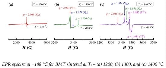
Temperature-dependent EPR spectra for BM1T are shown in Figure 5. For BM1T sintered at Ts = 1200 °C, only the g = 2.004 signal existed over the measuring temperature (T) range of −188 to 150 °C (Figure 5a). This signal was assigned as ionized Ti vacancies [21,22,23]. The g = 2.004 signal was activated in the cubic phase above Tm and in the rhombohedral phase below T = −100 °C. This activation confirmed the nature of Ti vacancies [23]. The pair of weak lines denoted as g1 = 1.944 and g3 = 2.060 appeared at T = −188 °C, forming a centrosymmetric pattern around g2 = 2.004. This phenomenon is similar to the low-temperature EPR spectrum observed for (Ba0.85Sr0.15)TiO3 [24], which may relate to the occupation of Mg2+ on the A site.
When Ts = 1300 °C, except for the g = 2.004 signal, two additional signals at g = 1.974 and 1.956 observed at T = −188 °C (Figure 5b) were assigned as ionized Ba [14,22] and oxygen ( V O + e V O ) [19] vacancies, respectively.
BM1T sintered at Ts = 1400 °C existed in mixed forms of the hexagonal and tetragonal phases. Five EPR signals appeared below T = −100 °C and their intensity increased with decreasing T (Figure 5c). The presence of three signals at g = 2.004, 1.974, and 1.957 implies the coexistence of V Ba , V Ti , and V O . We attributed two additional signals at g = 1.934 and 1.942 to a hexagonally distorted d1 ion from Ti3+ ( Ti Ti ) because low temperatures can effectively prolong the spin–lattice relaxation time (τ) [19,22]. This indicates that during high-temperature sintering of Ts = 1400 °C, the electrons in BM1T can be trapped by Ti4+ ions to cause a reduction from Ti4+ to Ti3+. It has been reported that the (Ba1−xCax)TiO3 (x = 0.03) ceramic sintered at Ts = 1500 °C showed a more ordered tetragonal structure, and only a Ti3+-related signal at g = 1.932 was observed at T = −188 °C [19,22]. However, this signal did not appear in the tetragonal BMT sintered at Ts = 1300 °C (Figure 5b). In the mixed hexagonal and tetragonal phases of BMT sintered at Ts = 1400 °C, the Ti3+-related signal split into two signals at g = 1.934 and 1.942. It is obvious that these two signals originated from the hexagonal phase in BM1T.

4. Discussion

4.1. Site Occupation of Mg2+ in BM1T at Different Sintering Temperatures

On the basis of a simple comparison of 12-CN ionic size between Ba2+ (1.61 Å) and Mg2+ (1.23 Å) and 6-CN ionic size between Ti4+ (0.605 Å) and Mg2+ (0.72 Å), a continuous decrease in V0 with x (≤0.05) for BM1T sintered at Ts = 1200 °C (Figure 3, inset) provides sufficient evidence for occupation of the A site by Mg2+. When x is higher than the solubility limit of 0.05, Mg2+ cannot continuously enter the A site, accompanied by separation of Mg-rich phases (Figure 3). The appearance of V O can be considered as an indication of the existence of Mg2+ at the B site, i.e., forming Mg Ti V O pairs [10,11,12,13]. BO6 octahedrons are characteristic of the perovskite lattice. Hence, higher energy is required to incorporate doping ions into the B site. It is inferred that the sintering temperature of Ts = 1200 °C is too low to incorporate Mg2+ into the B site because the V O -related EPR signal was not observed (Figure 5a). On the other hand, BM1T has a pseudocubic structure and its V0 (= 64.40 Å3) is equal to the tetragonal BaTiO3 (V0 = 64.41 Å3, JCPDS Card No. 6-526). This implies that Mg2+ tends to remain close to the surfaces of the grains and plays an important role in the temperature-stable X8S behavior in BM1T, as suggested by Chang et al. [5]. At this time, Mg2+ exists only at the A site as Mg Ba × .
El Ghadraoui et al. indicated that the solubility limit of Mg2+ in (Ba1−xMgx)TiO3 was 0.15. They neglected a small amount of the secondary phases of BaMg6Ti6O19 and MgTiO3, which also appeared in their samples with x ≥ 0.05 [25]. Their report undoubtedly supports that Mg2+ may exist at the A site.
When Ts was increased to 1300 °C, V O and V Ba were detected (Figure 5b), revealing that some Mg2+ ions transferred from the A site to the B site during the cooling process of ceramic sintering, accompanied by the creation of V O . However, the numbers of Mg Ti and V O were too small to induce the hexagonal phase.
When Ts = 1400 °C, more Mg2+ ions enter the B site. The concentration of Mg Ti V O was high enough to cause phase splitting into hexagonal and tetragonal (Figure 5c). The hexagonal phase in BM1T originated from Mg Ti V O defect complexes. Kirianov et al. and Dang et al. also reported a similar result on the mixed phases for Ba(Ti1−xMnx)O3 with x < 0.03 [26,27]. The Jahn–Teller distortion encased by the Mn Ti ions is proposed to be the driving force of the phase transition from tetragonal to hexagonal [28]. This implies that Mg Ti and Mn Ti acceptors on the Ti sites in BaTiO3 play the same role in the formation of the hexagonal phase. Thus, Mg Ti and V O mainly exist in the hexagonal phase, and Mg Ba × is predominant in the tetragonal phase.
As a whole, Mg2+ ions in BM1T sintered at Ts ≥ 1300 °C exhibited amphoteric behavior, i.e., Mg2+ existed at the A site as Mg Ba × and at the B site as Mg Ti .

4.2. Oxygen Vacancies and Dielectric Loss

The V O can be detected by the EPR technique for Mg-doped BaTiO3. It is not easy to create V O when Ts is lower than 1200 °C and tan δ at Ts = 1300 °C is astonishingly low over the T range of −55 to 150 °C (tan δ ≤ 0.03).
The increase in Ts can create V O and Ti Ti , giving rise to phase splitting into hexagonal and tetragonal at Ts = 1400 °C. The high value of tan δ is attributed to the high concentrations of V O and Ti Ti in the hexagonal phase in BM1T (Figure 4).

5. Conclusions

The solubility of Mg2+ in (Ba1−xMgx)TiO3 ceramics sintered at 1200 °C was 0.05, and rhombohedral MgTiO3 and hexagonal BaMg6Ti6O19 phases were observed with higher doping content. The evolution of unit cell volume provided sufficient evidence for the A-site occupation of Mg2+. The x = 0.015 ceramic had a pseudocubic crystal structure when the sintered temperature was 1200 °C and exhibited a temperature-stable X8S dielectric specification with εRT = 1200. The structure transformed into a tetragonal phase when sintered at 1300 °C, and tetragonal and hexagonal phases coexisted when sintered at 1400 °C.
For x = 0.015 sintered above 1300 °C, the g = 1.956 signal observed at T = −188 °C was assigned as ionized oxygen vacancies ( V O ). Mg2+ acted as an amphoteric doping ion with Mg Ba × and Mg Ti . Mg Ti and V O mainly existed in the hexagonal phase and Mg Ba × was predominant in the tetragonal phase. Two EPR signals at g = 1.934 and 1.942 originated from the hexagonal phase in x = 0.015 and were related to Ti3+ which, along with V O , is mainly responsible for the higher tan δ value.

Author Contributions

Conceptualization, D.L.; Methodology, D.L.; Software, Y.Z. and L.Y.; Validation, D.L., Y.Z. and L.Y.; Formal analysis, D.L. and Y.Z.; Investigation, D.L., Y.Z. and L.Y.; Resources, D.L.; Data curation, D.L. and Y.Z.; Writing—original draft preparation, D.L.; Writing—review and editing, D.L.; Supervision, D.L.; Project administration, D.L.; Funding acquisition, D.L.

Funding

This research was funded by the National Natural Science Foundation of China, grant number 21271084; a project of the Jilin Province Development and Reform Commission, grant number 2019C044-1; and a project of the Changbai Mountain Scholar Distinguished Professor, grant number 2015047.

Acknowledgments

The authors would like to thank Dandan Han and Qiaoli Liu in Jilin Institute of Chemical Technology for their support and help.

Conflicts of Interest

The authors declare no conflict of interest.

References

  1. Huang, X.; Liu, H.; Hao, H.; Zhang, S.; Sun, Y.; Zhang, W.; Zhang, L.; Cao, M. Microstructure effect on dielectric Properties of MgO-doped BaTiO3–BiYO3 ceramics. Ceram. Int. 2015, 41, 7489–7495. [Google Scholar]
  2. Lin, Y.T.; Ou, S.F.; Lin, M.H.; Song, Y.R. Effect of MgO addition on the microstructure and dielectric properties of BaTiO3 ceramics. Ceram. Int. 2018, 44, 3531–3535. [Google Scholar]
  3. Li, J.-H.; Wang, S.-F.; Hsu, Y.-F.; Chung, T.-F.; Yang, J.-R. Effects of Sc2O3 and MgO additions on the dielectric properties of BaTiO3-based X8R materials. J. Alloy. Compd. 2018, 768, 122–129. [Google Scholar] [CrossRef]
  4. Wang, S.; Zhang, S.; Zhou, X.; Li, B.; Chen, Z. Effect of sintering atmospheres on the microstructure and dielectric properties of Yb/Mg co-doped BaTiO3 ceramics. Mater. Lett. 2005, 59, 2457–2460. [Google Scholar] [CrossRef]
  5. Kim, C.H.; Park, K.J.; Yoon, Y.J.; Hong, M.H.; Hong, J.O.; Hur, K.H. Role of Yttium and magnesium in the formation of core-shell structure of BaTiO3 grains in MLCC. J. Eur. Ceram. Soc. 2008, 28, 1213–1219. [Google Scholar] [CrossRef]
  6. Chang, C.Y.; Wang, W.N.; Huang, C.Y. Effect of MgO and Y2O3 doping on the formation of core-shell structure in BaTiO3 ceramics. J. Am. Ceram. Soc. 2013, 96, 2570–2576. [Google Scholar] [CrossRef]
  7. Nagai, T.; Iijima, K.; Hwang, H.J.; Sando, M.; Sekino, T.; Niihara, K. Effect of MgO doping on the phase transformations of BaTiO3. J. Am. Ceram. Soc. 2000, 83, 107–112. [Google Scholar] [CrossRef]
  8. Hagemann, H.J.; Hennings, D. Reversible weight change of acceptor-doped BaTiO3. J. Am. Ceram. Soc. 1981, 64, 590–594. [Google Scholar]
  9. Lu, D.; Ji, L.; Liu, J. Aging-resistant behavior and room-temperature electron spin resonance of Nd3+ in singly and doubly doped BaTiO3 ceramics associated with preservation history. Materials 2019, 12, 451. [Google Scholar] [CrossRef]
  10. Cha, S.H.; Han, Y.H. Effects of Mn doping on dielectric properties of Mg-doped BaTiO3. J. Appl. Phys. 2006, 100, 104102. [Google Scholar]
  11. Dong, M.; Miao, H.; Tan, G.; Pu, Y. Effects of Mg-doping on the microstructure and properties of BaTiO3 ceramics prepared by hydrothermal method. J. Electroceram. 2008, 21, 573–576. [Google Scholar] [CrossRef]
  12. Yoon, S.H.; Randall, C.A.; Hur, K.H. Effects of acceptor concentration on the bulk electrical conduction in acceptor (Mg)-doped BaTiO3. J. Appl. Phys. 2010, 107, 103721. [Google Scholar] [CrossRef]
  13. Kishi, H.; Mizuno, Y.; Chazono, H. Base-metal electrode-multilayer ceramic capacitors: past, present and future perspectives. Jpn. J. Appl. Phys. 2003, 42, 1–15. [Google Scholar] [CrossRef]
  14. Lu, D.; Cui, S. Defects characterisation of Dy-doped BaTiO3 ceramics via electron paramagnetic resonance. J. Eur. Ceram. Soc. 2014, 34, 2217–2227. [Google Scholar] [CrossRef]
  15. Cui, S.; Lu, D. Study of the solubility of La3+-Dy3+ defect complexes and dielectric properties of (Ba1−xLax)(Ti1−xDyx)O3 ceramics. Ceram. Int. 2015, 41, 2301–2308. [Google Scholar] [CrossRef]
  16. Lu, D.; Yin, S.; Cui, S. A fine-grained and low-loss X8R (Ba1–xDyx)(Ti1–x/2Cax/2)O3 ceramic. J. Alloy. Compd. 2018, 762, 282–288. [Google Scholar] [CrossRef]
  17. Shannon, R.D. Revised effective ionic radii and systematic studies of interatomic distances in halides and chalcogenides. Acta Crystallogr. Sect. A 1976, 32, 751–767. [Google Scholar] [CrossRef]
  18. Bernard, J.; Houivet, D.; El Fallah, J.; Haussonne, J.M. MgTiO3 for Cu base metal multilayer ceramic capacitors. J. Eur. Ceram. Soc. 2004, 24, 1877–1881. [Google Scholar] [CrossRef]
  19. Lu, D.; Yuan, L.; Liang, W.; Zhu, Z. Characterization of oxygen vacancy defects in Ba1−xCaxTiO3 insulating ceramics using electron paramagnetic resonance technique. Jpn. J. Appl. Phys. 2016, 55, 011501. [Google Scholar] [CrossRef]
  20. Jayanthi, S.; Kutty, T.R.N. Dielectric properties of barium magnesiotitanate ceramics, BaMg6Ti6O19. Mater. Lett. 2006, 60, 796–800. [Google Scholar] [CrossRef]
  21. Dunbar, T.D.; Warren, W.L.; Tuttle, B.A.; Randall, C.A.; Tsur, T. Electron paramagnetic resonance investigations of lanthanide-doped barium titanate: dopant site occupancy. J. Phys. Chem. B 2004, 108, 908–917. [Google Scholar] [CrossRef]
  22. Kolodiazhnyi, T.; Petric, A. Analysis of point defects in polycrystalline BaTiO3 by electron paramagnetic resonance. J. Phys. Chem. Solids 2003, 64, 953–960. [Google Scholar] [CrossRef]
  23. Lu, D.; Liu, T. Dielectric properties and defect chemistry of (Ba1−xLax)(Ti1−xLux)O3 ceramics. J. Alloy. Compd. 2017, 698, 967–976. [Google Scholar] [CrossRef]
  24. Lu, D.; Sun, X. A novel electron paramagnetic resonance phenomenon in a barium stronium titanate ceramic. Adv. Mater. Res. 2011, 295–297, 1050–1054. [Google Scholar] [CrossRef]
  25. El Ghadraoui, O.E.; Zouhairi, M.; Abdi, F.; Lamcharfi, T.; Bali, H.; El Ghadraoui, E.; Aillerie, M. Preparation and physico-chemical study of Mg doped BaTiO3 (Ba1−xMgxTiO3) prepared by the sol-gel method. Int. J. Curr. Res. 2016, 8, 42815–42820. [Google Scholar]
  26. Kirianov, A.; Ozaki, N.; Ohsato, H.; Kohzu, N.; Kishi, H. Studies on the solid solution of Mn in BaTiO3. Jpn. J. Appl. Phys. 2001, 40, 5619–5623. [Google Scholar] [CrossRef]
  27. Dang, N.V.; Phan, T.-L.; Thanh, T.D.; Lam, V.D.; Hong, L.V. Structural phase separation and optical and magnetic properties of Ba(Ti1−xMnx)O3 multiferroics. J. Appl. Phys. 2012, 111, 113913. [Google Scholar] [CrossRef]
  28. Langhammer, H.T.; Muller, T.; Felgner, K.H.; Abicht, H.P. Crystal structure and related properties of manganese-doped barium titanate ceramics. J. Am. Ceram. Soc. 2000, 83, 605–611. [Google Scholar] [CrossRef]
Figure 1. (a) Powder XRD patterns of (Ba1−xMgx)TiO3 (x = 0.015) (BM1T) ceramics prepared at Ts = 1200–1400 °C. Insets show enlarged diffraction peaks in the vicinity of 45°. The lattice parameters are given. (b) Simulated XRD patterns of BaTiO3 with cubic, tetragonal, and hexagonal structures.
Materials 12 01525 g001
Figure 2. SEM images of polished and thermally etched surfaces of BM1T sintered at Ts = (a) 1200, (b) 1300, and (c) 1400 °C.
Materials 12 01525 g002
Figure 3. XRD patterns of (a) (Ba1−xMgx)TiO3 (x = 0.03‒0.20) (BMT) ceramics sintered at Ts = 1200 °C. Inset depicts variation in V0 as a function of x. Simulated XRD patterns of (b) hexagonal BaMg6Ti6O19 (JCPDS Cards No. 42‒0441) and (c) rhombohedral MgTiO3 (JCPDS Cards No. 06‒0494).
Materials 12 01525 g003
Figure 4. Temperature dependencies of (a) dielectric permittivity (ε’) and (b) dielectric loss (tan δ) for BM1T sintered at Ts = 1200, 1300, and 1400 °C.
Materials 12 01525 g004
Figure 5. Temperature-dependent EPR spectra for BM1T sintered at Ts = (a) 1200, (b) 1300, and (c) 1400 °C.
Materials 12 01525 g005
Table 1. Ionic radius as a function of coordinate number (CN).
IonCNr (Å)
Ba2+121.61
Ti4+60.605
Ti3+60.67
Mg2+121.23
Mg2+60.72