
Structural Studies of the Phage G Tail Demonstrate an Atypical Tail Contraction


 ,
,  and
and
Abstract
:1. Introduction
2. Materials and Methods
2.1. Phage G Propagation and Purification
2.2. Negative Stain EM of Phage G–Host Interaction
2.3. Cryo-EM Data Collection
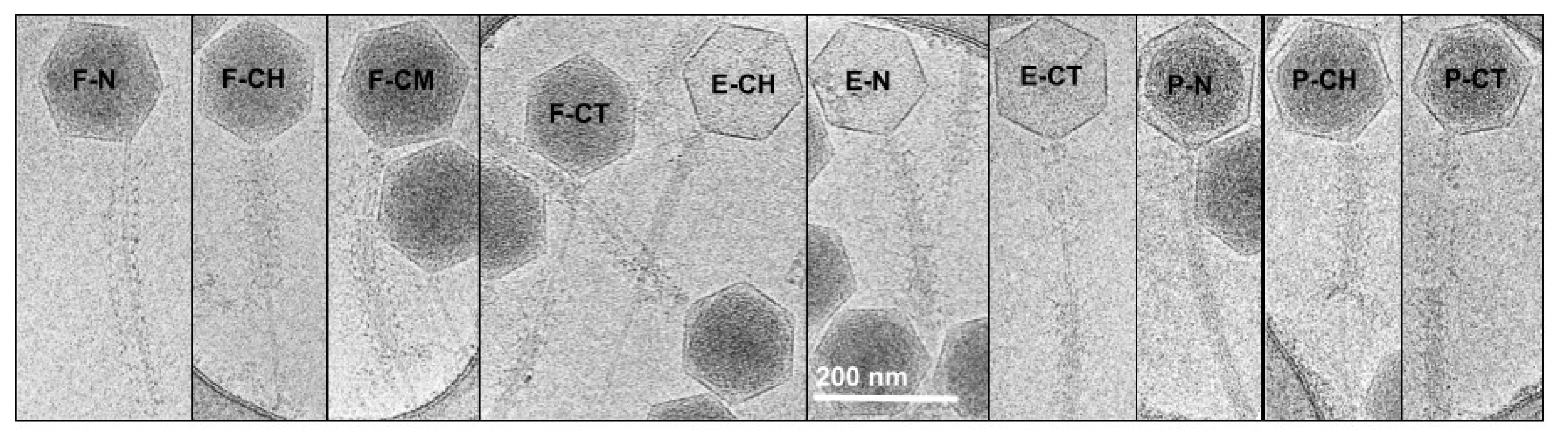
2.4. Categorization of Tail Contraction States from Cryo-EM Micrographs
2.5. Helical Reconstruction of Non-Contracted and Contracted Phage G Tail Sheath
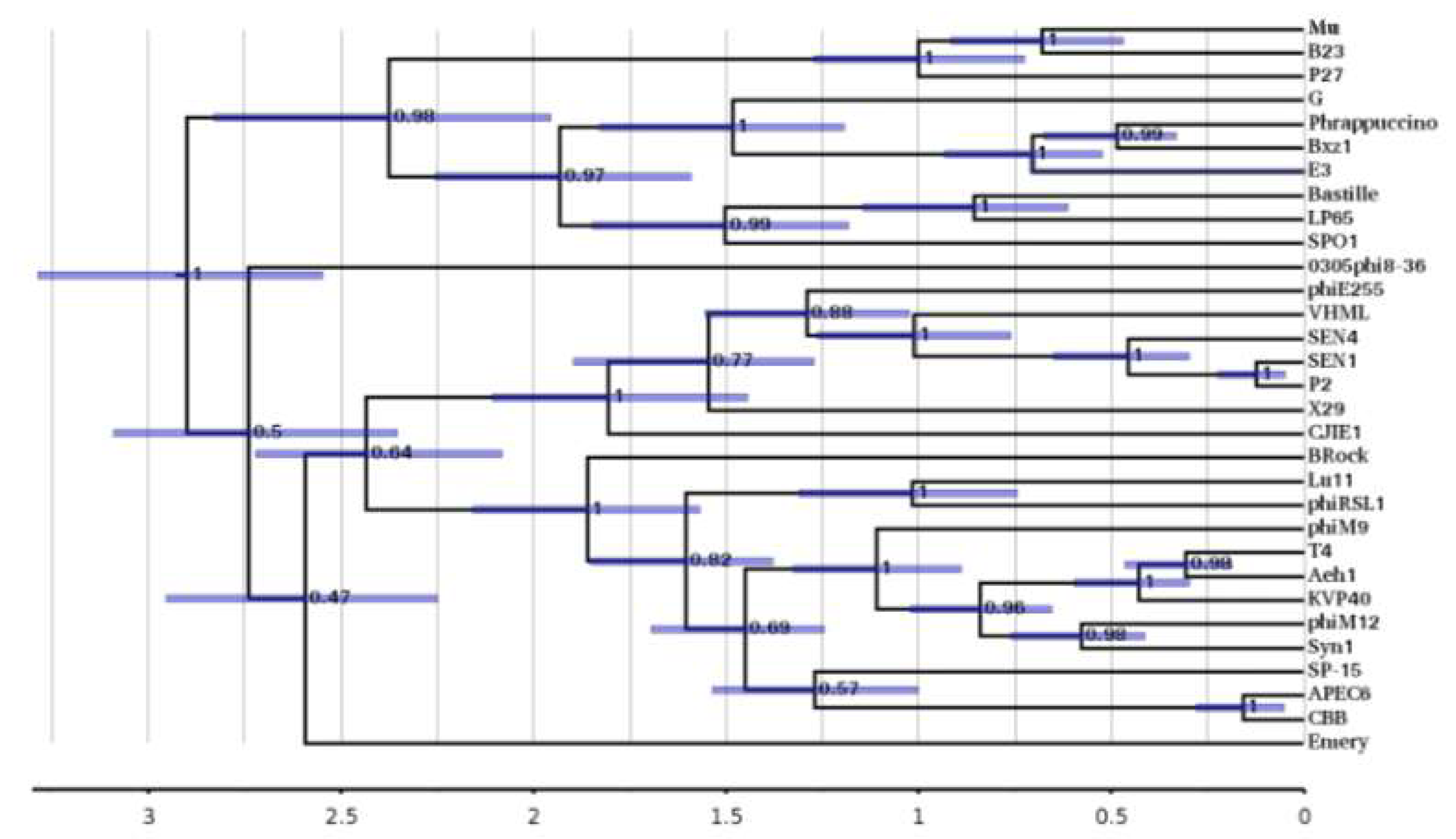
2.6. Bioinformatic Evolutionary Analysis of Phage G Tail Sheath
3. Results
3.1. Negative Stain EM of Phage G Host Attachment
3.2. Tail Contraction States in Our Cryo-EM Data
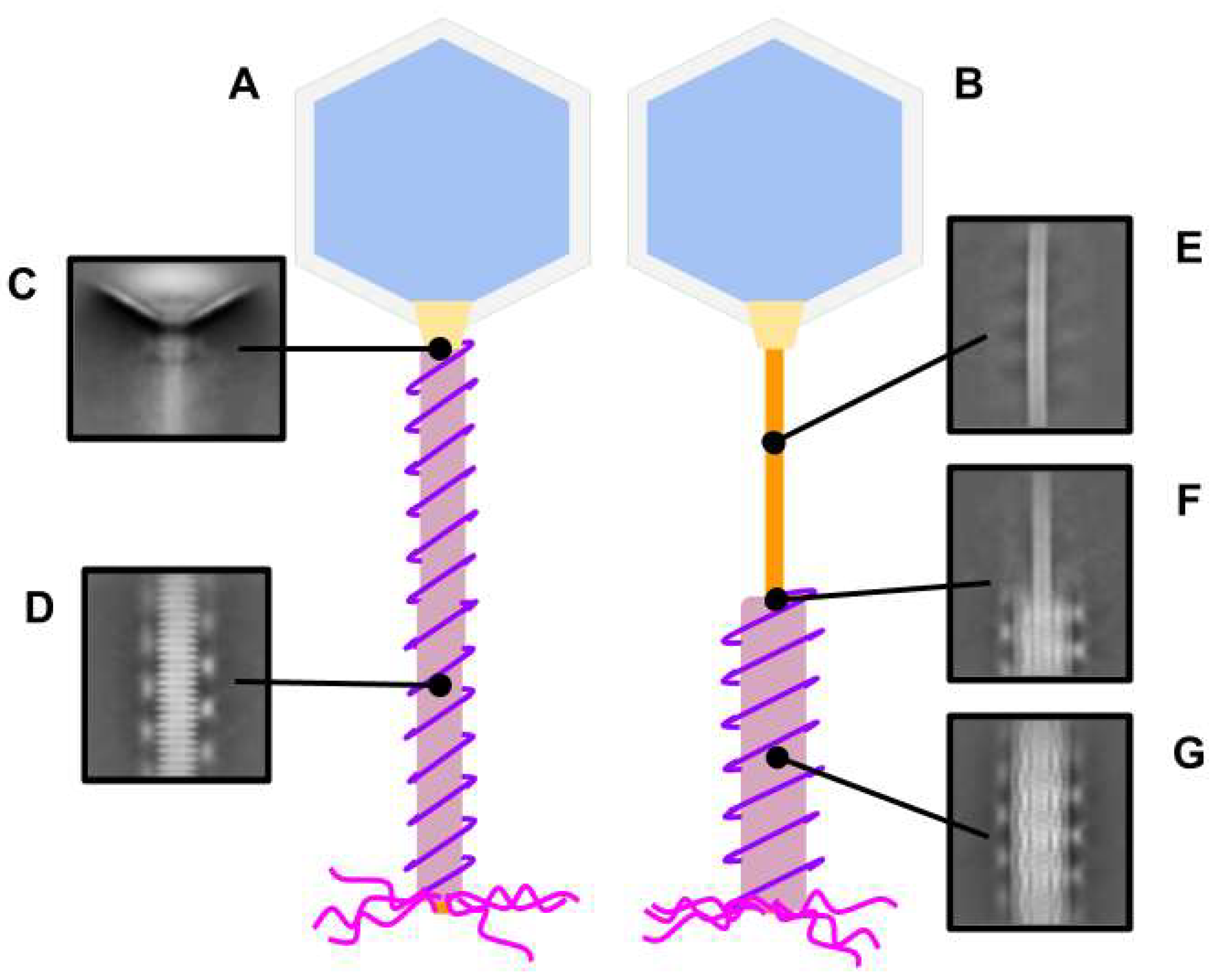
3.3. Phage G Tail Components from 2D Classification
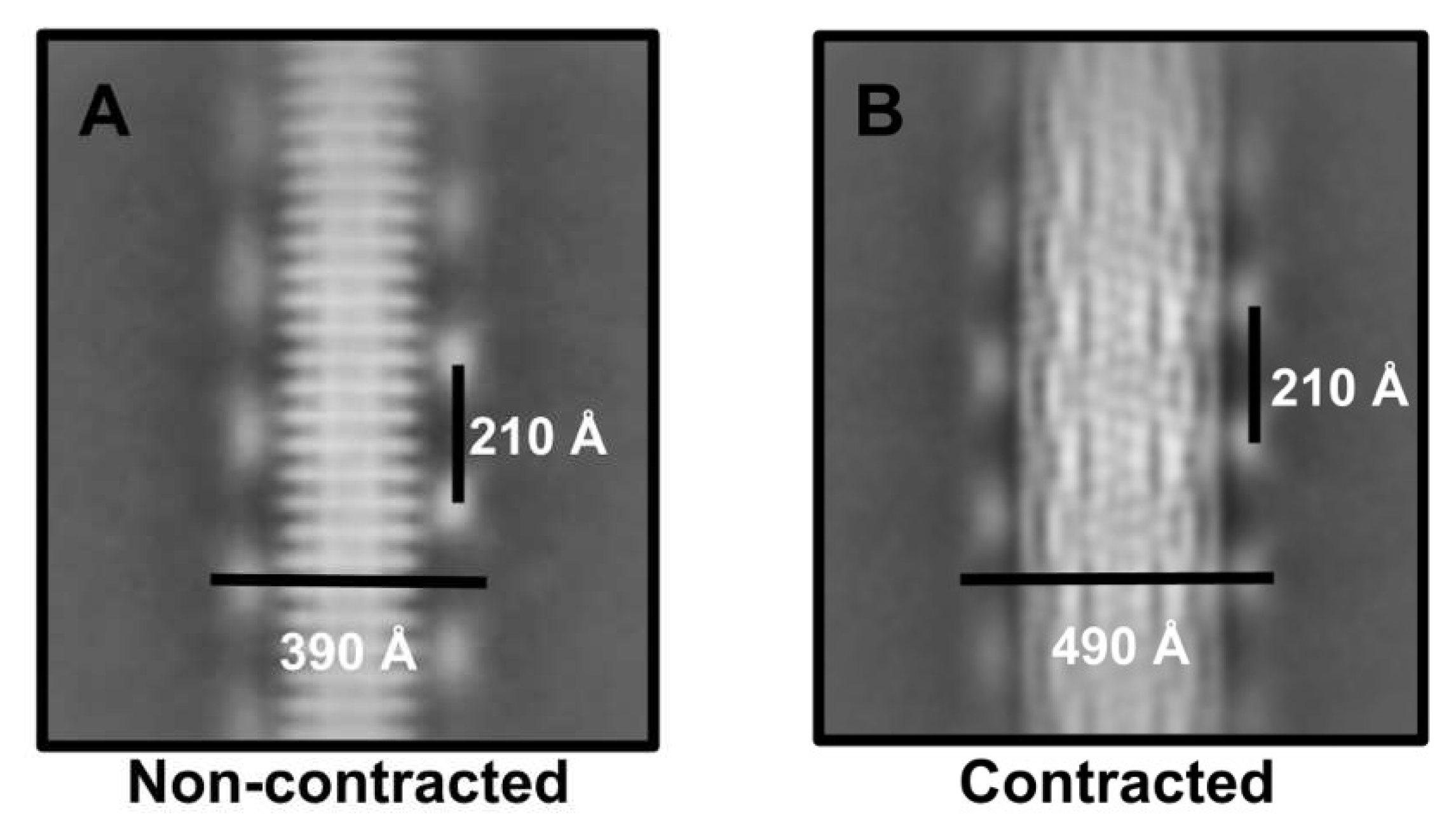
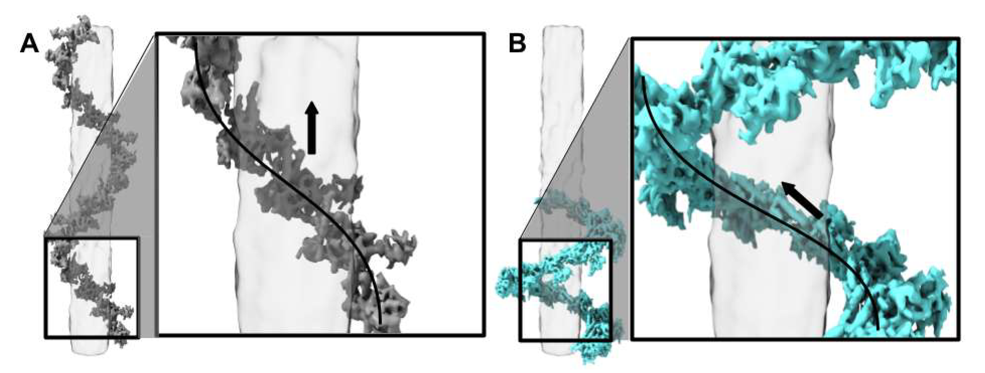
3.4. 3D Cryo-EM Structure of the Non-Contracted and Contracted Phage G Tail Sheath
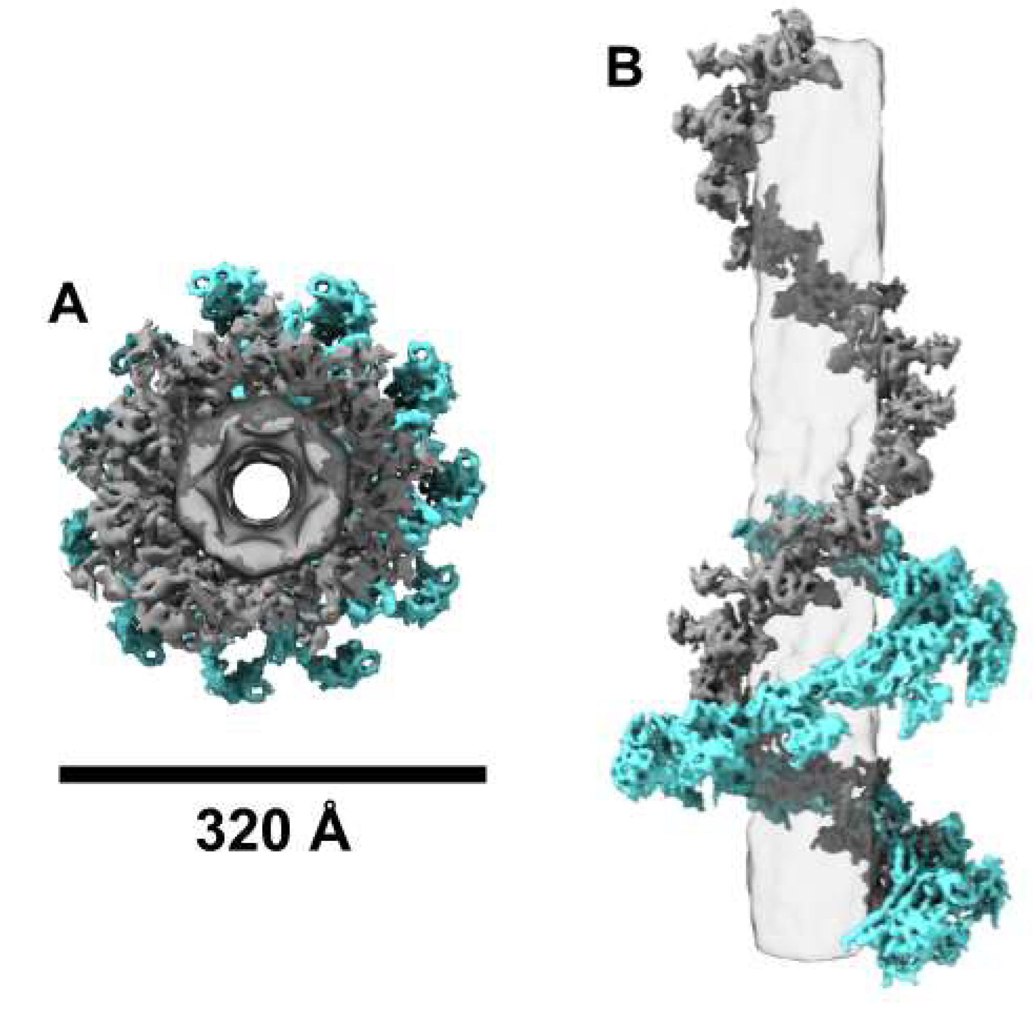
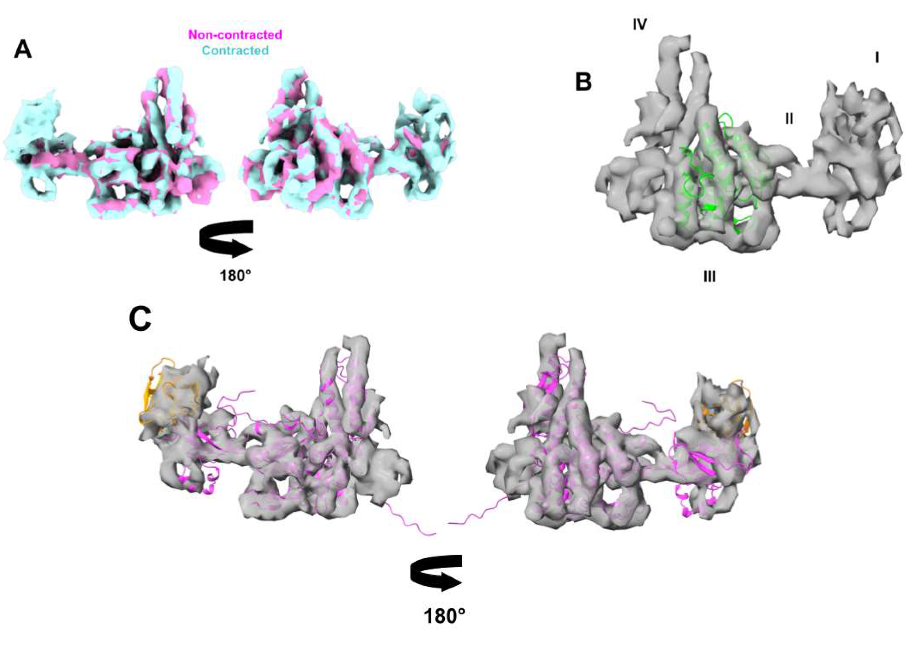
3.5. Phage G Tail Sheath Subunit Structure and Arrangement

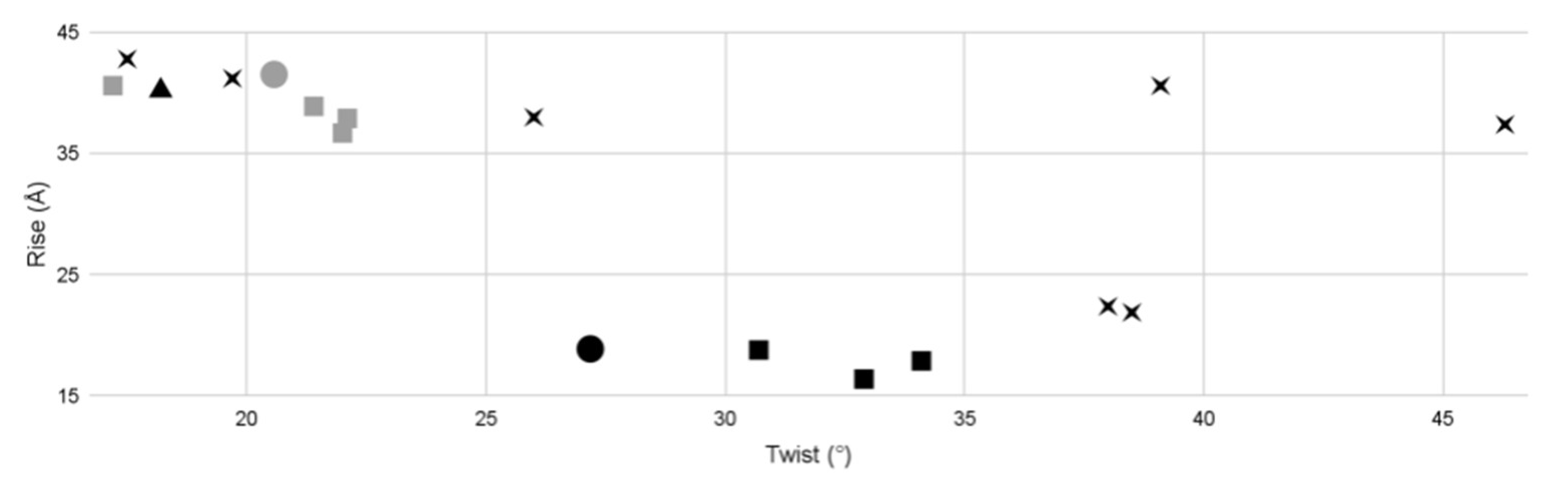
3.6. Phage G Tail Sheath Helical Symmetry Compared to Other Known Phages


| Tail Morphology | Virus | Structure | Twist (°) | Rise (Å) | Reported Resolution (Å) | EMDB Entry | Host Gram (−/+) | Citation |
|---|---|---|---|---|---|---|---|---|
| Myophage | phage G | non-contracted sheath | 20.57 | 41.53 | 7–8 | 25155 | + | Current study |
| contracted sheath | 27.18 | 18.89 | 6–7 | 25154 | ||||
| phi812K1-420 | non-contracted sheath | 21.4 | 38.9 | 6.2 | 4051 | – | [11] | |
| contracted sheath | 30.7 | 18.8 | 4.2 | 4052 | ||||
| phiRSL1 | non-contracted sheath | 22.1 | 37.9 | 9.6 | 2244 | – | [10] | |
| phiKZ | contracted poly sheath | 34.1 | 17.9 | 19.0 | 5331 | – | [33] | |
| non-contracted sheath | 22 | 36.7 | 18.0 | 5332 | ||||
| T4 | contracted sheath | 32.9 | 16.4 | N/A | N/A | – | [44,45] | |
| non-contracted | 17.2 | 40.6 | N/A | N/A | ||||
| tube | 18.2 | 40.2 | 3.4 | 8767 | [45] | |||
| Siphophage | SPP1 | tube | 38.5 | 21.9 | 4.0 | 10792 | + | [35] |
| YSD1 | 19.7 | 41.2 | 3.5 | 22183 | – | [38] | ||
| lambda | 17.5 | 42.8 | 6.4 | 20242 | – | [46] | ||
| T5 | 39.1 | 40.6 | 6.0 | 3692 | – | [39] | ||
| p2 | 46.3 | 37.4 | 22.0 | 2464 | – | [42] | ||
| Araucaria | 26 | 38 | 24.0 | 2337 | + | [36] | ||
| TP901-1 | 38 | 22.4 | 20.0 | 2228 | + | [47] |
3.7. Evolutionary Analysis of Phage G Tail Sheath Protein gp178
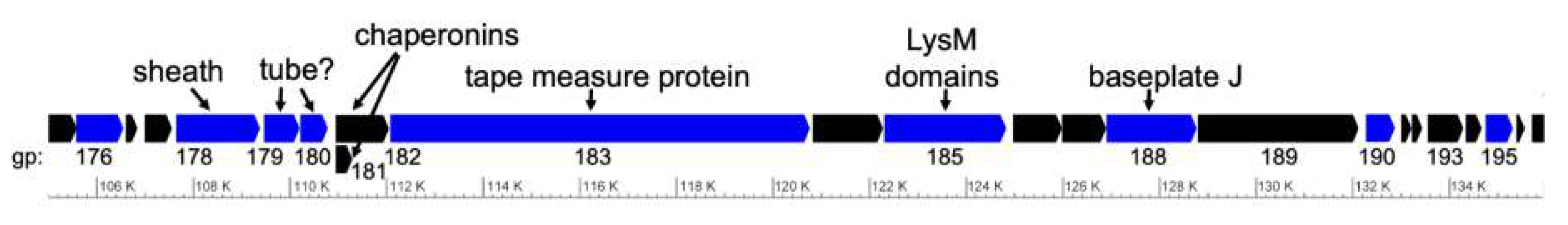
3.8. The Phage G Tail Sheath Gene Is Located in a Syntenous Tail Morphogenesis Gene Module
4. Discussion
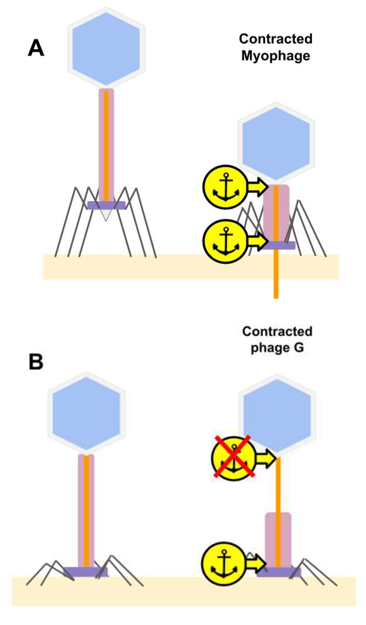
4.1. Unusual Tail Sheath Contraction in Phage G
4.2. Missing Tail Sheath Anchor Point at the Neck Region in Phage G
4.3. Evolutionary Implication of Phage Tail Mediated Infection Mechanisms and Future Directions
Supplementary Materials
Author Contributions
Funding
Institutional Review Board Statement
Informed Consent Statement
Data Availability Statement
Acknowledgments
Conflicts of Interest
References
- Yuan, Y.; Gao, M. Jumbo Bacteriophages: An Overview. Front. Microbiol. 2017, 8, 403. [Google Scholar] [CrossRef] [Green Version]
- Hua, J.; Huet, A.; Lopez, C.A.; Toropova, K.; Pope, W.H.; Duda, R.L.; Hendrix, R.W.; James, F. ConwayCapsids and genomes of jumbo-sized bacteriophages reveal the evolutionary reach of the HK97 fold. mBio 2017, 8, e01579-17. [Google Scholar] [CrossRef] [Green Version]
- Donelli, G. Isolation of a bacteriophage of exceptional dimensions active in r megatherium. Atti Della Accad. Naz. Dei Lincei Rend. Cl. Di Sci. Fis. Mat. Nat. 1968, 44, 95. [Google Scholar]
- González, B.; Monroe, L.; Li, K.; Yan, R.; Wright, E.; Walter, T.; Kihara, D.; Weintraub, S.T.; Thomas, J.A.; Serwer, P. Phage G structure at 6.1 AA resolution, condensed DNA, and host identity revision to a lysinibacillus. J. Mol. Biol. 2020, 432, 4139–4153. [Google Scholar] [CrossRef]
- Leiman, P.G.; Chipman, P.R.; Kostyuchenko, V.A.; Mesyanzhinov, V.V.; Rossmann, M.G. Three-dimensional rearrangement of proteins in the tail of bacteriophage T4 on infection of its host. Cell 2004, 118, 419–429. [Google Scholar] [CrossRef] [PubMed] [Green Version]
- Aksyuk, A.A.; Leiman, P.G.; Kurochkina, L.P.; Shneider, M.M.; Kostyuchenko, V.A.; Mesyanzhinov, V.V.; Rossmann, M.G. The tail sheath structure of bacteriophage T4: A molecular machine for infecting bacteria. EMBO J. 2009, 28, 821–829. [Google Scholar] [CrossRef]
- Amos, L.; Klug, A. Three-dimensional image reconstructions of the contractile tail of T4 bacteriophage. J. Mol. Biol. 1975, 99, 51–64. [Google Scholar] [CrossRef] [Green Version]
- Hu, B.; Margolin, W.; Molineux, I.J.; Liu, J. Structural remodeling of bacteriophage T4 and host membranes during infection initiation. Proc. Natl. Acad. Sci. USA 2015, 112, E4919–E4928. [Google Scholar] [CrossRef] [Green Version]
- Moody, M. Sheath of bacteriophage T4: III. Contraction mechanism deduced from partially contracted sheaths. J. Mol. Biol. 1973, 80, 613–635. [Google Scholar] [CrossRef]
- Effantin, G.; Hamasaki, R.; Kawasaki, T.; Bacia, M.; Moriscot, C.; Weissenhorn, W.; Yamada, T.; Schoehn, G. Cryo-electron microscopy three-dimensional structure of the jumbo phage ΦRSL1 infecting the phytopathogen Ralstonia solanacearum. Structure 2013, 21, 298–305. [Google Scholar] [CrossRef] [Green Version]
- Nováček, J.; Šiborová, M.; Benešík, M.; Pantůček, R.; Doškař, J.; Plevka, P. Structure and genome release of Twort-like Myoviridae phage with a double-layered baseplate. Proc. Natl. Acad. Sci. USA 2016, 113, 9351–9356. [Google Scholar] [CrossRef] [PubMed] [Green Version]
- Ageno, M.; Donelli, G.; Guglielmi, F. Structure and physico-chemical properties of bacteriophage G. II, The shape and symmetry of the capsid. Micron 1973, 4, 376–403. [Google Scholar] [CrossRef]
- Donelli, G.; Dore, E.; Frontali, C.; Grandolfo, M.E. Structure and physico-chemical properties of bacteriophage G: III. A homogeneous DNA of molecular weight 5 × 10⁸. J. Mol. Biol. 1975, 94, 555–565. [Google Scholar] [CrossRef]
- Donelli, G.; Griso, G.; Paoletti, L.; Rebessi, S. Capsomeric Arrangement in the Bacteriophage G Head. In Proceedings of the Sixth European Congress on Electron Microscopy, Jerusalem, Israel, 14–20 September 1976; pp. 502–503. [Google Scholar]
- Donelli, G.; Guglielmi, F.; Paoletti, L. Structure and physico-chemical properties of bacteriophage G. I. Arrangement of protein subunits and contraction process of tail sheath. J. Mol. Biol. 1972, 71, 113–125. [Google Scholar] [CrossRef]
- Egelman, E.H. Ambiguities in helical reconstruction. eLife 2014, 3, e04969. [Google Scholar] [CrossRef] [Green Version]
- Fangman, W.L. Separation of very large DNA molecules by gel electrophoresis. Nucleic Acids Res. 1978, 5, 653–665. [Google Scholar] [CrossRef] [Green Version]
- Sun, M.; Serwer, P. The conformation of DNA packaged in bacteriophage G. Biophys. J. 1997, 72, 958–963. [Google Scholar] [CrossRef] [Green Version]
- Serwer, P.; Estrada, A.; Harris, R.A. Video light microscopy of 670-kb DNA in a hanging drop: Shape of the envelope of DNA. Biophys. J. 1995, 69, 2649–2660. [Google Scholar] [CrossRef] [Green Version]
- Li, X.; Mooney, P.; Zheng, S.; Booth, C.R.; Braunfeld, M.B.; Gubbens, S.; Agard, D.A.; Cheng, Y. Electron counting and beam-induced motion correction enable near-atomic-resolution single-particle cryo-EM. Nat. Methods 2013, 10, 584–590. [Google Scholar] [CrossRef] [PubMed] [Green Version]
- He, S.; Scheres, S.H.W. Helical reconstruction in RELION. J. Struct. Biol. 2017, 198, 163–176. [Google Scholar] [CrossRef] [PubMed]
- Punjani, A.; Rubinstein, J.L.; Fleet, D.J.; Brubaker, M.A. cryoSPARC: Algorithms for rapid unsupervised cryo-EM structure determination. Nat. Methods 2017, 14, 290–296. [Google Scholar] [CrossRef] [PubMed]
- Guo, F.; Jiang, W. Single particle cryo-electron microscopy and 3-D reconstruction of viruses. In Electron Microscopy; Springer: New York, NY, USA, 2014; pp. 401–443. [Google Scholar]
- Hardies, S.C.; Thomas, J.A.; Black, L.; Weintraub, S.T.; Hwang, C.Y.; Cho, B.C. Identification of structural and morphogenesis genes of Pseudoalteromonas phage φRIO-1 and placement within the evolutionary history of Podoviridae. Virology 2016, 489, 116–127. [Google Scholar] [CrossRef] [Green Version]
- Hughey, R.; Krogh, A. Hidden Markov models for sequence analysis: Extension and analysis of the basic method. Bioinformatics 1996, 12, 95–107. [Google Scholar] [CrossRef] [Green Version]
- Karplus, K.; Barrett, C.; Hughey, R. Hidden Markov models for detecting remote protein homologies. Bioinformatics 1998, 14, 846–856. [Google Scholar] [CrossRef]
- Steinegger, M.; Meier, M.; Mirdita, M.; Vöhringer, H.; Haunsberger, S.J.; Söding, J. HH-suite3 for fast remote homology detection and deep protein annotation. BMC Bioinform. 2019, 20, 473. [Google Scholar] [CrossRef] [Green Version]
- Ronquist, F.; Teslenko, M.; Van Der Mark, P.; Ayres, D.L.; Darling, A.; Höhna, S.; Larget, B.; Liu, L.; Suchard, M.A.; Huelsenbeck, J.P. MrBayes 3.2: Efficient Bayesian phylogenetic inference and model choice across a large model space. Syst. Biol. 2012, 61, 539–542. [Google Scholar] [CrossRef] [Green Version]
- Bouckaert, R.; Heled, J.; Kühnert, D.; Vaughan, T.; Wu, C.-H.; Xie, D.; Suchard, M.A.; Rambaut, A.; Drummond, A.J. BEAST 2: A software platform for Bayesian evolutionary analysis. PLoS Comput. Biol. 2014, 10, e1003537. [Google Scholar] [CrossRef] [Green Version]
- Arisaka, F.; Engel, J.; Klump, H. Contraction and dissociation of the bacteriophage T4 tail sheath induced by heat and urea. Prog. Clin. Biol. Res. 1981, 64, 365–379. [Google Scholar]
- Arisaka, F.; Nakako, T.; Takahashi, H.; Ishii, S.-L. Nucleotide sequence of the tail sheath gene of bacteriophage T4 and amino acid sequence of its product. J. Virol. 1988, 62, 1186–1193. [Google Scholar] [CrossRef] [Green Version]
- Jumper, J.; Evans, R.; Pritzel, A.; Green, T.; Figurnov, M.; Ronneberger, O.; Tunyasuvunakool, K.; Bates, R.; Žídek, A.; Potapenko, A. Highly accurate protein structure prediction with AlphaFold. Nature 2021, 596, 583–589. [Google Scholar] [CrossRef] [PubMed]
- Aksyuk, A.A.; Kurochkina, L.P.; Fokine, A.; Forouhar, F.; Mesyanzhinov, V.V.; Tong, L.; Rossmann, M.G. Structural conservation of the myoviridae phage tail sheath protein fold. Structure 2011, 19, 1885–1894. [Google Scholar] [CrossRef] [Green Version]
- Pettersen, E.F.; Goddard, T.D.; Huang, C.C.; Meng, E.C.; Couch, G.S.; Croll, T.I.; Morris, J.H.; Ferrin, T.E. UCSF ChimeraX: Structure visualization for researchers, educators, and developers. Protein Sci. 2021, 30, 70–82. [Google Scholar] [CrossRef]
- Zinke, M.; Sachowsky, K.A.; Öster, C.; Zinn-Justin, S.; Ravelli, R.; Schröder, G.F.; Habeck, M.; Lange, A. Architecture of the flexible tail tube of bacteriophage SPP1. Nat. Commun. 2020, 11, 5759. [Google Scholar] [CrossRef]
- Sassi, M.; Bebeacua, C.; Drancourt, M.; Cambillau, C. The first structure of a mycobacteriophage, the Mycobacterium abscessus subsp. bolletii phage Araucaria. J. Virol. 2013, 87, 8099–8109. [Google Scholar] [CrossRef] [Green Version]
- Katsura, I. Mechanism of length determination in bacteriophage lambda tails. Adv. Biophys. 1990, 26, 1–18. [Google Scholar] [CrossRef]
- Hardy, J.M.; Dunstan, R.A.; Grinter, R.; Belousoff, M.J.; Wang, J.; Pickard, D.; Venugopal, H.; Dougan, G.; Lithgow, T.; Coulibaly, F. The architecture and stabilisation of flagellotropic tailed bacteriophages. Nat. Commun. 2020, 11, 3748. [Google Scholar] [CrossRef]
- Arnaud, C.-A.; Effantin, G.; Vivès, C.; Engilberge, S.; Bacia, M.; Boulanger, P.; Girard, E.; Schoehn, G.; Breyton, C. Bacteriophage T5 tail tube structure suggests a trigger mechanism for Siphoviridae DNA ejection. Nat. Commun. 2017, 8, 1953. [Google Scholar] [CrossRef] [Green Version]
- Büttner, C.R.; Wu, Y.; Maxwell, K.L.; Davidson, A.R. Baseplate assembly of phage Mu: Defining the conserved core components of contractile-tailed phages and related bacterial systems. Proc. Natl. Acad. Sci. USA 2016, 113, 10174–10179. [Google Scholar] [CrossRef] [PubMed] [Green Version]
- Mahony, J.; Alqarni, M.; Stockdale, S.; Spinelli, S.; Feyereisen, M.; Cambillau, C.; Sinderen, D.V. Functional and structural dissection of the tape measure protein of lactococcal phage TP901-1. Sci. Rep. 2016, 6, 36667. [Google Scholar] [CrossRef] [Green Version]
- Bebeacua, C.; Tremblay, D.; Farenc, C.; Chapot-Chartier, M.-P.; Sadovskaya, I.; van Heel, M.; Veesler, D.; Moineau, S.; Cambillau, C. Structure, adsorption to host, and infection mechanism of virulent lactococcal phage p2. J. Virol. 2013, 87, 12302–12312. [Google Scholar] [CrossRef] [PubMed] [Green Version]
- Fokine, A.; Kostyuchenko, V.A.; Efimov, A.V.; Kurochkina, L.P.; Sykilinda, N.N.; Robben, J.; Volckaert, G.; Hoenger, A.; Chipman, P.R.; Battisti, A.J.; et al. A three-dimensional cryo-electron microscopy structure of the bacteriophage ϕKZ head. J. Mol. Biol. 2005, 352, 117–124. [Google Scholar] [CrossRef]
- De Rosier, D.; Klug, A. Reconstruction of three dimensional structures from electron micrographs. Nature 1968, 217, 130–134. [Google Scholar] [CrossRef] [PubMed]
- Zheng, W.; Wang, F.; Taylor, N.M.; Guerrero-Ferreira, R.C.; Leiman, P.G.; Egelman, E.H. Refined cryo-EM structure of the T4 tail tube: Exploring the lowest dose limit. Structure 2017, 25, 1436–1441. [Google Scholar] [CrossRef] [PubMed] [Green Version]
- Campbell, P.L.; Duda, R.L.; Nassur, J.; Conway, J.F.; Huet, A. Mobile loops and electrostatic interactions maintain the flexible tail tube of bacteriophage lambda. J. Mol. Biol. 2020, 432, 384–395. [Google Scholar] [CrossRef] [PubMed]
- Bebeacua, C.; Lai, L.; Vegge, C.S.; Brøndsted, L.; van Heel, M.; Veesler, D.; Cambillau, C. Visualizing a complete Siphoviridae member by single-particle electron microscopy: The structure of lactococcal phage TP901-1. J. Virol. 2013, 87, 1061–1068. [Google Scholar] [CrossRef] [PubMed] [Green Version]
- Morgan, G.J.; Hatfull, G.F.; Casjens, S.; Hendrix, R.W. Bacteriophage Mu genome sequence: Analysis and comparison with Mu-like prophages in Haemophilus, Neisseria and Deinococcus. J. Mol. Biol. 2002, 317, 337–359. [Google Scholar] [CrossRef]
- Hardies, S.C.; Thomas, J.A.; Serwer, P. Comparative genomics of Bacillus thuringiensis phage 0305φ8-36: Defining patterns of descent in a novel ancient phage lineage. Virol. J. 2007, 4, 97. [Google Scholar] [CrossRef] [Green Version]
- Kaliniene, L.; Šimoliūnas, E.; Truncaitė, L.; Zajančkauskaitė, A.; Nainys, J.; Kaupinis, A.; Valius, M.; Meškys, R. Molecular analysis of Arthrobacter myovirus vB_ArtM-ArV1: We blame it on the tail. J. Virol. 2017, 91, e00023-17. [Google Scholar] [CrossRef] [Green Version]
- Levin, M.E.; Hendrix, R.W.; Casjens, S.R. A programmed translational frameshift is required for the synthesis of a bacteriophage λ tail assembly protein. J. Mol. Biol. 1993, 234, 124–139. [Google Scholar] [CrossRef]
- Abuladze, N.K.; Gingery, M.; Tsai, J.; Eiserling, F.A. Tail Length Determination in Bacteriophage T4. Virol. 1994, 199, 301–310. [Google Scholar] [CrossRef]
- Vianelli, A.; Wang, G.; Gingery, M.; Duda, R.; Eiserling, F.; Goldberg, E. Bacteriophage T4 self-assembly: Localization of gp3 and its role in determining tail length. J. Bacteriol. 2000, 182, 680–688. [Google Scholar] [CrossRef] [Green Version]
- King, J. Assembly of the tau of bacteriophage T4. J. Mol. Biol. 1968, 32, 231–262. [Google Scholar] [CrossRef]
- Leiman, P.G.; Arisaka, F.; Van Raaij, M.J.; A Kostyuchenko, V.; A Aksyuk, A.; Kanamaru, S.; Rossmann, M.G. Morphogenesis of the T4 tail and tail fibers. Virol. J. 2010, 7, 355. [Google Scholar] [CrossRef] [Green Version]
- Rodríguez-Rubio, L.; Gutiérrez, D.; Martínez, B.; Rodríguez, A.; Götz, F.; García, P. The tape measure protein of the Staphylococcus aureus bacteriophage vB_SauS-phiIPLA35 has an active muramidase domain. Appl. Environ. Microbiol. 2012, 78, 6369–6371. [Google Scholar] [CrossRef] [Green Version]
- Loessner, M.J.; Calendar, R. The Listeria bacteriophages. In The Bacteriophages; Oxford University Press: Oxford, UK, 2006; pp. 593–601. [Google Scholar]
- Kostyuchenko, V.A.; Chipman, P.R.; Leiman, P.G.; Arisaka, F.; Mesyanzhinov, V.V.; Rossmann, M.G. The tail structure of bacteriophage T4 and its mechanism of contraction. Nat. Struct. Mol. Biol. 2005, 12, 810–813. [Google Scholar] [CrossRef] [PubMed]
- Taylor, N.M.I.; Prokhorov, N.; Guerrero-Ferreira, R.; Shneider, M.M.; Browning, C.; Goldie, K.N.; Stahlberg, H.; Leiman, P. Structure of the T4 baseplate and its function in triggering sheath contraction. Nat. Cell Biol. 2016, 533, 346–352. [Google Scholar] [CrossRef] [PubMed]
- Lynch, K.H.; Stothard, P.; Dennis, J.J. Genomic analysis and relatedness of P2-like phages of the Burkholderia cepacia complex. BMC Genom. 2010, 11, 599. [Google Scholar] [CrossRef] [PubMed] [Green Version]
- Yordpratum, U.; Tattawasart, U.; Wongratanacheewin, S.; Sermswan, R.W. Novel lytic bacteriophages from soil that lyse Burkholderia pseudomallei. FEMS Microbiol. Lett. 2011, 314, 81–88. [Google Scholar] [CrossRef] [Green Version]


| Capsid State | Non-Contracted | Contracted Tail Location | ||
|---|---|---|---|---|
| Near Head | Middle | Near Tip | ||
| DNA full | 196 | 25 | 3 | 104 |
| Partial DNA | 3 | 1 | 0 | 7 |
| Empty | 12 | 1 | 0 | 12 |
| Helical Structure Features | Non-Contracted | Contracted |
|---|---|---|
| Outer diameter (Å) | 240 | 320 |
| Inner diameter (Å) | 60 | 120 |
| Pitch (Å) | 706.01 | 245.57 |
| Rise (Å) | 41.53 | 18.89 |
| Twist (°) | 20.57 | 27.13 |
Publisher’s Note: MDPI stays neutral with regard to jurisdictional claims in published maps and institutional affiliations. |
© 2021 by the authors. Licensee MDPI, Basel, Switzerland. This article is an open access article distributed under the terms and conditions of the Creative Commons Attribution (CC BY) license (https://creativecommons.org/licenses/by/4.0/).
Share and Cite
González, B.; Li, D.; Li, K.; Wright, E.T.; Hardies, S.C.; Thomas, J.A.; Serwer, P.; Jiang, W. Structural Studies of the Phage G Tail Demonstrate an Atypical Tail Contraction. Viruses 2021, 13, 2094. https://doi.org/10.3390/v13102094
González B, Li D, Li K, Wright ET, Hardies SC, Thomas JA, Serwer P, Jiang W. Structural Studies of the Phage G Tail Demonstrate an Atypical Tail Contraction. Viruses. 2021; 13(10):2094. https://doi.org/10.3390/v13102094
Chicago/Turabian StyleGonzález, Brenda, Daoyi Li, Kunpeng Li, Elena T. Wright, Stephen C. Hardies, Julie A. Thomas, Philip Serwer, and Wen Jiang. 2021. "Structural Studies of the Phage G Tail Demonstrate an Atypical Tail Contraction" Viruses 13, no. 10: 2094. https://doi.org/10.3390/v13102094
APA StyleGonzález, B., Li, D., Li, K., Wright, E. T., Hardies, S. C., Thomas, J. A., Serwer, P., & Jiang, W. (2021). Structural Studies of the Phage G Tail Demonstrate an Atypical Tail Contraction. Viruses, 13(10), 2094. https://doi.org/10.3390/v13102094