Communication

Self-Assembled Microwires of Terephthalic Acid and Melamine

Department of Chemistry, Drexel University, Philadelphia, PA 19104, USA
*
Author to whom correspondence should be addressed.
Crystals 2017, 7(8), 236; https://doi.org/10.3390/cryst7080236
Submission received: 12 July 2017 / Revised: 26 July 2017 / Accepted: 26 July 2017 / Published: 31 July 2017
(This article belongs to the Special Issue Advance in Crystalline Thin Wires)

Abstract

:
Self-assembled microwires of terephthalic acid (TPA) and melamine are prepared through the evaporation of water in a solution mixture of TPA and melamine. The microwires were characterized by using scanning electron microscope (SEM), attenuated total reflection infrared (ATR-IR) spectra, and cross-polarized optical microscopy (CPOM). The TPA•M microwires showed semi-conductive properties.

Graphical Abstract

Introduction

Many inorganic and organic complexes have been prepared under varying conditions in order to enable the self-assemblage of micro- and nanostructures [1,2]. ‘Supramolecular chemistry’ refers to chemicals systems that are made up of self-assembled molecular components through weaker noncovalent interactions between molecules [3,4]. The potential applications for these structures are largely uncharted, but could be useful if applied properly to fields such as electronics or biological medicine. Depending on the compounds used, the fibers that compose these structures can vary in diameter, length, structure, and other characterizing properties [5,6,7,8]. In our previous work, we assembled an array of well-defined hexagonal nanopillars of cyanuric acid (CA) and melamine complex on a gold surface by means of a solvent evaporation approach [9].
In this work, we prepared a crystalline microwire of melamine (M) with terephthalic acid (TPA) (Figure 1) by using a similar approach. The noncovalent interactions between these compounds when mixing, largely due to hydrogen bonding, could be observed. Furthermore, these interactions can be successfully produced at room temperature under normal conditions. Here we report the self-assemblage of melamine with terephthalic acid and some properties of the resultant fibrous microstructure.
In our experiments, 1 × 10⁻³ M TPA and melamine water solution were prepared in advance. A 10-mL sample of TPA and melamine solution was mixed in a 2:1 ratio in a V-shaped vial. The vial was left alone at room temperature for the water to evaporate until the volume of the solution decreased to 0.1 mL. The microwires of TPA and melamine were collected on a porous Al2O3 filter membrane (pore size 0.2 μm) for analysis. Figure 2 shows a scanning electron microscopy (SEM) image of microwires of TPA•M complexes. The diameter of these wires is of 2–5 µm and the length is up to mm. These wires do not disintegrated in water after formation.
Figure 3 shows the infrared spectra of melamine powder, TPA powder, and TPA•M microwires. In the IR spectrum of the melamine powder, the major transmission peaks of the IR spectrum appear at 3500–3000 and 1700–1300 cm−1. The peaks at 3472 and 3420 cm−1 are typical NH2 stretch peaks of melamine. For the TPA powder, the characteristic peaks appear at 1666, 1282, and 925 cm−1, which are attributed to C=O stretching and COO bending [10]. In particular, the peak at 925 cm−1 is assigned to the formation of TPA dimmer via hydrogen bonds. Compared to the IR spectra of TPA, the peak at 925 cm−1 disappears, suggesting the formation of hydrogen bonds between COO in TPA molecules and NH2 in melamine molecules in the microwires. The appearance of a broad band around 3000 cm−1 is attributed to the inclusion of water molecules in the hydrogen-bound network of melamine and TPA in the microwires [11,12].
We could not obtain the crystal structure of the TPA•M microwires because their diameter is not large enough for X-ray diffraction (XRD) measurement. The crystalline property of the TPA•M microwires obtained from water solutions could be determined by cross-polarized optical microscopy (CPOM). Figure 4A shows the illumination intensity of CPOM images of TPA•M microwires changes on adjusting the angle between the microwire and the polarization light. The intensity increases significantly from 0° to 45° angles. The intensity versus the angle (Figure 4B) shows four similar profiles in a 360° circle. The intensity changes from the minimum to the maximum every 45°. This periodical change of the intensity vs. the angle suggests the birefringent crystallinity of the TPA•M microwires [13,14], in which there are two distinct indices of refraction resulting in the splitting of one light beam into two beams.
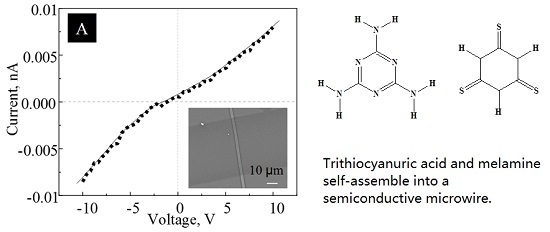
Figure 5 shows the conductivity of a single TPA•M microwire at 25 °C. The microwire shows a semi-conductive property in dry N2 at 25 °C. The conductivity originates from electron transportation. Out of five tested samples, two TPA•M microwires showed undetectable conductivity, i.e. signals not distinguishable from the noises, while the other three showed conductivity from 0.01 mS·cm−1 to 0.017 S·cm−1. This inconsistivity may be caused from defects or cracks in the TPA•M microwires. Avoiding defects is indeed the greatest challenge to develop electronics based on supramolecular systems.
In this short communication, we report the formation of a crystalline, semi-conductive TPA•M microwire by means of a facile self-assembly process and some properties of the resultant microwire. Our current efforts focus on fine-tuning interaction conditions, such as slower evaporation and higher concentrations, in order to grow wider wires for XRD. We are also investigating the orientation of molecules in the wires, the mechanism of the electron conductivity, and the correlation between the conductivity and the structures of the microwires. These studies will be reported in due course.

Acknowledgments

This work is partially supported by the Drexel-SARI Grant.

Author Contributions

Hong Wang conducted most of the experiments; Arben Kojtari wrote part of the introduction and also conducted some work, but figures not included in the paper; Xiaohe Xu took the SEM images. Hai-Feng Ji edited and confirmed the writing.

Conflicts of Interest

The authors declare no conflict of interest.

References

  1. Zimmerman, S.C.; Zeng, F.; Reichert, D.F.C.; Kolotuchin, C.V. Self-assembling dendrimers. Science 1996, 271, 1095–1098. [Google Scholar] [CrossRef] [PubMed]
  2. Schenning, A.P.H.J.; Meijer, E.W. Supramolecular electronics; nanowires from self-assembled π-conjugated systems. Chem. Commun. 2005, 41, 3245–3258. [Google Scholar] [CrossRef] [PubMed]
  3. Oshovsky, G.V.; Reinhoudt, D.N.; Verboom, W. Supramolecular chemistry in water. Angew. Chem. Int. Ed. 2007, 46, 2366–2393. [Google Scholar] [CrossRef] [PubMed]
  4. Nakamura, O.; Kodama, T.; Ogino, J.; Migake, Y. High-conductivity solid proton conductors: Dodecamolybdophosphoric acid and dodecatungstophosphoric acid crystals. Chem. Lett. 1979, 1, 17–18. [Google Scholar] [CrossRef]
  5. Zerkowski, J.A.; Seto, C.T.; Wierda, D.A.; Whitesides, G.M. The design of organic structures in the solid state: Hydrogen-bonded molecular “tapes”. J. Am. Chem. Soc. 1990, 112, 9025–9026. [Google Scholar] [CrossRef]
  6. Seto, C.T.; Whitesides, G.M. Molecular self-assembly through hydrogen bonding: Supramolecular aggregates based on the cyanuric acid-melamine lattice. J. Am. Chem. Soc. 1993, 115, 905–916. [Google Scholar] [CrossRef]
  7. Seto, C.T.; Whitesides, G.M. Self-assembly based on the cyanuric acid-melamine lattice. J. Am. Chem. Soc. 1990, 112, 6409–6411. [Google Scholar] [CrossRef]
  8. Hoeben, F.J.M.; Jonkheijm, P.; Meijer, E.W.; Schenning, A.P.H.J. About supramolecular assemblies of π-conjugated systems. Chem. Rev. 2005, 105, 1491–1546. [Google Scholar] [CrossRef] [PubMed]
  9. Ji, H.F.; Xu, X. Hexagonal organic nanopillar array from the melamine−cyanuric acid complex. Langmuir 2010, 26, 4620–4622. [Google Scholar] [CrossRef] [PubMed]
  10. Lee, W.; Kim, M.S.; Kim, K. Infrared and raman spectroscopic study of terephthalic acid adsorbed on silver surfaces. J. Mol. Struct. 1997, 415, 93–100. [Google Scholar] [CrossRef]
  11. Christian, P.; Isabel, H.; Karl, K.; Eyal, N.; Mattanjah, S.V. Pairing of isolated nucleobases: Double resonance laser spectroscopy of adenine–thymine. ChemPhysChem 2003, 4, 838–842. [Google Scholar]
  12. Habeeb, M.M.; Al-Wakil, H.A.; El-Dissouky, A.; Refat, N.M. Vibrational spectroscopic studies of hydrogen-bonded complexes between 2,5-dihydroxy-P-benzoquinone and amines. Spctroscopy 2001, 15, 33–44. [Google Scholar] [CrossRef]
  13. Kim, D.H.; Han, J.T.; Park, Y.D.; Jang, Y.; Cho, J.H.; Hwang, M.; Cho, K. Single-crystal polythiophene microwires grown by self-assembly. Adv. Mater. 2006, 18, 719. [Google Scholar] [CrossRef]
  14. Pisula, W.; Kastler, M.; Wasserfallen, D.; Pakula, T.; Mullen, K. Exceptionally long-range self-assembly of hexa-peri-hexabenzocoronene with dove-tailed alkyl substituents. J. Am. Chem. Soc. 2004, 126, 8074. [Google Scholar] [CrossRef] [PubMed]
Figure 1. Molecular structure of terephthalic acid (left) and melamine (right).
Crystals 07 00236 g001
Figure 2. A SEM image of 2:1 TPA•M microwires. Insert shows an optical image of the wires.
Crystals 07 00236 g002
Figure 3. Infrared (IR) spectra of melamine, TPA, and TPA•M.
Crystals 07 00236 g003
Figure 4. (A) A cross-polarized optical microscopy (CPOM) image of TPA•M microwires. (B) Brightness versus the angle between the direction of the polarization light and the long axis of a TPA•M microwire.
Crystals 07 00236 g004
Figure 5. I–V curve of a single TPA•M microwire measured in dry nitrogen. Insert shows an optical image of the wires.
Crystals 07 00236 g005