Article

Characteristics of Phomopsis juglandina (Sacc.) Hohn. Associated with Dieback of Walnut in the Climatic Conditions of Southern Romania

by
Cristina Mihaescu
1,*,
Daniel Dunea
2,*,
Adrian Gheorghe Bășa
3,* and
Loredana Neagu Frasin
2
1
Faculty of Sciences, University of Pitesti, Targu din Vale Street 1, 110040 Pitesti, Romania
2
Faculty of Environmental Engineering and Food Science, Valahia University of Targoviste, Aleea Sinaia, no 13, 130004 Targoviste, Romania
3
Faculty of Agriculture, University of Agronomic Sciences and Veterinary Medicine of Bucharest, 59 Marasti Blvd, District 1, 011464 Bucharest, Romania
*
Authors to whom correspondence should be addressed.
Agronomy 2021, 11(1), 46; https://doi.org/10.3390/agronomy11010046
Submission received: 12 November 2020 / Revised: 21 December 2020 / Accepted: 23 December 2020 / Published: 28 December 2020

Abstract

:
Phomopsis juglandina (Sacc.) Höhn., which is the conidial state of Diaporthe juglandina (Fuckel) Nitschke, and the main pathogen causing the dieback of branches and twigs of walnut was recently detected in many orchards from Romania. The symptomatological, morphological, ultrastructural, and cultural characteristics, as well as the pathogenicity of an isolate of this lignicolous fungus, were described and illustrated. The optimum periods for infection, under the conditions prevailing in Southern Romania, mainly occur in the spring (April) and autumn months (late September-beginning of October). Strong inverse correlations (p < 0.001) were found between potential evapotranspiration and lesion lengths on walnut branches in 2019. The pathogen forms two types of phialospores: alpha and beta; the role of beta phialospores is not well known in pathogenesis. In Vitro, the optimal growth temperature of mycelial hyphae was in the range of 22–26 °C, and the optimal pH is 4.4–7. This pathogen should be monitored continuously due to its potential for damaging infestations of intensive plantations.

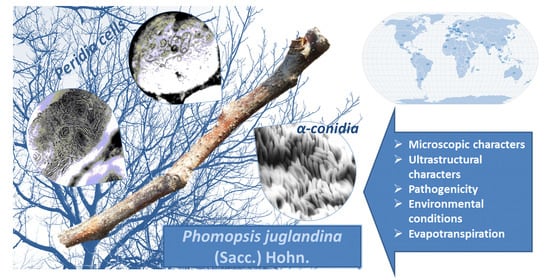
Graphical Abstract

1. Introduction

Although worldwide it does not have the same weight as the culture of apple, peach or plum, walnut (Juglans regia L.) culture plays an important role in fruit growing in the temperate zone. It is highly valued mainly for its fruits, which are eaten both fresh and processed, as well as for the high quality of its wood, both products being in high demand both on the domestic markets and for export. In Romania, it was cultivated since ancient times still attracting a special interest from fruit growers. The development of the walnut cropping on a modern agroecological basis supposes the establishment of commercial intensive plantations in which new, efficient technologies must be applied, thus, ensuring the obtaining of consistent yields. Modifying these ecosystems by intensifying and reducing the biodiversity of biocoenoses requires a radical change of the maintenance technologies, including those of integrated production [1].
Species of the genus Phomopsis belong to the class Coelomycetes, order Sphaeropsidales [2,3]. Their mycoma is of the mycelial type, and the conidioms are of the pycnidial, stromatic type, in which two types of phialospores are usually formed: α and β. Few species of the genus Phomopsis, about 20%, are associated with Diaporthe Nitschke teleomorphs [4,5]. In this case, in the life cycle of these fungi, together with pycnospores, ascospores have an important role, ensuring the perpetuation of pathogens from one year to another and the production of primary infections [1].
Phomopsis species are spread on virtually all continents. The frequency of taxa varies in relation to climatic factors, host susceptibility and, finally yet importantly, to the size of the areas included in the biodiversity studies [6].
The highest abundance of taxa has been reported in Europe and Asia, especially in Germany, England, Italy, Czechia, Slovakia and India, respectively, where mycology studies have been much more extensive compared to other countries in the world [7]. It is noteworthy that some species have also been found in the Scandinavian Peninsula (Sweden and Finland), where annual average temperatures are low. In North America, studies have been conducted mainly on pathogenic species of juniper (Ph. virginiana), conifers (Ph. occulta), vines (Ph. viticola), blueberries (Ph. vaccinii) and elm (Ph. oblonga). In Australia, Africa and South America, the number of described species is lower, probably due to limited mycological research on this pathogen [8,9,10,11].
Most of the parasitic hosts of different Phomopsis species are woody (63%). Their affinity for this habitat is also evidenced by parasitic organs, which are rarely leaves, but most often, juvenile branches. The incidence of these species on branches is undoubtedly related to a certain chemical composition of these organs that does not exist in the leaves [12,13].
Along with the two key diseases caused by the bacterium Xanthomonas campestris pv. juglandis (Pierce) Dye and the fungus Gnomonia leptostyla (Fr.) Ces. et De Not, there has been an increasing incidence of infections caused by the fungus Phomopsis juglandina (Sacc.) Hohn, in both scattered trees, and especially in plantations in different fruit growing areas of Romania (e.g., Pitesti-Maracineni, Geoagiu, Focsani). Disease symptoms include the drying of branches and premature death of saplings leading to increased attention on this pathogen [10,13].
In the phytopathological and mycological literature, the biotypes of Phomopsis identified as wilting agents of walnut branches are attributed to various species of Diaporthe. Thus, Green (1977) reported a 30% loss of Juglans nigra rootstocks in nurseries due to infections caused by Phomopsis elaeagni Sacc. [14]. In Italy, where the presence of the species Diaporthe juglandina (Fuckel) Nitschke was also reported, Vercersi connected Phomopsis sp. on walnut with teleomorphic Diaporthe medusae Nitschke, a species originally described on Cytisus laburnum L. [15].
Current knowledge regarding the Phomopsis genus is largely due to the electron-microscopic investigations performed by a plethora of mycologists [16,17,18]. Yet, compared to other taxonomic groups of fungi, electron-microscopic research on the various taxa of the genus Phomopsis appears sporadic, far from forming a comprehensive picture of the ultrastructural aspects defining the various somatic and reproductive formations in their various ontogenetic stages. Such research seems even more necessary, if we keep in mind that many species are represented exclusively by orphan anamorphs [19,20].
The main aim of the study was to describe and illustrate the symptomatological, morphological, ultrastructural and cultural characteristics as well as the pathogenicity of Phomopsis juglandina. Furthermore, the optimum periods for infection under the conditions prevailing in the south of Romania i.e., Arges region, were established based on field observations correlated with meteorological data.

2. Materials and Methods

Pathogen. Phomopsis juglandina (PJ) was isolated from the infected fruits and branches of walnut (Budeasa 2 variety) collected from Arges region: PJ1 on fruit, PJ2 and PJ3 on branches. The pathogen was isolated from the fruits and grown on differential culture media such as malt agar (MEA: malt extract-25 g, agar-20 g-ROKO, Asturias, Spain) in a vertical laminar flow hood (Asalair 700, ASAL, Cernusco, Italy). In all experiments, incubation was at 23 °C for 21 days. In our studies, five repetitions were used for each variant (PJ) for a correct statistical correlation. The PJ3 isolate was used for carrying out the pathogenicity test and the ultrastructural examinations.
The observations were performed for 2–3 weeks, and the observation sheet included: mycelium growth rate; the appearance of the mycelium during growth and color change; morphology of conidioms: consistency, size, color, grouping mode; type and size of phialospores; and the quantitative ratio between the different types of phialospores. In the current study, a corroboration of the data provided by photonic microscopy (Optech, Oxfordshire OX9 3EX, UK) with those obtained under the electron microscope (TEM- Philips-200 electron acceleration voltage of 80 and 100 kV, SEM JSM 6390) was considered to achieve a better morphological and structural typification of the asexual reproductive organs of the species [21,22].
Microscopic characters. The following steps were performed to obtain the envisaged microscopic preparations: sampling of the biological material; fixing samples in Carnoy mixture; dehydration of the successive material in ethyl alcohol of 70%, 96%, 100%; paraffin embedding and sectioning; deparaffining the sections by inserting the slides into toluene (xylol, benzene) beakers for 3–4 min; hydration of the sections by gradual transition to series of decreasing concentrations of ethyl alcohol (100%, 96%, 70%); staining with cotton blue; dehydration, by passing the slides through a series of: 70% alcohol—2 min, 96% alcohol—2 min, 100% alcohol—3 min; alcohol substitution with toluene: toluene I—1–2 min, toluene II—3–5 min; mounting in Canada conditioner [17].
Ultrastructural characters. Examination using the transmission electron microscope was used in order to highlight ultrastructural features of vegetative hyphae, pycnidia and conidia. The processing of the samples was performed following the working protocol stipulated by the ultrasecting method (inclusion in synthetic resins), to which some adjustments were made. Fixation of the parts was performed in 2% glutaraldehyde, in cacodilate buffer, for one hour, substituted with 3.5% glutaraldehyde for another 3 h, at a temperature of + 4 °C (Kirsch refrigerator). Fixation was interrupted by 3 washes in the same cold swab, followed by 15 min, after which the samples were left overnight in the swab in the refrigerator. The next day, another 4 washes were performed at 30-min intervals. Postfixation was performed in 1% osmic acid for 90 min in the refrigerator and in the dark. The operation was interrupted by 3 successive washes at 15-min intervals; the first two were made with cold 0.1 M cacodilated buffer, and the third with cold, sterile distilled water. Dehydration of the double-fixed samples was performed in the series of alcohols (15% → 100%) up to a concentration of 70%; dehydration was carried out in cold conditions, and then, at room temperature.
Biological samples were maintained at each concentration for 30 min. Absolute alcohol was changed three times. The substitution of alcohol with propylene oxide was performed by successively passing the samples through a series of mixtures of these substances with the following proportion: 3/1; 2/1; 1/1; 1/2; 1/3, and finally in propylene oxide (0/1 × 3). In each mixture, as in propylene oxide, samples were kept for 30 min. The inclusion in the resin was performed by successively passing the samples through a series of mixtures of propylene oxide-resin (Epon-812), with the following proportions: 3/1; 2/1; 1/1; 1/2; 1/3, and finally in pure resin. In each mixture, samples were kept for two hours. The samples processed in this way were included in the resin using special polyester capsules. Prepolymerization was performed at 400 °C for 24 h and polymerization at 600 °C for 48 h. Ultrathin serial sections were performed on the LKB microtome. The ultrasections, which were mounted (collected) on copper and / or gold grids, were contrasted (stained) following the Reynold procedure (with uranyl acetate and lead citrate) and viewed at a TEM-Philips-200 transmission electron microscope [23].
Pathogenicity. The symptoms of the disease were followed in the competition cultures and in the nursery of grafted branches of the Research and Production Institute for Fruit Growing Piteşti-Mărăcineni in 2019. In this experiment, 5 branches of 1 year and 5 of 2 years were infected on each of 10 trees from Budeasa 2 biotype to determine the conditions that define the infection with this fungus. The pathogenicity test was performed in the field on 1 and 2-years old branches of some trees from a local biotype (Budeasa 2). The PJ3 culture fragments, made in Petri dishes, aged 7 days, were applied both on fresh sections of branches and “in windows” with a size of 1.5–2 × 1 cm, made between free and secondary wood and protected for 10 days with a thin polyethylene film. Prior to infection, the bark was disinfected with 70% ethyl alcohol to remove other saprophytic fungi. The evaluation of the lesions was monitored throughout the growing season, in the control variant. The colony fragment was replaced with a sterile culture medium [18].
Environmental conditions. In Argeș region, the annual average temperature was 11.9 °C and the precipitations sum was 634.7 mm during the 2019 year. An uneven distribution of precipitations was observed with an excess of 215 mm in June, and a rainfall deficiency of 309 mm between August and October. Potential evapotranspiration was calculated using the Penman-Monteith algorithm.
Statistical analyses. SPSS software (SPSS Inc., Chicago, IL, USA, 2011) was used to perform data inferences [24]. Correlations between the variables were assessed using the Pearson product-moment correlation (2-tailed level) [25].

3. Results

3.1. Pathogenicity

To evaluate the pathogenicity of the fungus during the summer of the year 2019, two series of tests were performed, on the branches of a local walnut variety. Each tree was artificially infected on five 1-year, and five 2-years branches (n = 50), respectively, and the length of the lesions was measured (Figure 1). Twenty days after inoculation, the lesions characteristic of the disease appeared while the branches that served as control remained intact. After 30 days, on 1-year-old branches inoculated on fresh cross-sections, the lesions were 3.5–4 cm from the site of application of the pathogen, and the appearance of the first pycnidia was observed. In September, about 90 days after inoculation, parts of the branches become infected between 10 and 15 cm (Figure 2). On 1-year-old branches, the infection progressed faster than on older ones. The branches incised in the window showed ellipsoidal lesions of different sizes, while in the control variant the wounds have healed. In the pycnidia formed on the lesion, we observed the formation of alpha and beta phialospores.
The virulence and aggressiveness of the pathogen were correlated with the environmental conditions in the studied area and during the experimental period (Figure 3). The rapid development of necrosis in the open field during the summer and autumn months is correlated with an average of 60% humidity and 19 °C temperature. During the vegetative dormancy, when the ambient temperature is below 0 °C, the necrotic areas grow at a low speed, so the fungus continues to act through a mechanical and enzymatic activity on the bark.
Table 1 shows the correlation coefficients between the environmental factors and lesion lengths on infected walnut branches. Strong inverse correlations (p < 0.001) were found between potential evapotranspiration and lesion lengths on both types of branches. Air temperature showed the same type of association (p < 0.01 for 1-year branches and p < 0.05 for 2-year branches). Precipitations did not correlate with lesion lengths, nor the excess of precipitations above climatic normal, or the rainfall deficiency below climatic normal.

3.2. Cultural Characteristics

The dynamics of the Phomopsis juglandina pathogen was observed using the cultures on the malt-agar medium of some isolates from the branches of Juglans regia. For each isolate, 20 repetitions were used, from which we have selected for presentation the PJ3 isolate being considered representative.
On the malt-agar medium, the PJ3 isolate has formed a whitish, woolly, abundant mycelium, which in 4 days reached a diameter of 2.5/3 cm. After 10 days, the mycelium that occupies the surface of the Petri dish with a diameter of 7.5 cm, started to turn brown, presenting hyphae with thickened walls, with a structure similar to chlamydospores. Also during this period, the first pycnidal buds appeared, which were arranged in concentric circles (Figure 4A). They grew further, and after 3 weeks from inoculation, they become mature pycnidia. They were stromatous, grouped, but well individualized, rarely isolated, with a concentric disposition at the inoculation point. Their wall was thick, pseudoparenchymal, blackish in color, covered up to the upper third by slightly browned mycelial hyphae (Figure 4B).
Both types of phialospores formed in pycnidia. The α-phialospores were ellipsoidal to fusiform, acute at both ends, hyaline, straight, supported, with two or more oily drops, around 6–10 × 2–3 μm. β-phialospores were filiform, curved, sharp at one end, hyaline, without oily drops, 18–28 × 1.1–1.2 μm (Figure 4C,D). Phialospores were included in gelatinous masses, in the form of yellowish streaks, called waxes. It was observed that they contained both types of spores, α and β, not forming separate waxes with α or β spores.
Figure 5 shows that there are no relevant differences in the dynamics of the fungus between repetitions after 3 days of observations, or 7 days and 10 days, respectively, which indicates the accuracy of in vitro observations.

3.3. Ultrastructural Characteristics

3.3.1. Ultrastructure of the Vegetative Hyphae Cells

The general plan of ultrastructural organization of Phomopsis anamorphic hyphae cells is similar to that of cells from different mycelial (filamentous) taxa of fungi with pycnidia. The ultrastructural organization of the cells changes progressively, making the transition from the apical cell of embryonic type, to the fully matured cells, majority in all hyphae, and from these to the senescent ones, always present towards the hyphae, due to permanent transfer of the cytoplasm to the apical areas.
The structural-functional components of a mature cell are the following: I. the cell wall; II. protoplast: A. protoplasm: (a) membrane (plasmalemma); (b) cytoplasm: 1. cytosol (cell matrix + cytoskeleton); 2. organelles: ribosomes, mitochondria, endoplasmic reticulum, Golgi bodies, lysosomes, microbodies, vacuoma, paramural bodies; chitosomes, vialosomes, multivesicular bodies, paramural bodies, concentric bodies; (c) nucleus; B. Ergastic inclusions: glycogen, lipids (Figure 6).
The cell membrane (plasma membrane, plasmalemma) represents the second coating of living cells, essentially a lipo-protein structure, with a molecular organization that coincides with the principles of the fluid mosaic model. It adheres closely, through almost its entire surface, to the cell wall with which it functionally cooperates (Figure 7). Like the cell wall, the plasmalemma is a dynamic envelope, whose properties (ultrastructural, biochemical and functional) change significantly in a programmed way, in relation to the stage of the cell cycle and under the influence of environmental factors [26,27]. In the electron-microscopic images of the ultrasections, the plasmalemma appears in the form of an electron-dense line, with a relief that changes in parallel with the progress of cell differentiation.
In apical cells, the plasmalemma has a rugged, convoluted relief due to extensive exocytosis processes, a process by which carbohydrate and protein polymers are excreted in the periplasmic space that will be integrated into the cell wall structure, contributing to the rapid expansion of its surface and increasing thickness. In subapical cells, the plasmalemma relief attenuates, and in the mature cells below, the plasmalemma has a smooth surface, which denotes the cessation of exocytosis processes, the biogenesis of the cell wall being completed. In the senescent cells at the base, plasmatic hyphae often appear in a state of contiguity and even continuity with endomembrane systems with varying degrees of structural complexity, a state that suggests the role of plasmalemma in their biogenesis.
Like other ultrastructural components, the cytoplasm differs depending on the degree of cell differentiation. In the apical cell, the cytoplasm (cytoplasmic matrix/cytosol) is abundant; hosts a small set of organelles: a nucleus, Golgi bodies and sporadic profiles of the endoplasmic reticulum, all located in the lower third, numerous relatively evenly distributed ribosomes and a consistent population of vesicles of different sizes, located in the upper third of the cell, considered by us to be involved in the exocytosis of parietal polymers. We identified the smallest vesicles as chytosomes. The structure known as the apical body (Spitzenkorper), described from some ascomycetes and deuteromycetes, has not been highlighted.
Cytoribosomes have been shown to be present in all living cells, but the size of the ribosomal population decreases as cytodifferentiation advances. Therefore, they are visible in a very large number in apical cells, in the form of tiny electron-dense granules, scattered randomly or linearly ordered, suggesting the existence of polysomal formations engaged in proteasynthesis.
Mitochondria are absent from the apical cell whose metabolism is supported by the transfer of macroergic products from the subapical cell, where their number is considerable. From some calculations, based on the analysis of serial sections, it results that a mature cell contains between 18 and 24 mitochondrial profiles, with variable dimensions (0.8 × 1.2) 1.5 × 2.3 (3.2 × 4.5) μm. The ultrastructural organization plan coincides with that of the mitochondria from other species of fungi imperfecti, as well as with that of the mitochondria from the conidia. The details will be provided in the next section.
The endoplasmic reticulum (ER) is the main component of the endomembranous system, of any cell type, but with the greatest development in cells undergoing differentiation. It has a rudimentary organization in both apical and senescent cells (Figure 7D). In the latter, it seems to be involved in the biogenesis of autophagic vacuoles, a way in which a “consumption” of its profiles takes place, especially of rough endoplasmic reticulum (RER) tanks.
Some electron-microscopic images reveal the intimate vicinity of Golgi bodies with RE profiles, lysosomes and provacuoles, which suggests some biogenetic relationships and functional cooperation between these components of the endomembranous system. Through the exocytic vesicles they generate, Golgi bodies also participate in the expansion of plasmalemma.
None of the ultrastructural types of peroxisomes, described so far in fungi in general, could be identified in our electro-microscopic preparations.
Some of the vesicles of about 0.1–0.2 μm, delimited by a simple membrane, with an electron-transparent and very stable content, present in the apical and subapical cells, were identified as primary lysosomes based on their proximity to the Golgi cistern and frequent contact, followed by their coalescence with other micro- and macrovessional formations (Figure 7D).
Vacuoles are “true lakes” in the cellular landscape, essential for maintaining an optimal chemical climate for the development of vital processes. Homeostasis of cellular metabolism is largely dependent on the physiology of the vacuole [28]. There is a close correlation between the structure of the vacuoma and the stage of ontogenetic development of the cell (Figure 7D). Similar to other ascomycetes and basidiomycetes, the cells in the hyphal apexes of the species we have investigated contain numerous vesicles / vacuoles spherical or ovoid. The set of these vesicles is called the apical vesicular complex (Figure 7C).
Concentric bodies are cellular organs with a role in the storage of proteins, in a crystalloid form, arranged in concentric layers. Each layer having a different electronic density from the adjacent ones, makes it possible to discriminate concentric bodies very easily. The presence of concentric bodies, even if only sporadic, is a curiosity, because so far they have been highlighted in a very small number of free fungal species; however, they are quite common in lichenizing fungi (Figure 8A,B).
The cells of the somatic hyphae are almost entirely uninucleate, haploid; only very rarely, the dicaryotic cells were observed. Dicariotization relies on two causes: (1) asynchrony between karyokinesis and cytokinesis or (2) a fusion phenomenon between two compatible, uninucleate cells, a phenomenon that triggers the process of parasexuality.

3.3.2. Ultrastructure of the Peridia

The peridia is thick, paraplectic, not very compact, consisting of several layers of iso- or slightly heterodiametric cells. The intercellular spaces are frequent and quite voluminous, representing about 1/5 of the general volume of the period. From the analysis of the serial sections, it results that they are communicating and form within the period a true aerial labyrinth (Figure 9A). The functional significance of these channels filled with air is important from two points of view: it facilitates the passage of air to the pycnidal cavity, while the living cells, conidiogenic, being consuming a large amount of oxygen. Due to the shape of this labyrinth, the construction seems more viable both in terms of mechanical strength and in terms of function, in the sense that it achieves a more efficient thermal insulation, protecting the conidiogenic structures from heat.
The peridia of mature pycnidia, not the aged ones, consist of a mixture of living and dead cells in a ratio of about 2/5. Dead cells have a thin wall, with unequal thickness in its various sectors, always unilayered and high electron-dense (Figure 9B). Electronic density suggests the presence of a considerable amount of lipids. It can be deemed that these lipids are of the type of suberins, sporopolenins and/or cutins from plants. Based on the analysis of living cell phenotypes from juvenile, mature and senescent pycnidia, we concluded that cell death is genetically programmed (it is, therefore, an apoptotic process), occurring after multiple successive changes in cell structure and metabolism, completed by another chemical composition and structure of the cell wall and by a hydrolysis of the cell contents, followed by the transfer of the substances to the neighboring compartment located further inwards.
These cells give the physical-mechanical and chemical resistance necessary for conidiomas.
Living cells have a structure comparable to that of mature cells in the median area of the hyphae. Their number is gradually increasing inwards. We consider that their presence makes it is possible to supply the conidiogenic cells with nutrients until the conidia are fully mature. The terminal cells of such hyphae are conidiophore-generating.
The species in our study, namely Phomopsis juglandina forms two types of conidia: α and β, with a different morphology, ultrastructural organization and physiological potential. In the electronic images of the ultrasections through conidia in various developmental stages, the main organs characteristic of a heterotrophic, dermatoplastic eucite are evident, which have some peculiarities, on which our attention was focused with predilection where the data obtained allowed us to try to make a description in an ontogenetic-evolutionary vision (Figure 9C,D). β-conidia form in the same conidioms as α-conidia. Their simultaneous or successive genesis is still uncertain.

4. Discussion

Most pathogen species reported in Romania are hemibiotrophic [17]. There are also necrotrophic species, secondary or wound pathogens that attack weakened plants, weakened for other reasons of abiotic or biotic nature [15]. In agricultural practice, special importance is given to pathogens in vines, sunflowers, walnuts, strawberries, blueberries, fruit trees (apple, pear, apricot, and almond) and conifers, because they can cause significant qualitative and quantitative crop losses [29]. The virulence and aggressiveness of the different species of Phomopsis reflect their parasitic capacity, and there is often no positive correlation between them. Some pathogens are particularly virulent, but have low aggressiveness (P. elaeagni), while others have low virulence and high aggressiveness (P. malvacearum) [30].
Species of the genus Phomopsis parasitize cultivated and spontaneous woody (63%) and grassy (37%) plants [29]. Some species of Phomopsis are of high socio-economic importance, as they parasitize crop plants particularly valuable for human existence, namely: Phomopsis helianthi on Helianthus annus, Phomopsis viticola on Vitis vinifera, Phomopsis juglandina on various Juglans sp., Phomopsis occulta conifers, Phomopsis perniciosa on Mallus domestica, Phomopsis obscurans on Fragaria sp., Phomopsis inconstans on Juniperus communis [31].
The studies regarding the Phomopsis juglandina characteristics are rarely reported in the literature. The search in Web of Sciences database yielded no results, while the Scopus database provided an article related to in vitro fungicide testing against this pathogen [32]. A comprehensive description of the symptomatological, morphological, ultrastructural and cultural characteristics was provided by the current report, but more research is required to correlate the microclimate influence on the development of the pathogen.
The pathogen Phomopsis juglandina is a primary pathogen, involved in the drying of branches and in the premature loss of nuts, with a wide spreading in Romania. Infections can occur throughout the year, the optimal period of contamination being represented in spring (April) and autumn months (late September-beginning of October), the main gateways of the pathogen being the lesions caused by various abiotic and biotic agents. In this study, we found that evapotranspiration was associated with the length of lesions observed on walnut branches. When potential evapotranspiration rates increase, the lengths of lesions decrease. The same trend but with lower correlation coefficients was present for air temperature. It is important to understand the fungal response to regional climate for the assessment of future fungal production in walnut orchards under various climate change scenarios [33]. The relative humidity of 70% and the temperatures between 20–25 °C favor the development of the pathogen in the walnut plantations. Abundant rainfalls recorded between May and June increased the pathogen’s ability to develop in both spring and autumn.
In this context, Phomopsis jugladina (Sacc.) Hohn. is a particularly virulent wound pathogen. The pathogen can infect fresh wounds throughout the growing season, causing young branches to dry out and perennial ulcers on older ones. In the area of study, the shoots presented infections especially in autumn, following the penetration of the pathogen through the leaf scars and the pedicel of the recently fallen fruits, a process favored by the wet conditions and rainy weather. In most cases, the lesions became visible during the following spring. The fractures of the anamorph were differentiated under the periderm, at first submerged in the tissues, becoming eruptive and visible with the naked eye [34,35]. Despite the careful examination of numerous samples, from different places or taken throughout the year, we were unable to identify a teleomorph (Diaporthe) that could have been associated with the imperfect form of the pathogen [36].
Together with this pathogen, other fungi such as Cytospora juglandina, Diplodia juglandis, Melanconium juglandinum, and Phoma juglandina have been associated with the drying of walnut branches [37]. The lack of phytosanitary treatments, the environmental factors that are favorable to the attack (large variations in temperature and air relative humidity), the wounds on the bark of biotic or abiotic nature lead to the easy installation of these secondary pathogens that can cause significant damage to walnut crops. The identification of these necrotrophs is difficult to achieve due to their similar symptoms on the affected branches. However, applied research at the structural, ultrastructural and pathogenic levels in correlation with detailed monitoring of environmental factors [38] is of major importance for the correct diagnosis of these spheropsidal fungi [36]. Problems related to the management of walnut cancer induced by various secondary pathogens have been presented in [39]. The associated effect of these pathogens should be further studied in conjunction with crop yield effects based on the susceptibility of different parts of the plant, in coordination with favorable environmental conditions. Such an approach may provide important data for disease management strategies and we consider continuing our experiments in the next years to further assess the climatic variability effects on pathogen development [40].
In vitro, we found that Phomopsis juglandina grows and develops well on the malt-agar culture medium, easily forming numerous pycnidia at temperatures between 22 and 24 °C with alpha and beta pycnospores. In future studies we aim to study, in vitro, the ecophysiology of this pathogen corroborated with the efficacy of systemic and contact fungicides [41].
Electron-microscopic investigations (SEM, TEM) required for interpreting the structural features of various biological samples have always had a special character. The main results are images with multiple highlights of these structures, most of them being used as diagnostic in taxonomy: pycnidial conidioms, enteroblastic conidiogenesis, the presence of the two types of alpha and beta phialospores, which is a unique character not found in other types of imperfect fungi. The fungal cells of the investigated specimens had an exterior cell wall, which is a strong, flexible, elastic structure that can be defined as a complex mixture of organic and mineral substances [42]. Under the cell wall, the plasmalemma was outlined as an electron-dense line, with a relief convoluted in apical cells due to the exocytosis processes [43]. In subapical cells and mature cells, the relief acquired a smooth aspect due to the cessation of these processes of cell biogenesis. The cytoplasm looked differently depending on the degree of cell differentiation. The cells of the somatic hyphae of the investigated specimens contained the set of organelles that are characteristic for fungal cells in general, namely: mitochondria, endoplasmic reticulum, Golgi bodies, lysosomes, concentric bodies, vacuoles, and cytoribosomes [44].
Cytoribosomes were visible in a very large number within the apical cells. Mitochondria were missing from the apical cell, and in the mature cells, there were between 18 and 24 mitochondrial profiles. The endoplasmic reticulum from the apical and subapical cells participates together with the Golgi bodies in the formation of secretory vesicles. Although this biogenetic process is of considerable magnitude, RE retains its organization, suggesting that there is a close correlation between the rate of expansion of its profiles and the formation of exocytic vesicles [45]. Golgi bodies were plentiful in apical cells of the expanding hyphae, in which they were located in the basal part, having an important role in the biosynthesis of parietal carbohydrate polymers and in the glycosylation of parietal proteins. In the apical and subapical cells, primary lysosomes were highlighted that will evolve into secondary lysosomes with a role in the processes of cellular phagocytosis [46].
The cells from the hyphal apexes contained numerous vacuoles involved in cellular homeostasis. Despite some detailed analysis, we did not manage to identify chitosomes and filasomes in our electro-microscopic images. Instead, concentric organ bodies have been found that are less common in fungi and are involved in protein storage [47]. The peridia of mature pycnidia, not the aged ones, consisted of a mixture of living and dead cells. The peridia were thick, paraplectenchymatic, having several layers of iso- or slightly heterodiametric cells. Intercellular spaces were frequent and quite voluminous. The pathogen formed two types of conidia (α and β) with different morphology and structural organization [17].

5. Conclusions

In the case of Phomopsis juglandina, the reaction of the environment influences its parasitic properties as well as its life cycle. Both subliminal and supraliminal values create stress conditions, which negatively influence the intimate mechanisms of cell membranes, the expression of certain genes, epigenetic changes, and the activity of certain enzymes. Optimal pH values for growth and development of Phomopsis juglandina are in the range of 4.4–7, so slightly acidic to neutral. On the malt-agar culture medium, the pathogen showed exceptional growth dynamics, also forming abundant pycnidia with the two types of alpha and beta spores at an optimal temperature in the 22–24 °C range. The comparative analysis of the two types of conidia revealed the existence of strong morphological and physiological differences that will be the subject of our future studies.
Electron microscopy research in the genus Phomopsis is rarely mentioned in the literature and thus we consider that the present study is a starting point in the detailed study of this pathogen.The electron-microscopic investigations (e.g., SEM, TEM) are very useful to interpret the structural features of the pathogen in the form of images with multiple highlights of these structures, most of them supporting the diagnostic in taxonomy. It was found that the ultrastructural organizations of the component cells of the somatic structures (vegetative hyphae) or of the reproductive organs (conidioms, conidia) are unitary.

Author Contributions

Conceptualization, C.M. and D.D.; methodology, C.M.; software, D.D. and A.G.B.; validation, C.M.; formal analysis, C.M., D.D., A.G.B. and L.N.F.; investigation, C.M., D.D., A.G.B. and L.N.F.; writing—review and editing, C.M., D.D., A.G.B. and L.N.F. All authors have read and agreed to the published version of the manuscript.

Funding

The work of D.D. was supported by a grant of the Romanian National Authority for Scientific Research, CNDI-UEFISCDI, project number PN-III-1.2-PCCDI-2017-0721.

Institutional Review Board Statement

Not applicable.

Informed Consent Statement

Not applicable.

Data Availability Statement

The data presented in this study are available on request from the corresponding authors. The data are not publicly available due to the potential for further development of research method.

Acknowledgments

The authors thank the two reviewers for comments that consistently improved the manuscript.

Conflicts of Interest

The authors declare no conflict of interest.

References

  1. Botu, I.; Botu, M.; Achim, G. Cultura Nucului in Exploataţii Nucicole Moderne; Phoenix: Bucharest, Romania, 2001; p. 174. ISBN 973-85474-2-3. [Google Scholar]
  2. Index Fungorum. Available online: http://www.indexfungorum.org/names/names.asp (accessed on 1 November 2020).
  3. Kirk, P.M.; Cannon, P.F.; David, J.C.; Stalpers, J.A. Dictionary of the Fungi, 9th ed.; CAB International: Wallingford, UK, 2001. [Google Scholar]
  4. Allescher, A. Rabenhorst’s Kryptogamen-Flora von Deutschland, Oesterreich und der Schweiz–Fungi Imperfecti; Verlag von Eduard Kummer: Leipzig, Germany, 1900; Volume VI, IX. [Google Scholar]
  5. Diego, E.L.F.; Santos, J.M.; Phillips, A.J.L. Phylogeny, morphology and pathogenicity of Diaporthe and Phomopsis species on almond in Portugal. Fungal Divers. 2010, 44, 107–115. [Google Scholar] [CrossRef]
  6. USDA. Index of Plant Diseases in the United States; USDA Agric. Handb.: Washington, DC, USA, 1960; pp. 1–531.
  7. Benschop, K.; Tewari, J.P.; Toop, E.W. Phomopsis twig die-back of some woody interior ornamentals in Alberta. Can. Plant. Dis. Surv. 1984, 64, 29–31. [Google Scholar]
  8. Claydon, N.; Grove, J.F.; Pople, M. Elm bark beetle boring and feeding deterrents from Phomopsis oblonga. Phytochemistry 1985, 24, 937–943. [Google Scholar] [CrossRef]
  9. Farr, D.F.; Castlebury, L.A.; Rossman, A.Y. Morphological and molecular characterization of Phomopsis vaccinii and additional isolates of Phomopsis from blueberry and cranberry in the eastern United States. Mycologia 2002, 94, 494–504. [Google Scholar] [CrossRef] [PubMed]
  10. Georgescu, C.C. Bolile Criptogamice din Pepiniere şi Plantaţii; ICES: Bucharest, Romania, 1955; Volume II, p. 6. [Google Scholar]
  11. Boddy, L.; Griffith, G.S. Role of endophytes and latent invasion in the development of decay communities in sapwood of angiospermous trees. Sydowia 1989, 41, 41–73. [Google Scholar]
  12. Charudattan, R. Current status of biological control of weeds. In Emerging Technologies for Integrated Pest Management: Concepts, Research, and Implementation; Kennedy, G.G., Sutton, T.B., Eds.; APS: St. Paul, MN, USA, 2000; pp. 269–288. [Google Scholar]
  13. Bechet, M.; Silaghi, G.; Turcu, L.; Lörinczi, F. Flora micologică. In Flora şi Vegetaţia Rezervaţiei Naturale Defileul Crişului Repede; Contributii Botanice Universitatea Babes-Bolyai din Cluj-Napoca: Cluj-Napoca, Romania, 1966; Volume I, pp. 31–82. [Google Scholar]
  14. Green, R.J. Dieback of black walnut seedlings caused by Phomopsis elaeagni. Plant. Dis. Rep. 1977, 61, 5825–5884. [Google Scholar]
  15. Vercesi, A.M. Disseccamenti da Phomopsis su noce. Inf. Fitopatol. 1982, 12, 51–54. [Google Scholar]
  16. Kanematsu, S.; Kobayashi, T.; Kudo, A.; Ohtsu, Y. Conidial morphology, pathogenicity and culture characteristics of Phomopsis isolates from peach, Japanese pear and apple in Japan. Ann. Phytopathol. Soc. Jpn. 1999, 65, 264–273. [Google Scholar] [CrossRef]
  17. Mihaescu, C. Ultrastructural aspect of structure somatic and reproductive of the species of Phomopsis (Sacc.) Bubák. Curr. Trends Nat. Sci. 2017, 6, 311–314. [Google Scholar]
  18. Cristescu, C. The morphology and anatomy of structure somatic and reproductive of species of Phomopsis Sacc. Bubak. Bul. Grădinii Bot. Iaşi. 2007, 14, 19–27. [Google Scholar]
  19. Sutton, B.C. The Coelomycetes: Fungi Imperfecti with Pycnidia, Acervuli and Stromata; Commonwealth Mycological Institute: Kew, UK, 1980. [Google Scholar]
  20. Chen, Y.Q.; Jiang, Z. Application of RAPD and ITS region sequence analyses on classification and identification of Phomopsis. Mycosystema 2002, 21, 39–46. [Google Scholar]
  21. Luo, L.J.; Xi, P.G.; Jiang, Z.; Qi, P.K. Sporulation conditions of Phomopsis in pure culture. Mycosystema 2004, 23, 219–225. [Google Scholar]
  22. Uecker, F.A.; Johnson, D.A. Morphology and taxonomy of species of Phomopsis on Asparagus. Mycologia 1991, 83, 192–199. [Google Scholar] [CrossRef]
  23. Montañola-Cvetković, M.; Bojovic-Cvetic, D.; Vukojevic, J. An ultrastructural study of α and β conidia in the fungal genus. Phomopsis. Cryptogam. Mycol. 1985, 6, 171–184. [Google Scholar]
  24. Botu, I.; Botu, M. Metode şi Tehnici de Cercetare în Pomicultură; Editura Conphys: Rm. Valcea, Romania, 1997; p. 327. ISBN 973-9334-08-03. [Google Scholar]
  25. SPSS Tutorials: Home. Available online: https://libguides.library.kent.edu/SPSS/PearsonCorr (accessed on 1 November 2020).
  26. Bartnicki-Garcia, S. Role of Vesicle in Apical Growth and a New Mathematical Model of Hyphal Morphogenesis; Heath, I.B., Ed.; Academic: San Diego, CA, USA, 1990; pp. 211–232. [Google Scholar]
  27. Coppin, E.; Debuchy, R.; Arnaise, S.; Picard, M. Mating types and sexual development in filamentous ascomycetes. Microbiol. Mol. Biol. Rev. 1997, 61, 411–428. [Google Scholar] [CrossRef] [PubMed]
  28. Hess, W.M. Fungal Organelles and other Cell Structures; Academic Press: New York, NY, USA, 1981. [Google Scholar]
  29. Uecker, F.A. A world list of Phomopsis names with notes on nomenclature, morphology and biology. Mycol. Mem. 1988, 13, 1–231. [Google Scholar]
  30. Guido, M.A.D.; Pollastro, S.; Carlucci, A.; Angelini, R.M.D.M.; Faretra, F. Phomopsis viticola is easily transformed with hph and Bmlr genes. J. Plant. Pathol. 2003, 85, 43–52. [Google Scholar]
  31. Udayanga, D.; Liu, X.; McKenzie, E.H.C.; Chukeatirote, E.; Bahkali, A.H.A.; Hyde, K.D. The genus Phomopsis: Biology, applications, species concepts and names of common phytopathogens. Fungal Divers. 2011, 50, 189–225. [Google Scholar] [CrossRef]
  32. Simon, F.; Fischl, G.; Kadlicskó, S.; Pintér, C.S.; Dankó, J.; Süle, S. Phytopatological problems and solutions in the walnut orchards along Lake Balaton. Commmun. Agric. Appl. Biol. Sci. 2007, 72, 765–770. [Google Scholar]
  33. Ágreda, T.; Águeda, B.; Olano, J.M.; Vicente-Serrano, S.M.; Fernández-Toirán, M. Increased evapotranspiration demand in a Mediterranean climate might cause a decline in fungal yields under global warming. Glob. Chang. Biol. 2015, 21, 3499–3510. [Google Scholar] [CrossRef] [Green Version]
  34. Kendrick, W.B. The Fifth Kingdom, 3rd ed.; Focus Publishing, R. Pullins Company Newburyport: Newburyport, MA, USA, 2000. [Google Scholar]
  35. Hilario, S.; Amaral, I.A.; Goncalves, M.F.M.; Lopes, A.; Santos, L.; Alves, A. Diaporthe species associated with twig blight and dieback of Vaccinium corymbosum in Portugal, with description of four new species. Mycologia 2020, 112, 293–308. [Google Scholar] [CrossRef] [PubMed]
  36. León, M.; Berbegal, M.; Rodríguez-Reina, J.M.; Elena, G.; Abad-Campos, P.; Ramón-Albalat, A.; Olmo, D.; Vicent, A.; Luque, J.; Miarnau, X.; et al. Identification and Characterization of Diaporthe spp. Associated with Twig Cankers and Shoot Blight of Almonds in Spain. Agronomy 2020, 10, 1062. [Google Scholar] [CrossRef]
  37. Eichmeier, A.; Pecenka, J.; Spetik, M.; Necas, T.; Ondrasek, I.; Armengol, J.; León, M.; Berlanas, C.; Gramaje, D. Fungal Trunk Pathogens Associated with Juglans regia in the Czech Republic. Plant. Dis. 2020, 104, 761–771. [Google Scholar] [CrossRef] [PubMed]
  38. Oprea, M.; Dunea, D. SBC-MEDIU: A multi-expert system for environmental diagnosis. Environ. Eng. Manag. J. 2010, 9, 205–213. [Google Scholar] [CrossRef]
  39. Michailides, T.J. Botryopshaeria, Phomopsis and Anthracnose Management in Walnuts; Quad-County Walnut: Davis, UC, USA, 2019. [Google Scholar]
  40. Dunea, D.; Dincă, N. Improving land utilization using intensive grass-clover mixtures in forage production systems. Rom. Agric. Res. 2014, 31, 147–158. [Google Scholar]
  41. Sittisart, P.; Yossan, S.; Prasertsan, P. Antifungal property of chili, shallot and garlic extracts against pathogenic fungi, Phomopsis spp., isolated from infected leaves of para rubber (Hevea brasiliensis Muell. Arg.). Agric. Nat. Resour. 2017, 51, 485–491. [Google Scholar] [CrossRef]
  42. Wessels, J.G.H. Ultrastructural analysis of hyphal tip cell growth in fungi: Spitzenkorper, cytoskeleton and endomembranes after freeze-substitution. J. Cell Sci. 1981, 48, 89–103. [Google Scholar]
  43. Howard, R.J. Wall growth, protein excretion and morphogenesis in fungi. New Phytol. 1993, 3, 397–413. [Google Scholar]
  44. Vargas, M.; Aronson, J.M.; Roberson, R.W. The cytoplasmic organization of hyphal tip cell in the fungus Allomyces macrogynus. Protoplasma 1993, 176, 43–52. [Google Scholar] [CrossRef]
  45. Cole, L.; Orlovich, D.; Ashfors, A.E. Structure, function and motility of vacuoles in Filamentous Fungi. Fungal Genet. Biol. 1998, 24, 86–100. [Google Scholar] [CrossRef]
  46. Lousa, C.M.; Denecke, J. Lysosomal and vacuolar sorting: Not so different after all! Biochem. Soc. Trans. 2016, 44, 891–897. [Google Scholar] [CrossRef] [PubMed] [Green Version]
  47. Griffiths, H.B.; Greenwood, A. The concentric bodies of lichenized fungi. Biology 2004, 87, 285–302. [Google Scholar] [CrossRef]
Figure 1. Phomopsis juglandina (Sacc.) Höhn. (A)—attack on fruit; (B)—the stage with abundant pycnidia on the branches, 30 days after inoculation.
Agronomy 11 00046 g001
Figure 2. The development of the lesions (cm) from the date of inoculation of the branches with Phomopsis juglandina.
Agronomy 11 00046 g002
Figure 3. Meteorological data recorded in 2019 (monthly averages), potential evapotranspiration and the periods with the highest frequency of Phomopsis juglandina (red arrows) in the area of study (data obtained from the meteorological station of the Institute of Research and Development for Pomiculture Maracineni).
Agronomy 11 00046 g003
Figure 4. Phomopsis juglandina: (A) 21-days culture on malt agar; (B) P—mature pycnidia; Py—young pycnidia; M—mycelium; (C) Morphology (SEM) of alfa-conidia 2850x; (D) drawings of α and β conidia.
Agronomy 11 00046 g004
Figure 5. Dynamics of the pathogen Phomopsis juglandina on malt agar (x-axis shows the repetition number).
Agronomy 11 00046 g005
Figure 6. Details regarding the ultrastructural organization of some sectors from the subapical cells of some vegetative hyphae (transversal section): L—lipids; Lz—lysosomes; mt—mitochondria; N—nucleus; Pc—cell wall; Pl—plasmalema; RE—endoplasmic reticulum; V—vacuole; (bar—1 µm).
Agronomy 11 00046 g006
Figure 7. Electron-microscopic images of longitudinal (A,B) and transversal (C,D) ultrasections through the apical area of some vegetative hyphae. (A): Pc—cell wall; Pl—plasmalema; (bar-1µm). (B): N—nucleus; n—nucleol; mt—mitochondria; V—vacuole; (bar-1µm). (C): Fm—myelin formations (concentric membranes); Lo—lomasomes; Va—autophagic vacuole; L—lipids. (D): RE—endoplasmic reticulum; Va—autophagic vacuole; L—lipids; Lz—lisosome; (bar-500 nm).
Agronomy 11 00046 g007
Figure 8. Electron-microscopic appearance of concentric bodies in the median cells of some vegetative (transversal ultrasections). (A): Fm—myelin formations (concentric membranes); V—vacuole; Cc—concentric bodies; (bar-400 nm). (B): Cc—concentric bodies; (bar-200 nm).
Agronomy 11 00046 g008
Figure 9. The ultrastructural organization of the pycnidia peridia—(A,B) longitudinal sections: Cm—dead cells; Spe—intercellular spaces, (bar = 10 µm); Cv—living cells; (bar = 10 µm); (C,D) —Cross-sections and longitudinal sections through α-conidia in different ontogenetic stages: α-c-young α-conidia, Pc—cellular wall, L—lipids, Co—enteroblastic conidiogenesis; (bar = 1 µm).
Agronomy 11 00046 g009
Table 1. Correlation matrix of the environmental factors and lesion lengths on walnut branches; ETo—potential evapotranspiration, P (mm) -precipitations, T (°C)—average temperature, Exc (mm)—excess of precipitations above climatic normal, Def (mm)—rainfall deficiency below climatic normal, LL1Y—lesion length on 1-year branches and LL2Y—lesion length on 2-year branches; correlation is significant at the 0.01 2-tailed level (**) and 0.05 (*); σ is highlighted in italic fonts; statistically significant correlations were highlighted in bold fonts.
EToP (mm)T (°C)Exc (mm)Def (mm)LL1YLL2Y
ETo10.5030.949 **0.0860.530−0.986 **−0.962 **
-0.2500.0010.8540.2210.0000.001
P (mm)-10.3900.807 *−0.411−0.617−0.622
--0.3870.0280.3600.1400.136
T (°C)--10.0380.613−0.909 **−0.870 *
---0.9360.1440.0050.011
Exc (mm)---1−0.499−0.229−0.284
----0.2540.6210.538
Def (mm)----1−0.427−0.428
-----0.3390.338
LL1Y-----10.988 **
------0.000
LL2Y------1
-------
Publisher’s Note: MDPI stays neutral with regard to jurisdictional claims in published maps and institutional affiliations.