Article

Quantification of Hypochlorite in Water Using the Nutritional Food Additive Pyridoxamine

by
Kamilla M. S. Kaarsholm
1,
Argyro Kokkoli
1,2,
Eleni Keliri
1,3,
Paul D. Mines
1,4,
Maria G. Antoniou
3,
Mogens Havsteen Jakobsen
5 and
Henrik R. Andersen
1,*
1
Department of Environmental Engineering, Technical University of Denmark, Bygningstorvet, Building 115, 2800 Kongens Lyngby, Denmark
2
FUJIFILM Diosynth Biotechnologies, Biotek Allé 1, 3400 Hillerød, Denmark
3
Department of Chemical Engineering, Cyprus University of Technology, Limassol 3036, Cyprus
4
International and Intelligent Society Division, Sweco Danmark A/S, Ørestads Boulevard 41, 2300 København, Denmark
5
Department of Chemistry, Technical University of Denmark, Produktionstorvet, Building 423, 2800 Kongens Lyngby, Denmark
*
Author to whom correspondence should be addressed.
Water 2021, 13(24), 3616; https://doi.org/10.3390/w13243616
Submission received: 22 November 2021 / Revised: 10 December 2021 / Accepted: 10 December 2021 / Published: 16 December 2021
(This article belongs to the Special Issue Healthy Recreational Waters: Sanitation and Safety Issues)

Abstract

:
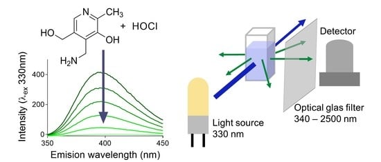
Chlorine is a widely used disinfectant and oxidant used for an array of municipal and industrial applications, including potable water, swimming pools, and cleaning of membranes. The most popular method to verify the concentration of free chlorine is the colorimetric method based on DPD (N, N-diethyl-p-phenylenediamine), which is fast and reasonably cheap, but DPD and its product are potentially toxic. Therefore, a novel, environmentally friendly colorimetric method for the quantification of residual chlorine based on the food additive pyridoxamine (4-(aminomethyl)-5-(hydroxymethyl)-2-methylpyridin-3-ol) was investigated. Pyridoxamine is a B6 vitamin with an absorption maximum at 324 nm and fluorescence emission at 396 nm. Pyridoxamine reacts rapidly and selectively with free chlorine, resulting in a linear decrease both in absorbance and in emission, giving therefore calibration curves with a negative slope. The pyridoxamine method was successfully applied for the quantification of free chlorine from 0.2 to 250 mg/L. Using 1 cm cuvettes, the limit of quantification was 0.12 mg Cl2/L. The pyridoxamine and the DPD methods were applied to actual environmental samples, and the deviation between results was between 4% and 9%. While pyridoxamine does not react with chloramine, quantification of monochloramine was possible when iodide was added, but the reaction is unfavourably slow.

Graphical Abstract

1. Introduction

The need for disinfection of water has long been recognized as paramount in industrialized society, and it can be implemented through multiple routes. These include chemical agents (e.g., chlorine, bromine, ozone, etc.), physical agents (e.g., heat, UV, and sound waves), mechanical means (e.g., filtration and sedimentation), and gamma rays from cobalt-60 isotopes, although irradiation from high-energy sources is neither practical nor commercially in use. Chlorination, in particular, is a common and effective disinfectant used for potable water, wastewater and swimming pools [1,2], as it provides multiple mechanisms of disinfection. These include oxidation, reactivity with available chlorine, protein precipitation, disruption of cell wall permeability, and hydrolysis. It is generally preferred over other disinfection processes because of its low cost, simplicity, and residual effect [3]. Chlorine has a strong oxidizing potential and kills or inactivates pathogens [4]. Depending on the situation, chlorine is used as chlorine (Cl2), sodium hypochlorite (NaOCl), calcium hypochlorite (Ca(OCl)2), or chlorine dioxide (ClO2). Once in water and depending on the pH and temperature, chlorine exists in the forms of hypochlorous acid (HOCl) and hypochlorite ions (OCl), which are termed as “free chlorine”. If ammonia is present in the water, free chlorine will react with it to form three types of chloramines in successive reactions, monochloramine (NH2Cl), dichloramine (NHCl2), and trichloramine (NCl3). These chloramines also act as disinfectants; however, they are less reactive than free chlorine and are thus more stable and long lasting in an applied area. Chloramines existing by themselves as the only disinfects present are referred to as “combined chlorine residual,” while the sum of free and combined chlorine is described as “total chlorine” [1,5,6]. Disinfection by chlorine is not without its side effects, however, as chloramines have been found to pose a threat for human health when present in high levels in swimming pools [7]. Furthermore, depending on the make-up of the water being disinfected, many harmful disinfection by-products will be formed, including trihalomethanes such as chloroform, haloacetic acids, haloacetonitriles, among others [8].
The quantification of free and combined chlorine in a system is usually monitored online as to ensure adequate dosing, constant disinfection, and regulatory compliance [1,9]. Over the years, various analytical methods have been developed for online measurements, the most frequently applied to be amperometric and colorimetric [9,10], with the latter ones to be more user friendly. Despite their successful market penetration, none of the typically implemented methods are highly specific and completely robust to interferences [9]. Limiting factors such as reaction time, chemical waste generation and treatment, cost, and availability prevented their universal application for all types of water matrices, making the development of novel, rapid, cheap and environmentally friendly methods for chlorine detection a high priority task. Online amperometric methods function based on sensors utilizing electrochemical principles. These sensors consist of two electrodes, an anode and a cathode, that measure the change in current due to the redox reactions of chlorine. However, these types of sensors require frequent calibration and an analytical method for a reference [9]; thus, this sensor is not useful for routine analytical applications, and it is generally only used for online control. Most often, the reference method used is that which is based on N,N-diethyl-p-phenylenediamine (DPD) [9].
Of all the colorimetric analysis methods being used, the DPD method is the most typically employed [5]. This method relies on the reaction of residual chlorine with DPD and the formation of pink-coloured products, which are measured by spectroscopy in the visual range. Free chlorine readily reacts with DPD, while chloramines require the addition of potassium iodide in order to catalyse the reaction. Iodide reacts with chloramines to form triiodide ion, which consequently reacts with DPD [5,9,11]. However, this method has serious disadvantages, in particular, that it requires the use of expensive chemicals and results in the production of a waste stream that cannot be released into the environment without further processing [9].
Herein, we investigate pyridoxamine as an alternative colorimetric reagent for chlorine quantification. Pyridoxamine is a form of vitamin B6 with multiple beneficial mechanisms for the human health since one of its basic actions is to trap reactive oxygen species, [12,13]. The main advantage of making use of the food grade chemical pyridoxamine for chlorine identification compared with DPD is the production of innocuous waste that do not require costly post-treatment for safe disposal. Furthermore, we anticipate that pyridoxamine would be an excellent candidate for chlorine quantification and one worth exploring because it is commercially readily available, inexpensive, and an eatable material. Daumer et al. (2000) used pyridoxamine as a model molecule in their investigation on how reactive oxygen species produced by activated neutrophils and monocytes mediate the loss of collagen and other matrix proteins at sites of inflammation. They found, through absorbance and fluorescence spectroscopy, investigations that hypochlorous acid could oxidize pyridoxamine at acidic and neutral pH. They found that pyridoxamine was consumed by HOCl/OCl to form N-chloramine, which resulted in a decrease of its fluorescence intensity.
In this study, we investigated the reaction of chlorine with pyridoxamine, evaluated its performance, determined the optimum reaction conditions for various quantification ranges and tested the function of the method in different water matrixes containing chlorine, covering an array of potential applications.

2. Materials and Methods

2.1. Reagents

All the chemicals were purchased from Sigma-Aldrich (Brøndby, Denmark). A stock solution of 0.1 mM pyridoxamine was made in 20 mM saline phosphate buffer (P5368, 0.02 M PBS pH 7.4, NaCl 0.276 M, KCl 0.0054 M). Chlorine stock solution was diluted in ultrapure water to a concentration of 1 g Cl2/L and the actual chlorine concentration was determined by titration with sodium thiosulfate (Na2S2O3) which had been standardized against potassium dichromate (4500-Cl B. iodometric method in the standard methods for examination of water and wastewater [14]). The chlorine solution was stored at 5 °C and the concentration of free chlorine was measured by the DPD colorimetric assay (LCK310, Hach, Denmark) on a daily basis. Working solutions of chlorine were prepared by diluting the solution with ultrapure water immediately before experiments. For the conduction of the experiments, pyridoxamine was always diluted in saline phosphate buffer, whereas chlorine was diluted in ultrapure water.

2.2. UV-Vis and Fluorescence Spectroscopy

The spectrophotometric analysis of pyridoxamine was conducted with a Varian Cary 50Bio UV-Vis spectrophotometer (Agilent, Thomastown, Victoria, Australia) and a Cary Eclipse fluorescence spectrophotometer (Agilent, Thomastown, Victoria, Australia). Pyridoxamine was initially analysed in the scanning range of 200–370 nm for UV absorbance, and then further analysis was conducted at the single wavelength of 324 nm. Regarding fluorescence, the reagent was scanned for a range of excitation wavelengths from 270 to 350 nm and emission wavelength 350–500 nm. The fluorescence scanning was conducted in three different voltages: 400, 500 and 800 V to determine the optimal combination of parameters. The highest signal was acquired with emission wavelength 396 nm, excitation wavelength 330 nm, excitation slit 5 nm, emission slit 5 nm and voltage 500 V; therefore, these were the running conditions of the fluorescence spectrophotometer. The sample volume for analysis was 2 mL in a Helma® Analytics Fluorescence Cell, H: 45 mm, Macro 111-QS (Sigm-Aldrich, Müllheim, Germany).

2.3. Method Development

All experiments were conducted under temperature-controlled conditions and by maintaining reagents and samples in a cooling bath at 25 °C.

2.3.1. Linearity

To test for the linearity of the method, in a pyridoxamine solution (100 µM) at pH 7.0 a small amount of solution with high chlorine concentration (10 mM) was added. After each addition, UV absorbance and fluorescence were measured.

2.3.2. pH Effects

The effect of pH in the reaction between pyridoxamine and chlorine was investigated for pH values of 4, 5, 6, 7 and 8, in 25 µM pyridoxamine solutions. The solution pH was adjusted with either NaOH or HCl. Additionally, several free chlorine solutions with concentrations from 0 to 3 mg Cl2/L were prepared. For the fluorescence measurements, 1 mL of each pyridoxamine solution was mixed with 1 mL of each free chlorine solution in the cuvette and then measured.

2.3.3. Stability

To check the stability of the formed complex between pyridoxamine and chlorine, a solution of 25 µM pyridoxamine at pH = 6 was prepared and split in two glass vials. It was then mixed in volume ratio 1:1 with ultrapure water in the first vial and with 1.5 mg/L free chlorine solution in the second one. Several samples of 2 mL were collected from the two mixtures over a period of 65 min and their fluorescence intensity was measured. The chemical stability of pyridoxamine alone in water as well as in presence of free chlorine was observed.

2.3.4. Measuring Range

In order to determine the optimum conditions for each measuring range, free chlorine solutions with different concentrations were prepared and mixed with pyridoxamine of different concentrations or volumes. Overall, four different measuring ranges of Cl2 were designed and determined by fluorescence spectroscopy. The lowest range included solutions of free chlorine with concentration from 0 to 3 mg Cl2/L. For this case, each solution was mixed with pyridoxamine (25 µM, pH 6) in a volume ratio 1:1 and measured in the fluorescence spectrophotometer. Similarly, for the next measuring range, 1 mL of chlorine solutions with concentration from 0 to 6 mg Cl2/L reacted with 1 mL pyridoxamine (50 µM, pH 6). For the third range, 1 mL pyridoxamine 200 µM pH 6 was mixed 1 mL of chlorine solutions with 0 to 20 mg Cl2/L. For the highest measuring range, the free chlorine solutions had concentrations from 0 to 220 mg Cl2/L. In the cuvette, 2 mL pyridoxamine (100 µM pH 6) reacted with 50 µL of each chlorine solution.

2.3.5. Combined Chlorine

Monochloramine was prepared for combined chlorine reaction, by mixing free chlorine and ammonium chloride (NH4Cl) solutions with a molar ratio of 1:5 Cl2:NH4+ in alkalified ultrapure water (pH 8) which should result in a complete reaction within seconds [10]. Three solutions of 18 µM monochloramine were prepared and mixed with pyridoxamine (25 µM, pH 6) in a volume ratio 1:1. Since the reaction between the reagents is expected to be slow, KI was added to accelerate the reaction. The effect of the concentration of KI in the rate of the reaction was investigated, by adding KI at a concentration of 0–600 µM KI. The volume of the added KI was insignificant to change the concentration of the reagents. The reaction was monitored over time by collecting 2 mL samples from the mixtures and measuring the fluorescence over a 1 h period.

2.3.6. Method Application in Field Samples

The chlorine concentration in samples of different water types (i.e., water from a membrane cleaning-in-place process, from a public swimming pool, and drinking water) was quantified using the developed pyridoxamine method. Depending on expected free chlorine concentration, the optimum conditions determined in Section 2.3.4 were applied. Background absorbance and fluorescence of all samples was measured.

3. Results

3.1. UV-Vis Absorption and Fluorescence Spectroscopy

Pyridoxamine absorbs in the UV range (Figure 1a) with local maximum at 218, 251 and 324 nm but no absorbance in the visible range; hence, its solution is colourless. Pyridoxamine also has fluorescence properties, and the highest fluorescence intensity was found at excitation at 330 nm with emission at 396 nm. Similar findings have been reported by Daumer et al. [13].
To investigate the reaction of chlorine with pyridoxamine, a small amount (4–24 µL) of a high concentration chlorine was added to the solution of pyridoxamine (2 mL). This way the total volume change was negligible as after several additions the maximal volume increase was less than 2%. Following chlorine addition, mixing, and the measured spectroscopically, a decrease in the absorbance was observed, which was stable for at least 65 min (data not shown). The reaction of chlorine with pyridoxamine was fast and was completed in less than one minute. The addition of chlorine resulted in a decrease of the UV absorbance spectra at all three peaks (Figure 2a) as well as a decrease of the fluorescence intensity when exciting at 330 nm (Figure 2b).
Based on the above, it was decided to plot the absorbance at 324 nm against the added amount of chlorine, resulting a linear correlation (Figure 2c). The slope has a negative sign as the signal decreased with increasing chlorine addition, which is opposite to the DPD method where the signal increases with increasing chlorine concentration. When developing a colorimetric kit with a negative slope, the precision of the in initial value (without chlorine) is of high importance and may pose a challenge. However, there are commercially available colorimetric kits that have a negative slope, such as Ozone Accuvac from Hach, Denmark.
When using DPD method for chlorine determination, the absence of signal does not necessarily mean the absence of chlorine, as it has been reported that at high chlorine to DPD ratios, chlorine can react with the oxidized DPD resulting in sample decolourisation and give a false negative [5]. However, the main benefit of a quantification method based on a negative slope is that in the presence of any chlorine concentration the signal drops immediately and if not, then there is no chlorine in the sample. This also means that, in high chlorine concentrations, the sample will have to be diluted to fit the quantification range but at least the presence of chlorine will not be questionable.

3.1.1. Effect of pH

The effect of pH on the reaction of chlorine with pyridoxamine was investigated next (Figure 3). The fluorescence intensity in the sample without chlorine was pH dependent. This is due to different protonated forms of pyridoxamine. Pyridoxamine has three pKa values (3.1, 7.9 and 10.3) determined by adsorption titration and nuclear magnetic resonance (NMR) experiments [15]. The different protonated forms of pyridoxamine have different adsorption maximum, and the fluorescence spectra are found to be affected by pH [15]. Calibration curves was obtained for pH range between 4–8 and it can be easily concluded that the calibration curve is affected by pH as shown on Figure 3. The optimal pH is around pH 6 to 7 where a high initial fluorescence signal is observed, and the calibration curve gives a steep slope, therefore allowing for more accurate sample quantification.
Furthermore, pH also affected the stoichiometry of the reaction. At pH 7 and 8, no or low fluorescence was measured at 2 mg Cl2/L (28 µmol Cl2/L) which was corresponding to a stoichiometry at 1:1 pyridoxamine to chlorine. At pH 6, the stoichiometry was 1:2 and at lower pH it was close to 1:3.

3.2. Method Optimization and Validation

In this section, the method was optimized for its linearity at four different quantification levels, and the limit of detection (LOD) and quantification of the method (LOQ) was determined for each range.

3.2.1. Quantification Range

Four calibration curves were made with different concentration of pyridoxamine (Figure 4). For the initial sample without chlorine addition, the fluorescence intensity did not double when the pyridoxamine concentration was doubled (e.g., fluorescence intensity was 170 at 25 µM, 290 at 50µM and 520 at 100 µM). This was probably due to self-quenching [16]. However, all four curves showed linear correlation to the amount of added chlorine in sample. Thus, by changing the concentration of pyridoxamine as well as the sample volume, it was possible to quantify chlorine from 0.2 to 250 mg/L.

3.2.2. Determination of LOD and LOQ

To investigate the limit of detection (LOD) and quantification of the method (LOQ), seven replicates of 0.0, 0.1 and 1.0 mg/L chlorine solutions were prepared and quantified with 1 mL of pyridoxamine 25 μM based on the calibration curve in Figure 4a. It was found that the standard deviation decreased with increasing chlorine concentration. The LOD was calculated as three times standard deviations and was determined to be 0.12, 0.08 and 0.04 mg/L for the samples with 0.0, 0.1 and 1.0 mg/L chlorine, respectively, while LOQ, which was calculated as 10 standard deviations, was 0.42, 0.25 and 0.12 mg/L, respectively.
Other available methods for quantification of chlorine have a little lower LOQ. For example, the kit (LCK310) based on DPD from Hach stated a LOQ at 0.05 mg/L. The same concentration is given as lowest concentration in the calibration curve in the method 4500-Cl G (DPD colorimetric method) in the standard methods for examination of water and wastewater [14]. An alternative method based on ABTS (2,2′-azino-bis(3-ethylbenzothiazoline-6-sulphonic acid-diammonium salt) has been published by Pinkernell et al. [10], where free chlorine is measured indirectly by determined the total and the combined chlorine. The LOQ of the ABTS method was estimated at 0.04 mg/L.

3.3. Quantification of Chloramines

Besides measuring free chlorine, it would also be useful if the method was able to measure combined chlorine. To investigate the reaction of pyridoxamine for combined chlorine quantification, monochloramine was used as model compound. Therefore, the recovery of monochloramine was calculated based on fluorescence development of a sample containing monochloramine (measured as chlorine 1.2 mg Cl2/L) under different condition tested and calibration curve for chlorine (Figure 5).
Based on the spectra obtained from the reaction between monochloramine and pyridoxamine it is concluded that combined chlorine can react with pyridoxamine. However, the reaction is slow and after 1 h of reaction time the quantification of monochloramine was about 30% of expected for a 1 mg/L chlorine solution. No decrease in fluorescence was measured when measuring within 1 min after mixing with pyridoxamine, while after 10 min of reaction low decrease was observed. Thus, monochloramine will not act as interference when measuring free chlorine in present of monochloramine if the samples is measured immediately after mixing.
When monochloramine is measured using the DPD method, monochloramine reacts with iodide to form triiodide, which then reacts with DPD [5]. The same method was in this paper investigated for pyridoxamine. However, triiodide absorbs light at 324 nm [17] and therefore only the measurement of fluorescence intensity can be used for quantification. At pH 6, the recovery of monochloramine in general was low (20–50%) within 60 min of reaction without significant difference of added amount of iodide (Figure 5). By adding a small amount of iodide, >85% recovery was observed at pH 6.4 and 7.4 after 60 min of reaction, while higher concentration of iodide resulted in faster reaction but lower recovery. With the high concentration of iodide (600 µM), 30% recovery was measured after 1 min and found stable for up to at least 60 min.

3.4. Quantification of Free Chlorine in Different Water Matrices

To verify that reaction of free chlorine with pyridoxamine can lead to a reliable quantification method, the method was tested in three different water matrices where chlorine presence is expected: (A) water during a clean-in-place process of a membrane system in a food preparation facility, (B) swimming pool water from a public facility and (C) tap water. Note that the last one was a chlorine-free tap water sample that had to be spiked with chlorine for the validation of this method.

3.4.1. Membrane Clean-in-Place Monitoring

Samples from a cleaning-in-place (CIP) process of a membrane system in a food production facility in Denmark were received and analysed with pyridoxamine for the quantification of residual chlorine. The CIP treatment began with flushing of the membrane system, followed by the addition of high concentration chlorine to the feed tank with recirculation of the retentate and permeate (Figure 6 insert). During chlorination, the temperature of the water was increased to 50 °C within the first 10 min which was maintained for the following 20 min. After chlorination, the system was flushed before a second chlorination was performed, followed by two times flushing. The water used during the CIP process was demineralized water. Samples from different steps of the cleaning process were collected (Figure 6) and kept in dark in refrigerator until they were analysed.
As far as the analytical procedure followed for these samples, initially the background signal for UV absorbance and fluorescence was measured (prior to pyridoxamine addition), and it was observed that some of the samples had significant adsorption at λ = 324 nm, which interferes with our method. The samples that gave absorbance at A324 were those taken form the tanks during the first flushing of the system before chlorination. Most probably this occurred because of the presence of residuals from the food production that contained organic and inorganic compounds with absorbance in the wavelength range that we tested in. The samples taken from the tank after the chlorination process had no interference with the absorbance wavelengths of pyridoxamine. However, no background fluorescence signal was detected for all the samples collected during the CIP process. Thus, we went on and quantified the concentration of free chlorine, based on the decrease of the fluorescence intensity.
A high amount of chlorine was added (estimated to >170 mg/L) where most of the chlorine was most likely consumed by the organic matter present during the CIP. The method was able to quantify the residual chlorine in samples from the chlorination steps (Figure 6).
Since the membrane system is used in a food production facility, the company has to be sure that no residual chlorine or chlorination by-products can end up in the produced products. Today, it is performed by repeatedly flushing of the system and having an alkaline followed by an acidic cleaning treatment after chlorination. Currently, the company does not use any method to measure the chlorine level during cleaning since DPD is not allowed inside the production area (possible genotoxic compound). However, having more knowledge on the concentration of chlorine could be beneficial when optimizing water consumption of the facility as well as time consumption for flushing. Therefore, a method based on pyridoxamine could be beneficial since it is a food additive and should be easy to handle without any restriction or threat within the production area.

3.4.2. Public Swimming Pool Chlorine Concentration Verification

Public swimming pools are generally required to have free chlorine residual in the pool and due to the reaction with organic matter and photolysis of chlorine constant dosing is preferred [18]. The accepted range of free chlorine in swimming pools is usually 0.4–6 mg/L while the concentration of combined chlorine should not exceed 0.2 mg/L. To be able to dose the appropriate amount of chlorine to the pool water, an online chlorine sensor is needed. A frequent used sensor is the amperometric electrochemical sensor with three-electrode setup: (1) working electrode; (2) counter electrode; (3) reference electrode. However, it is relatively expensive and requires maintenance and frequent calibration checks. In many countries, it is required to be checked at least three times per day to verify the chlorine concentration by an independent analysis, such as the DPD method [19]. An available alternative to the electrode system is online colorimetry with DPD; however, it is expense to apply, and it requires high consumption of hazardous chemicals resulting in a toxic waste stream that needs to be properly handled leading to additional costs for the facility.
In order to verify the method on a real sample, a water sample was collected from a public swimming pool in the Capital region of Denmark. The sample was brought immediately to the laboratory for analysis. First, the background absorbance and fluorescence were measured. The pool water had no absorbance at λ = 324 nm as well as no fluorescence signal at λexcitation/emission 330/396 nm was observed. Fluorescence determination was chosen for the analysis of the pool samples by mixing 1 mL sample with 1 mL pyridoxamine solution (200 µM, pH 6). The concentration was determined to be 0.58 mg/L, while the DPD kit gave 0.56 mg/L. Thus, there was a good agreement of the two methods.

3.4.3. Chlorination of Drinking Water

Chlorination is a standard method that many countries around the globe implement to disinfect water intended for drinking purposes. Disinfection reduces pathogenic microorganisms to levels that are safe for environmental and public health and align with the regulations on the water quality standards. However, in Denmark, the drinking water supply is based entirely on groundwater which only needs simple treatment before it is released into the distribution system. In most cases, aeration and rapid sand filtration is sufficient treatment, but in all cases the water is not chlorinated before entering the distribution system. Thus, the tap water collected from the campus of the Technical University of Denmark did not contain chlorine, and its absorbance and emission spectra were obtained in order to verify no interference with pyridoxamine. Regardless, the background signal was measured for the unchlorinated tap water, and it had no UV absorbance at λ = 324 nm, while a low signal for fluorescence was observed corresponding to 0.5% of initial fluorescence signal. To ensure the lack of chlorine from tap water both DPD method and fluorescence was utilized concluding that the sample was chlorine-free.
A chlorine stock solution was added to the tap water sample to prepare a 1 mg/L chlorinated solution. The reaction of 1 mL pyridoxamine solution (200 µM, pH 6) and 1 mL of chlorinated sample resulted a reduction of the emission peak at 396 nm equal to 1.2 mg/L chlorine while the DPD method gave 1.1 mg/L. The samples were measured only by fluorescence spectrophotometer at excitation wavelength of 330 nm and emission of 396 nm. The method seems to have a good coherence and verifies the application in different water types.

3.5. Construction of a Free Chlorine Meter

Spectrophotometers for absorption measurements are well known apparatus which can be found in many laboratories working on environmental science. The spectrophotometer can also be found as portable meters for field measurements, both as handheld meter for a specific quantification method and as a bit bigger multimethod meter. In Figure 7a, a conceptual drawing of a photometer is given.
Fluorescence spectrophotometer is a meter available in research laboratories but is not found as frequently as absorption spectrophotometers. Its working principle is based on fluorescence where a fluorescent light is emitted from a sample after an excitation with a Xenon flash lamp. Both excitation and emission wavelengths can be optimized to a single wavelength by collecting the fluorescence fingerprint of the compound in interest. In this case the optimal wavelength found to be λexcitation/emission 330/396 nm which can be fixed on a portable meter as a method for measuring chlorine as shown on Figure 7b.
Fluorescence is more specific when the sample can excite and emit at a specific wavelength than absorbance of a sample. In general, there is a lower risk of interference when using specific excitation/emission wavelength. However, some types of water might have fluorescence at this combination of excitation and emission wavelength [20]; for example, river water and recycled water have been reported to have some fluorescence signal while potable water had almost no signal [20]. If the sample is coloured or has a high UV absorbance, then a high level of self-quenching is expected. However, the initial level of fluorescence can easily be determined by removing chlorine with a reducing agent such as thiosulfate measuring the natural background fluorescence before measuring the chlorine content using the pyridoxamine method.
With the commercial availability in the last decade of cheap LED light sources which emit light in a narrow range of the UV spectra of typically only 10 nm new simple photometers and fluorometers which operate at a fixed wavelength can be constructed cheaply as illustrated in Figure 7. A photometer using the pyridoxamine method based in the absorbance mode would require a cuvette made of quarts and a LED emitting at 324 nm and a simple photometer diode (Figure 7a). A fluorometer could be made from similar components if the photodetector were placed at an angle to the light path and the window of the cuvette on the emission side were made of optical glass which cuts off UV light below about 340 nm (Figure 7b).

4. Conclusions

To conclude, this study focused on the reaction of pyridoxamine with free and combined chlorine and demonstrated that pyridoxamine has the potential to be a highly specific reagent for the determination of chlorine concentration in many different water applications. Our method, using a food compatible chemical, does not require any hazardous reagents or complex procedures and it does not have a demanding waiting time for the analysis to be completed. It is convenient, inexpensive, and rapid and is an environmentally friendly method. More importantly, the proposed method using pyridoxamine with maximum emission wavelength at 396 nm can be easily monitored with a standard fluorescence spectrophotometer, available in most laboratories. Moreover, we envision a portable device that can be set up with fixed wavelengths based on a pyridoxamine method for having a fast, easy, and portable chlorine meter. This would be of a great value for monitoring chlorine in many different systems such as drinking water, swimming pools, cleaning of membranes and chlorine as a wastewater effluent.

Author Contributions

Conceptualization, H.R.A.; methodology, H.R.A., K.M.S.K. and M.H.J.; validation, A.K., P.D.M. and E.K.; investigation, A.K., P.D.M. and E.K.; writing—original draft preparation, K.M.S.K. and A.K.; writing—review and editing, K.M.S.K., P.D.M., M.G.A., M.H.J. and H.R.A.; visualization, K.M.S.K. and A.K.; supervision, H.R.A., K.M.S.K. and M.G.A.; funding acquisition, H.R.A. All authors have read and agreed to the published version of the manuscript.

Funding

This research received no external funding.

Acknowledgments

The authors thank the swimming pool and the food production facility for delivering samples to test our method.

Conflicts of Interest

The authors declare no conflict of interest.

References

  1. Bergendahl, J.A.; Stevens, L. Oxidation reduction potential as a measure of disinfection effectiveness for chlorination of wastewater. Environ. Prog. 2005, 24, 214–222. [Google Scholar] [CrossRef]
  2. Cheema, W.A.; Manasfi, T.; Kaarsholm, K.M.; Andersen, H.R.; Boudenne, J.-L. Effect of medium-pressure UV-lamp treatment on disinfection by-products in chlorinated seawater swimming pool waters. Sci. Total Environ. 2017, 599–600, 910–917. [Google Scholar] [CrossRef] [PubMed] [Green Version]
  3. Abbas, S.; Hashmi, I.; Rehman, M.S.U.; Qazi, I.A.; Awan, M.A.; Nasir, H. Monitoring of chlorination disinfection by-products and their associated health risks in drinking water of Pakistan. J. Water Health 2015, 13, 270–284. [Google Scholar] [CrossRef] [PubMed] [Green Version]
  4. Tchobanoglous, G.; Burton, F.L.; Stensel, H.D. Wastewater Engineering, Treatment and Reuse, 4th ed.; Metcalf & Eddy Inc.: Boston, MA, USA; McGraw Hill: New York, NY, USA, 2003. [Google Scholar]
  5. Harp, D.L. Current Technology of Chlorine Analysis for Water and Wastewater; Hach Company: Loveland, CO, USA, 2002. [Google Scholar]
  6. Ibanez, J.G.; Balderas-Hernandez, P.; Garcia-Pintor, E.; Espinosa-Marvan, L.; Ruiz-Martin, R.M.; Arrieta, J.J.; Ramirez-Silva, M.T.; Casillas, N. Microscale environmental chemistry. Part 8: Properties, preparation and characterization of chloramines. Chem. Educ. 2006, 11, 402–405. [Google Scholar]
  7. Kaydos-Daniels, S.C.; Beach, M.J.; Shwe, T.; Magri, J.; Bixler, D. Health effects associated with indoor swimming pools: A suspected toxic chloramine exposure. Public Health 2008, 122, 195–200. [Google Scholar] [CrossRef] [PubMed]
  8. Boorman, G.A. Drinking water disinfection byproducts: Review and approach to toxicity evaluation. Environ. Health Perspect. 1999, 107, 207–217. [Google Scholar] [CrossRef] [PubMed]
  9. Malkov, V.B.; Zachman, B.; Gordon, S. Comparison of On-line Chlorine Analysis Methods and Instrumentation Built on Amperometric and Colorimetric Technologies. In Proceedings of the AWWA Water Quality Conference and Exposition, Seattle, WA, USA, 15–19 November 2009; pp. 1–22. [Google Scholar]
  10. Pinkernell, U. Methods for the photometric determination of reactive bromine and chlorine species with ABTS. Water Res. 2000, 34, 4343–4350. [Google Scholar] [CrossRef]
  11. Moberg, L.; Karlberg, B. An improved N,N′-diethyl-p-phenylenediamine (DPD) method for the determination of free chlorine based on multiple wavelength detection. Anal. Chim. Acta 2000, 407, 127–133. [Google Scholar] [CrossRef]
  12. Voziyan, P.A.; Hudson, B. Pyridoxamine as a multifunctional pharmaceutical: Targeting pathogenic glycation and oxidative damage. Cell. Mol. Life Sci. 2005, 62, 1671–1681. [Google Scholar] [CrossRef] [PubMed]
  13. Daumer, K.M.; Khan, A.U.; Steinbeck, M.J. Chlorination of Pyridinium Compounds. J. Biol. Chem. 2000, 275, 34681–34692. [Google Scholar] [CrossRef] [PubMed] [Green Version]
  14. APHA; AWWA; WEF. Standard Methods for the Examination of Water and Waste Water, 22nd ed.; American Public Health Association: Washington, DC, USA, 2012. [Google Scholar]
  15. Bueno, C.; Encinas, M.V. Photophysical and Photochemical Studies of Pyridoxamine. Helvetica Chim. Acta 2003, 86, 3363–3375. [Google Scholar] [CrossRef]
  16. Skoog, D.A.; West, D.M.; Holler, F.J. Fundamentals of Analytical Chemistry, 7th ed.; Harcourt, Inc.: San Diego, CA, USA, 1996. [Google Scholar]
  17. Awtrey, A.D.; Connick, R.E. The Absorption Spectra of I2, I3-, I-, IO3-, S4O6= and S2O3=. Heat of the Reaction I3- = I2 + I-. J. Am. Chem. Soc. 1951, 73, 1842–1843. [Google Scholar] [CrossRef]
  18. World Health Organisation. Guidelines for Safe Recreational Water Environments. In Swimming Pools and Similar Environments; WHO Press: Geneva, Switzerland, 2006; Volume 2. [Google Scholar]
  19. Pool Water Treatment Advisory Group. Swimming Pool Water: Treatment and Quality Standards for Pools and Spas, 2nd ed.; Countrywide Publications: Suffolk, UK, 2009. [Google Scholar]
  20. Hambly, A.C.; Henderson, R.K.; Baker, A.; Stuetz, R.M.; Khan, S.J. Cross-connection detection in Australian dual reticulation systems by monitoring inherent fluorescent organic matter. Environ. Technol. Rev. 2012, 1, 67–80. [Google Scholar] [CrossRef]
Figure 1. Absorption spectra and structure (a) and fluorescence fingerprint ((b) red, high intensity; blue, low intensity) of pyridoxamine (100 mM, pH 7.0).
Water 13 03616 g001
Figure 2. UV absorbance spectra (a) and fluorescence emission spectra with excitation at 330 nm (b) of pyridoxamine solution (100 µM, pH 7.0) dosed with different amount of chlorine. Linearity of absorbance at 324 nm (c) and fluorescence emission signal at λex = 330 nm and λem = 396 nm (d) with chlorine addition to a solution of pyridoxamine (100 µM, pH 7.0). The R2 was in both cases >0.998. Fluorescence intensity on vertical axis shown with arbitrary units.
Water 13 03616 g002
Figure 3. Calibration curves at different pH values measured by fluorescence spectrophotometer (1 mL pyridoxamine 25 µM, 1 mL sample, λex 330 nm, λem 396 nm). The R2 was >0.97 for all lines. Fluorescence intensity on vertical axis shown with arbitrary units.
Water 13 03616 g003
Figure 4. Calibration curves, measured by fluorescence spectrophotometer, with different concentrations and volumes of pyridoxamine and chlorine. (a) 1 mL pyridoxamine 25 µM and 1 mL sample, (b) 1 mL pyridoxamine 50 µM and 1 mL sample, (c) 1 mL pyridoxamine 200 µM and 1 mL sample, and (d) 2 mL pyridoxamine 100 µM and 50 µL sample. (pH 6, λex 330 nm, λem 396 nm, R2 > 0.98). Fluorescence intensity on vertical axis shown with arbitrary units.
Water 13 03616 g004
Figure 5. Recovery of monochloramine based on fluorescence measurements at different pH (pH 6.0 (a), pH 6.4 (b), and pH 7.4 (c)) values with different amount of rotation iodide added (25 µM pyridoxamine and 17.6 µM (=1 mg Cl2/L) monochloramine, ratio 1:1, λex 330 nm, λem 396 nm).
Water 13 03616 g005
Figure 6. Concentration of free chlorine in samples acquired from the cleaning process in the food industry. Insert (stipple box): Schematic of a membrane system and flow during recycling of water in the cleaning process.
Water 13 03616 g006
Figure 7. Conceptual drawing of the construction of meter for absorbance measurement (a) and fluorescence measurement (b).
Water 13 03616 g007
Publisher’s Note: MDPI stays neutral with regard to jurisdictional claims in published maps and institutional affiliations.