Next Issue
Volume 12, February
Previous Issue
Volume 11, December


Diagnostics, Volume 12, Issue 1 (January 2022) – 226 articles

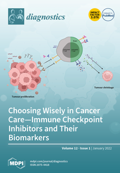
Cover Story (view full-size image): Immune checkpoint inhibitors are monoclonal antibodies which unmask cancer to the immune system by blocking negative feedback signalling in T cells. These drugs have revolutionised cancer care and prolonged survival and increased cure rates in many malignancies. However, they are expensive, cause autoimmune toxicity and help only 40% of patients who receive them. The Healey Bird et al. review licensed tissue biomarkers of response and their limitations as well as emerging tissue, blood, and stool biomarkers which may help to predict both response and toxicity as a new generation of immune checkpoint inhibitors becomes available in cancer care. View this paper.
  • Issues are regarded as officially published after their release is announced to the table of contents alert mailing list.
  • You may sign up for e-mail alerts to receive table of contents of newly released issues.
  • PDF is the official format for papers published in both, html and pdf forms. To view the papers in pdf format, click on the "PDF Full-text" link, and use the free Adobe Reader to open them.
Order results
Result details
Section
Select all
Export citation of selected articles as:
11 pages, 632 KiB  
Article
Comparison of HE4, CA125, ROMA and CPH-I for Preoperative Assessment of Adnexal Tumors
by Núria Carreras-Dieguez, Ariel Glickman, Meritxell Munmany, Georgina Casanovas, Núria Agustí, Berta Díaz-Feijoo, Adela Saco, Beatriz Sánchez, Lydia Gaba, Martina Aida Angeles, Jaume Pahisa, Esther Fernández-Galán, Aureli Torné and Pere Fusté
Diagnostics 2022, 12(1), 226; https://doi.org/10.3390/diagnostics12010226 - 17 Jan 2022
Cited by 15 | Viewed by 3662
Abstract
(1) OBJECTIVE: To assess the performance of CA125, HE4, ROMA index and CPH-I index to preoperatively identify epithelial ovarian cancer (EOC) or metastatic cancer in the ovary (MCO). (2) METHODS: single center retrospective study, including women with a diagnosis of adnexal mass. We [...] Read more.
(1) OBJECTIVE: To assess the performance of CA125, HE4, ROMA index and CPH-I index to preoperatively identify epithelial ovarian cancer (EOC) or metastatic cancer in the ovary (MCO). (2) METHODS: single center retrospective study, including women with a diagnosis of adnexal mass. We obtained the AUC, sensitivity, specificity and predictive values were of HE4, CA125, ROMA and CPH-I for the diagnosis of EOC and MCO. Subgroup analysis for women harboring adnexal masses with inconclusive diagnosis of malignancy by ultrasound features and Stage I EOC was performed. (3) RESULTS: 1071 patients were included, 852 (79.6%) presented benign/borderline tumors and 219 (20.4%) presented EOC/MCO. AUC for HE4 was higher than for CA125 (0.91 vs. 0.87). No differences were seen between AUC of ROMA and CPH-I, but they were both higher than HE4 AUC. None of the tumor markers alone achieved a sensitivity of 90%; HE4 was highly specific (93.5%). ROMA showed a sensitivity and specificity of 91.1% and 84.6% respectively, while CPH-I showed a sensitivity of 91.1% with 79.2% specificity. For patients with inconclusive diagnosis of malignancy by ultrasound features and with Stage I EOC, ROMA showed the best diagnostic performance (4) CONCLUSIONS: ROMA and CPH-I perform better than tumor markers alone to identify patients harboring EOC or MCO. They can be helpful to assess the risk of malignancy of adnexal masses, especially in cases where ultrasonographic diagnosis is challenging (stage I EOC, inconclusive diagnosis of malignancy by ultrasound features). Full article
Show Figures

Figure 1

14 pages, 3879 KiB  
Article
AI Denoising Significantly Improves Image Quality in Whole-Body Low-Dose Computed Tomography Staging
by Andreas S. Brendlin, David Plajer, Maryanna Chaika, Robin Wrazidlo, Arne Estler, Ilias Tsiflikas, Christoph P. Artzner, Saif Afat and Malte N. Bongers
Diagnostics 2022, 12(1), 225; https://doi.org/10.3390/diagnostics12010225 - 17 Jan 2022
Cited by 17 | Viewed by 2880
Abstract
(1) Background: To evaluate the effects of an AI-based denoising post-processing software solution in low-dose whole-body computer tomography (WBCT) stagings; (2) Methods: From 1 January 2019 to 1 January 2021, we retrospectively included biometrically matching melanoma patients with clinically indicated WBCT staging from [...] Read more.
(1) Background: To evaluate the effects of an AI-based denoising post-processing software solution in low-dose whole-body computer tomography (WBCT) stagings; (2) Methods: From 1 January 2019 to 1 January 2021, we retrospectively included biometrically matching melanoma patients with clinically indicated WBCT staging from two scanners. The scans were reconstructed using weighted filtered back-projection (wFBP) and Advanced Modeled Iterative Reconstruction strength 2 (ADMIRE 2) at 100% and simulated 50%, 40%, and 30% radiation doses. Each dataset was post-processed using a novel denoising software solution. Five blinded radiologists independently scored subjective image quality twice with 6 weeks between readings. Inter-rater agreement and intra-rater reliability were determined with an intraclass correlation coefficient (ICC). An adequately corrected mixed-effects analysis was used to compare objective and subjective image quality. Multiple linear regression measured the contribution of “Radiation Dose”, “Scanner”, “Mode”, “Rater”, and “Timepoint” to image quality. Consistent regions of interest (ROI) measured noise for objective image quality; (3) Results: With good–excellent inter-rater agreement and intra-rater reliability (Timepoint 1: ICC ≥ 0.82, 95% CI 0.74–0.88; Timepoint 2: ICC ≥ 0.86, 95% CI 0.80–0.91; Timepoint 1 vs. 2: ICC ≥ 0.84, 95% CI 0.78–0.90; all p ≤ 0.001), subjective image quality deteriorated significantly below 100% for wFBP and ADMIRE 2 but remained good–excellent for the post-processed images, regardless of input (p ≤ 0.002). In regression analysis, significant increases in subjective image quality were only observed for higher radiation doses (≥0.78, 95%CI 0.63–0.93; p < 0.001), as well as for the post-processed images (≥2.88, 95%CI 2.72–3.03, p < 0.001). All post-processed images had significantly lower image noise than their standard counterparts (p < 0.001), with no differences between the post-processed images themselves. (4) Conclusions: The investigated AI post-processing software solution produces diagnostic images as low as 30% of the initial radiation dose (3.13 ± 0.75 mSv), regardless of scanner type or reconstruction method. Therefore, it might help limit patient radiation exposure, especially in the setting of repeated whole-body staging examinations. Full article
(This article belongs to the Special Issue Novel Approaches in Oncologic Imaging)
Show Figures

Figure 1

11 pages, 2734 KiB  
Article
Artificial Intelligence Application in Assessment of Panoramic Radiographs
by Łukasz Zadrożny, Piotr Regulski, Katarzyna Brus-Sawczuk, Marta Czajkowska, Laszlo Parkanyi, Scott Ganz and Eitan Mijiritsky
Diagnostics 2022, 12(1), 224; https://doi.org/10.3390/diagnostics12010224 - 17 Jan 2022
Cited by 36 | Viewed by 7976
Abstract
The aim of this study was to assess the reliability of the artificial intelligence (AI) automatic evaluation of panoramic radiographs (PRs). Thirty PRs, covering at least six teeth with the possibility of assessing the marginal and apical periodontium, were uploaded to the Diagnocat [...] Read more.
The aim of this study was to assess the reliability of the artificial intelligence (AI) automatic evaluation of panoramic radiographs (PRs). Thirty PRs, covering at least six teeth with the possibility of assessing the marginal and apical periodontium, were uploaded to the Diagnocat (LLC Diagnocat, Moscow, Russia) account, and the radiologic report of each was generated as the basis of automatic evaluation. The same PRs were manually evaluated by three independent evaluators with 12, 15, and 28 years of experience in dentistry, respectively. The data were collected in such a way as to allow statistical analysis with SPSS Statistics software (IBM, Armonk, NY, USA). A total of 90 reports were created for 30 PRs. The AI protocol showed very high specificity (above 0.9) in all assessments compared to ground truth except from periodontal bone loss. Statistical analysis showed a high interclass correlation coefficient (ICC > 0.75) for all interevaluator assessments, proving the good credibility of the ground truth and the reproducibility of the reports. Unacceptable reliability was obtained for caries assessment (ICC = 0.681) and periapical lesions assessment (ICC = 0.619). The tested AI system can be helpful as an initial evaluation of screening PRs, giving appropriate credibility reports and suggesting additional diagnostic methods for more accurate evaluation if needed. Full article
(This article belongs to the Special Issue Artificial Intelligence in Oral Health)
Show Figures

Figure 1

15 pages, 4011 KiB  
Article
The Evaluation of the Four-Chamber Cardiac Dissection Method of the Fetal Heart as an Alternative to Conventional Inflow–Outflow Dissection in Small Gestational-Age Fetuses
by Camelia Albu, Adelina Staicu, Roxana Popa-Stanilă, Cosmina Bondor, Bogdan Pop, Liviu Chiriac, Dan Gheban, Romeo Micu, Romulus Valeriu Flaviu Turcu, Simion Simon, Doinița Crișan and Florin Stamatian
Diagnostics 2022, 12(1), 223; https://doi.org/10.3390/diagnostics12010223 - 17 Jan 2022
Cited by 5 | Viewed by 2689
Abstract
The examination of very small fetal hearts requires special equipment and a specialist that are not available in many general pathology laboratories. Compared to conventional examination, the four-chamber cardiac dissection (4CCD) method can be performed by any pathologist using instruments generally available in [...] Read more.
The examination of very small fetal hearts requires special equipment and a specialist that are not available in many general pathology laboratories. Compared to conventional examination, the four-chamber cardiac dissection (4CCD) method can be performed by any pathologist using instruments generally available in pathology services. The aim of this study is to evaluate the efficiency of the 4CCD method in the examination of small fetal hearts using post-mortem magnetic resonance imaging (pm-MRI) at 7T as the standard. Twelve fetuses with gestational ages between 13 and 19 weeks have been included in this study. All fetuses underwent pm-MRI examination prior to pathologic examination. The 4CCD method was used for the cardiac examination in all cases following the same guidelines for cardiac sectioning. The 4CCD was able to identify all cardiac anatomic structures as compared to pm-MRI at 7T, demonstrating a sensibility of 95.8% (95% CI, 94.5–95.8) and specificity of 100% (95% CI, 32.3–100). The overall accuracy in identifying cardiac anatomic structures was 95.8% (95% CI, 93.4–95.8). Additionally, the 4CCD method was able to detect cardiac anomalies with an overall diagnostic accuracy of 91% (95% CI, 85.8–94.2), sensibility of 67.6% (95% CI, 54.5–75.3), and specificity of 97% (95% CI, 93.7–99) as compared to pm-MRI at 7T. The four-chamber view dissection method can be considered as an alternative to the conventional inflow–outflow dissection method in selected cases. Full article
(This article belongs to the Section Medical Imaging and Theranostics)
Show Figures

Figure 1

11 pages, 1242 KiB  
Article
Comparison of FDG PET/CT and Bone Marrow Biopsy Results in Patients with Diffuse Large B Cell Lymphoma with Subgroup Analysis of PET Radiomics
by Eun Ji Han, Joo Hyun O, Hyukjin Yoon, Seunggyun Ha, Ie Ryung Yoo, Jae Won Min, Joon-Il Choi, Byung-Ock Choi, Gyeongsin Park, Han Hee Lee, Young-Woo Jeon, Gi-June Min and Seok-Goo Cho
Diagnostics 2022, 12(1), 222; https://doi.org/10.3390/diagnostics12010222 - 17 Jan 2022
Cited by 11 | Viewed by 2619
Abstract
Whether FDG PET/CT can replace bone marrow biopsy (BMBx) is undecided in patients with diffuse large B cell lymphoma (DLBCL). We compared the visual PET findings and PET radiomic features, with BMBx results. A total of 328 patients were included; 269 (82%) were [...] Read more.
Whether FDG PET/CT can replace bone marrow biopsy (BMBx) is undecided in patients with diffuse large B cell lymphoma (DLBCL). We compared the visual PET findings and PET radiomic features, with BMBx results. A total of 328 patients were included; 269 (82%) were PET-negative and 59 (18%) were PET-positive for bone lesions on visual assessment. A fair degree of agreement was present between PET and BMBx findings (ĸ = 0.362, p < 0.001). Bone involvement on PET/CT lead to stage IV in 12 patients, despite no other evidence of extranodal lesion. Of 35 discordant PET-positive and BMBx-negative cases, 22 (63%) had discrete bone uptake on PET/CT. A total of 144 patients were eligible for radiomic analysis, and two grey-level zone-length matrix derived parameters obtained from the iliac crests showed a trend for higher values in the BMBx-positive group compared to the BMBx-negative group (mean 436.6 ± 449.0 versus 227.2 ± 137.8, unadjusted p = 0.037 for high grey-level zone emphasis; mean 308.8 ± 394.4 versus 135.7 ± 97.2, unadjusted p = 0.048 for short-zone high grey-level emphasis), but statistical significance was not found after multiple comparison correction. Visual FDG PET/CT assessment and BMBx results were discordant in 17% of patients with newly diagnosed DLBCL, and the two tests are complementary in the evaluation of bone involvement. Full article
(This article belongs to the Special Issue The Diagnosis and Treatment of Lymphomas)
Show Figures

Figure 1

9 pages, 1294 KiB  
Article
Feasibility of Recombinant Human TSH as a Preparation for Radioiodine Therapy in Patients with Distant Metastases from Papillary Thyroid Cancer: Comparison of Long-Term Survival Outcomes with Thyroid Hormone Withdrawal
by Hsi-Chen Tsai, Kung-Chu Ho, Shih-Hsin Chen, Jing-Ren Tseng, Lan-Yan Yang, Kun-Ju Lin, Ju-Chin Cheng and Miaw-Jene Liou
Diagnostics 2022, 12(1), 221; https://doi.org/10.3390/diagnostics12010221 - 17 Jan 2022
Cited by 7 | Viewed by 2364
Abstract
Background: this study was designed to compare the long-term survival outcomes of patients prepared for radioiodine (RAI) therapy using either thyroid hormone withdrawal (THW) or recombinant human thyrotropin (rhTSH) stimulation, by specifically focusing on cases with distant metastases from papillary thyroid cancer (PTC). [...] Read more.
Background: this study was designed to compare the long-term survival outcomes of patients prepared for radioiodine (RAI) therapy using either thyroid hormone withdrawal (THW) or recombinant human thyrotropin (rhTSH) stimulation, by specifically focusing on cases with distant metastases from papillary thyroid cancer (PTC). Methods: A retrospective analysis was performed on 88 patients with distant metastases from PTC. Fifty-one and thirty-seven patients were prepared for RAI treatment by either THW or rhTSH stimulation, respectively. The primary endpoints were progression-free survival (PFS) and disease-specific survival (DSS). Results: The 10-year DSS rates of patients prepared for RAI therapy using either THW or rhTSH stimulation were 62.2% and 73.3%, respectively. Using multivariate analysis, RAI-avid metastases (p = 0.025) and preparation with rhTSH (p = 0.041) were identified as independent prognostic factors for PFS. Notably, PFS in the group of patients with RAI-avid metastases and preparation with rhTSH was significantly better than that in the other groups (p = 0.025). Conclusions: Preparation for RAI therapy using rhTSH stimulation is not inferior to THW preparation in terms of long-term survival outcomes experienced by patients with PTC and distant metastasis. Patients with RAI-avid metastases and preparation with rhTSH had the most favorable PFS. Full article
(This article belongs to the Topic Cancer Biology and Therapy)
Show Figures

Figure 1

8 pages, 2144 KiB  
Article
Immunohistochemical Expression Patterns of CD45RO, p105/p50, JAK3, TOX, and IL-17 in Early-Stage Mycosis Fungoides
by Tariq N. Aladily, Tasnim Abushunar, Ahmad Alhesa, Raneen Alrawi, Noor Almaani and Maram Abdaljaleel
Diagnostics 2022, 12(1), 220; https://doi.org/10.3390/diagnostics12010220 - 17 Jan 2022
Cited by 4 | Viewed by 2274
Abstract
The morphologic changes in early-stage mycosis fungoides (MF) might overlap with benign inflammatory dermatitis (BID). Previous studies have described altered expression patterns of several proteins in MF, but their diagnostic significance is uncertain. This study aims at examining the frequency of expression of [...] Read more.
The morphologic changes in early-stage mycosis fungoides (MF) might overlap with benign inflammatory dermatitis (BID). Previous studies have described altered expression patterns of several proteins in MF, but their diagnostic significance is uncertain. This study aims at examining the frequency of expression of CD45RO, NFkB-p105/p50, JAK3, TOX, and IL-17 proteins by immunohistochemistry. The cohorts included 21 patients of early-stage MF and 19 with benign BID as a control group. CD45RO was positive in all patients of MF and BID. NFkB-p105/p50 showed normal cytoplasmic staining, indicating an inactive status in all patients of both groups. JAK3 was positive in 3 (14%) MF and in 17 (89%) BID patients (p = 0.003). TOX was expressed in 19 (90%) and 13 (68%) patients of MF and BID, respectively (p = 0.120). IL-17 was detected in 13 (62%) MF and in 7 (37%) BID patients (p = 0.056). Co-expression of TOX and IL-17 was seen in 11 (52%) MF patients but in only 3 (16%) BID patients, which was statistically significant (p = 0.021). We conclude that a double expression of TOX and IL-17 may support the diagnosis of MF in the right clinicopathologic setting, while none of the immunohistochemical stains alone provided a significant discrimination between MF and BID. Full article
(This article belongs to the Topic Cancer Biology and Therapy)
Show Figures

Figure 1

12 pages, 3158 KiB  
Article
Evaluation of Clinical and Immunohistochemical Factors Relating to Melanoma Metastasis: Potential Roles of Nestin and Fascin in Melanoma
by Yumiko Yamamoto, Yoshihiro Hayashi, Hideyuki Sakaki and Ichiro Murakami
Diagnostics 2022, 12(1), 219; https://doi.org/10.3390/diagnostics12010219 - 17 Jan 2022
Cited by 3 | Viewed by 2158
Abstract
For melanoma treatment, an early diagnosis and a complete resection of the primary tumor is essential. In addition, detection of factors that may be related to metastasis is indispensable. A total of 30 Japanese patients with Stage I or II melanoma, diagnosed according [...] Read more.
For melanoma treatment, an early diagnosis and a complete resection of the primary tumor is essential. In addition, detection of factors that may be related to metastasis is indispensable. A total of 30 Japanese patients with Stage I or II melanoma, diagnosed according to the classification of the American Joint Committee on Cancer, are included in this study. Clinical background (sex, onset age, primary tumor area, existence of remaining cancer cells at the resected tissue margin, and treatment after the primary surgery) and immunohistochemical staining (Nestin and Fascin) on the resected tissue were examined to detect factors statistically related to metastasis. The analysis result has shown that older onset age and positive immunohistochemical expressions of Nestin and Fascin are statistically related to metastasis. To facilitate meticulous observation of Nestin and Fascin expression at different timing (e.g., onset and metastasis), double immunofluorescence staining was performed. Nestin is a class VI intermediate filament protein, initially detected in neural stem cells. Fascin is an actin-bundling protein which regulates cell adhesion, migration and invasion. Nestin and Fascin are suggested to relate to melanoma metastasis, however, the potential role of Fascin is controversial. Analysis of variations in Fascin expression detected in this study may contribute to further investigations concerning potential roles of Fascin for progression of melanoma. This is the first study to report double immunofluorescent staining of Nestin and Fascin in melanoma. Nestin and Fascin double-positive melanoma cells were detected. Full article
(This article belongs to the Section Pathology and Molecular Diagnostics)
Show Figures

Figure 1

19 pages, 7402 KiB  
Article
Sudden Cardiac Death and Ex-Situ Post-Mortem Cardiac Magnetic Resonance Imaging: A Morphological Study Based on Diagnostic Correlation Methodology
by Giuseppe Bertozzi, Francesco Pio Cafarelli, Michela Ferrara, Nicola Di Fazio, Giuseppe Guglielmi, Luigi Cipolloni, Federico Manetti, Raffaele La Russa and Vittorio Fineschi
Diagnostics 2022, 12(1), 218; https://doi.org/10.3390/diagnostics12010218 - 17 Jan 2022
Cited by 11 | Viewed by 2513
Abstract
During the last years, post-mortem imaging has gradually been assumed within research in the field of forensic pathology. This role appears to be clearly and simply applied in the trauma field with the use of conventional radiography or Post Mortem Computed Tomography (PMCT). [...] Read more.
During the last years, post-mortem imaging has gradually been assumed within research in the field of forensic pathology. This role appears to be clearly and simply applied in the trauma field with the use of conventional radiography or Post Mortem Computed Tomography (PMCT). Recently, particular attention was paid to cardiovascular imaging using Post Mortem Magnetic Resonance Imaging (PMMRI). The present experimental study aims to: (i) confirm the efficacy of a Post Mortem Cardiac Resonance Imaging (PMCRI) study protocol for the study of human hearts collected during the autopsy; (ii) apply the defined protocol on subjects who died of “SCD (sudden cardiac death)”, to identify alterations that could guide subsequent sampling. Two hearts of healthy subjects (A: male 22 years; B: female 26 years), who died from causes other than SCD were collected and compared to hearts that belonged to SCD individuals (C: male, 47 years old; D: female, 44 years old; E: male; 72 years old). The exams were performed on a 1.5 T scanner (Philips Intera Achieva, Best, the Netherlands) on hearts collected during autopsy and after a 30-day formalin fixation. Two readers analyzed the obtained images blindly and after randomization. From the comparison between the data from imaging and the macroscopic and histological investigations carried out, the present study proved the effectiveness of a PMMRI protocol to study ex-situ hearts. Moreover, it suggested the following semeiology in post-mortem SCD cases: the hyperintense area with indistinct margins in the Short Tau Inversion Recovery (STIR) sequence was linked to edema or area of pathological fibers, whereas the hypointense area in the T2-FFE sequences was linked to fibrosis. PMMRI can provide a valuable benefit to post-mortem investigations, helping to distinctly improve the success rate of histological sampling and investigations, which remains the gold standard in the diagnosis of sudden death. Full article
(This article belongs to the Section Pathology and Molecular Diagnostics)
Show Figures

Figure 1

2 pages, 159 KiB  
Editorial
Special Issue “Advancement in Breast Diagnostic and Interventional Radiology”
by Graziella Di Grezia
Diagnostics 2022, 12(1), 217; https://doi.org/10.3390/diagnostics12010217 - 17 Jan 2022
Viewed by 1238
Abstract
A multimodality approach in breast imaging is a unique solution to guarantee to the patient a complete diagnosis [...] Full article
(This article belongs to the Special Issue Advancement in Breast Diagnostic and Interventional Radiology)
19 pages, 6117 KiB  
Case Report
How Many Times Can One Go Back to the Drawing Board before the Accurate Diagnosis and Surgical Treatment of Glucagonoma?
by Carmen Sorina Martin, Ovidiu Dumitru Parfeni, Liliana Gabriela Popa, Mara Madalina Mihai, Dana Terzea, Vlad Herlea, Mirela Gherghe, Razvan Adam, Osama Alnuaimi, Valentin Calu, Adrian Miron, Silvius Negoita, Cornelia Nitipir and Simona Fica
Diagnostics 2022, 12(1), 216; https://doi.org/10.3390/diagnostics12010216 - 16 Jan 2022
Cited by 6 | Viewed by 4967
Abstract
Glucagonomas are neuroendocrine tumors (NETs) that arise from the alpha cells of the pancreatic islets. They are typically slow-growing tumors associated with abnormal glucagon secretion, resulting in one or more non-specific clinical features, such as necrolytic migratory erythema (NME), diabetes, diarrhea, deep vein [...] Read more.
Glucagonomas are neuroendocrine tumors (NETs) that arise from the alpha cells of the pancreatic islets. They are typically slow-growing tumors associated with abnormal glucagon secretion, resulting in one or more non-specific clinical features, such as necrolytic migratory erythema (NME), diabetes, diarrhea, deep vein thrombosis, weight loss, and depression. Here, we report the case of a 44-year-old male with a history of diabetes mellitus, presenting with a pruritic and painful disseminated cutaneous eruption of erythematous plaques, with scales and peripheral pustules, misdiagnosed as disseminated pustular psoriasis and treated for 2 years with oral retinoid and glucocorticoids. During this period, the patient complained of weight loss of 32 kg and diarrhea and developed deep vein thrombosis. These symptoms, together with an inadequate response to therapy of the skin lesions, led to the reassessment of the initial diagnosis. Laboratory tests confirmed elevated plasma glucagon levels (>1000 pg/mL) and computed tomography (CT) scans revealed a 35/44 mm tumor in the pancreatic tail. Due to considerable disease complications and the COVID-19 pandemic, the surgical removal of the tumor was delayed for nearly 2 years. During this time, somatostatin analogue therapy efficiently controlled the glucagonoma syndrome and likely prevented tumor progression. As in other functional pancreatic NETs, the early clinical recognition of hormonal hypersecretion syndrome and the multidisciplinary approach are the keys for best patient management. Full article
(This article belongs to the Special Issue Abdominal Surgical Diseases: Diagnosis, Treatment and Management)
Show Figures

Figure 1

14 pages, 2010 KiB  
Review
Vertical Artifacts in Lung Ultrasonography: Some Common Clinician Questions and the Related Engineer Answers
by Marcello Demi, Natalia Buda and Gino Soldati
Diagnostics 2022, 12(1), 215; https://doi.org/10.3390/diagnostics12010215 - 16 Jan 2022
Cited by 14 | Viewed by 2802
Abstract
Introduction: Vertical artifacts, including B lines, are frequently seen in a variety of lung diseases. Their sonomorphology varies in length, width, shape, and internal reverberations. The reason for this diversity is still unknown and is the cause of discussion between clinicians and ultrasound [...] Read more.
Introduction: Vertical artifacts, including B lines, are frequently seen in a variety of lung diseases. Their sonomorphology varies in length, width, shape, and internal reverberations. The reason for this diversity is still unknown and is the cause of discussion between clinicians and ultrasound physics engineers. Aim: The aim of this work is to sum up the most common clinician observations and provide an explanation to each of them derived from ultrasound physics. Materials and Methods: Based on clinical and engineering experiences as well as data collected from relevant literature, the sonomorphology of vertical artifacts was analyzed. Thirteen questions and answers were prepared on the common sonomorphology of vertical artifacts, current nomenclature, and clinical observations. Conclusions: From a clinical standpoint, the analysis of vertical artifacts is very important and requires that further clinical studies be conducted in cooperation with engineers who specialize in physics. Full article
(This article belongs to the Special Issue Lung Ultrasound: A Leading Diagnostic Tool)
Show Figures

Figure 1

17 pages, 735 KiB  
Systematic Review
Microbial Translocation and Perinatal Asphyxia/Hypoxia: A Systematic Review
by Dimitra-Ifigeneia Matara, Abraham Pouliakis, Theodoros Xanthos, Rozeta Sokou, Georgios Kafalidis, Zoi Iliodromiti, Theodora Boutsikou, Nicoletta Iacovidou and Christos Salakos
Diagnostics 2022, 12(1), 214; https://doi.org/10.3390/diagnostics12010214 - 16 Jan 2022
Cited by 11 | Viewed by 5399
Abstract
The microbiome is vital for the proper function of the gastrointestinal tract (GIT) and the maintenance of overall wellbeing. Gut ischemia may lead to disruption of the intestinal mucosal barrier, resulting in bacterial translocation. In this systematic review, according to PRISMA (Preferred Reporting [...] Read more.
The microbiome is vital for the proper function of the gastrointestinal tract (GIT) and the maintenance of overall wellbeing. Gut ischemia may lead to disruption of the intestinal mucosal barrier, resulting in bacterial translocation. In this systematic review, according to PRISMA (Preferred Reporting Items for Systematic Review and Meta-Analysis) guidelines, we constructed a search query using the PICOT (Patient, Intervention, Comparison, Outcome, Time) framework. Eligible studies reported in PubMed, up to April 2021 were selected, from which, 57 publications’ data were included. According to these, escape of intraluminal potentially harmful factors into the systemic circulation and their transmission to distant organs and tissues, in utero, at birth, or immediately after, can be caused by reduced blood oxygenation. Various factors are involved in this situation. The GIT is a target organ, with high sensitivity to ischemia–hypoxia, and even short periods of ischemia may cause significant local tissue damage. Fetal hypoxia and perinatal asphyxia reduce bowel motility, especially in preterm neonates. Despite the fact that microbiome arouse the interest of scientists in recent decades, the pathophysiologic patterns which mediate in perinatal hypoxia/asphyxia conditions and gut function have not yet been well understood. Full article
(This article belongs to the Special Issue Maternal-Fetal Medicine)
Show Figures

Figure 1

11 pages, 258 KiB  
Article
Resistome and Virulome of Multi-Drug Resistant E. coli ST131 Isolated from Residents of Long-Term Care Facilities in the Northern Italian Region
by Sabrina Cherubini, Mariagrazia Perilli, Anna Maria Azzini, Evelina Tacconelli, Laura Maccacaro, Alda Bazaj, Laura Naso, Gianfranco Amicosante, LTCF-Veneto Working Group, Giuliana Lo Cascio and Alessandra Piccirilli
Diagnostics 2022, 12(1), 213; https://doi.org/10.3390/diagnostics12010213 - 16 Jan 2022
Cited by 13 | Viewed by 2274
Abstract
Long-term care facilities (LTCFs) are important reservoirs of antimicrobial-resistant (AMR) bacteria which colonize patients transferred from the hospital, or they may emerge in the facility as a result of mutation or gene transfer. In the present study, we characterized, from a molecular point [...] Read more.
Long-term care facilities (LTCFs) are important reservoirs of antimicrobial-resistant (AMR) bacteria which colonize patients transferred from the hospital, or they may emerge in the facility as a result of mutation or gene transfer. In the present study, we characterized, from a molecular point of view, 43 E. coli strains collected from residents of LTCFs in Northern Italy. The most common lineage found was ST131, followed by sporadic presence of ST12, ST69, ST48, ST95, ST410 and ST1193. All strains were incubators of several virulence factors, with iss, sat, iha and senB being found in 84%, 72%, 63% and 51% of E. coli, respectively. Thirty of the ST131 analyzed were of the O25b:H4 serotype and H30 subclone. The ST131 isolates were found to be mainly associated with IncF plasmids, CTX-M-1, CTX-M-3, CTX-M-15, CTX-M-27 and gyrA/parC/parE mutations. Metallo-β-lactamases were not found in ST131, whereas KPC-3 carbapenemase was found only in two ST131 and one ST1193. In conclusion, we confirmed the spread of extended-spectrum β-lactamase genes in E. coli ST131 isolated from colonized residents living inside LTCFs. The ST131 represents an incubator of fluoroquinolones, aminoglycosides and other antibiotic resistance genes in addition to different virulence factors. Full article
(This article belongs to the Special Issue Molecular Diagnostics of Antimicrobial Resistance)
19 pages, 1830 KiB  
Article
Development and Validation of an Insulin Resistance Predicting Model Using a Machine-Learning Approach in a Population-Based Cohort in Korea
by Sunmin Park, Chaeyeon Kim and Xuangao Wu
Diagnostics 2022, 12(1), 212; https://doi.org/10.3390/diagnostics12010212 - 16 Jan 2022
Cited by 23 | Viewed by 3397
Abstract
Background: Insulin resistance is a common etiology of metabolic syndrome, but receiver operating characteristic (ROC) curve analysis shows a weak association in Koreans. Using a machine learning (ML) approach, we aimed to generate the best model for predicting insulin resistance in Korean adults [...] Read more.
Background: Insulin resistance is a common etiology of metabolic syndrome, but receiver operating characteristic (ROC) curve analysis shows a weak association in Koreans. Using a machine learning (ML) approach, we aimed to generate the best model for predicting insulin resistance in Korean adults aged > 40 of the Ansan/Ansung cohort using a machine learning (ML) approach. Methods: The demographic, anthropometric, biochemical, genetic, nutrient, and lifestyle variables of 8842 participants were included. The polygenetic risk scores (PRS) generated by a genome-wide association study were added to represent the genetic impact of insulin resistance. They were divided randomly into the training (n = 7037) and test (n = 1769) sets. Potentially important features were selected in the highest area under the curve (AUC) of the ROC curve from 99 features using seven different ML algorithms. The AUC target was ≥0.85 for the best prediction of insulin resistance with the lowest number of features. Results: The cutoff of insulin resistance defined with HOMA-IR was 2.31 using logistic regression before conducting ML. XGBoost and logistic regression algorithms generated the highest AUC (0.86) of the prediction models using 99 features, while the random forest algorithm generated a model with 0.82 AUC. These models showed high accuracy and k-fold values (>0.85). The prediction model containing 15 features had the highest AUC of the ROC curve in XGBoost and random forest algorithms. PRS was one of 15 features. The final prediction models for insulin resistance were generated with the same nine features in the XGBoost (AUC = 0.86), random forest (AUC = 0.84), and artificial neural network (AUC = 0.86) algorithms. The model included the fasting serum glucose, ALT, total bilirubin, HDL concentrations, waist circumference, body fat, pulse, season to enroll in the study, and gender. Conclusion: The liver function, regular pulse checking, and seasonal variation in addition to metabolic syndrome components should be considered to predict insulin resistance in Koreans aged over 40 years. Full article
(This article belongs to the Special Issue Artificial Intelligence Approaches for Medical Diagnostics in Korea)
Show Figures

Figure 1

10 pages, 967 KiB  
Article
Impact of Antibiotic Prophylaxis on Infection Rate after Endoscopic Ultrasound Through-the-Needle Biopsy of Pancreatic Cysts: A Propensity Score-Matched Study
by Antonio Facciorusso, Martha Arevalo-Mora, Maria Cristina Conti Bellocchi, Laura Bernardoni, Daryl Ramai, Paraskevas Gkolfakis, Domenico Loizzi, Nicola Muscatiello, Antonio Ambrosi, Nicola Tartaglia, Carlos Robles-Medranda, Elisa Stasi, Andrew Ofosu and Stefano Francesco Crinò
Diagnostics 2022, 12(1), 211; https://doi.org/10.3390/diagnostics12010211 - 16 Jan 2022
Cited by 4 | Viewed by 2262
Abstract
Background: Despite weak evidence, antibiotic prophylaxis prior to endoscopic ultrasound-guided through-the-needle biopsy (EUS-TTNB) of pancreatic cystic lesions (PCLs) is routinely used in clinical practice. We aim to compare a group of patients treated with antibiotics before EUS-TTNB of PCLs and a group who [...] Read more.
Background: Despite weak evidence, antibiotic prophylaxis prior to endoscopic ultrasound-guided through-the-needle biopsy (EUS-TTNB) of pancreatic cystic lesions (PCLs) is routinely used in clinical practice. We aim to compare a group of patients treated with antibiotics before EUS-TTNB of PCLs and a group who did not undergo antimicrobial prophylaxis. Methods: Out of 236 patients with pancreatic cystic lesions referred to two high-volume centers between 2016 and 2021, after propensity score matching, two groups were compared: 98 subjects who underwent EUS-TTNB under antibiotic prophylaxis and 49 subjects without prophylaxis. Results: There was no difference in terms of baseline parameters between groups. Final diagnosis was serous cystadenoma in 36.7% of patients in the group not treated with prophylaxis and in 37.7% of patients in the control group, whereas IPMN and mucinous cystadenoma were diagnosed in 3 (6.1%) and 16 (32.6%) versus 6 (6.1%) and 32 (32.6%) patients in the two groups, respectively (p = 0.23). Overall, the adverse event rate was 6.1% in the group not treated with antibiotic prophylaxis and 5.1% in the control group (p = 0.49). Only a single infectious adverse event occurred in each group (p = 0.48). The diagnostic yields were 89.7% and 90.8% in the two groups (p = 0.7), and the diagnostic accuracy rate was 81.6% in both groups (p = 1.0). Conclusions: Prophylactic antibiotics do not seem to influence the risk of infection, and their routine use should be discouraged. Full article
(This article belongs to the Special Issue Endoscopic Ultrasound Guided Tissue Sampling of Tumors)
Show Figures

Figure 1

11 pages, 415 KiB  
Article
Cervical Cytology–Histology Correlation Based on the American Society of Cytopathology Guideline (2017) at the Russian National Medical Research Center for Obstetrics, Gynecology, and Perinatology
by Aleksandra Asaturova, Darya Dobrovolskaya, Alina Magnaeva, Anna Tregubova, Guldana Bayramova and Gennady Sukhikh
Diagnostics 2022, 12(1), 210; https://doi.org/10.3390/diagnostics12010210 - 15 Jan 2022
Cited by 8 | Viewed by 2566
Abstract
Recent evidence suggests that a cytology–histology correlation (CHC) with discrepancy detection can both evaluate errors and improve the sensitivity and specificity of the cytologic method. We aimed to analyze the errors in cytologic–histologic discrepancies according to the CHC protocol guideline of the American [...] Read more.
Recent evidence suggests that a cytology–histology correlation (CHC) with discrepancy detection can both evaluate errors and improve the sensitivity and specificity of the cytologic method. We aimed to analyze the errors in cytologic–histologic discrepancies according to the CHC protocol guideline of the American Society of Cytopathology (2017). This retrospective study included 273 patients seen at the National Medical Research Center of Obstetrics, Gynecology and Perinatology (Moscow, Russia) between January 2019 and September 2021. The patients’ mean age was 34 ± 8.1 years. The cytology–histology agreement was noted in 158 cases (57.9%). Major discrepancies were found in 21 cases (7.6%), while minor discrepancies were noted in 93 cases (34.1%). The reason for 13 (4.8%) discrepancies was a colposcopy sampling error and, in 46 (16.8%) cases, the reason was a Papanicolaou (PAP) test sampling error. The discrepancy between primary and reviewed cytology was due interpretive errors in 13 (4.8%) cases and screening errors in 42 (15.4%) cases. We demonstrated that the ASC guidelines facilitate cervical CHC. A uniform application of these guidelines would standardize cervical CHCs internationally, provide a scope for the inter-laboratory comparison of data, and enhance self-learning and peer learning. Full article
(This article belongs to the Special Issue Cyto-Histopathological Correlations in Pathology Diagnostics)
Show Figures

Figure 1

9 pages, 1067 KiB  
Case Report
Isolated Splenic Metastasis of Primary Lung Cancer Presented as Metachronous Oligometastatic Disease—A Case Report
by Milorad Reljic, Boris Tadic, Katarina Stosic, Milica Mitrovic, Nikola Grubor, Stefan Kmezic, Miljan Ceranic and Vladimir Milosavljevic
Diagnostics 2022, 12(1), 209; https://doi.org/10.3390/diagnostics12010209 - 15 Jan 2022
Cited by 4 | Viewed by 2490
Abstract
Modern oncology practice and new antitumor drugs prolonged disease-free intervals in patients with lung cancer. Patients with distant metastatic disease are treated only with palliative intent. The International Association for the Study of Lung Cancer, in the 8th edition of the TNM classification, [...] Read more.
Modern oncology practice and new antitumor drugs prolonged disease-free intervals in patients with lung cancer. Patients with distant metastatic disease are treated only with palliative intent. The International Association for the Study of Lung Cancer, in the 8th edition of the TNM classification, for the first time includes oligometastatic disease as a clinical state that describes the patients with distant metastasis, limited in number and organ sites, who may have more indolent biology. In this paper, we present a case of a 56-year-old man who was admitted to our clinic regarding a radiologically diagnosed splenic lesion of uncertain nature, and who underwent a left upper lobectomy for primary lung cancer 12 years before. After a detailed radiological diagnosis, it was concluded that it is highly suspected metastatic lesion of the spleen and the patient underwent a splenectomy. While no definitive protocols exist on the management of isolated splenic metastasis from lung cancer, splenectomy, in suitable patients, with reasonable survival expectations, improves patient disease-free survival and can prevent potentially life-threatening complications, such as splenic rupture. 18F-FDG PET has very high sensitivity and specificity for differentiating benign and malignant splenic lesions especially in patients who are in the follow up protocol due to primary malignancy. Full article
(This article belongs to the Section Pathology and Molecular Diagnostics)
Show Figures

Figure 1

33 pages, 1228 KiB  
Review
Recent Developments in Phenotypic and Molecular Diagnostic Methods for Antimicrobial Resistance Detection in Staphylococcus aureus: A Narrative Review
by Andrea Sanchini
Diagnostics 2022, 12(1), 208; https://doi.org/10.3390/diagnostics12010208 - 15 Jan 2022
Cited by 25 | Viewed by 7706
Abstract
Staphylococcus aureus is an opportunistic pathogen responsible for a wide range of infections in humans, such as skin and soft tissue infections, pneumonia, food poisoning or sepsis. Historically, S. aureus was able to rapidly adapt to anti-staphylococcal antibiotics and become resistant to several [...] Read more.
Staphylococcus aureus is an opportunistic pathogen responsible for a wide range of infections in humans, such as skin and soft tissue infections, pneumonia, food poisoning or sepsis. Historically, S. aureus was able to rapidly adapt to anti-staphylococcal antibiotics and become resistant to several classes of antibiotics. Today, methicillin-resistant S. aureus (MRSA) is a multidrug-resistant pathogen and is one of the most common bacteria responsible for hospital-acquired infections and outbreaks, in community settings as well. The rapid and accurate diagnosis of antimicrobial resistance in S. aureus is crucial to the early initiation of directed antibiotic therapy and to improve clinical outcomes for patients. In this narrative review, I provide an overview of recent phenotypic and molecular diagnostic methods for antimicrobial resistance detection in S. aureus, with a particular focus on MRSA detection. I consider methods for resistance detection in both clinical samples and isolated S. aureus cultures, along with a brief discussion of the advantages and the challenges of implementing such methods in routine diagnostics. Full article
Show Figures

Figure 1

8 pages, 2661 KiB  
Case Report
Identification of the First Single GSDME Exon 8 Structural Variants Associated with Autosomal Dominant Hearing Loss
by Luke Mansard, Christel Vaché, Julie Bianchi, Corinne Baudoin, Isabelle Perthus, Bertrand Isidor, Catherine Blanchet, David Baux, Michel Koenig, Vasiliki Kalatzis and Anne-Françoise Roux
Diagnostics 2022, 12(1), 207; https://doi.org/10.3390/diagnostics12010207 - 15 Jan 2022
Cited by 8 | Viewed by 2061
Abstract
GSDME, also known as DFNA5, is a gene implicated in autosomal dominant nonsyndromic hearing loss (ADNSHL), affecting, at first, the high frequencies with a subsequent progression over all frequencies. To date, all the GSDME pathogenic variants associated with deafness lead to [...] Read more.
GSDME, also known as DFNA5, is a gene implicated in autosomal dominant nonsyndromic hearing loss (ADNSHL), affecting, at first, the high frequencies with a subsequent progression over all frequencies. To date, all the GSDME pathogenic variants associated with deafness lead to skipping of exon 8. In two families with apparent ADNSHL, massively parallel sequencing (MPS) integrating a coverage-based method for detection of copy number variations (CNVs) was applied, and it identified the first two causal GSDME structural variants affecting exon 8. The deleterious impact of the c.991-60_1095del variant, which includes the acceptor splice site sequence of exon 8, was confirmed by the study of the proband’s transcripts. The second mutational event is a complex rearrangement that deletes almost all of the exon 8 sequence. This study increases the mutational spectrum of the GSDME gene and highlights the crucial importance of MPS data for the detection of GSDME exon 8 deletions, even though the identification of a causal single-exon CNV by MPS analysis is still challenging. Full article
(This article belongs to the Special Issue Genetic Testing for Rare Diseases)
Show Figures

Figure 1

11 pages, 2501 KiB  
Article
Preclinical Validation of a Novel Injection-Molded Swab for the Molecular Assay Detection of SARS-CoV-2
by Chiara E. Ghezzi, Devon R. Hartigan, Justin P. Hardick, Rebecca Gore, Miryam Adelfio, Anyelo R. Diaz, Pamela D. McGuinness, Matthew L. Robinson, Bryan O. Buchholz and Yukari C. Manabe
Diagnostics 2022, 12(1), 206; https://doi.org/10.3390/diagnostics12010206 - 15 Jan 2022
Cited by 4 | Viewed by 2948
Abstract
During the COVID-19 public health emergency, many actions have been undertaken to help ensure that patients and health care providers have timely and continued access to high-quality medical devices to respond effectively. The development and validation of new testing supplies and equipment, including [...] Read more.
During the COVID-19 public health emergency, many actions have been undertaken to help ensure that patients and health care providers have timely and continued access to high-quality medical devices to respond effectively. The development and validation of new testing supplies and equipment, including collection swabs, has helped to expand the availability and capability for various diagnostic, therapeutic, and protective medical devices in high demand during the COVID-19 emergency. Here, we report the initial validation of a new injection-molded anterior nasal swab, ClearTip™, that was experimentally validated in a laboratory setting as well as in independent clinical studies in comparison to gold standard flocked swabs. We have also developed an in vitro anterior nasal tissue model which offers a novel, efficient, and clinically relevant validation tool to replicate the clinical swabbing workflow with high fidelity, while being accessible, safe, reproducible, and time- and cost-effective. ClearTip™ displayed greater inactivated virus release in the benchtop model, confirmed by its greater ability to report positive samples in a small clinical study in comparison to flocked swabs. We also quantified the detection of biological materials, as a proxy for viral material, in multi-center pre-clinical and clinical studies which showed a statistically significant difference in one study and a reduction in performance in comparison to flocked swabs. Taken together, these results emphasize the compelling benefits of non-absorbent injection-molded anterior nasal swabs for COVID-19 detection, comparable to standard flocked swabs. Injection-molded swabs, as ClearTip™, could have the potential to support future swab shortages, due to its manufacturing advantages, while offering benefits in comparison to highly absorbent swabs in terms of comfort, limited volume collection, and potential multiple usage. Full article
Show Figures

Figure 1

8 pages, 1323 KiB  
Article
Assessing SARS-CoV-2 Neutralizing Antibodies after BNT162b2 Vaccination and Their Correlation with SARS-CoV-2 IgG Anti-S1, Anti-RBD and Anti-S2 Serological Titers
by Angélica Ramos, Maria João Cardoso, Luís Ribeiro and João Tiago Guimarães
Diagnostics 2022, 12(1), 205; https://doi.org/10.3390/diagnostics12010205 - 15 Jan 2022
Cited by 12 | Viewed by 2605
Abstract
The humoral response through neutralizing antibodies (NAbs) is a key component of the immune response to COVID-19. However, the plaque reduction neutralization test (PRNT), the gold standard for determining NAbs, is technically demanding, time-consuming and requires BSL-3 conditions. Correlating the NAbs and total [...] Read more.
The humoral response through neutralizing antibodies (NAbs) is a key component of the immune response to COVID-19. However, the plaque reduction neutralization test (PRNT), the gold standard for determining NAbs, is technically demanding, time-consuming and requires BSL-3 conditions. Correlating the NAbs and total antibodies levels, assessed by generalized and automated serological tests, is crucial. Through a commercial surrogate virus neutralization test (sVNT), we aimed to evaluate the production of SARS-CoV-2 NAbs in a set of vaccinated healthcare workers and to correlate these NAbs with the SARS-CoV-2 IgG anti-S1, anti-RBD and anti-S2 serological titers. We found that 6 months after vaccination, only 74% maintain NAbs for the Wuhan strain/UK variant (V1) and 47% maintain NAbs for the South African and Brazil variants (V2). Through Spearman’s correlation, we found the following correlations between the percentage of inhibition of NAbs and the SARS-CoV-2 IgG II Quant (Abbott Laboratories, Chicago, IL, USA) and BioPlex 2200 SARS-CoV-2 IgG Panel (Bio-Rad, Hercules, CA, USA) immunoassays: rho = 0.87 (V1) and rho = 0.73 (V2) for anti-S1 assessed by Abbott assay; rho = 0.77 (V1) and rho = 0.72 (V2) for anti-S1, rho = 0.88 (V1) and rho = 0.82 (V2) for anti-RBD, and rho = 0.68 (V1) and rho = 0.60 (V2) for anti-S2 assessed by BioPlex assay (p < 0.001 for all). In conclusion, we found a strong correlation between this fast, user-friendly, mobile and bio-safe sVNT and the serological immunoassays. Full article
(This article belongs to the Section Diagnostic Microbiology and Infectious Disease)
Show Figures

Figure 1

15 pages, 3848 KiB  
Article
Preliminary Clinical Experience with a Novel Optical–Ultrasound Imaging Device on Various Skin Lesions
by Gergely Csány, László Hunor Gergely, Norbert Kiss, Klára Szalai, Kende Lőrincz, Lilla Strobel, Domonkos Csabai, István Hegedüs, Péter Marosán-Vilimszky, Krisztián Füzesi, Miklós Sárdy and Miklós Gyöngy
Diagnostics 2022, 12(1), 204; https://doi.org/10.3390/diagnostics12010204 - 15 Jan 2022
Cited by 8 | Viewed by 4276
Abstract
A compact handheld skin ultrasound imaging device has been developed that uses co-registered optical and ultrasound imaging to provide diagnostic information about the full skin depth. The aim of the current work is to present the preliminary clinical results of this device. Using [...] Read more.
A compact handheld skin ultrasound imaging device has been developed that uses co-registered optical and ultrasound imaging to provide diagnostic information about the full skin depth. The aim of the current work is to present the preliminary clinical results of this device. Using additional photographic, dermoscopic and ultrasonic images as reference, the images from the device were assessed in terms of the detectability of the main skin layer boundaries and characteristic image features. Combined optical-ultrasonic recordings of various types of skin lesions (melanoma, basal cell carcinoma, seborrheic keratosis, dermatofibroma, naevus, dermatitis and psoriasis) were taken with the device (N = 53) and compared with images captured with a reference portable skin ultrasound imager. The investigator and two additional independent experts performed the evaluation. The detectability of skin structures was over 90% for the epidermis, the dermis and the lesions. The morphological and echogenicity information observed for the different skin lesions were found consistent with those of the reference ultrasound device and relevant ultrasound images in the literature. The presented device was able to obtain simultaneous in-vivo optical and ultrasound images of various skin lesions. This has the potential for further investigations, including the preoperative planning of skin cancer treatment. Full article
(This article belongs to the Special Issue Advances in Diagnostics of Skin Diseases)
Show Figures

Figure 1

17 pages, 10890 KiB  
Article
Machine Learning in Prediction of Bladder Cancer on Clinical Laboratory Data
by I-Jung Tsai, Wen-Chi Shen, Chia-Ling Lee, Horng-Dar Wang and Ching-Yu Lin
Diagnostics 2022, 12(1), 203; https://doi.org/10.3390/diagnostics12010203 - 14 Jan 2022
Cited by 21 | Viewed by 5320
Abstract
Bladder cancer has been increasing globally. Urinary cytology is considered a major screening method for bladder cancer, but it has poor sensitivity. This study aimed to utilize clinical laboratory data and machine learning methods to build predictive models of bladder cancer. A total [...] Read more.
Bladder cancer has been increasing globally. Urinary cytology is considered a major screening method for bladder cancer, but it has poor sensitivity. This study aimed to utilize clinical laboratory data and machine learning methods to build predictive models of bladder cancer. A total of 1336 patients with cystitis, bladder cancer, kidney cancer, uterus cancer, and prostate cancer were enrolled in this study. Two-step feature selection combined with WEKA and forward selection was performed. Furthermore, five machine learning models, including decision tree, random forest, support vector machine, extreme gradient boosting (XGBoost), and light gradient boosting machine (GBM) were applied. Features, including calcium, alkaline phosphatase (ALP), albumin, urine ketone, urine occult blood, creatinine, alanine aminotransferase (ALT), and diabetes were selected. The lightGBM model obtained an accuracy of 84.8% to 86.9%, a sensitivity 84% to 87.8%, a specificity of 82.9% to 86.7%, and an area under the curve (AUC) of 0.88 to 0.92 in discriminating bladder cancer from cystitis and other cancers. Our study provides a demonstration of utilizing clinical laboratory data to predict bladder cancer. Full article
(This article belongs to the Special Issue Intelligent Data Analysis for Medical Diagnosis)
Show Figures

Figure 1

10 pages, 1475 KiB  
Article
Experimental Approach of Quadriceps Strength Measurement: Implications for Assessments in Critically Ill Survivors
by Anne-Françoise Rousseau, Isabelle Kellens, Pierre Delanaye, Olivier Bruyère, Benoit Misset and Jean-Louis Croisier
Diagnostics 2022, 12(1), 202; https://doi.org/10.3390/diagnostics12010202 - 14 Jan 2022
Cited by 2 | Viewed by 2385
Abstract
(1) Background: The supine testing position is suitable for early quadriceps strength (QS) assessment in intensive care unit, while a seated position is more appropriate for survivors who have regained mobility. Acquiring consistent measurements is essential for longitudinal follow-up. We compared the QS [...] Read more.
(1) Background: The supine testing position is suitable for early quadriceps strength (QS) assessment in intensive care unit, while a seated position is more appropriate for survivors who have regained mobility. Acquiring consistent measurements is essential for longitudinal follow-up. We compared the QS generated in different settings in healthy volunteers. (2) Methods: Isometric QS was assessed using a MicroFet2 and standardised protocols comparing different modalities. Hip and knee flexion angles were, respectively, 45° and 40° (H45-K40) in the supine position, and both at 90° (H90-K90) in the seated position. Dynamometer was either handheld (non-fixed configuration, NFC), or fixed (FC) in a cubicle. (3) Results: QS in H90–K90 and H45-K40 positions were strongly correlated, but QS was higher in the later position regardless of the configuration. Compared to H45-K40, biases of 108.2N (or 28.05%) and 110.3N (27.13%) were observed in H90-K90 position, respectively, in the NFC and FC. These biases were independently and positively associated with QS (p < 0.001). For both position, there were no significant differences between QS measured in NFC or FC. (4) Conclusions: The quadriceps was less efficient in the seated position, compared to the supine position, in healthy volunteers. These findings have practical implications for further assessments and research in critically ill patients. Full article
(This article belongs to the Section Pathology and Molecular Diagnostics)
Show Figures

Figure 1

15 pages, 315 KiB  
Review
Osseous Infections Caused by Aspergillus Species
by Christos Koutserimpas, Ifigeneia Chamakioti, Konstantinos Raptis, Kalliopi Alpantaki, Georgia Vrioni and George Samonis
Diagnostics 2022, 12(1), 201; https://doi.org/10.3390/diagnostics12010201 - 14 Jan 2022
Cited by 12 | Viewed by 2497
Abstract
Background: Osteomyelitis caused by Aspergillus spp. is a severe, but rare, clinical entity. However, clear guidelines regarding the most effective medical management have not yet been established. The present study is a literature review of all such cases, in an effort to elucidate [...] Read more.
Background: Osteomyelitis caused by Aspergillus spp. is a severe, but rare, clinical entity. However, clear guidelines regarding the most effective medical management have not yet been established. The present study is a literature review of all such cases, in an effort to elucidate epidemiology, as well as the therapeutic management and the infection’s outcome. Methods: A thorough review of all reports of osteomyelitis of the appendicular and the axial skeleton, without the skull and the spine, caused by Aspergillus spp. was undertaken. Data about demographics, imaging techniques facilitating diagnosis, causative Aspergillus, method of mold isolation, antifungal treatment (AFT), surgical treatment, as well as the infection’s outcome were recorded and evaluated. Results: A total of 63 cases of osseous infection due to Aspergillus spp. were identified. The studied population’s mean age was 37.9 years. The most commonly affected site was the rib cage (36.8%). Most hosts suffered immunosuppressive conditions (76.2%). Regarding imaging methods indicating diagnosis, computer tomography (CT) was performed in most cases (42.9%), followed by plain X-ray (41.3%) and magnetic resonance imaging (MRI) (34.9%). The most frequent isolated mold was Aspergillus fumigatus (49.2%). Cultures and/or histopathology were used for definite diagnosis in all cases, while galactomannan antigen test was additionally used in seven cases (11.1%), polymerase chain reaction (PCR) in four cases (6.3%), and beta-d-glucan testing in three cases (4.8%). Regarding AFT, the preferred antifungal was voriconazole (61.9%). Most patients underwent surgical debridement (63.5%). The outcome was successful in 77.5%. Discussion: Osteomyelitis due to Aspergillus spp. represents a severe infection. The available data suggest that prolonged AFT in combination with surgical debridement is the preferred management of this infection, while identification of the responsible mold is of paramount importance. Full article
(This article belongs to the Special Issue Advances in Fungal Infections: Special Issue in Diagnostics Journal)
5 pages, 1427 KiB  
Interesting Images
Significant Stenosis of the Brachiocephalic Trunk and Moderate Stenosis of the Left Circumflex Artery in Computed Tomography Angiography Images
by Paweł Gać and Rafał Poręba
Diagnostics 2022, 12(1), 200; https://doi.org/10.3390/diagnostics12010200 - 14 Jan 2022
Viewed by 2563
Abstract
Atherosclerosis, as a civilization disease, is a serious epidemiological problem. Significant carotid disease and significant coronary artery disease result in acute consequences, such as ischemic stroke and myocardial infarction, which are the major causes of cardiovascular mortality. Typically, atherosclerosis of the aortic arch [...] Read more.
Atherosclerosis, as a civilization disease, is a serious epidemiological problem. Significant carotid disease and significant coronary artery disease result in acute consequences, such as ischemic stroke and myocardial infarction, which are the major causes of cardiovascular mortality. Typically, atherosclerosis of the aortic arch branches involves the bulbs of the common carotid arteries and the proximal segments of the internal carotid arteries, and can be effectively assessed by ultrasonography. Computed tomography angiography enables the identification of patients with less typical clinical manifestations of atherosclerosis, e.g., brachiocephalic trunk stenosis with symptoms of the steal syndrome and moderate stenosis in the coronary arteries. We present examples of computed tomography angiography images of this type of changes. Full article
(This article belongs to the Collection Interesting Images)
Show Figures

Figure 1

22 pages, 975 KiB  
Review
Gait Alterations in Adults after Ankle Fracture: A Systematic Review
by Marta Mirando, Corrado Conti, Federica Zeni, Fabio Pedicini, Antonio Nardone and Chiara Pavese
Diagnostics 2022, 12(1), 199; https://doi.org/10.3390/diagnostics12010199 - 14 Jan 2022
Cited by 10 | Viewed by 4940
Abstract
(1) Background: Ankle fracture results in pain, swelling, stiffness and strength reduction, leading to an altered biomechanical behavior of the joint during the gait cycle. Nevertheless, a common pattern of kinematic alterations has still not been defined. To this end, we analyzed the [...] Read more.
(1) Background: Ankle fracture results in pain, swelling, stiffness and strength reduction, leading to an altered biomechanical behavior of the joint during the gait cycle. Nevertheless, a common pattern of kinematic alterations has still not been defined. To this end, we analyzed the literature on instrumental gait assessment after ankle fracture, and its correlation with evaluator-based and patient-reported outcome measures. (2) Methods: We conducted a systematic search, according to the Preferred Reporting Items for Systematic Reviews and Meta-analyses guidelines, of articles published from January 2000 to June 2021 in PubMed, Embase and PEDro on instrumental gait assessment after ankle fracture. (3) Results: Several changes in gait occur after ankle fracture, including a reduction in step length, swing time, single support time, stride length, cadence, speed and an earlier foot-off time in the affected side. Additionally, trunk movement symmetry (especially vertical) is significantly reduced after ankle fracture. The instrumental assessments correlate with different clinical outcome measures. (4) Conclusions: Instrumental gait assessment can provide an objective characterization of the gait alterations after ankle fracture. Such assessment is important not only in clinical practice to assess patients’ performance but also in clinical research as a reference point to evaluate existing or new rehabilitative interventions. Full article
(This article belongs to the Special Issue Advance in the Diagnostics and Management of Musculoskeletal Diseases)
Show Figures

Figure 1

22 pages, 4631 KiB  
Article
Epigenome-Wide DNA Methylation Profiling in Colorectal Cancer and Normal Adjacent Colon Using Infinium Human Methylation 450K
by Rashidah Baharudin, Muhiddin Ishak, Azliana Muhamad Yusof, Sazuita Saidin, Saiful Effendi Syafruddin, Wan Fahmi Wan Mohamad Nazarie, Learn-Han Lee and Nurul-Syakima Ab Mutalib
Diagnostics 2022, 12(1), 198; https://doi.org/10.3390/diagnostics12010198 - 14 Jan 2022
Cited by 11 | Viewed by 4233
Abstract
The aims were to profile the DNA methylation in colorectal cancer (CRC) and to explore cancer-specific methylation biomarkers. Fifty-four pairs of CRCs and the adjacent normal tissues were subjected to Infinium Human Methylation 450K assay and analysed using ChAMP R package. A total [...] Read more.
The aims were to profile the DNA methylation in colorectal cancer (CRC) and to explore cancer-specific methylation biomarkers. Fifty-four pairs of CRCs and the adjacent normal tissues were subjected to Infinium Human Methylation 450K assay and analysed using ChAMP R package. A total of 26,093 differentially methylated probes were identified, which represent 6156 genes; 650 probes were hypermethylated, and 25,443 were hypomethylated. Hypermethylated sites were common in CpG islands, while hypomethylated sites were in open sea. Most of the hypermethylated genes were associated with pathways in cancer, while the hypomethylated genes were involved in the PI3K-AKT signalling pathway. Among the identified differentially methylated probes, we found evidence of four potential probes in CRCs versus adjacent normal; HOXA2 cg06786372, OPLAH cg17301223, cg15638338, and TRIM31 cg02583465 that could serve as a new biomarker in CRC since these probes were aberrantly methylated in CRC as well as involved in the progression of CRC. Furthermore, we revealed the potential of promoter methylation ADHFE1 cg18065361 in differentiating the CRC from normal colonic tissue from the integrated analysis. In conclusion, aberrant DNA methylation is significantly involved in CRC pathogenesis and is associated with gene silencing. This study reports several potential important methylated genes in CRC and, therefore, merit further validation as novel candidate biomarker genes in CRC. Full article
(This article belongs to the Special Issue Epigenetics in Diagnosis and Treatment of Cancer)
Show Figures

Figure 1

8 pages, 276 KiB  
Article
Comparison of Three Expanded-Spectrum Cephalosporin Hydrolysis Assays and the NG-Test CTX-M Multi Assay That Detects All CTX-M-Like Enzymes
by Camille Gonzalez, Christian Moguet, Arnaud Chalin, Saoussen Oueslati, Laurent Dortet, Stéphanie Simon, Hervé Volland and Thierry Naas
Diagnostics 2022, 12(1), 197; https://doi.org/10.3390/diagnostics12010197 - 14 Jan 2022
Cited by 3 | Viewed by 1987
Abstract
Rapid detection of expanded-spectrum cephalosporins (ESC) hydrolysing enzymes is crucial to implement infection control measures and antibiotic stewardship. Here, we have evaluated three biochemical ESC hydrolysis assays (ESBL NDP test, β-LACTA™ test, LFIA-CTX assay) and the NG-Test® CTX-M MULTI that detects CTX-M [...] Read more.
Rapid detection of expanded-spectrum cephalosporins (ESC) hydrolysing enzymes is crucial to implement infection control measures and antibiotic stewardship. Here, we have evaluated three biochemical ESC hydrolysis assays (ESBL NDP test, β-LACTA™ test, LFIA-CTX assay) and the NG-Test® CTX-M MULTI that detects CTX-M enzymes, on 93 well-characterized Gram-negative isolates, including 60 Enterobacterales, 21 Pseudomonas spp. and 12 Acinetobacter spp. The performances were good for all three hydrolysis assays, with the LFIA-CTX being slightly more sensitive and specific on the tested panel of isolates especially with Enterobacterales, without ambiguous results. This study showed that LFIA-CTX may be used for the detection of ESC hydrolysis as a competitive alternative to already available assays (β-LACTA™ test and ESBL NDP test) without any specific equipment and reduced hands-on-time. The lateral flow immunoassay NG-Test® CTX-M MULTI has proven to be a useful, easy, rapid, and reliable confirmatory test in Enterobacterales for detection of CTX-M-type ESBLs, which account for most of the resistance mechanisms leading to ESC resistance in Enterobacterales, but it misses rare ESC hydrolysing β-lactamases (AmpC, minor ESBLs, and carbapenemases). Combining it with the LFIA-CTX assay would yield an assay detecting the most frequently-encountered ESBLs (CTX-M-like β-lactamases) together with ESC hydrolysis. Full article
(This article belongs to the Section Diagnostic Microbiology and Infectious Disease)
Previous Issue
Next Issue
Back to TopTop