Article

The Potential Role of Everlasting Flower (Helichrysum stoechas Moench) as an Antihypertensive Agent: Vasorelaxant Effects in the Rat Aorta

by
Marta Sofía Valero
1,2,*,
Sonia Nuñez
3,
Francisco Les
2,3,
Marta Castro
1,2,4,
Carlota Gómez-Rincón
2,3,
María Pilar Arruebo
1,2,4,
Miguel Ángel Plaza
1,2,4,
Ralf Köhler
5 and
Víctor López
2,3,*
1
Departamento de Farmacología, Fisiología y Medicina Legal y Forense, Universidad de Zaragoza, 50009 Zaragoza, Spain
2
Instituto Agroalimentario de Aragón, IA2, Universidad de Zaragoza-CITA, 50830 Zaragoza, Spain
3
Facultad de Ciencias de la Salud, Universidad San Jorge, Villanueva de Gállego, 50830 Zaragoza, Spain
4
Instituto de Investigación Sanitaria Aragón (IIS Aragón), 50009 Zaragoza, Spain
5
Instituto Aragonés de Ciencias de la Salud (IACS), Agencia Aragonesa de Investigación y Desarrollo (ARAID), 50009 Zaragoza, Spain
*
Authors to whom correspondence should be addressed.
Antioxidants 2022, 11(6), 1092; https://doi.org/10.3390/antiox11061092
Submission received: 4 May 2022 / Revised: 26 May 2022 / Accepted: 27 May 2022 / Published: 31 May 2022

Abstract

:
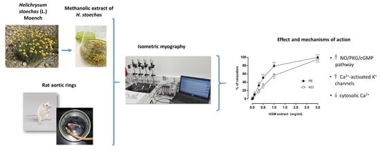
Helichrysum stoechas (L.) Moench (H. stoechas) is a medicinal plant traditionally used in the Iberian Peninsula to treat different disorders such as arterial hypertension. The aim of this study was to investigate the vascular effects of a polyphenolic methanolic extract of H. stoechas, which has high antioxidant activity, and its mechanism of action. Isometric myography studies were performed in an organ bath with rat aortic rings with intact endothelium. The H. stoechas extract produced vasorelaxation in the aortic rings that were precontracted by phenylephrine or KCl. L-NAME and Rp-8-Br-PET-cGMPS but not indomethacin or H-89; it also reduced the relaxant response evoked by H. stoechas extract on the phenylephrine-induced contractions. H. stoechas extract reduced the response to CaCl2 similar to verapamil and reduced the phenylephrine-induced contractions comparable with heparin. TRAM-34, apamin and glibenclamide reduced relaxation induced by the H. stoechas extract. The combination of L-NAME+TRAM-34+apamin almost completely inhibited the H. stoechas-induced effect. In conclusion, the relaxant effect of the H. stoechas extract is partially mediated by endothelium through the activation of the NO/PKG/cGMP pathway and the opening of Ca2+-activated K+ channels. Furthermore, the decrease in the cytosolic Ca2+ by the inhibition of Ca2+ influx through the L-type Ca2+ channels and by the reduction of Ca2+ release from the sarcoplasmic reticulum via the IP3 pathway is also involved.

Graphical Abstract

1. Introduction

Since ancient times, natural products have been a valuable resource of therapeutic agents for the prevention and treatment of diseases, with many of the current and developing drugs being natural products or derivatives. These natural products have been gaining more and more popularity, being widely used by the population because of their therapeutic effects and because they are affordable [1].
Natural products offer broad chemical and structural diversity, providing a wide range of nutrients and bioactive compounds including vitamins, minerals, fibre and phytochemical components, among others. Phytochemicals are very important from a health-promoting point of view because they are responsible for the medicinal properties of plants and act as functional components; these compounds are natural molecules derived from plants that are nonnutritive but biologically active and associated with the reduction of risk of important chronic diseases. So far, more than 5000 phytochemicals have been identified, but a high percentage remain to be isolated and identified. These compounds are obtained during the secondary metabolism of plants and are generally divided into polyphenols, terpenoids and alkaloids [2,3]. Polyphenols are the phytochemicals that have a broader spectrum of bioactivity, even being considered therapeutic agents with useful applications as medicines, food supplements, cosmetic products, or agrochemicals for food or plant protection.
Numerous studies support that phytochemicals have a beneficial effect on health due to their antioxidant, anti-inflammatory, antimicrobial, antiallergic, antispasmodic, anticancer, cardioprotective, neuroprotective, antidiabetic and antiobesity activity and because they maintain the homeostasis of the intestinal microbiota [3,4,5]. Due to these biological effects, phytochemicals are powerful therapeutic tools; that is why the bioactive compounds of plants must be identified, their biochemical and physiological effects known, as well as their mechanisms of action, and their efficacy and therapeutic safety guaranteed [6].
The genus Helichrysum belongs to the Compositae (or Asteraceae) family and is characterized by a large number of species, approximately 500–600, that are widely distributed throughout southern Europe and Africa. Species of this genus have yellow flowers and a rich amount of secondary metabolites, which provide a great variety of bioactive properties. Within this genus, Helichrysum stoechas (L.) Moench (H. stoechas) is one of the least known. In the Iberian Peninsula, this species, commonly known as everlasting flower (siempreviva, perpetua or sol de oro in Spanish) is used in traditional medicine. Herbal teas of these flowers are used to treat disorders of the cardiovascular, digestive (bloating, hepatic and pancreatic problems) and respiratory systems (influenza and cold) and have anti-urolithiatic and diuretic effects as well [7,8,9,10].
Previous studies have reported antioxidant and antimicrobial properties [11,12] and anti-inflammatory capacity [13] as well as antidiabetic and neuroprotective properties [12]. Recently, this plant has been used to assess the degree of soil contamination in a region of Spain, suggesting its utility for the phytostabilization of contaminated soils [14].
A recent study carried out by our group reported the phytochemical analysis of a metanolic extract of H. stoechas and showed a high content of polyphenolic compounds (166.6 μg gallic acid equivalents (GAE)/mg by the Folin-Ciocalteu method) with a particular phenolic profile that was isolated by chromatography and identified using NMR and MS [12]. The main components isolated were arzanol, homodimeric α-pyrone (helipyrone and p-hydroxybenzoic acids), phenolic acids (caffeic acids, neochlorogenic acids and 5-7-dihydroxy-3,6,8-trimethoxyflavone), flavonoids (isoquercitrin, quercetagetin-7-O-glucopyranoside) and diacylglycerol (santinol B). Furthermore, this phenolic profile demonstrated high antioxidant properties [12].
Arzanol pyranone is one of the most characteristic phenols found in this genus, in the extract of both H. stoechas and another species known as H. itallicum [12]. This bioactive compound explained the different properties such as antioxidant, antiviral (anti-HIV-1), and anti-inflammatory (by inhibiting NF-κB and mPGES-1 activity, as well as the production of pro-inflammatory cytokines) [15,16,17].
It is known that phenolic compounds exert antioxidant activity [18,19,20]. They improve nitric oxide (NO) bioavailability and reduce its degradation [21], so they could have an antihypertensive effect by reducing the production of reactive oxygen species (ROS) and thereby preventing vascular alteration. Studies have shown that polyphenols have a cardiovascular protective effect, improving the functions of endothelial cells due to its antioxidant, anti-inflammatory, antihypertensive, antithrombotic and antiproliferative properties [22,23]. Endothelial dysfunction represents the loss of the endothelium’s ability to modulate vascular tone by releasing relaxation factors. This dysfunction is thought to promote cardiovascular pathologies such as hypertension, atherosclerosis, stroke, heart disease, peripheral vascular disease, diabetes, chronic kidney failure, thrombosis and metastasis [24,25].
In keeping with the potentially beneficial compounds in H. stoechas extracts and the lack of mechanistic studies investigating its physiological effects at the vascular level, the objective of this study was to evaluate the effect of a methanolic H. stoechas extract on the vascular smooth musculature and its mechanism of action, as well as to study whether these effects are due to arzanol, the main biologically active component of the extract.

2. Materials and Methods

2.1. Reagents and Chemicals

The preparation of methanolic H. stoechas extract was previously described [12], and a plant voucher was deposited at the Universidad San Jorge herbarium (ref. 002–2014). The phenolic profile of this extract was previously determined [12] (see Introduction).
The compositions of the buffers used were as follows: Krebs buffer (in millimolar): NaCl 120, KCl 4.7, CaCl2 2.4, MgSO4 1.2, NaHCO3 24.5, KH2PO4 1, and glucose 5.6. Ca2+-free Krebs: NaCl 120, KCl 4.7, CaCl2 2.40, MgSO4 1.2, NaHCO3 24.5, KH2PO4 1, glucose 5.6, and ethyleneglycoltetraacetic acid (EGTA) 1. Ca2+-free high K+ Krebs ([K+]o = 50 mM). All buffers were adjusted to pH 7.4. Apamin (AP), arzanol, barium chloride dihydrate (BaCl2), glibenclamide (Glib), heparin (H), H-89 dihydrochloride hydrate, indomethacine, Nω-Nitro-L-arginine methyl ester hydrochloride (L-NAME), phenylephrine (PE), ruthenium red (RR) and verapamil (V) were obtained from Sigma (Madrid, Spain).
Tetraethylammonium (TEA), 1-[(2-chlorophenyl) diphenylmethyl]-1H-pyrazole (TRAM-34) and Rp-8-Br-PET-cGMPS were purchased from Tocris (Madrid, Spain). AP was diluted in acetic acid. Glib, TRAM-34 and arzanol were dissolved in dimethyl sulfoxide (DMSO). All other chemicals were dissolved in distilled water.

2.2. Animals

The care and use of animals were in accordance with the Spanish Policy for Animal Protection RD 53/2013, RD1386/2018 and RD118/2021, which meet the European Union Directive 2010/63 on the protection of animals used for experimental and other scientific purposes. All protocols were approved by the Ethics Committee for Animal Experiments of the University of Zaragoza (Spain) (reference: PI66/17, 18 January 2018). Male Wistar albino rats (200–250 g) were purchased from Janvier (LeGenest St. Isle, France) and were maintained with a standard feed and free access to water. The animals were euthanized by pentobarbital sodium (60 mg/Kg i.p.) followed by cervical dislocation.

2.3. Preparation of the Aortic Rings and Isometric Myography

Myography on the rat aorta artery rings was performed as described previously [26]. After sacrifice, the thoracic aorta was removed carefully, placed in ice-cold Krebs buffer, and cleaned of fat and adherent connective tissue. The aorta was cut into rings (3 mm long), and the rings were individually mounted to an isometric force transducer (Pioden UF1, Graham Bell House, Canterbury, UK) for tension measurement. The organ bath contained 5 mL Krebs buffer maintained at 37 °C and gassed with 95% O2–5% CO2. Tension was recorded and digitalized at a sample rate of 0.5 samples per second using a data acquisition system (ED410 e-corder 410, eDAQ, Cibertec, Madrid, Spain). An initial tension of 1 g was applied to the preparations to achieve spontaneous tone, and rings were allowed to equilibrate for 60 min with changing the bath buffer every 20 min.

2.4. The Experimental Protocols

After the stabilization of spontaneous tone, PE (1 µM) or KCl (80 mM) were used to induce sustained contractions. Fifteen minutes thereafter, the effects of H. stoechas extract, methanol (MeOH, solvent of the extract), arzanol and DMSO (solvent of the arzanol) were assessed, and cumulative concentration-response curves were constructed on endothelium-intact aortic rings. The vasorelaxant effect was calculated as the percentage change of precontraction to basal tone in the absence of the contractile agent.
To specifically measure endothelium-mediated vasorelaxation, the NO-synthase blocker L-NAME (10 µM) and the cyclooxygenase blocker indomethacin (10 µM) were added to the bath 20 min prior to the addition of PE. At stable PE-induced contraction, a concentration-response curve for the H. Stoechas extract was performed.
Next, the involvement was investigated of secondary messengers such as cAMP and cGMP and potassium channels in response to the H. stoechas extract. 20 min before addition of PE (1 µM), aortic rings were preincubated with H-89 (200 nM), Rp-8-Br-PET-cGMPS (10 µM), TRAM-34 (1 µM), AP (1 µM), Glib (10 µM), BaCl2 (30 mM) and TEA (tetraethylammonium, 0.5 mm). At stable contraction, the H. stoechas extract (0.3 and 1 mg/mL) was added for 15 min. The relaxant effect was compared with the response obtained by control (H. stoechas extract alone).
To examine the effects of H. stoechas extract on PE-induced contraction, the rings were preincubated with the extract or the solvent for 20 min, and a PE concentration-response curve was made. The contractile response of each concentration was compared to its control (baseline).
The role of Ca2+ influx in relaxation to the H. stoechas extract was studied. After prior incubation with Krebs buffer, the medium was replaced with Ca2+-free Krebs buffer for 15 min and then with Ca2+-free high-K+ buffer. Then, the rings were preincubated with methanol, H. stoechas extract (0.3 and 1 mg/mL) or V (1 µM) for 15 min, and after that time, cumulative concentration-response curves for CaCl2 (10−5–10−2 M) were constructed. The responses obtained to CaCl2 in the presence of methanol served as control.
In order to know if the response of the extract involves intracellular Ca2+, we evaluated the effect of the H. stoechas extract on the contractile response of PE (1 µM). For this, the rings were incubated for 15 min with verapamil (V) and the combination of V with RR and H before the addition of PE. Contractions were compared with controls.
Each protocol was performed on aortic rings from six or eight different animals.

2.5. Analysis of the Data

Data are expressed as mean ± standard error of mean (SEM). Statistical significance was tested using one-way analysis of variance (ANOVA) followed by the Dunnett test (when parametric distribution is observed), Kruskal-Wallis test (nonparametric) and paired two-tailed Student’s t test, if appropriate. A two-way ANOVA was performed to assess the interaction between two variables and the effects of each one, separately. p values <0.05 were considered statistically significant. The concentration of compound that inhibited 50% of the maximal contraction (EC50) was calculated as the geometric mean with 95% confidence intervals (CI). All statistical analyses were performed and all figures were created with GraphPad Prism 6.

3. Results

3.1. The Effects of H. stoechas Extract on PE- and KCl-Induced Contractions

PE (1 µM) or KCl (80 mM) induced a sustained contraction over time in the rat aortic rings. The H. stoechas extract induced a concentration-dependent vasorelaxation in the PE- or KCl-precontracted rings, with EC50 of 0.49 mg/mL (0.47–0.55, 95% CI) and 0.87 mg/mL (0.77–0.99, 95% CI), respectively. However, at the maximum concentration, 3 mg/mL, the extract produced a similar relaxation in the PE- or KCl-precontracted rings (PE: 99.9 ± 1.9% vs. KCl: 95.1 ± 1.4%) (Figure 1a,b). To exclude the possibility that the effect of the extract was caused by the solvent (MeOH), we studied the per se effect of MeOH in the precontracted rings and found that at the concentrations used, MeOH did not produce changes in muscle tone (Figure 1a).
To test whether the relaxing effect of H. stoechas extract was produced by one of its most characteristic components, an arzanol-concentration-response curve was performed (10−9 M–10−5 M) (Figure 1c). To exclude the possibility that the relaxing effect was caused by the per se relaxing effects of the solvent DMSO, we tested the same concentrations of DMSO (Figure 1c). The greatest relaxing effect of arzanol was found at a concentration of 10−5 M. Arzanol showed a stronger vasorelaxant effect than DMSO at concentrations of 10−7 and 10−6 M. However, at the highest concentration tested (10−5 M), arzanol showed the same effect as that obtained by the corresponding concentration of the solvent alone. Considering these results, arzanol was discarded as the main phenolic involved in the vasorelaxant effects, with other compounds being responsible for the observed activity.

3.2. The Effects of Indomethacin and L-NAME on H. stoechas Extract-Induced Vasorelaxation

To study the involvement of endothelium-derived factors in the relaxation evoked by the H. stoechas extract on the PE-contracted rings, intact aortic rings were preincubated with the nitric oxide synthase inhibitor L-NAME (10 µM) and with the nonselective cyclooxygenase inhibitor indomethacin (10 µM). As shown in Figure 2, indomethacin had no effect on the H. stoechas extract-induced vasorelaxation on rings contracted with PE. However, preincubation with L-NAME significantly inhibited H. stoechas extract-induced vasorelaxation compared with control. The maximum blocking response to the relaxing effect of H. stoechas extract by L-NAME was 30% at the concentration of 1 mg/mL (Figure 2).

3.3. The Effects of PKA and PKG Inhibitors on H. stoechas Extract-Induced Vasorelaxation

Figure 3 shows the vasorelaxant responses to different concentrations of H. sotechas extract (0.3 and 1 mg/mL) in aortic rings precontracted with PE (control) and PE-precontracted rings in the presence of the protein kinase A inhibitor H-89 (200 nM) or the protein kinase G inhibitor Rp-8-Br-cGMP (10 µM). While Rp-8-Br-cGMP significantly reduced the vasorelaxation by 79.3% and 50.64% at 0.3 and 1 mg/mL H. stoechas extract, respectively, H-89 had no effect on the response.

3.4. The Effects of H. stoechas Extract on the Contractile Response to PE

To assess the effect of the H. stoechas extract on a contractile agonist, we preincubated the rings with the extract (0.3 and 1 mg/mL) and then performed a cumulative PE curve (10−9–3 × 10⁻⁶ M). The contractile response of PE decreased in a concentration-dependent manner (Figure 4). Thus, preincubation with H. stoechas extract at 0.3 and 1 mg/mL reduced the contraction induced by the highest concentration of PE by 23% and by 70% respectively, compared with the contraction in absence of the extract (Figure 4).

3.5. The Effects of Ca2+ Influx on H. stoechas Extract-Induced Vasorelaxation

To examine the role of extracellular Ca2+ in the vasorelaxation induced by H. stoechas extract, the aortic rings were preincubated in a Ca2+-free K+-rich buffer with the H. stoechas extract at 0.3 or 1 mg/mL or verapamil, a Ca2+ channel blocker. After the incubation period, a cumulative Ca2+ curve was performed by adding increasing concentrations of CaCl2. As a control, the rings were preincubated with the solvent. The H. stoechas extract at 0.3 and 1 mg/mL reduced the maximum response to CaCl2 by 32.3% and 67.1%, respectively (Figure 5). Verapamil at 10−6 M produced a similar relaxation of 78.7% (Figure 5).

3.6. Role of Intracellular Ca2+ on the Effect of H. stoechas Extract on the Contractile Response Induced by Phenylephrine

In a series of experiments, we evaluated whether intracellular Ca2+ release is involved in the vasorelaxant response to H. stoechas extract. For this, aortic rings were preincubated with the L-type Ca2+ channel blocker verapamil and the ryanodine receptor inhibitor, ruthenium red or verapamil and the inositol 1,4,5-triphosphate (IP3) receptor inhibitor heparin before the addition of PE by ≈22% (control). This response was not modified when ruthenium red was added along with verapamil. However, the addition of heparin induced a greater reduction in the contractile response to PE (31.7%). The decrease in the contractile response to PE in the presence of verapamil and heparin was similar to that produced by 3 mg/mL of H. stoechas extract (36.9%) (Figure 6).

3.7. The Effects of K+ Channels Inhibitors on the H. stoechas Extract-Induced Vasorelaxation

The role of K+ channels in the relaxing response to H. stoechas extract was studied by preincubating the rings with various K+ channel blocking agents such as TRAM-34 (1 µM) or apamin (AP, 1 µM) or selective inhibitors of Ca2+-activated K+ channels of either intermediate (IKCa) or small-conductance (SKCa): respectively, glibenclamide (Glib, 10 µM), an inhibitor of ATP-sensitive K+ channel (KATP); TEA (0.5 mM), a nonspecific K+ channel inhibitor; and barium chloride (BaCl2, 30 mM), a nonspecific inhibitor of inward rectifier K+ channels (KIR).
The vasorelaxant effects of the H. stoechas extract at 0.3 and 1 mg/mL were inhibited by TRAM-34 (84.7% and 54.8%, respectively) and AP (61% and 44.6%, respectively). Glib significantly reduced the vasorelaxant effect only at the highest concentration of extract (45.3%). In contrast, TEA and BaCl2 did not affected the vasorelaxation response of the H. stoechas extract (Figure 7). At the concentrations used, the K+ channels inhibitors did not show any per se effect on the basal muscle tone.

3.8. The Inhibition of H. stoechas Extract-Induced Vasorelaxation by the Combination of TRAM-34 and Apamin with or without L-NAME

We investigated possible additive effects of the combination of the K+ channel inhibitors (TRAM-34 and AP) and the NO inhibitor, L-NAME, on the relaxant response evoked by the H. stoechas extract.
The most interesting result was that the combination of TRAM-34 and AP did not have additive effects on the vasorelaxation produced by the extract. However, L-NAME showed additive effects in the presence of these K+ channel inhibitors (Figure 8).

4. Discussion

H. stoechas (L.) Moench is used in traditional medicine for the treatment of various pathologies, and especially those of the cardiovascular system. Until now, in vitro studies have shown that this plant has strong antioxidant and anti-inflammatory activities and antimicrobial capacity as well as antiproliferative and antidiabetic effects [12]. Recently, neuroprotective effects were observed in in vitro and in vivo studies [12,27]. However, the vascular activity of H. stoechas is unknown.
Here, we demonstrated for the first time the capacity of a methanolic extract of H. stoechas to relax rat aortic rings, revealing its vascular mechanisms of action. According to our data, the vasorelaxation depended on the activation of the NO/PKG/cGMP pathway, opening of K+ channels and inhibition of Ca2+ signalling.
Our results show that the H. stoechas extract counteracts contraction to PE (an α1-adrenergic agonist) and KCl (a classical membrane depolarizing agent) in endothelium-intact aortic rings. The relaxant effect of the H. stoechas extract on PE-induced contraction was reduced by the inhibition of NO synthesis but not by inhibition of cyclooxygenase activity. In addition, preincubation with the cGMP inhibitor Rp-8-Br-cGMP blocked the vasorelaxant effect of the extract, supporting that the NO and cGMP pathways are involved mechanistically. In contrast, protein kinase A activity did not seem to be required. These results demonstrated that the vasorelaxant effects of the H. stoechas extract on rat aortic rings involve the NO/PKG/cGMP pathway and are thus partly endothelium dependent.
It is well-known that the endothelium plays an important role in maintaining vascular homeostasis through the secretion and release of vasodilator substances such as NO and prostacyclin as well as endothelium-dependent hyperpolarization (EDH) [24,25,28]. EDH is an additional major endothelial vasodilator system that acts in many vessels and species mainly through the opening of endothelial KCa channels, causing hyperpolarization. This potential change spreads through presumably direct electrical gap-junctional coupling, either K+-release or the smooth-muscle K+ channel activating diffusible factors to the smooth muscle and closes VDCCs, causing a decrease in cytosolic Ca2+ and, finally, vasodilation. So, K+ channels also play a central role in the regulation of vascular smooth muscle tone [29,30]. In this study, we show that TRAM-34 and AP, selective inhibitors of endothelial Ca2+-activated K+ channels of intermediate (IKCa) and small conductance (SKCa) respectively, significantly decreased the relaxant response to the H. stoechas extract in these rat aortic rings. Glib, an inhibitor of ATP-sensitive K+ channels (KATP), only reduced the response at the highest concentration of the extract tested. However, TEA, a nonspecific inhibitor of K+ channels, and BaCl2, an inhibitor of inward rectifier K+ channels (KIR) and the large-conductance K+ channels expressed in smooth muscles, did not alter the response to the H. stoechas extract. These findings implicate the endothelial K+ channels IKCa and SKCa and, to a lesser extent, KATP in the relaxing response to the H. stoechas extract. Therefore, the EDH mechanism also contributes to the H. stoechas-induced vasorelaxation.
It is noteworthy that in addition to initiating the EDH vasodilator response, endothelial SKCa and IKCa also contribute to the generation of NO, presumably by promoting endothelial signalling [28,31]. This occurs because these channels produce hyperpolarization of the endothelial cell, which induces an influx of Ca2+ through voltage-independent Ca2+ channels. This cation binds to NOS through calmodulin, which activates and thereby induces the synthesis of NO by this enzyme [31,32]. Thus, the effect of the combination of the SKCa and IKCa inhibitors together with or without the NO release inhibitor was assessed. Treatment with TRAM-34 and apamin in the presence of NOS inhibited the effect induced by the H. stoechas extract in a stronger way than in the absence of NOS, showing that hyperpolarization and NO release are additive.
In vascular smooth muscle, Ca2+ mobilization has an important role in the regulation of contraction and relaxation. Cytosolic Ca2+ results from Ca2+ influx from the extracellular space through receptor-operated Ca2+ channels (ROCCs) or VDCCs and from the release of Ca2+ from intracellular stores [32,33]. In our experiments, H. stoechas extract seemed to affect both Ca2+ pathways and by this reduced the contractile response to KCl and PE. The contraction produced by KCl in VSMCs is caused by the opening of VDCCs and therefore by the entry of Ca2+ from the extracellular medium, whereas the contraction caused by PE is due to both the influx of extracellular Ca2+ through ROCCs and the release of intracellular Ca2+ from the SR after the activation of IP3 receptors [33,34]. At low concentrations, the extract showed a greater vasorelaxant effect in the rings precontracted with PE than in those precontracted with KCl (see Figure 1), suggesting that the H. stoechas extract acts as an inhibitor of Ca2+ efflux from intracellular stores. In addition, the preincubation of the rings with the extract strongly reduced the contractions produced by increasing concentrations of PE in a concentration-dependent manner.
Furthermore, the H. stoechas extract inhibited the contraction caused by the influx of extracellular Ca2+ evoked by the addition of increasing concentrations of CaCl2 to a Ca2+-free medium. This vasorelaxant effect induced by the highest concentration of the H. stoechas extract was similar to that produced by verapamil, an inhibitor of L-type voltage-dependent Ca2+ channels. This finding suggests that the H. stoechas extract inhibits Ca2+ influx into smooth muscle cells through the VDCCs and thus reduces Ca2+-dependent contraction. On the other hand, our results suggest that H. stoechas extract also blocks intracellular Ca2+ release from the SR. Thus, the H. stoechas extract reduced the PE-evoked contraction in a similar manner to that produced by heparin, an IP3 receptor inhibitor, suggesting that the vasorelaxant response to H. stoechas extract involves the inhibition of the IP3 signalling pathway. All these results suggest that a mechanism of action by which the H. stoechas extract produces vascular relaxation is the inhibition of vascular smooth muscle Ca2+ signalling by blocking both Ca2+ entry and release from SR. Together, the H. stoechas extract uses multiple and likely additive endothelial and vascular smooth muscle mechanisms to produce vasorelaxation in this vessel.
To shed additional light on the active components in the extract, we studied whether a main component, arzanol, mimics the effect of the extract. However, we found that arzanol was only responsible for a small percentage of the vasorelaxant effect produced by the H. stoechas extract, suggesting that in addition to arzanol, other not yet identified vasorelaxing polyphenols or other components are involved here.
Concerning phenols, most of them have been reported to cause blood vessel dilation predominantly through activation of the NO/cGMP pathway. For instance, a study that analysed the direct relationship between NO release and the relaxant effect on porcine coronary rings induced by plant phenols showed that endothelium-dependent vasorelaxation is limited to those compounds that induce nitric oxide elevation > 5 nM (=Km value of soluble guanylate cyclase). Caffeic acid and isoquercitrin, compounds present in our extract, moderately increased the formation of NO (5 nM < [NO] < 8.5 nM) [35]. In this study, the vasorelaxant effect was significantly attenuated after treatment with L-NAME, and relaxations were almost completely abolished on the endothelium-denuded rings. To the contrary, indomethacin was without effect.
In the same way as our extract, the flavonoid isoquercitrin induces both endothelium-dependent and endothelium-independent vasodilation in the arteries of the mesenteric vascular bed of rats, through the release of NO but not PGI2 and through the opening of K+ channels in the cells of the smooth muscle [36]. Another study showed that caffeic acid exerted a vasorelaxant effect in isolated rat thoracic aorta precontracted with PE and KCl. This effect was partially dependent on NO and on blocking the increase in cytosolic Ca2+ elicited by Ca2+ influx and from intracellular stores [37].
Still, the phenol or component(s), responsible for the vasorelaxation capability of H. stoechas reported here remain to be elucidated in future studies.
In phytopharmacology, polyphenols are considered beneficial for relieving harmful oxidative stress and enhanced contractility in the vasculature. This enhanced contractility caused by ROS probably relies on increased cytosolic Ca2+ in VSMCs as a result of excessive Ca2+ influx through Ca2+ channels and release from the SR; inhibition of the SERCA pump; and other impairments like modification of cytoskeletal proteins [33]. It can be speculated that antioxidant compounds, like those that are present in our extract [12], could relieve oxidative and hypercontractility by counteracting excessive Ca2+ signaling and could therefore exert a cardiovascular protective effect.
Although there are no studies on the cardioprotective and vascular effects of H. stoechas, there are studies with other species of the same genus that suggest such cardioprotective utility. For instance, a crude aqueous extract of the root or leaves of Helicrysum ceres in rats induced hypotension and natriuresis [38]. Moreover, ethanolic extract of Helichrysum ceres leaves lowered blood pressure by producing negative inotropic and chronotropic effects in vivo experiments and evoked vasorelaxant effects in vascular tissue in isolated aortic rings involving both endothelium-dependent and endothelium-independent mechanisms [39]. The oral administration of aqueous and ethanolic extracts of Helichrysum plicatum (500 mg/Kg/bw) and ethanolic extracts of Helichrysum graveolens (500 mg/Kg/bw) showed significant antihyperglycaemic and antioxidant activity in streptozotocin-induced-diabetic rats, possibly preventing the onset of cardiovascular pathologies [40,41]. The study of flavonoids from the flowers of Helichrysum arenarium showed anti-atherosclerotic activities and fewer anti-inflammatory molecules [42]. Additionally, other plants in the Asteraceae family have been extensively studied and suggested to improve cardiovascular health, at least in part because they show vasodilator action involving pathways similar to those used by our extract [43]. In more detail, an aqueous extract of Artemisia campestris L. aerial parts showed a vasorelaxant effect on isolated rat aorta by activating the calmodulin-NO-cGC-PKG pathway and evoked a decrease in intracellular calcium by blocking VOCC channels and activating the SERCA pump. Kir, KATP, Kv and nonspecific K+ channels were not involved here [44]. Another study showed that organic extracts of Achillea millefolium produced vasodilation through endothelium-dependent NO release by increasing cGMP levels and by calcium channel blockade [45]. An aqueous extract of leaves of Tridax procumbens causes vasodiltation in the isolated rat aortic artery though the NO-cGMP and cAMP pathways and the dephosphorylation of MLC [46]. An ethanolic extract of Vernonia amygdalina Del. showed a pronounced vasorelaxant effect by activating the NO/cGMP and PGI2 pathways; reducing cytosolic calcium by blocking VOCC and inhibiting the intracellular release of calcium through IP3 receptor; opening K+ channels (KCa, Kir, KATP, Kv); and activating the muscarinic and β2-adrenoreceptor pathways [47]. A Jasonia glutinosa extract produces a vasorelaxant effect by preventing increases in cytosolic calcium, mainly by blocking L-type calcium channels [26].
The endothelium is important for vasorelaxation and, of particular note, also for angiogenesis in tissue repair. Our extract produces vasorelaxation by activating the NO/cGMP pathway and by cellular hyperpolarization by opening endothelial IKCa and SKCa channels. These signalling pathways not only play a role in endothelium-dependent vascular relaxation but also are important in neo-angiogenesis, as indicated elsewhere [48,49]. For this reason, it would be interesting to study whether H. stoechas promotes angiogenesis, particularly during tissue repair. In fact, there are already healing creams containing an extract of this plant. It is tempting to speculate that the healing effect could rely on the antioxidant and vasorelaxing properties of H. stoechas, which may help to improve blood circulation in the damaged tissue and may also help to vascularize the tissue by promoting angiogenesis.

5. Conclusions

In this study, a methanolic extract of H. stoechas has been shown to relax vascular smooth muscle mediated by the endothelium-dependent NO/cGMP pathway and endothelial IKCa and SKCa channels. Moreover, the extract’s relaxing actions also involves an endothelium-independent mechanism as it blocks increases in intracellular Ca2+ through the inhibition of Ca2+ influx via ROCCs or VDCCs as well as the Ca2+ release from intracellular stores through the IP3 pathway. Considering its relaxing capacity at the endothelial and smooth muscle levels, this medicinal plant could be a useful candidate for the development of herbal medicines for the treatment or prevention of hypertension and other cardiovascular pathologies.

Author Contributions

Conceptualization, M.S.V., M.P.A., R.K. and V.L.; Formal analysis, M.S.V., M.P.A., R.K. and V.L.; Funding acquisition, M.S.V., F.L., C.G.-R. and V.L.; Investigation, M.S.V., S.N., F.L., M.C., M.Á.P., M.P.A., R.K. and V.L.; Methodology, M.S.V., C.G.-R., M.Á.P., M.P.A., R.K. and V.L.; Project administration, M.S.V., M.P.A. and V.L.; Resources, M.S.V., C.G.-R. and V.L.; Supervision, M.S.V. and V.L.; Visualization, M.S.V., M.P.A., R.K. and V.L.; Writing—original draft, M.S.V., M.P.A., R.K. and V.L.; Writing—review & editing, S.N., F.L., M.C., C.G.-R., M.Á.P., M.P.A., R.K. and V.L. All authors have read and agreed to the published version of the manuscript.

Funding

This research was funded by Government of Aragon (financial support of the group B44_20D-Phyto-Pharm: Principios vegetales bioactivos y ciencias farmacéuticas) and Universidad de Zaragoza (JIUZ-2018-BIO-09).

Institutional Review Board Statement

The animal study protocol was approved by the Ethics Committee for Animal Experiments of the University of Zaragoza (Spain) (reference: PI66/17, 18 January 2018).

Informed Consent Statement

Not applicable.

Data Availability Statement

Data is contained within the article.

Conflicts of Interest

The authors declare no conflict of interest. The funders had no role in the design of the study; in the collection, analyses or interpretation of data; in the writing of the manuscript or in the decision to publish the results.

References

  1. Veiga, M.; Costa, E.M.; Silva, S.; Pintado, M. Impact of plant extracts upon human health: A review. Crit. Rev. Food Sci. Nutr. 2020, 60, 873–886. [Google Scholar] [CrossRef] [PubMed]
  2. Özaslan, M.; Oguzkan, S.B. Use of Plant Extracts in Alternative Medicine. Pakistan J. Biol. Sci. 2018, 21, 1–7. [Google Scholar] [CrossRef] [PubMed]
  3. Santos-Buelga, C.; González-Paramás, A.M.; Oludemi, T.; Ayuda-Durán, B.; González-Manzano, S. Plant phenolics as functional food ingredients. Adv. Food Nutr. Res. 2019, 90, 183–257. [Google Scholar] [CrossRef] [PubMed]
  4. Ullah, A.; Munir, S.; Badshah, S.L.; Khan, N.; Ghani, L.; Poulson, B.G.; Emwas, A.H.; Jaremko, M. Important Flavonoids and Their Role as a Therapeutic Agent. Molecules 2020, 25, 5243. [Google Scholar] [CrossRef]
  5. Wang, Z.; Li, S.; Ge, S.; Lin, S. Review of Distribution, Extraction Methods, and Health Benefits of Bound Phenolics in Food Plants. J. Agric. Food Chem. 2020, 68, 3330–3343. [Google Scholar] [CrossRef]
  6. Thomford, N.E.; Senthebane, D.A.; Rowe, A.; Munro, D.; Seele, P.; Maroyi, A.; Dzobo, K. Natural Products for Drug Discovery in the 21st Century: Innovations for Novel Drug Discovery. Int. J. Mol. Sci. 2018, 19, 1578. [Google Scholar] [CrossRef] [Green Version]
  7. Benítez, G.; González-Tejero, M.R.; Molero-Mesa, J. Pharmaceutical ethnobotany in the western part of Granada province (southern Spain): Ethnopharmacological synthesis. J. Ethnopharmacol. 2010, 129, 87–105. [Google Scholar] [CrossRef]
  8. Carmona, M.D.; Llorach, R.; Obon, C.; Rivera, D. “Zahraa”, a Unani multicomponent herbal tea widely consumed in Syria: Components of drug mixtures and alleged medicinal properties. J. Ethnopharmacol. 2005, 102, 344–350. [Google Scholar] [CrossRef]
  9. Orhan, N.; Onaran, M.; Şen, İ.; Işık Gönül, İ.; Aslan, M. Preventive treatment of calcium oxalate crystal deposition with immortal flowers. J. Ethnopharmacol. 2015, 163, 60–67. [Google Scholar] [CrossRef]
  10. Onaran, M.; Orhan, N.; Farahvash, A.; Ekin, H.N.; Kocabıyık, M.; Gönül, İ.I.; Şen, İ.; Aslan, M. Successful treatment of sodium oxalate induced urolithiasis with Helichrysum flowers. J. Ethnopharmacol. 2016, 186, 322–328. [Google Scholar] [CrossRef]
  11. Albayrak, S.; Aksoy, A.; Sagdic, O.; Hamzaoglu, E. Compositions, antioxidant and antimicrobial activities of Helichrysum (Asteraceae) species collected from Turkey. Food Chem. 2010, 119, 114–122. [Google Scholar] [CrossRef]
  12. Les, F.; Venditti, A.; Cásedas, G.; Frezza, C.; Guiso, M.; Sciubba, F.; Serafini, M.; Bianco, A.; Valero, M.S.; López, V. Everlasting flower (Helichrysum stoechas Moench) as a potential source of bioactive molecules with antiproliferative, antioxidant, antidiabetic and neuroprotective properties. Ind. Crops Prod. 2017, 108, 295–302. [Google Scholar] [CrossRef]
  13. Bremner, P.; Rivera, D.; Calzado, M.A.; Obón, C.; Inocencio, C.; Beckwith, C.; Fiebich, B.L.; Muñoz, E.; Heinrich, M. Assessing medicinal plants from South-Eastern Spain for potential anti-inflammatory effects targeting nuclear factor-Kappa B and other pro-inflammatory mediators. J. Ethnopharmacol. 2009, 124, 295–305. [Google Scholar] [CrossRef] [PubMed]
  14. Mykolenko, S.; Liedienov, V.; Kharytonov, M.; Makieieva, N.; Kuliush, T.; Queralt, I.; Marguí, E.; Hidalgo, M.; Pardini, G.; Gispert, M. Presence, mobility and bioavailability of toxic metal(oids) in soil, vegetation and water around a Pb-Sb recycling factory (Barcelona, Spain). Environ. Pollut. 2018, 237, 569–580. [Google Scholar] [CrossRef] [PubMed]
  15. Appendino, G.; Ottino, M.; Marquez, N.; Bianchi, F.; Giana, A.; Ballero, M.; Sterner, O.; Fiebich, B.L.; Munoz, E. Arzanol, an anti-inflammatory and anti-HIV-1 phloroglucinol α-pyrone from Helichrysum italicum ssp. microphyllum. J. Nat. Prod. 2007, 70, 608–612. [Google Scholar] [CrossRef] [PubMed]
  16. Bauer, J.; Koeberle, A.; Dehm, F.; Pollastro, F.; Appendino, G.; Northoff, H.; Rossi, A.; Sautebin, L.; Werz, O. Arzanol, a prenylated heterodimeric phloroglucinyl pyrone, inhibits eicosanoid biosynthesis and exhibits anti-inflammatory efficacy in vivo. Biochem. Pharmacol. 2011, 81, 259–268. [Google Scholar] [CrossRef] [PubMed] [Green Version]
  17. Rosa, A.; Deiana, M.; Atzeri, A.; Corona, G.; Incani, A.; Melis, M.P.; Appendino, G.; Dessì, M.A. Evaluation of the antioxidant and cytotoxic activity of arzanol, a prenylated α-pyrone-phloroglucinol etherodimer from Helichrysum italicum subsp. microphyllum. Chem. Biol. Interact. 2007, 165, 117–126. [Google Scholar] [CrossRef]
  18. Hussain, T.; Tan, B.; Yin, Y.; Blachier, F.; Tossou, M.C.B.; Rahu, N. Oxidative stress and inflammation: What polyphenols can do for us? Oxid. Med. Cell. Longev. 2016, 2016, 7432797. [Google Scholar] [CrossRef] [Green Version]
  19. Nani, A.; Murtaza, B.; Khan, A.S.; Khan, N.A.; Hichami, A. Antioxidant and Anti-Inflammatory Potential of Polyphenols Contained in Mediterranean Diet in Obesity: Molecular Mechanisms. Molecules 2021, 26, 985. [Google Scholar] [CrossRef]
  20. Zhou, Y.; Zhang, S.; Fan, X. Role of Polyphenols as Antioxidant Supplementation in Ischemic Stroke. Oxid. Med. Cell. Longev. 2021, 2021, 5471347. [Google Scholar] [CrossRef]
  21. Forte, M.; Conti, V.; Damato, A.; Ambrosio, M.; Puca, A.A.; Sciarretta, S.; Frati, G.; Vecchione, C.; Carrizzo, A. Targeting Nitric Oxide with Natural Derived Compounds as a Therapeutic Strategy in Vascular Diseases. Oxid. Med. Cell. Longev. 2016, 2016, 7364138. [Google Scholar] [CrossRef] [PubMed] [Green Version]
  22. Biegańska-Hensoldt, S.; Rosołowska-Huszcz, D. Polyphenols in preventing endothelial dysfunction. Postepy Hig. Med. Dosw. 2017, 71, 227–235. [Google Scholar] [CrossRef] [PubMed]
  23. Behl, T.; Bungau, S.; Kumar, K.; Zengin, G.; Khan, F.; Kumar, A.; Kaur, R.; Venkatachalam, T.; Tit, D.M.; Vesa, C.M.; et al. Pleotropic Effects of Polyphenols in Cardiovascular System. Biomed. Pharmacother. 2020, 130, 110714. [Google Scholar] [CrossRef] [PubMed]
  24. Godo, S.; Shimokawa, H. Endothelial Functions. Arterioscler. Thromb. Vasc. Biol. 2017, 37, e108–e114. [Google Scholar] [CrossRef] [PubMed] [Green Version]
  25. Rajendran, P.; Rengarajan, T.; Thangavel, J.; Nishigaki, Y.; Sakthisekaran, D.; Sethi, G.; Nishigaki, I. The Vascular Endothelium and Human Diseases. Int. J. Biol. Sci. 2013, 9, 1057–1069. [Google Scholar] [CrossRef] [Green Version]
  26. Valero, M.S.; Oliván-Viguera, A.; Garrido, I.; Langa, E.; Berzosa, C.; López, V.; Gómez-Rincón, C.; Murillo, M.D.; Köhler, R. Rock Tea extract (Jasonia glutinosa) relaxes rat aortic smooth muscle by inhibition of L-type Ca2+ channels. J. Physiol. Biochem. 2015, 71, 785–793. [Google Scholar] [CrossRef]
  27. Borgonetti, V.; Les, F.; López, V.; Galeotti, N. Attenuation of Anxiety-Like Behavior by Helichrysum stoechas (L.) Moench Methanolic Extract through Up-Regulation of ERK Signaling Pathways in Noradrenergic Neurons. Pharmaceuticals 2020, 13, 472. [Google Scholar] [CrossRef]
  28. Félétou, M.; Köhler, R.; Vanhoutte, P.M. Nitric oxide: Orchestrator of endothelium-dependent responses. Ann. Med. 2012, 44, 694–716. [Google Scholar] [CrossRef]
  29. Duboscq, C. Vascular endothelium. Hematología 2017, 21, 19–30. [Google Scholar]
  30. Dogan, M.F.; Yildiz, O.; Arslan, S.O.; Ulusoy, K.G. Potassium channels in vascular smooth muscle: A pathophysiological and pharmacological perspective. Fundam. Clin. Pharmacol. 2019, 33, 504–523. [Google Scholar] [CrossRef]
  31. Köhler, R.; Kaistha, B.P.; Wulff, H. Vascular KCa-channels as therapeutic targets in hypertension and restenosis disease. Expert Opin. Ther. Targets 2010, 14, 143–155. [Google Scholar] [CrossRef] [PubMed] [Green Version]
  32. Ottolini, M.; Sonkusare, S.K. The Calcium Signaling Mechanisms in Arterial Smooth Muscle and Endothelial Cells. Compr. Physiol. 2021, 11, 1831–1869. [Google Scholar] [CrossRef] [PubMed]
  33. Brozovich, F.V.; Nicholson, C.J.; Degen, C.V.; Gao, Y.Z.; Aggarwal, M.; Morgan, K.G. Mechanisms of Vascular Smooth Muscle Contraction and the Basis for Pharmacologic Treatment of Smooth Muscle Disorders. Pharmacol. Rev. 2016, 68, 476–532. [Google Scholar] [CrossRef] [PubMed] [Green Version]
  34. Touyz, R.M.; Alves-Lopes, R.; Rios, F.J.; Camargo, L.L.; Anagnostopoulou, A.; Arner, A.; Montezano, A.C. Vascular smooth muscle contraction in hypertension. Cardiovasc. Res. 2018, 114, 529–539. [Google Scholar] [CrossRef] [Green Version]
  35. Taubert, D.; Berkels, R.; Klaus, W.; Roesen, R. Nitric oxide formation and corresponding relaxation of porcine coronary arteries induced by plant phenols: Essential structural features. J. Cardiovasc. Pharmacol. 2002, 40, 701–713. [Google Scholar] [CrossRef] [Green Version]
  36. Gasparotto, A.; dos Reis Piornedo, R.; Assreuy, J.; Da Silva-Santos, J.E. Nitric oxide and Kir6.1 potassium channel mediate isoquercitrin-induced endothelium-dependent and independent vasodilation in the mesenteric arterial bed of rats. Eur. J. Pharmacol. 2016, 788, 328–334. [Google Scholar] [CrossRef]
  37. Cicala, C.; Morello, S.; Iorio, C.; Capasso, R.; Borrelli, F.; Mascolo, N. Vascular effects of caffeic acid phenethyl ester (CAPE) on isolated rat thoracic aorta. Life Sci. 2003, 73, 73–80. [Google Scholar] [CrossRef]
  38. Musabayane, C.T.; Munjeri, O.; Mdege, N.D. Effects of Helichrysum ceres extracts on renal function and blood pressure in the rat. Ren. Fail. 2003, 25, 5–14. [Google Scholar] [CrossRef] [Green Version]
  39. Musabayane, C.T.; Kamadyaapa, D.R.; Gondwe, M.; Moodley, K.; Ojewole, J.A. Cardiovascular effects of Helichrysum ceres S Moore [Asteraceae] ethanolic leaf extract in some experimental animal paradigms. Cardiovasc. J. Afr. 2008, 19, 246–253. [Google Scholar]
  40. Aslan, M.; Deliorman Orhan, D.; Orhan, N.; Sezik, E.; Yesilada, E. In vivo antidiabetic and antioxidant potential of Helichrysum plicatum ssp. plicatum capitulums in streptozotocin-induced-diabetic rats. J. Ethnopharmacol. 2007, 109, 54–59. [Google Scholar] [CrossRef]
  41. Aslan, M.; Orhan, D.D.; Orhan, N.; Sezik, E.; Yeşilada, E. A study of antidiabetic and antioxidant effects of Helichrysum graveolens capitulums in streptozotocin-induced diabetic rats. J. Med. Food 2007, 10, 396–400. [Google Scholar] [CrossRef] [PubMed]
  42. Mao, Z.; Gan, C.; Zhu, J.; Ma, N.; Wu, L.; Wang, L.; Wang, X. Anti-atherosclerotic activities of flavonoids from the flowers of Helichrysum arenarium L. MOENCH through the pathway of anti-inflammation. Bioorg. Med. Chem. Lett. 2017, 27, 2812–2817. [Google Scholar] [CrossRef] [PubMed]
  43. Michel, J.; Abd Rani, N.Z.; Husain, K. A Review on the Potential Use of Medicinal Plants from Asteraceae and Lamiaceae Plant Family in Cardiovascular Diseases. Front. Pharmacol. 2020, 11, 852. [Google Scholar] [CrossRef] [PubMed]
  44. Dib, I.; Tits, M.; Angenot, L.; Wauters, J.N.; Assaidi, A.; Mekhfi, H.; Aziz, M.; Bnouham, M.; Legssyer, A.; Frederich, M.; et al. Antihypertensive and vasorelaxant effects of aqueous extract of Artemisia campestris L. from Eastern Morocco. J. Ethnopharmacol. 2017, 206, 224–235. [Google Scholar] [CrossRef] [PubMed] [Green Version]
  45. Arias-Durán, L.; Estrada-Soto, S.; Hernández-Morales, M.; Millán-Pacheco, C.; Navarrete-Vázquez, G.; Villalobos-Molina, R.; Ibarra-Barajas, M.; Almanza-Pérez, J.C. Antihypertensive and vasorelaxant effect of leucodin and achillin isolated from Achillea millefolium through calcium channel blockade and NO production: In vivo, functional ex vivo and in silico studies. J. Ethnopharmacol. 2021, 273, 113948. [Google Scholar] [CrossRef] [PubMed]
  46. Salahdeen, H.M.; Idowu, G.O.; Salami, S.A.; Murtala, B.A.; Alada, A.A. Mechanism of vasorelaxation induced by Tridax procumbens extract in rat thoracic aorta. J. Intercult. Ethnopharmacol. 2016, 5, 174–179. [Google Scholar] [CrossRef]
  47. Ch’ng, Y.S.; Loh, Y.C.; Tan, C.S.; Ahmad, M.; Asmawi, M.Z.; Omar, W.M.W.; Yam, M.F. Vasorelaxant properties of Vernonia amygdalina ethanol extract and its possible mechanism. Pharm. Biol. 2017, 55, 2083–2094. [Google Scholar] [CrossRef] [Green Version]
  48. Coletta, C.; Papapetropoulos, A.; Erdelyi, K.; Olah, G.; Módis, K.; Panopoulos, P.; Asimakopoulou, A.; Gerö, D.; Sharina, I.; Martin, E.; et al. Hydrogen sulfide and nitric oxide are mutually dependent in the regulation of angiogenesis and endothelium-dependent vasorelaxation. Proc. Natl. Acad. Sci. USA 2012, 109, 9161–9166. [Google Scholar] [CrossRef] [Green Version]
  49. Wong, W. Two Gases Required for Vasodilation and Angiogenesis. Sci. Signal. 2012, 5, ec163. [Google Scholar] [CrossRef]
Figure 1. (a) Representative recordings showing the precontractions evoked in intact isolated rat aorta rings by PE (1 µM) or KCl (80 mM) and the effects induced by MeOH and H. stoechas extract (HSM; 0.01–3 mg/mL) on these contractions. (b) The relaxant effects of different concentrations of extract (mg/mL) on PE- or K+-induced contractions of isolated rat aorta rings (PE, F = 219, p < 0.0001; KCl, F = 187.6, p = 0.0001). * p < 0.05, ** p < 0.01 and *** p < 0.001 vs. control values (basal tone). (c) The relaxant effects of arzanol and DMSO on PE-induced contractions of isolated rat aorta rings. * p < 0.05 vs. DMSO values. Data points are mean ± SEM (n = 6–8).
Antioxidants 11 01092 g001
Figure 2. The relaxant effects of increasing concentrations of H. stoechas (HSM) extract in the absence (control) and presence of L-NAME (10 µM) or indomethacin (10 µM) on PE-induced contractions evoked in isolated rat aorta rings. Data points are mean ± SEM (n = 6–8). * p < 0.05 and ** p < 0.01 vs. control.
Antioxidants 11 01092 g002
Figure 3. The relaxant effects of H. stoechas extract (HSM; 0.3 and 1 mg/mL) in the absence (control) and presence of the protein kinase A inhibitor H-89 (200 nM) or the protein kinase G inhibitor Rp-8-Br-cGMP (10 µM) on PE-induced contractions evoked in isolated rat aorta rings (Interaction: F = 0.32, p = 0.7277; Dose: F = 52.40, p < 0.0001, Way: F = 18.42, p < 0.0001). Data points are mean ± SEM (n = 6–8). ** p < 0.01 vs. control.
Antioxidants 11 01092 g003
Figure 4. The contractile response to PE after the incubation of the rings with methanol and H. sotechas extract (HSM). (a) Representative recordings showing the contractions produced by PE (10−9–3 × 10⁻⁶ M) in rings preincubated with methanol (control) or HSM (0.3 and 1 mg/mL). (b) The tension (g) produced by each concentration of PE. Data are expressed as mean ± SEM (n = 3–6).
Antioxidants 11 01092 g004
Figure 5. The effects of preincubation with H. stoechas extract (HSM, 0.3 or 1 mg/mL) or verapamil (V, 10−6 M) on tension produced by different concentrations of CaCl2 in rat aorta. Data points are mean ± SEM (n = 6–8). * p < 0.05 and *** p < 0.001 vs. control (HSM extract solvent alone).
Antioxidants 11 01092 g005
Figure 6. The effects of preincubation with verapamil (V, 1 µM), the combinations of V with ruthenium red (10 µM) (V + RR) or V with heparin (50 mg/mL) (V + H), and H. stoechas extract (HSM, 3 mg/mL) on contraction to PE (1 µM) in rat aorta. The Kruskal-Wallis test was performed. Data points are mean ± SEM (n = 6–8). ** p < 0.01 and *** p < 0.001 vs. control (response alone to PE).
Antioxidants 11 01092 g006
Figure 7. The effects of TRAM-34 (1 µM), apamin (AP, 1 µM), glibenclamide (Glib, 10 µM), tetraethylamonium (TEA, 0.5 mM) and BaCl2 (30 mM) on the vasorelaxant response of H. stoechas extract (HSM, 0.3 and 1 mg/mL) in isolated rat rings precontracted with PE (Interaction: F = 0.68, p = 0.6426; Dose: F = 11.08, p < 0.0001, Way: F = 61.59, p < 0.0001). Data points are mean ± SEM (n = 6–8). * p < 0.05 and ** p < 0.01 vs. control.
Antioxidants 11 01092 g007
Figure 8. The effects of preincubation with L-NAME (10 µM), TRAM-34 (1 µM), apamin (AP, 1 µM) and the combination of TRAM-34+AP and L-NAME+TRAM-34+AP on the vasorelaxant response evoked by H. stoechas (HSM; 0.3 and 1 mg/mL) in isolated rat rings precontracted with PE (HSM 0.3 mg/mL; F = 51.45, p < 0.0001 and HSM 1 mg/mL; F = 48.19, p < 0.0001). Data are mean ± SEM (n = 6–8). ** p < 0.01 vs. control.
Antioxidants 11 01092 g008
Publisher’s Note: MDPI stays neutral with regard to jurisdictional claims in published maps and institutional affiliations.