Article

Synthetic Tuning of CoII-Doped Silica Nanoarchitecture Towards Electrochemical Sensing Ability

1
Arbuzov Institute of Organic and Physical Chemistry, FRC Kazan Scientific Center, Russian Academy of Sciences, Arbuzov str., 8, 420088 Kazan, Russia
2
Kazan Federal University, Kremlevskaya str. 29/1, 420008 Kazan, Russia
*
Author to whom correspondence should be addressed.
Nanomaterials 2020, 10(7), 1338; https://doi.org/10.3390/nano10071338
Submission received: 17 June 2020 / Revised: 2 July 2020 / Accepted: 7 July 2020 / Published: 9 July 2020
(This article belongs to the Special Issue Synthesis and Functionalization of Colloidal Nanoparticles)

Abstract

:
The present work introduces both synthesis of silica nanoparticles doped with CoII ions by means of differently modified microemulsion water-in-oil (w/o) and Stöber techniques and characterization of the hybrid nanoparticles (CoII@SiO2) by TEM, DLS, XRD, ICP-EOS, SAXS, UV-Vis, and UV-Vis/DR spectroscopy and electrochemical methods. The results reveal the lack of nanocrystalline dopants inside the hybrid nanoparticles, as well as no ligands, when CoII ions are added to the synthetic mixtures as CoII(bpy)3 complexes, thus pointing to coordination of CoII ions with Si-O- groups as main driving force of the doping. The UV-Vis/DR spectra of CoII@SiO2 in the range of d-d transitions indicate that Stöber synthesis in greater extent than the w/o one stabilizes tetrahedral CoII ions versus the octahedral ions. Both cobalt content and homogeneity of the CoII distribution within CoII@SiO2 are greatly influenced by the synthetic technique. The electrochemical behavior of CoII@SiO2 is manifested by one oxidation and two reduction steps, which provide the basis for electrochemical response on glyphosate and HP(O)(OEt)2 with the LOD = 0.1 μM and the linearity within 0.1–80 μM. The Stöber CoII@SiO2 are able to discriminate glyphosate from HP(O)(OEt)2, while the w/o nanoparticles are more efficient but nonselective sensors on the toxicants.

Graphical Abstract

1. Introduction

Silica nanoparticles provide a good platform for uploading of metal ions and complexes through different synthetic approaches, including doping into silica matrix or deposition at a surface of nanoparticles [1,2,3,4,5,6,7,8,9,10,11,12,13,14,15,16]. Such hybrid silica nanoparticles uploaded by transition d-metal ions exhibit electrochemical behavior controlled by both inner-sphere environments of the metal ions and nano-architecture of the nanoparticles [14,15,17,18]. Controllable electrochemical behavior is a prerequisite for an application of the hybrid nanoparticles in electrochemical catalysis or sensing. In turn, nanoheterogeneous catalysts and sensors are of particular importance due to their greater stability and reusability versus the molecular complexes [14,15,17,18]. The substrate dependent electrochemical behavior of transition metal ions and complexes is the basis for electrochemical detection of many pollutants, including organophosphorous ones [19,20,21,22,23,24,25,26,27,28,29]. Electrochemical methods have many advantages over other sensing techniques such as reproducibility, good stability, high sensitivity, measuring trace level of samples and cost-effectiveness [19,30,31]. This requires an engineering of new materials as components of electrodes with advanced electrochemical functions [32,33,34]. Both unique redox activity and easy encapsulation into polymeric materials make hybrid silica nanoparticles rather promising components of the electrodes for electrochemical sensing [35], although metal oxide or metal-based nanomaterials have got wider application in the engineering of the devices for electrochemical sensing [36,37]. Thus, design and synthesis of the hybrid nanoparticles with controllable inner-sphere environment of d-metal ions and their distribution within the nanoparticles is a rather challenging task from both fundamental and practical points of view.
However, successful doping or deposition of d-metal complexes without significant changes in the ligand environment can be performed only for kinetically inert complexes, which are exemplified in literature by tris-dipyridyles of CoIII and RuII [16,38,39,40]. The labile d-metal complexes, including those of NiII and CoII, can also be doped into silica nanoparticles, although their ligand environment scarcely remains unchanged after the doping [14,15]. Moreover, literature data introduce both works reporting on safe doping of the CoII complexes [2,9] and those revealing complex formation of CoII ions with Si–O-groups as the reason for the ligand substitution [41]. The mutual impacts of electrostatic attraction of the complexes or coordinative bonding of CoII ions with Si-O-groups to the doping procedure can be recognized by comparative structural analysis of the CoII-dopants inside silica spheres. Thus, both CoII(bpy)3 (bpy is 2,2′-bipyridine) and CoCl2 will be used as the precursors for the synthesis of CoII-doped silica nanoparticles.
The water-in-oil microemulsion (w/o) and Stöber procedures are most widely applied for doping of hydrophilic dopants such as d-metal salts or complexes into silica spheres [14,15,16,38,39,40]. As it was previously reported, the choice of w/o or Stöber procedure for doping of metal ions into silica nanoparticles has a great impact on both content of the dopants and their electrochemical and catalytic activity [14,15,16]. Thus, the present work is aimed at highlighting a role of the synthetic procedure and nature of the precursor on the structure of the CoII-dopants and nanoarchitecture of CoII-doped silica nanoparticles. In turn, the structural features of CoII-dopants and CoII-doped silica nanoparticles are correlated with their electrochemical behavior and sensing ability in order to reveal an applicability of the differently synthesized CoII-doped silica nanoparticles for the sensing purposes. The water-in-oil microemulsion and Stöber procedures were chosen as the synthetic methods with the use of both CoII(bpy)3 (bpy is 2,2′-bipyridine) and CoCl2 as the precursors. The structural features of the hybrid silica nanoparticles were analyzed on both molecular and nano-levels by dynamic light and small angle X-ray scattering, electronic microscopy, elemental analysis and UV-Vis, UV-Vis DR, and IR spectroscopy methods. Electrochemical behavior of the hybrid nanoparticles was revealed in order to evaluate their applicability for the sensing. The sensing ability of the hybrid nanoparticles was exemplified by two organophosphorous compounds (OPC), which are the well-known pollutant glyphosate [42] and diethyl phosphite. The latter was chosen as a model of decomposition products of OPC, which are often no less toxic than the precursors.

2. Materials and Methods

2.1. Materials

Tetraethyl orthosilicate (TEOS, 98%), ammonium hydroxide (28–30%), n-heptanol (98%), cyclohexane (99%), 2.2′-bipyridine (bpy, 99%), diethyl phosphite (98%) were obtained from Acros Organics (Geel, Belgium); Triton X-100, cobalt(II) chloride (97%), cobalt(II) tetrafluoroborate hexahydrate (99%), N-(Phosphonomethyl)glycine (Glyphosate) were purchased from Sigma-Aldrich (St. Louis, Missouri, USA). CH3CN (CHROMASOLV® Plus, ≥99.9%, by Acros Organics, Geel, Belgium) was used without any preliminary purification. The Ethanol, Acetone, and TEOS were purified by distillation.
[Co(bpy)3](BF4)2 was synthesized according to the known procedure [43]. To an ethanol solution of Co(BF4)2·6H2O (2 × 10⁻² mol·L−1) (100 mL), an ethanol solution (50 mL) of bpy ligand (6 × 10⁻² mol·L−1) was added under stirring. The resulting solution was stirred for 1 h, then filtered and left to evaporate at room temperature. After several days, air-stable, brown crystals of this complex were obtained. Yield: 83%. The elemental analysis data for C30H24B2CoF8N6 are presented in Table 1. The spectroscopic data for [Co(BF4)2(bpy)3] matched that reported in the literature [43].
Et4NBF4 was obtained by mixing an aqueous solution of Et4NOH (30–35%) with HBF4 to a neutral indicator reaction. Et4NBF4 precipitated from the reaction mixture as white crystals, which were separated by filtering. The powder salt was further recrystallized from diethyl ether and dried for 2 to 3 days in a vacuum at 55 °C for dehydration.

Silica Nanoparticles Preparation

Water-in-oil (w/o) microemulsion procedure [44] was the following. A mixture of Triton X-100 (2.38 g), n-heptanol (2.29 mL), cyclohexane (9.32 mL), TEOS (0.2 mL), and 1.1 mL of bidistilled water (for “empty” silica nanoparticles) or aqueous solution (1.1 mL) of the CoII-precursor (Co(bpy)3(BF4)2 or CoCl2, C = 4.5 × 10⁻³ mol·L−1) was prepared and stirred for 30 min. The obtained w/o microemulsion was mixed with a microemulsion containing Triton X-100 (2.38 g), n-heptanol (2.29 mL), cyclohexane (9.32 mL) and aqueous solutions of NH3 (28–30%) with stirring. After 24 h of stirring, both CoII-doped and empty silica nanoparticles were precipitated from the microemulsion by adding acetone with further centrifugation. The nanoparticle precipitate was washed by solutions of ethanol-acetone (1:1), ethanol (two times), and water (several times) to remove any residual surfactant molecules and organic solvents. Physically adsorbed CoII ions or complexes and Triton X-100 molecules at nanoparticles surface were removed by ultrasonication during the washing procedure. Pink-colored CoII-doped and white “empty” silica nanoparticles were obtained.
Stöber synthesis [14] was done through the following steps. A solution of TEOS (1.14 mL) in EtOH (11.36 mL) was added to NH4OH (28–30%) (0.38 mL) in EtOH (8.05 mL)-H2O (4.05 mL) at the speed 2 mL per min (thought syringe pump) under continuous stirring (750 rpm). After 6 min, the solution of [Co(bpy)3](BF4)2 or CoCl2 in EtOH (2.5 mL, C = 4.5 × 10⁻³ mol·L−1) was injected into this mixture. In the case of the “empty” silica nanoparticles, the solution of TEOS (1.14 mL) in EtOH (11.36 mL) was added to NH4OH (28–30%) (0.38 mL) in EtOH (10.55 mL)-H2O (4.05 mL) at the speed 2 mL per min (thought syringe pump) under continuous stirring (750 rpm). After 6 h of stirring, silica nanoparticles were precipitated by centrifuging and washing several times by ethanol and water. To remove physically absorbed CoII complexes or ions from the particles surfaces, an ultrasonication was used during the washing procedure. Blue-colored CoII-doped and white “empty” silica nanoparticles were obtained.
The electrochemical, PXRD, SAXS, IR, and Raman experiments were performed with dried samples of the silica nanoparticles.

2.2. Samples Characterization

Transmission electron microscopy (TEM) measurements were carried out on Hitachi HT7700 transmission electron microscope (Chiyoda-ku, Japan) with energy-dispersive X-ray detector from Thermo Scientific. The accelerating voltage was equal to 80 kV. The accumulation time of one spectrum was 300 s.
Co and Si were identified in the silica nanoparticles colloids using a simultaneous inductively coupled plasma atomic emission spectrometry (ICP-AES) model iCAP 6300 DUO by Varian Thermo Scientific Company equipped with a CID detector. This spectrometer enables the simultaneous measurement of peak heights within the 166 to 867 nm range. The optical resolution is less than 0.007 nm to 200 nm. As for the working frequency, it is 27.12 MHz. Together, the radial and axial view configurations enable optimal peak height measurements with suppressed spectral noises.
UV-Vis spectra of solutions and silica nanoparticles dispersions were recorded on SpecordR50 Plus (Analytikjena, Germany). SNs dispersions were ultrasonicated within 10 min before using.
Powdered samples were characterized by UV-Vis/DR technique using a Jasco V-650 spectrophotometer (Jasco International Co. Ltd., Hachioji, Tokyo, Japan) equipped with an integrating sphere accessory for diffuse reflectance spectra acquisition. BaSO4 powder was used as the reference for baseline correction.
The dynamic light scattering (DLS) measurements were performed by means of the Malvern Mastersize 2000 particle analyzer (Malvern, UK). A He-Ne laser operating at 633 nm wavelength and emitting vertically polarized light was used as a light source. The measured autocorrelation functions were analyzed by Malvern DTS software and the second-order cumulant expansion methods. The effective hydrodynamic radius (RH) was calculated by the Einstein–Stokes relation from the first cumulant: D = kBT/6phRH, where D is the diffusion coefficient, kB is the Boltzmann constant, T is the absolute temperature, and h is the viscosity. The diffusion coefficient was measured at least three times for each sample. The average error in these experiments was approximately 4%. All samples were prepared from the bidistilled water with prior filtering through the PVDF membrane using the Syringe Filter (0.45 µm). Zeta potential “Nano-ZS” (MALVERN) using laser Doppler velocimetry and phase analysis light scattering was used for zeta potential measurements. Silica nanoparticles dispersions were ultrasonicated within 10 min before using.
IR spectra of solid samples have been registered using a Bruker Vector-27 FTIR spectrometer in the 400–4000 cm−1 range (optical resolution 4 cm−1). The samples were prepared as KBr pellets.
Raman spectra were registered at room temperature using a BRUKER RAM II module attached to a BRUKER VERTEX 70 FTIR spectrometer (excitation 1064 nm, Ge detector at liquid nitrogen temperature, back-scattering configuration; range 10–4000 cm−1, optical resolution 4 cm−1, scan number 1024).
Electrochemical measurements were taken on a BASiEpsilonE2P electrochemical analyzer (West Lafayette, IN, USA). The program concerned Epsilon-ECUSB-V200 waves. A conventional three-electrode system was used with glassy carbon for carbon paste electrode (CPE) solutions for powder samples as the working electrode, the An Fc/Fc system serving as reference electrode, and a Pt wire as the counter electrode. In addition, 0.1 M [Et4N][BF4] was used as the supporting electrolyte to determine the current voltage characteristics. The solvent for all measurements was water. To study the powder samples, a modified CPE working electrode was used, which was prepared as follows: the carbon particles/phosphonium salt (dodecyl(tri-tert-butyl)phosphonium tetrafluoroborate) composite electrode was prepared using a grinding a mixture of graphite powder and phosphonium salt with a 90/10 (w/w) ratio in mortar giving it a homogeneous mass [45]. A modified electrode was also devised in a similar manner except that a portion (ca. 5%) of the graphite powder was replaced by the CPs under study. As a result, a portion of the resulting paste was packed firmly into the (3 mm in diameter) Teflon holder cavity.
Powder X-ray diffraction (PXRD) measurements were performed on a Bruker D8 Advance diffractometer equipped with Vario attachment and Vantec linear PSD, using Cu radiation (40 kV, 40 mA) monochromated by the curved Johansson monochromator (λ Cu Kα1 1.5406 Å). Room-temperature data were collected in the reflection mode with a flat-plate sample. The samples were loaded on a standard zero diffraction silicon plate, which was kept spinning (15 rpm) throughout the data collection. Patterns were recorded in the 2Θ range between 3° and 93°, in 0.008° steps, with a step time of 0.1–4.0 s. Several diffraction patterns in various experimental modes were collected and summed for the sample. Processing of the obtained data performed using EVA software packages [46].
Small angle X-Ray scattering (SAXS) data for samples were collected with the Bruker AXS Nanostar system using CuKα (λ 1.5418Å) radiation from a 2.2 kW X-ray tube (40 kV, 35 mA) coupled with Gobbel mirrors optics and a HiStar 2D area detector. The beam was collimated using three pinholes with apertures of 800, 450, and 700 μm. The instrument was operated with a sample-to-detector distance of 63.5 cm to provide data at angles 0.1°< 2θ< 4.8°, which correspond to 0.007 Å−1 < s < 0.34 Å−1. The value of s is proportional to the inverse of the length scale (s = (4π/λ)sin(θ) in units of Å−1). Scattering patterns were obtained for the samples at 23 °C in an evacuated chamber. The measurements were performed in transmission mode with the use of glass capillaries filled by powder samples. The capillaries (2 mm diameter) were sealed and put into evacuated chamber by means of the holders. For each sample, several experiments were performed, allowing for controlling the quality of the experiments. The results of the experiments are summarized, so that the total time of each experiment was equal to 30,000 s. The 2D scattering patterns were integrated using the SAXS program package [47]. Calculation of structural parameters, simulation, and graphical representation of the results were performed using a PRIMUS [48] program package.

3. Results and Discussion

3.1. Size, Cobalt Content, and Spectral Properties of the Hybrid Nanoparticles (CoII@SiO2) in Correlation with the Synthetic Conditions

Stöber and water-in-oil microemulsion (w/o) techniques provide a facile approach for encapsulation of charged metal complexes into silica nanoparticles [2,9,14,15,16,38,39,40]. The use of the both techniques results in the synthesis of the hybrid silica nanoparticles. Figure 1 illustrates the TEM images of the hybrid nanoparticles, while their diameters evaluated from the images and the Si:Co ratios calculated from the ICP-EOS data are collected in Table 2. The data in Table 1 indicate that the size and the content of CoII evaluated by the Si:Co ratios are to a major extent dependent on the synthetic technique, while smaller differences derive from the doping of CoII(bpy)3 or CoCl2. The difference in the synthetic methods is illustrated by Scheme 1. The size of the nanoparticles synthesized by the w/o technique is controlled by the TX-100-based reverse micelles (Scheme 1). Thus, water:oil volume ratios along with surfactant and co-surfactant concentrations have a great impact on the size control in the framework of the w/o technique. The sizes of silica nanoparticles grown by the Stöber method are mainly affected by concentrations of TEOS and ammonia, while the ethanol:water volume ratio plays a smaller role in controlling the size (Scheme 1). Moreover, the water soluble CoII-dopants are concentrated within nano-droplets of the reverse micelles, which explains the greater uploading by CoII ions for the nanoparticles synthesized by the w/o procedure in comparison with those obtained by the Stöber method (Table 2, Scheme 1). It is also worth noting that the color of both aqueous colloids and dried samples of the hybrid nanoparticles are pink when they are synthesized by the w/o, while the Stöber procedure results in the blue nanoparticles. Similar to the above-mentioned factors the color of the hybrid nanoparticles seems to be independent from their doping with CoII(bpy)3 or CoCl2.
The presence of bpy ligands both coordinated with Co(II) and as “free” molecules inside silica nanoparticles can be revealed by UV-Vis spectroscopy measurements in both supernatants (Figure 2b) and aqueous dispersions of the synthesized silica nanoparticles (Figure 2a). The analysis of the UV-Vis spectral data reveals the lack of the bands peculiar for CoII(bpy)3 in the aqueous colloids for both types of CoII@SiO2 (curves 2 and 3 in Figure 2a). The supernatant after the synthesis by the Stöber procedure reveals the release of bpy from the complex (Figure 2b), while the electronic absorption bands of Triton X-100 mask the presence of bpy in the supernatant after the synthesis by the w/o method (Figure S1). Moreover, its concentration in the supernatant indicates the degradation of about 57% of CoII(bpy)3 added to the synthetic mixture.
Thus, the color changes during the synthesis of the nanoparticles by the w/o and Stöber procedures derive from the transformation of the spectral pattern from CoII(bpy)3 to that of bpy (Figure 3). In particular, the yellow color seems to remain unchanged in aqueous or alcohol solution of Co(bpy)3(BF4)2 exactly after addition of tetraethoxysilane and ammonia (4–8 in Figure 3), which is confirmed by the small changes in the spectral pattern of CoII(bpy)3. The color changes followed by the spectral transformations from that of CoII(bpy)3 to that of bpy (3 and 1 in Figure 3) is observed within 6 or 30 min after beginning of the Stöber (9 in Figure 3) or w/o synthesis. The time durations for 6 and 30 min correlate with the appearance of silica seeds in the framework of Stöber [14,39] and w/o [49] procedures. It is worth assuming that the growth of silica seeds with Si-O- groups exposed at their surface provides a reason for the transformations of CoII(bpy)3 to bpy and CoII ions due to a complex formation of the latter with Si-O- groups of the silica seeds. The assumption can be confirmed by comparison of the UV-Vis diffuse reflectance spectra in the long-wave spectral range derived from the d-d transitions of the hybrid nanoparticles doped with Co(dipy)3(BF4)2 or CoCl2 (Figure 4). For these purposes, the UV-Vis/DR spectra of CoII@SiO2 were recorded for their dried samples along with the powder of Co(dipy)3(BF4)2 (Figure 4) and “empty” silica spheres (SiO2) (Figure S2) for the comparison. The latter were synthesized by the same w/o and Stöber procedures without the addition of the CoII-dopants. The characterization of SiO2 synthesized in the same synthetic conditions as the CoII@SiO2 by TEM indicate that their sizes are greatly affected by the synthetic method (48 ± 5 and 97 ± 5 nm for SiO2 synthesized by w/o and Stöber methods correspondingly), while the effect of the dopants is rather small (compare with the sizes in Table 2).
The spectra in Figure 4 indicate the difference in the spectral pattern within 500–700 nm for the CoII@SiO2 synthesized by the w/o and Stöber procedures, while the nature of the CoII-precursor (Co(bpy)3(BF4)2 or CoCl2) insignificantly affects the d-d transitions of the CoII@SiO2. The spectra of Co(II)@SiO2 synthesized with the use of CoII(bpy)3 as precursor (2 and 3 in Figure 4) differ from the one of CoII(bpy)3 complex in the range associated with d-d transitions, but demonstrate the band at ~300 nm, absent in the spectra of Co(II)@SiO2 with CoCl2 (curve 4 and 5 in Figure 4), associated most probably with “free” bpy. The spectra of the Stöber CoII@SiO2 (curve 2 and 4 in Figure 4) demonstrate three strong bands at 528, 580, and 638 nm, which are characteristic for 4A2(F) → 4T1(P) transitions in tetrahedral CoII chromophores [50,51,52,53]. It should be mentioned that the same spectral pattern (bands at 547, 585, 633 nm) has been observed for tetrahedral CoII ion coordinated to four oxygen atoms in 1D coordination polymer of Co(fcdHp), based on 1,1′-ferrocenylenbis(H-phosphinic) acid (H2fcdHp) [54]. The similar bands in this region (curve 3 and 5 in Figure 4) are not so pronounced in the spectra of the w/o CoII@SiO2. They also demonstrate another rearrangement of intensities. Most probably, the spectral pattern of the w/o CoII@SiO2 samples derives from CoII ions in octahedral environment, for which weak bands at ca. 500–580 nm assigned to 4T1g4A2g(F) transitions are characteristic [50,53,55]. The absorbance intensity of tetrahedral CoII is more intense (∼100 times) than that of octahedral CoII due to the lack of an inversion center in the former symmetry [56]. Therefore, the contribution from the octahedral CoII to the spectral pattern could be masked by the bands arisen from the tetrahedral CoII chromophores. Nevertheless, both rearrangement of bands intensities at 500–700 nm and their lowering in spectra 3 and 5 compared to 2 and 4 (Figure 4) allows one to suggest the greater contribution of the octahedral CoII chromophores in the w/o CoII@SiO2 versus the Stöber CoII@SiO2. It is noteworthy that the coordinative bonds between the CoII ions and Si-O- groups at the silica surface are the key reason for the formation of the CoII@SiO2 by the w/o and the Stöber procedures. However, it is worth assuming that the extents of tetrahedral and octahedral Co2+ chromophores are greatly affected by the aqueous or alcohol environment of CoII ions in the frameworks of the w/o or Stöber techniques (Scheme 1).
The infrared and Raman spectra of CoII@SiO2 nanoparticles (Figure 5) synthesized by both methods are very similar and demonstrate characteristic for silica nanoparticles bands: 475 cm−1, attributed to bending modes between the Si–O–Si bonds, 800 cm−1, assigned to the symmetric stretching vibration between Si–O–Si bonds, ~960 cm−1 that is often linked with the stretching vibration of Si–OH bond and ~1100 cm−1, which corresponds to the asymmetric stretching vibration of Si–O–Si bond in the SiO4 tetrahedron as reported in literature [41,57,58,59,60]. The IR spectra of NPs obtained by the w/o technique (cyan and red in Figure 5a) contain additional bands at ~1370, 1701, and 2930 cm−1 most probably associated with CH bending and C=O and alkyl CH stretching vibrations of acetone and TX-100 molecules (cyan and red in Figure 5a). The presence of TX-100 molecules derives from their efficient adsorption at the surface of silica nanoparticles (Scheme 1) leading to the so-called surfactant-based corona similar with the previously reported F-127-based corona [61] of the silica nanoparticles synthesized by the w/o procedure with F-127 as the surfactant. Raman spectra of Stöber CoII@SiO2 based on CoII(bpy)3 contain very weak bands of bpy at ~1600 and 1570 cm−1 (magenta in Figure 5b), which are absent in the spectrum of “empty” SiO2 (black in Figure 5b), and are not visible in their IR spectra (magenta in Figure 5a).

3.2. Nanoarchitecture of CoII@SiO2 and SiO2 in Correlation with the Synthetic Procedure

Electrochemical behavior of metal ions encapsulated into silica nanoparticles is affected by the inner-sphere environment of metal ions and their preferable localization within interior (core) or exterior (shell) zones of the hybrid nanoparticles [15,62]. Thus, nanoarchitecture of the hybrid nanoparticles manifested by different distribution of the dopant within their core and shell zones was studied by powder X-ray diffraction (PXRD) and small angle X-ray scattering (SAXS) techniques. Combination of the techniques was used to reveal both supramolecular and nano-structuring in the dried samples of CoII@SiO2 and SiO2 synthesized by the both methods. It is well-known that silica spheres synthesized by w/o or Stöber techniques are manifested by widened peaks in PXRD spectra, while the presence of nano-crystallites derived from the cobalt salts or oxides can be revealed by the appearance of the specific peaks [41,62,63]. Literature data indicate that the amorphous nature of silica remains practically unchanged under coordination of Co(II) ions with Si-O- groups, while the narrow peaks revealing the nanocrystallites of cobalt oxide derive from the extra-amounts of the doped cobalt salt [41,62]. Such nanocrystallites can be revealed by comparative analysis of PXRD data of CoII@SiO2 nanoparticles synthesized by both methods and their empty analogous (SiO2). The PXRD spectra recorded for the dried samples of CoII@SiO2 and SiO2 nanoparticles synthesized by the both methods (Figure 6) demonstrate the PXRD patterns peculiar for amorphous silica nanoparticles, which agrees well with the concentration conditions in the synthesis of the CoII@SiO2 (see the Exp. Section for more details). However, the weak widened peaks in the range of small angles revealed in the PXRD curves of Stöber CoII@SiO2 and SiO2 derive from the inclusions of paracrystalline phase with short- and medium-range ordering into the amorphous nanoparticles.
SAXS has been previously documented as successful technique to reveal nano-structuring deriving from silica nanoparticles, as well as inhomogeneity in electron density distribution within nanoparticles [41,64,65,66]. It is worth noting in this connection that the sizes of CoII@SiO2 and SiO2 nanoparticles synthesized by Stöber method (Table 2 and Table 3) lie above the values available for correct evaluation by the method (1–60 nm), while the sizes of smaller CoII@SiO2 and SiO2 nanoparticles synthesized by the w/o procedure can be evaluated from the SAXS measurements. However, the X-ray scattering profiles of the CoII@SiO2 and SiO2 (Figure S3) reveal their nanostructuring manifested by the presence of nano-aggregates with the sizes below 60 nm even for the Stöber CoII@SiO2 and SiO2. The nanostructuring arisen from inhomogeneity of electron density distribution within the silica nanoparticles may be quantitatively described in a “core-shell” framework. However, the shell zones of such “core-shell” nanoparticles along with the interparticle space can be considered as isotropic homogeneous medium with the electron density lower than that of the core zone. It is worth assuming that the core zones of the CoII@SiO2 and SiO2 as nanoparticles encapsulated into an isotropic medium with distances between their centers above the sizes of the core zones. Thus, the quantitative analysis of the SAXS data was performed in the assumption that the scattering results from noninteracting spherical nanoparticles uniformly enclosed in a homogeneous medium which provides their local monodisperse environment. Two-dimensional scattering curves were obtained by averaging of the eight X-ray scattering measurements for both hybrid and empty silica nanoparticles, while no effect of the prolonged X-ray irradiation on the scattering data was revealed (Figure S4).
Table 3 collects the calculated from the SAXS data parameters characterizing nanoparticles, such as radius of gyration (Rg and Rg*) evaluated by the Guinier method and from analysis of distance distribution function respectively, biggest distances in the particles (Dmax), and average diameter of the particles in a sphere-shaped model framework (ds, ds = √(5/3) × Rg × 2). The latter can be compared with the d-values evaluated by TEM, which are also collected in Table 2 to facilitate the comparison. This difference is too great to be explained by the difference in the techniques. The ds values calculated from SAXS are at the level of 33–38 nm for CoII@SiO2 and SiO2 nanoparticles synthesized by the w/o technique; these values are lower than the d values evaluated by TEM on about 10 nm (Table 3). In the framework of the aforesaid assumption, the sizes revealed by the SAXS measurements refer to the core zones of the w/o CoII@SiO2 and SiO2. This doesn’t exclude the presence of an exterior layer exhibiting smaller electronic density than the silica spheres. The above-mentioned TX-100-corona at the surface of w/o CoII@SiO2 and SiO2 is one of the reasons for an exterior layer with the electron density smaller than that arisen from the silica matrix. Therefore, the deviation between the ds and d values can be explained by the fact that the sizes revealed by the TEM images derive from both core and shell zones of the w/o CoII@SiO2 and SiO2. It is also worth noting that the Dmax values exceed the ds ones on no more than 5 nm (Table 3), which, in turn, indicates that the shape of the w/o CoII@SiO2 and SiO2 revealed by SAXS deviates from an ideal spherical shape.
The evaluated from the SAXS data fractal dimensions (FD) of w/o CoII@SiO2 and SiO2 presented in Table 3 are above 3.0. Such FD values are peculiar for surficial fractality derived from the folded surface, whose density is lower than that of the silica matrix. This is in good agreement with the aforesaid assumption about the TX-100-based corona at the silica surface as schematically shown in Scheme 1. Comparative analysis of both FD and Rs values reveals the insignificant difference between “empty” SiO2 and CoII@SiO2 w/o nanoparticles (Table 3). This similarity indicates that the distribution of the CoII ions within CoII@SiO2 (w/o) is rather homogeneous without preferable localization within specific zones of the nanoparticles, while the lower electronic density of the exterior layer derives from the specificity of the w/o technique (Scheme 1).
The SAXS analysis of Stöber CoII@SiO2 and SiO2 is the peculiar case, since the greatest Rg values refer to the ds-values at the level of 34–41 nm which are far below the d-values evaluated from the TEM images (Table 3, Figure S5). Moreover, more smooth profiles of the scattering curves measured for Stöber CoII@SiO2 and SiO2 in comparison with the w/o ones (Figure S3) indicate greater polydispersity of the Stöber versus the w/o nanoparticles. This deviation points to inhomogeneity in electron density distribution within Stöber CoII@SiO2 and SiO2, which agrees well with the zones of short- and medium-range ordering revealed from the PXRD curves of the corresponding nanoparticles (Figure 6). The shapes of the revealed core zones also deviate from ideal spheres, which is evident from the deviation between Dmax и ds values (Table 3). Moreover, the FD value of the empty silica spheres made by Stöber procedure is on the level of surficial fractality (3.37), while the FD values of the Stöber CoII@SiO2 (2.6–2.7) refer to the so-called mass fractality which indicate the inhomogeneous distribution of CoII ions within the Stöber CoII@SiO2. This is quite different from the homogeneous distribution within the w/o CoII@SiO2, where both FD and ds values of the “empty” SiO2 and CoII@SiO2 nanoparticles are at the same level.
In summary, the SAXS data revealed that the synthesis of CoII@SiO2 in the framework of Stöber method results in the localization of CoII-dopant within specific zones of the silica spheres, while more homogeneous distribution of CoII-dopant within CoII@SiO2 is obtained by the w/o technique. Moreover, the surface fractality of CoII@SiO2 (w/o) can be explained by the porous silica surface decorated by TX-100-based corona.

3.3. Electrochemical Behavior of CoII@SiO2

The electrochemical behavior of CoII@SiO2 was measured with the use of carbon-paste electrode (CPE) [45]. The CPE-based technique enables avoiding effects of different colloid stability, which makes it most convenient for comparative analysis of electrochemical behavior of CoII encapsulated into CoII@SiO2 synthesized by the Stöber and w/o methods. Two types of CoII@SiO2, i.e., (Stöber) and (w/o) have identical first oxidation and reduction peaks. In particular, one-electron irreversible oxidation of CoII to CoIII is observed at a potential of 0.65 V (Figure 7). At −1.12–−1.15 V, a one-electron reversible reduction of CoII to CoI is observed for both types of nanoparticles, while a significant difference is observed at the second reduction of CoI to Co0 peak. The irreversible reduction of CoI to Co0 recorded at −1.69 V is revealed for Stöber CoII@SiO2, while, for w/o CoII@SiO2, the reduction at −1.38 V is reversible (Figure 7, Table 4).
It is well-known that reduction or oxidation potentials are to a great extent dependent on the inner-sphere environment of CoII. However, the previous report reveals that predominant localization of the NiII complexes within core zone of the NiII-doped silica nanoparticles shifts the electrochemical NiII to NiI reduction to more negative potentials [15]. Thus, the second CoI to Co0 reduction step is affected, while the CoII to CoI reduction and CoII to CoIII oxidation potentials are not influenced by the synthetic method (Table 4). The results are not enough to recognize the exact reasons for the aforesaid similarities and differences in electrochemical behavior of Stöber CoII@SiO2 and w/o CoII@SiO2. Nevertheless, the electrochemical activity of the CoII@SiO2 points to their applicability in electrochemical analysis of OPC.

3.4. CoII@SiO2 for Electrochemical Determination of OPC

The presentation of an electrochemical response of CoII@SiO2 on OPC is worth preceding by the discussion of main mechanisms responsible for the response. Both oxidation and reduction of CoII centers in CoII@SiO2 are manifested in the CVs by the peaks with the peak currents dependent on concentration of the CoII centers. The increased peak current of CoII/CoIII or CoII/CoI transformations results from regeneration of CoII centers due to rapid interaction of CoIII or CoI with OPC. In turn, the peak current can be significantly decreased in a case of unfavorable processes (the destruction of the catalytically active form or complex formation preventing a reestablishing of CoII centers).
Both OPC (glyphosate and diethyl phosphite) are electrochemically inactive in the studied interval of potentials. Thus, the enhanced peak current at the oxidation and reduction potentials of w/o CoII@SiO2 under the increased concentrations of the OPC (Figure 8a,c) indicates that oxidized CoIII@SiO2 or reduced CoI@SiO2 forms return to the initial form (CoII@SiO2) due to rapid redox reaction of CoIII or CoI centers with the OPC. The concentration dependent increase in the peak current (Figure 8a,c) provides the electrochemical response on the OPC. The comparative analysis of the substrate-induced electrochemical response of CoII@SiO2 synthesized by w/o and Stöber procedures reveal a significant difference between them. The difference is the highest at the reduction potential, when Stöber CoII@SiO2 nanoparticles demonstrate a significant increase in the peak current under the growth of diethylphosphite concentration (Figure 8c), while glyphosate in the same conditions drops the peak current to zero (Figure 8d). It is worth assuming that the aforesaid fact correlates with the differences in the inner- and outer-sphere of CoII ions within the silica spheres formed by the w/o and Stöber methods. Thus, the OPC-triggered electrochemical behavior of CoII@SiO2 is also greatly influenced by the method (Stöber or w/o) applied for their synthesis. Unfortunately, exact mechanisms responsible for the different electrochemical behavior of the hybrid nanoparticles in the presence of the OPC are not clear from the results. Additional studies lying beyond the present work scope are required for their clarification.
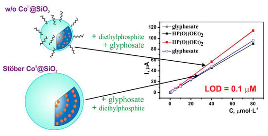
Nevertheless, the presented results clearly demonstrate the applicability of the substrate-induced electrochemical response for quantitative analysis of the OPC. The increase in the peak current within anodic potentials range is greater than that within the cathodic range. For evaluation of a lower detection level (LOD), the peak current of CoII@SiO2 at different concentrations of the OPC was analyzed by differential impulse voltammetry technique (Figure 9 and Figure S6). The results (Figure 9a,b) indicate that even small amounts of the OPC (0.1 μmol·L−1) induce the detectable peak increase; thus, this concentration level can be considered as the LOD-value. Moreover, the inset in Figure 9 indicates the linear relationship between the response currents and HP(O)(OEt)2 (red-black) and Glyphosate (gray-blue) concentrations in the range from 0.1 to 80 μM (with a correlation coefficient of 0.99). The slope of the linear dependencies is somewhat greater for w/o CoII@SiO2 versus Stöber CoII@SiO2, although the discrimination between glyphosate and diethylphosphite is rather poor for both types of CoII@SiO2. It is worth noting that glyphosate can be discriminated from diethylphosphite by the selective peak current increase in the cathodic range when the CPE was modified by Stöber CoII@SiO2.
The estimated LOD = 0.1 µmol·L−1 is greater than the values reported for most sensitive sensing systems [21], but lower than the values reported for the colorimetric sensor [67]. The linearity of the peak current increase is manifested in a wider concentration range than the linearity range reported in [21] for electrochemical sensors based on Cu-BTC/g-C3N4 nanosheets (g-C3N4 = graphite phase nitrogenized carbon, BTC = benzene-1,3,5-tricarboxylic acid). Moreover, the present LOD-value is enough for evaluation of dose limit of glyphosate in a drinking water. Furthermore, the CoII@SiO2 based sensors have the characteristics of short detection time, high stability and easy operation, which is of great impact on potential applications in electrochemical OPC analysis.

4. Conclusions

In summary, analysis of size and content of silica nanoparticles doped by CoII ions by means of water-in-oil microemulsion (w/o) or Stöber synthetic technique confirms the applicability of the both techniques for synthesis of the hybrid nanoparticles. Monitoring of the transformations of [Co(bpy)3](BF4)2 used as the precursor for the hybrid nanoparticles through the UV-Vis spectral analysis during the synthesis highlights the decomposition of the precursor due to efficient coordination of the CoII ions via Si-O- groups exposed at the silica seeds. Thus, the use of either [Co(bpy)3](BF4)2 or CoCl2 as the precursors results in the nanoparticles with the same inner-sphere environment of the CoII ions.
The analysis of the spectral properties in the wavelengths range of d-d transitions for the first time revealed preferable formation of tetrahedron CoII chromophores versus the octahedral ones in the the Stöber nanoparticles, while the predominance of octahedral CoII chromopheres was observed in the w/o nanoparticles. Both IR and Raman spectra indicate one more difference between the w/o and Stöber methods. The nanoparticles synthesized by the former method are decorated by the TX-100-based corona arisen from the TX-100 molecules adsorbed at the surface of the nanoparticles synthesized by the w/o method.
PXRD analysis of the synthesized nanoparticles indicated their amorphous nature, although he pracrystallinic inclusions into amorphous phase was revealed for the Stöber nanoparticles, which differentiate them from the w/o ones. The SAXS data revealed the heterogeneity of both nanoparticles, which is manifested by the presence of core zones with greater electron density than the shell zone. The sizes of the core zone are below those evaluated from the TEM images. The deviation between the sizes obtained by the different techniques for the w/o nanoparticles is no more than 20%. This enables to assume that the shell zone derives from the porous silica surface decorated by the TX-100-based corona. Both significant (above 50%) deviation revealed between the size values obtained by TEM and SAXS and specific PXRD profiles indicate the nano-structuring of the Stöber nanoparticles.
The electrochemical behavior of the CoII ions within the hybrid nanoparticles indicates the insignificant influence of the synthetic procedure at the first oxidation and reduction steps, while the second CoI to Co0 reduction step is greatly influenced by the synthetic procedure. In particular, the reversible CoI to Co0 reduction is facilitated within the w/o nanoparticles. The electrochemical behavior of the hybrid nanoparticles provides an efficient basis for electrochemical analysis of glyphosate and HP(O)(OEt)2 with the lower detection limit at the level of 0.1 µmol·L−1 with the linearity of the electrochemical response in the concentration range from 0.1 to 80 µmol·L−1, short detection time, high stability, and easy operation. The sensing properties of the hybrid nanoparticles are also influenced by the synthetic method: the electrochemical response of the Stöber nanoparticles is able to discriminate glyphosate from HP(O)(OEt)2, while the w/o nanoparticles are more efficient in the sensing of the both toxicants without discrimination between them.

Supplementary Materials

The following are available online at https://www.mdpi.com/2079-4991/10/7/1338/s1, Figure S1: UV-Vis spectra of supernatant diluted 300 times by ethanol after synthesis of w/o CoII@SiO2 (CoII(bpy)3 as precursor) (blue) and ethanol solutions of bpy (C = 5 × 10⁻⁵ mol·L−1, red) and Triton X-100 (C = 0.9 g·L−1, black), Figure S2: UV–Vis diffuse reflectance spectra of “empty” SiO2, Figure S3: SAXS diffraction intensity profiles at 23 °C (in logarithmic scale) top to bottom: (blue) w/o CoII@SiO2 (CoCl2 as precursor); (red) w/o CoII@SiO2 (CoII(bpy)3 as precursor); (magenta) Stöber CoII@SiO2 (CoCl2 as precursor); (green) Stöber CoII@SiO2 (CoII(bpy)3 as precursor); (dark) “empty” Stöber SiO2; (dark blue) “empty” w/o SiO2. Scattering vector s = 4πSinθ/λ, Å−1; λ = 1.5418Å is the wavelength of the X-ray beam, Figure S4: X-ray scattering curves of w/o CoII@SiO2 in logarithmic (left) and double logarithmic (right) scale. Different colors of curves correspond to eight consistent experiments, Figure S5: The fitting of experimental SAXS curve (points – experimental data, curves – simulation) and calculated distance distribution function p(r) for: (a) w/o CoII@SiO2 (CoCl2 as precursor); (b) w/o CoII@SiO2 (CoII(bpy)3 as precursor); (c) Stöber CoII@SiO2 (CoCl2 as precursor); (d) Stöber CoII@SiO2 (CoII(bpy)3 as precursor); (e) “empty” Stöber SiO2; (f) “empty” w/o SiO2, Figure S6: DPV voltammograms for w/o CoII@SiO2 (a) and Stöber CoII@SiO2 (b) in the absence and in the presence of increasing quantities of Glyphosate (from 0.1 µmol·L−1 to 80 µM: 0.1, 0.2, 0.3, 0.6, 1.2, 2.4, 5.0, 10.0, 20.0, 40.0, 80.0 µM). WE: CPE, H2O, 10−1 M Et4NBF4 Potentials vs. Ag/AgCl recalculated to Fc+/Fc. One-electron electron transfer during the oxidation and reduction of Co (II) is confirmed by the coincidence of the oxidation currents of ferrocene under the same conditions at the same analyte concentration (5 × 10⁻³ mol·L−1).

Author Contributions

Conceptualization, A.M., Y.B., and A.G.; investigation, O.B., M.K., A.G., T.G., I.N., A.L., and K.K.; supervision, A.M. and O.S. All authors analyzed and discussed the data and wrote the manuscript. All authors have read and agreed to the published version of the manuscript.

Funding

The authors would like to thank the financial support from the government assignments for realization of scientific priority projects by the FRC Kazan Scientific Center of RAS.

Acknowledgments

The authors acknowledge the CSF-SAC FRC KSC RAS for providing necessary facilities to carry out all X-ray diffraction experiments and to register infrared and Raman spectra.

Conflicts of Interest

The authors declare no conflict of interest.

References

  1. Antony, R.; Manickam, S.T.D.; Balakumar, S. Cu(II), Co(II) and Ni(II) Complexes Installed on Functionalized Silica Surface for Hydrogen Peroxide Assisted Cyclohexane Oxidation. J. Inorg. Organomet. Polym. Mater. 2017, 27, 418–426. [Google Scholar] [CrossRef]
  2. Vashurin, A.; Marfin, Y.; Tarasyuk, I.; Kuzmin, I.; Znoyko, S.; Goncharenko, A.; Rumyantsev, E. Sulfonated octa-substituted Co(II) phthalocyanines immobilized on silica matrix as catalyst for Thiuram E synthesis. Appl. Organomet. Chem. 2018, 32, e4482. [Google Scholar] [CrossRef]
  3. Faraji, A.R.; Mosazadeh, S.; Ashouri, F. Synthesis and characterization of cobalt-supported catalysts on modified magnetic nanoparticle: Green and highly efficient heterogeneous nanocatalyst for selective oxidation of ethylbenzene, cyclohexene and oximes with molecular oxygen. J. Colloid Interface Sci. 2017, 506, 10–26. [Google Scholar] [CrossRef] [PubMed]
  4. Rajabi, F.; Luque, R.; Clark, J.H.; Karimi, B.; Macquarrie, D.J. A silica supported cobalt (II) Salen complex as efficient and reusable catalyst for the selective aerobic oxidation of ethyl benzene derivatives. Catal. Commun. 2011, 12, 510–513. [Google Scholar] [CrossRef]
  5. Chen, L.; Li, B.-D.; Xu, Q.-X.; Liu, D.-B. A silica gel supported cobalt(II) Schiff base complex as efficient and recyclable heterogeneous catalyst for the selective aerobic oxidation of alkyl aromatics. Chin. Chem. Lett. 2013, 24, 849–852. [Google Scholar] [CrossRef]
  6. Islam, S.M.; Ghosh, K.; Molla, R.A.; Roy, A.S.; Salam, N.; Iqubal, M.A. Synthesis of a reusable polymer anchored cobalt(II) complex for the aerobic oxidation of alkyl aromatics and unsaturated organic compounds. J. Organomet. Chem. 2014, 774, 61–69. [Google Scholar] [CrossRef]
  7. Marfin, Y.S.; Vashurin, A.S.; Rumyantsev, E.V.; Puhovskaya, S.G. Sol–gel synthesis of highly effective catalyst based on cobalt tetrasulfophthalocyanine complex and silicon oxide. J. Sol Gel Sci. Technol. 2013, 66, 306–311. [Google Scholar] [CrossRef]
  8. Ranaweera, S.A.; Rowe, M.D.; Walters, K.B.; Henry, W.P.; White, M.G.; Rodriguez, J.M. Synthesis, characterization and catalytic activity of a cobalt catalyst: Silica-supported, bis(1,5-diphenyl-1,3,5-pentanetrionato)dicobalt(II)[Co2(dba)2]. Appl. Catal. A Gen. 2017, 529, 108–117. [Google Scholar] [CrossRef] [Green Version]
  9. Li-Juan, C.; Fu-Ming, M.; Guang-Xing, L. Co(II) Schiff base complexes on silica by sol–gel method as heterogeneous catalysts for oxidative carbonylation of aniline. Catal. Commun. 2009, 10, 981–985. [Google Scholar] [CrossRef]
  10. Trujillano, R.; Lambert, J.-F.; Louis, C. Chemistry of Silica-Supported Cobalt Catalysts Prepared by Cation Adsorption. 2. Neoformation of Cobalt Phyllosilicate. J. Phys. Chem. C 2008, 112, 18551–18558. [Google Scholar] [CrossRef]
  11. Yakubovich, T.N.; Ermokhina, N.I.; Bratushko, Y.I.; Zub, Y.L.; Chuiko, A.A. Cobalt complex with 2.2′-bipyridyl immobilized on disperse silica surface in the reaction of p-hydroquinone oxidation. Comparison of the homogeneous and heterogeneous catalysis results. In Dioxygen Activation and Homogeneous Catalytic Oxidation; Simindi, L.I., Ed.; Elsevier Science Publishers, B.V.: Amsterdam, The Netherlands, 1991; pp. 179–188. [Google Scholar]
  12. Rautiainen, A.; Lindblad, M.; Backman, L.B.; Puurunen, R.L. Preparation of silica-supported cobalt catalysts through chemisorption of cobalt(II) and cobalt(III) acetylacetonate. Phys. Chem. Chem. Phys. 2002, 4, 2466–2472. [Google Scholar] [CrossRef]
  13. Yuanfa, Y.; Renxian, Z.; Shaofen, Z.; Qiaoling, L.; Xiaoming, Z. Silica-supported poly-γ-aminopropylsilane Ni2+, Cu2+, Co2+ complexes: Efficient catalysts for Heck vinylation reaction. J. Mol. Catal. A Chem. 2003, 192, 303–306. [Google Scholar] [CrossRef]
  14. Khrizanforov, M.N.; Fedorenko, S.V.; Strekalova, S.O.; Kholin, K.V.; Mustafina, A.R.; Zhilkin, M.Y.; Khrizanforova, V.V.; Osin, Y.N.; Salnikov, V.V.; Gryaznova, T.V.; et al. A Ni(III) complex stabilized by silica nanoparticles as an efficient nanoheterogeneous catalyst for oxidative C–H fluoroalkylation. Dalton Trans. 2016, 45, 11976–11982. [Google Scholar] [CrossRef] [PubMed] [Green Version]
  15. Khrizanforov, M.N.; Fedorenko, S.V.; Mustafina, A.R.; Khrizanforova, V.V.; Kholin, K.V.; Nizameev, I.R.; Gryaznova, T.V.; Grinenko, V.V.; Budnikova, Y.H. Nano-architecture of silica nanoparticles as a tool to tune both electrochemical and catalytic behavior of NiII@SiO2. RSC Adv. 2019, 9, 22627–22635. [Google Scholar] [CrossRef] [Green Version]
  16. Budnikova, Y.; Bochkova, O.; Khrizanforov, M.; Nizameev, I.; Kholin, K.; Gryaznova, T.; Laskin, A.; Dudkina, Y.; Strekalova, S.; Fedorenko, S.; et al. Selective C(sp2)-H Amination Catalyzed by High-Valent Cobalt(III)/(IV)-bpy Complex Immobilized on Silica Nanoparticles. ChemCatChem 2019, 11, 5615–5624. [Google Scholar] [CrossRef]
  17. Ryabchuk, P.; Agostini, G.; Pohl, M.-M.; Lund, H.; Agapova, A.; Junge, H.; Junge, K.; Beller, M. Intermetallic nickel silicide nanocatalyst-A non-noble metal–based general hydrogenation catalyst. Sci. Adv. 2018, 4, eaat0761. [Google Scholar] [CrossRef] [Green Version]
  18. Dudkina, Y.B.; Gryaznova, T.V.; Osin, Y.N.; Salnikov, V.V.; Davydov, N.A.; Fedorenko, S.V.; Mustafina, A.R.; Vicic, D.A.; Sinyashin, O.G.; Budnikova, Y.H. Nanoheterogeneous catalysis in electrochemically induced olefin perfluoroalkylation. Dalton Trans. 2015, 44, 8833–8838. [Google Scholar] [CrossRef]
  19. Shabani-Nooshabadi, M.; Karimi-Maleh, H.; Tahernejad-Javazmi, F. Fabrication of an electroanalytical sensor for determination of deoxyepinephrine in the presence of uric acid using CuFe2O4 nanoparticle/ionic liquid amplified sensor. J. Electrochem. Soc. 2019, 166, H218. [Google Scholar] [CrossRef]
  20. Liu, X. Electrochemical sensor for determination of parathion based on electropolymerization poly (safranine) film electrode. Int. J. Electrochem. 2011, 2011, 986494. [Google Scholar] [CrossRef] [Green Version]
  21. Cao, Y.; Luona, W.; Chengyin, W.; Xiaoya, H.; Yunling, L.; Guoxiu, W. Sensitive detection of glyphosate based on a Cu-BTC MOF/g-C3N4 nanosheet photoelectrochemical sensor. Electrochim. Acta 2019, 317, 341–347. [Google Scholar] [CrossRef]
  22. Shelkovnikov, V.V.; Novolokov, K.Y. Voltammetric sensor for determining malathion and diazinon. Anal. Control 2019, 23, 362–369. [Google Scholar] [CrossRef] [Green Version]
  23. Tadayon, F.; Jahromi, M.N. A sensitive and selective electrochemical determination of Diazinon based on Au–Pd/rGO–MWCNTs composite: Analytical application in water, fruit and vegetable samples. J. Iran. Chem. Soc. 2020, 17, 847–857. [Google Scholar] [CrossRef]
  24. Song, D.; Li, Y.; Lu, X.; Sun, M.; Liu, H.; Yu, G.; Gao, F. Palladium-copper nanowires-based biosensor for the ultrasensitive detection of organophosphate pesticides. Anal. Chim. Acta 2017, 982, 168–175. [Google Scholar] [CrossRef] [PubMed]
  25. Zahirifar, F.; Rahimnejad, M.; Abdulkareem, R.A.; Najafpour, G. Determination of Diazinon in fruit samples using electrochemical sensor based on carbon nanotubes modified carbon paste electrode. Biocatal. Agric. Biotechnol. 2019, 20, 101245. [Google Scholar] [CrossRef]
  26. Mahmoudi, E.; Fakhri, H.; Hajian, A.; Afkhami, A.; Bagheri, H. High-performance electrochemical enzyme sensor for organophosphate pesticide detection using modified metal-organic framework sensing platforms. Bioelectrochemistry 2019, 130, 107348. [Google Scholar] [CrossRef] [PubMed]
  27. Sok, V.; Fragoso, A. Amperometric biosensor for glyphosate based on the inhibition of tyrosinase conjugated to carbon nano-onions in a chitosan matrix on a screen-printed electrode. Microchim. Acta 2019, 186, 569. [Google Scholar] [CrossRef]
  28. Hua, Q.T.; Ruecha, N.; Hiruta, Y.; Citterio, D. Disposable electrochemical biosensor based on surface-modified screen-printed electrodes for organophosphorus pesticide analysis. Anal. Methods 2019, 11, 3439–3445. [Google Scholar] [CrossRef]
  29. Hou, W.; Zhang, Q.; Dong, H.; Li, F.; Zhang, Y.; Guo, Y.; Sun, X. Acetylcholinesterase biosensor modified with ATO/OMC for detecting organophosphorus Pesticides. New J. Chem. 2019, 43, 946–952. [Google Scholar] [CrossRef]
  30. Tefera, M.; Admassie, S.; Tessema, M.; Mehretie, S. Electrochemical sensor for determination of fenitrothion at multi-wall carbon nanotubes modified glassy carbon electrode. Anal. Bioanal. Chem. Res. 2015, 2, 139–150. [Google Scholar] [CrossRef]
  31. Motaharian, A.; Motaharian, F.; Abnous, K.; Hosseini, M.R.M.; Hassanzadeh-Khayyat, M. Molecularly imprinted polymer nanoparticles-based electrochemical sensor for determination of diazinon pesticide in well water and apple fruit samples. Anal. Bioanal. Chem. 2016, 408, 6769–6779. [Google Scholar] [CrossRef]
  32. Budnikov, G.K.; Evtyugin, G.A.; Budnikova, Y.G.; Al′fonsov, V.A. Chemically modified electrodes with amperometric response in enantioselective analysis. J. Anal. Chem. 2008, 63, 2–12. [Google Scholar] [CrossRef]
  33. Khanmohammadi, A.; Ghazizadeh, A.J.; Hashemi, P.; Afkhami, A.; Arduini, F.; Bagheri, H. An overview to electrochemical biosensors and sensors for the detection of environmental contaminants. J. Iran. Chem. Soc. 2020, 1–19. [Google Scholar] [CrossRef]
  34. Kokkinos, C.; Economou, A. Recent advances in voltammetric, amperometric and ion-selective (bio)sensors fabricated by microengineering manufacturing approaches. Curr. Opin. Electrochem. 2020, 23, 21–25. [Google Scholar] [CrossRef]
  35. Wu., L.; Yan, H.; Wang, J.; Liu, G.; Xie, W. Tyrosinase Incorporated with Au-Pt@SiO2 Nanospheres for Electrochemical Detection of Bisphenol, A. J. Electrochem. Soc. 2019, 166, B562. [Google Scholar] [CrossRef]
  36. Jeerapan, I.; Poorahong, S. Review-Flexible and Stretchable Electrochemical Sensing Systems: Materials, Energy Sources, and Integrations. J. Electrochem. Soc. 2020, 167, 037573. [Google Scholar] [CrossRef]
  37. Patel, B.R.; Noroozifar, M.; Kerman, K. Review—Nanocomposite-Based Sensors for Voltammetric Detection of Hazardous Phenolic Pollutants in Water. J. Electrochem. Soc. 2020, 167, 037568. [Google Scholar] [CrossRef]
  38. Zhu, N.; Cai, H.; He, P.; Fang, Y. Tris(2,2′-bipyridyl)cobalt(III)-doped silica nanoparticle DNA probe for the electrochemical detection of DNA hybridization. Anal. Chim. Acta 2003, 481, 181–189. [Google Scholar] [CrossRef]
  39. Zhang, D.; Wu, Z.; Xu, J.; Liang, J.; Li, J.; Yang, W. Tuning the Emission Properties of Ru(phen)32+ Doped Silica Nanoparticles by Changing the Addition Time of the Dye during the Stöber Process. Langmuir 2010, 26, 6657–6662. [Google Scholar] [CrossRef]
  40. Lian, W.; Litherland, S.A.; Badrane, H.; Tan, W.; Wu, D.; Baker, H.V.; Gulig, P.A.; Lim, D.V.; Jin, S. Ultrasensitive detection of biomolecules with fluorescent dye-doped nanoparticles. Anal. Biochem. 2004, 334, 135–144. [Google Scholar] [CrossRef]
  41. Ahkam, Q.M.; Khan, E.U.; Iqbal, J.; Murtaza, A.; Khan, M.T. Synthesis and characterization of cobalt-doped SiO2 nanoparticles. Phys. B Condens. Matter 2019, 572, 161–167. [Google Scholar] [CrossRef]
  42. Bai, S.H.; Ogbourne, S.M. Glyphosate: Environmental contamination, toxicity and potential risks to human health via food contamination. Env. Sci. Pollut. Res. 2016, 23, 18988–19001. [Google Scholar] [CrossRef]
  43. Khrizanforov, M.; Gryaznova, T.; Sinyashin, O.; Budnikova, Y. Aromatic perfluoroalkylation with metal complexes in electrocatalytic conditions. J. Organomet. Chem. 2012, 718, 101–104. [Google Scholar] [CrossRef]
  44. Mustafina, A.R.; Fedorenko, S.V.; Konovalova, O.D.; Menshikova, A.Y.; Shevchenko, N.N.; Soloveva, S.E.; Konovalov, A.I.; Antipin, I.S. Novel Highly Charged Silica-Coated Tb(III) Nanoparticles with Fluorescent Properties Sensitive to Ion Exchange and Energy Transfer Processes in Aqueous Dispersions. Langmuir 2009, 25, 3146–3151. [Google Scholar] [CrossRef]
  45. Khrizanforov, M.N.; Arkhipova, D.M.; Shekurov, R.P.; Gerasimova, T.P.; Ermolaev, V.V.; Islamov, D.R.; Miluykov, V.A.; Kataeva, O.N.; Khrizanforova, V.V.; Sinyashin, O.G.; et al. Novel paste electrodes based on phosphonium salt room temperature ionic liquids for studying the redox properties of insoluble compounds. J. Solid State Electrochem. 2015, 19, 2883–2890. [Google Scholar] [CrossRef]
  46. DIFFRAC Plus Evaluation package EVA; Version 11; User’s Manual; Bruker AXS: Karlsruhe, Germany, 2005.
  47. Small Angle X-ray Scattering; Version 4.0; Software Reference Manual, M86-E00005-0600; Bruker AXS Inc.: Madison, WI, USA, 2000.
  48. Konarev, P.V.; Volkov, V.V.; Sokolova, A.V.; Koch, M.H.J.; Svergun, D.I. PRIMUS: A Windows PC-based system for small-angle scattering data analysis. J. Appl. Cryst. 2003, 36, 1277–1282. [Google Scholar] [CrossRef]
  49. Arriagada, F.J.; Osseo-Asare, K. Controlled hydrolysis of tetraethoxysilane in a nonionic water-in-oil microemulsion: A statistical model of silica nucleation. Colloids Surf. A Physicochem. Eng. Asp. 1999, 154, 311–326. [Google Scholar] [CrossRef]
  50. Liver, E. Elektronnaya Spektroskopiya Neorganicheskih Soedineniy (Electron Spectroscopy of Inorganic Compounds), 2nd ed.; Mir: Moscow, Russia, 1987. [Google Scholar]
  51. Alexandru, M.-G.; Velickovic, M.; Hrubaru, K.M.-M.; Draghici, C. Two complexes of Co(II) and Pd(II) formed in reaction with a mono-T.C. oxazoline derivative. Spectroscopic characterization and cytotoxic evaluation. J. Mol. Struct. 2013, 1041, 55–60. [Google Scholar] [CrossRef]
  52. Verberckmoes, A.A.; Weckhuysen, B.M.; Schoonheydt, R.A. Spectroscopy and coordination chemistry of cobalt in molecular sieves. Microporous Mesoporous Mater. 1998, 22, 165–178. [Google Scholar] [CrossRef] [Green Version]
  53. Gerasimova, T.; Shekurov, R.; Gilmanova, L.; Laskin, A.; Katsyuba, S.; Kovalenko, V.; Khrizanforov, M.; Milyukov, V.; Sinyashin, O. IR and UV study of reversible water-induced structural transformations of poly(manganese 1,10 -ferrocenediyl-bis(Hphosphinate)) and poly(cobalt 1,10-ferrocenediyl-bis(H-phosphinate)). J. Mol. Struct. 2018, 1166, 237–242. [Google Scholar] [CrossRef]
  54. Shekurov, R.; Khrizanforova, V.; Gilmanova, L.; Khrizanforov, M.; Miluykov, V.; Kataeva, O.; Yamaleeva, Z.; Burganov, T.; Gerasimova, T.; Khamatgalimov, A.; et al. Zn and Co redox active coordination polymers as efficient electrocatalysts. Dalton Trans. 2019, 48, 3601–3609. [Google Scholar] [CrossRef]
  55. Yousef, T.A.; El-Reash, G.M.A.; El-Gammal, O.A.; Bedier, R.A. Co(II), Cu(II), Cd(II), Fe(III) and U(VI) complexes containing a NSNO donor ligand: Synthesis, characterization, optical band gap, in vitro antimicrobial and DNA cleavage studies. J. Mol. Struct. 2012, 1029, 149–160. [Google Scholar] [CrossRef]
  56. Kang, W.; Spanjers, C.S.; Rioux, R.M.; Hoefelmeyer, J.D. Synthesis of brookite TiO2nanorods with isolated Co(II) surface sites and photocatalytic degradation of 5,8-dihydroxy-1,4-naphthoquinone dye. J. Mater. Chem. A 2013, 1, 7717–7728. [Google Scholar] [CrossRef]
  57. Islam, S.; Bakhtiar, H.; Aziz, M.S.B.A.; Duralima, M.B.; Riaz, S.; Naseemb, S.; Abdullha, M.B.; Osman, S.S. CR incorporation in mesoporous silica matrix for fiber optic pH sensing. Sens. Actuators A Phys. 2018, 280, 429–436. [Google Scholar] [CrossRef]
  58. Islam, S.; Bakhtiar, H.; Naseem, S.; Aziz, M.S.B.A.; Biden, N.; Riaz, S.; Ali, J. Surface functionality and optical properties impact of phenol red dye on mesoporous silica matrix for fiber optic pH sensing. Sens. Actuators A Phys. 2018, 276, 267–277. [Google Scholar] [CrossRef]
  59. Chen, C.; Xu, J.; Zhang, Q.; Ma, H.; Miao, H.; Zhou, L. Direct synthesis of bifunctionalized hexagonal mesoporous silicas and its catalytic performance for aerobic oxidation of cyclohexane. J. Phys. Chem. C 2009, 113, 2855–2860. [Google Scholar] [CrossRef]
  60. Fouad, O.A.; Makhlouf, S.A.; Ali, G.A.M.; El-Sayed, A.Y. Cobalt/silica nanocomposite via thermal calcination-reduction of gel precursors. Mater. Chem. Phys. 2011, 128, 70–76. [Google Scholar] [CrossRef]
  61. Genovese, D.; Rampazzo, E.; Bonacchi, S.; Montalti, M.; Zaccheroni, N.; Prodi, L. Energy transfer processes in dye-doped nanostructures yield cooperative and versatile fluorescent probes. Nanoscale 2014, 6, 3022–3036. [Google Scholar] [CrossRef] [PubMed]
  62. Koirala, R.; Safonova, O.V.; Pratsinis, S.E.; Baiker, A. Effect of cobalt loading on structure and catalytic behavior of CoOx/SiO2 in CO2-assisted dehydrogenation of ethane. Appl. Catal. A 2018, 552, 77–85. [Google Scholar] [CrossRef]
  63. Okamoto, Y.; Nagata, K.; Adachi, T.; Imanaka, T.; Inamura, K.; Takyu, T. Preparation and Characterization of Highly Dispersed Cobalt Oxide and Sulfide Catalysts Supported on SiO2. J. Phys. Chem. 1991, 95, 310–319. [Google Scholar] [CrossRef]
  64. Masalov, V.M.; Kudrenko, E.A.; Grigoryeva, N.A.; Ezdakova, K.V.; Roddatis, V.V.; Sukhinina, N.S.; Arefev, M.V.; Mistonov, A.A.; Grigoriev, S.V.; Emelchenko, G.A. Direct observation of the shell-like structure of SiO2 partices synthesized by the multistage Stober method. Nano Brief Rep. Rev. 2013, 8, 1350036. [Google Scholar] [CrossRef] [Green Version]
  65. Fouilloux, S.; Désert, A.; Taché, O.; Spalla, O.; Daillant, J.; Thill, A. SAXS exploration of the synthesis of ultra monodisperse silica nanoparticles and quantitative nucleation growth modeling. J. Colloid Interface Sci. 2010, 346, 79–86. [Google Scholar] [CrossRef] [PubMed]
  66. Fouilloux, S.; Daillant, J.; Thill, A. Single step synthesis of 5–30 nm monodisperse silica nanoparticles: Important experimental parameters and modeling. Colloids Surf. A Physicochem. Eng. Asp. 2012, 393, 122–127. [Google Scholar] [CrossRef]
  67. De Almeida, L.K.S.; Chigome, S.; Torto, N.; Frost, C.L.; Pletschke, B.I. A novel colorimetric sensor strip for the detection of glyphosate in water. Sens. Actuators B Chem. 2015, 206, 357–363. [Google Scholar] [CrossRef]
Figure 1. TEM-images of CoII@SiO2 nanoparticles obtained by: Stober procedure (a,c) and water-in-oil microemulsion technique (b,d) on the basis of CoII(bpy)3 (a,b) and CoCl2 (c,d) as dopants.
Nanomaterials 10 01338 g001
Scheme 1. Schematic representation of different techniques for synthesis of CoII@SiO2.
Nanomaterials 10 01338 sch001
Figure 2. UV-Vis absorbance spectra in ethanol of: (a) 1—Co(bpy)3(BF4)2 (C = 1.67 × 10⁻⁵ mol·L−1), CoII@SiO2 (C = 0.2 g·L−1) on the basis of CoII(bpy)3 obtained by different techniques: 2—Stöber, 3—w/o; (b) 1—bpy (C = 5 × 10⁻⁵ mol·L−1), 2—supernatant after Stöber synthesis diluted 10 times.
Nanomaterials 10 01338 g002
Figure 3. UV-Vis absorbance spectra in ethanol of: 1—bpy (C = 5 × 10⁻⁵ mol·L−1); 2—TEOS; 3—Co(bpy)3(BF4)2 (C = 4 × 10⁻⁵ mol·L−1); CoII(bpy)3 (C = 4 × 10⁻⁵ mol·L−1) in the presence of: 4—ammonia hydroxide, 5—TEOS, 6—NH4OH and TEOS, 7—NH4OH after 40 min of stirring, 8—NH4OH and TEOS after 40 min of stirring (Co:TEOS:NH3 ratio was the same as in the Stöber synthetic conditions); 9—CoII(bpy)3 after addition in the synthetic mixture according to synthesis condition (Stöber synthesis in Materials and Methods section) and this mixture was diluted 10 times.
Nanomaterials 10 01338 g003
Figure 4. UV–Vis diffuse reflectance spectra of: CoII(bpy)3 (1); and CoII@SiO2 synthesized by different techniques: Stöber with CoII(bpy)3 (2) and CoCl2 (4); w/o with CoII(bpy)3 (3) and CoCl2 (5).
Nanomaterials 10 01338 g004
Figure 5. IR (a) and Raman (b) spectra of CoII(bpy)3 (orange), SiO2 (black) and CoII@SiO2 synthesized by the w/o on the basis of CoII(bpy)3 (cyan) and CoCl2 (red) and by Stöber on the basis of CoII(bpy)3 (magenta) and CoCl2 (blue).
Nanomaterials 10 01338 g005
Figure 6. PXRD patterns: black—w/o SiO2, orange—Stöber SiO2, red—w/o CoII@SiO2, green—Stöber CoII@SiO2. The curves are shifted relative to each other along the intensity axis for clarity.
Nanomaterials 10 01338 g006
Figure 7. CVs for w/o CoII@SiO2 (red) and Stöber CoII@SiO2 (blue) WE: CPE, H2O, 10−1 mol·L−1 Et4NBF4 Potentials vs. Ag/AgCl recalculated to Fc+/Fc.
Nanomaterials 10 01338 g007
Figure 8. CVs for w/o CoII@SiO2 (a,c) and Stöber CoII@SiO2 (b,d) in the absence and in the presence of increasing quantities of HP(O)(OEt)2 (a,b) and Glyphosate (c,d) (1.0, 2.5, 5.0, 10.0, 25.0, 50.0 mmol·L−1). WE: CPE, H2O, 10−1 mol·L−1 Et4NBF4 Potentials vs. Ag/AgCl recalculated to Fc+/Fc. Arrows indicate the direction of current changes.
Nanomaterials 10 01338 g008
Figure 9. DPV voltammograms (on the left) of the CoII@SiO2 oxidation at CPE without and in the presence of increasing amounts of of phosphorus compounds (at the first addition of OPC). (a): w/o CoII@SiO2 (black); w/o CoII@SiO2 + 0.1 µmol·L−1 HP(O)(OEt)2 (blue); w/o CoII@SiO2 + 0.1 µmol·L−1 Glyphosate (red); (b): Stöber CoII@SiO2 (purpure); Stöber CoII@SiO2+ 0.1 µmol·L−1 HP(O)(OEt)2 (orange); Stöber CoII@SiO2+ 0.1 µmol·L−1 Glyphosate (Green); Potentials vs. Fc+/Fc. H2O, 10−1 mol·L−1 Et4NBF4. Corresponding plots of icat vs. OPC concentration (from 0.1 to 80 µmol·L−1: 0.1, 0.2, 0.3, 0.6, 1.2, 2.4, 5.0, 10.0, 20.0, 40.0, 80.0 µmol·L−1), the inset on the right: w/o CoII@SiO2 in the presence of Glyphosate (a, gray) and HP(O)(OEt)2 (c, red); Stöber CoII@SiO2 in the presence of Glyphosate (d, blue) and HP(O)(OEt)2 (b, black).
Nanomaterials 10 01338 g009
Table 1. The elemental analysis data for C30H24B2CoF8N6.
ElementsCHBCoFN
Anal. Calc., %51.393.453.088.4121.6811.99
Found, %51.413.41
Table 2. Average diameters (d) in dried samples (TEM), Si:Co molar ratio (ICP-OES), average hydrodynamic diameters (D), polydispersity index (PDI) and zeta potential values (ζ) (DLS) in aqueous colloids of the differently synthesized CoII@SiO2 (C = 0.2 × g·L−1).
Synthetic Method, Precursord, nmSi:Co Molar Ratio, %D, nmPDIζ, mV
Stöber, CoII(bpy)3109 ± 20100:0.243225 ± 20.139−51 ± 1
w/o, CoII(bpy)350 ± 5 1100:0.680144 ± 10.114−29 ± 1
Stöber, CoCl2118 ± 10100:0.130154 ± 10.144−51 ± 1
w/o, CoCl243 ± 3100:0.578118± 20.117−38 ± 1
1 TEM image (Figure 1b) reveals an admixture of the small (2–5 nm) nanoparticles.
Table 3. The Rg, Rg*, Dmax, ds, FD, and Vpart (averaged particle volume) values, the scattering intensity at q = 0 (I0, in arbitrary units), and the diameters of CoII@SiO2 and SiO2 nanoparticles synthesized by the both methods (dTEM obtained by TEM method) are presented for the comparison.
Sample (Technique, Precursor)Rg, ÅRg*, ÅDmax, nmVpart, Å3I0, a.u.ds, nmFDdTEM, nm
w/o, CoCl2131.0 ± 6130.9 ± 739.4 ± 0.6122 × 10516600033.8 ± 1.83.4143 ± 3
w/o, CoII(bpy)3145.5 ± 7145.6 ± 543.4 ± 0.7148 × 10522990037.6 ± 1.33.2950 ± 5
Stöber, CoCl2164.6 ± 7164.4 ± 850.1 ± 0.7236 × 1058466041.4 ± 2.12.61118 ± 10
Stöber, CoII(bpy)3139.4 ± 6139.4 ± 743.7 ± 0.6133 × 1056064036.0 ± 1.82.69109 ± 20
w/o, “empty”128.0 ± 6127.8 ± 736.4 ± 0.6114 × 10518360033.0 ± 1.83.5348 ± 5
Stöber, “empty”132.7 ± 8132.6 ± 637.1 ± 0.6101 × 10514320034.0 ± 1.53.3797 ± 5
Table 4. The potentials for Figure 7. Conditions: 25 °C, working electrode: CPE (graphite + ionic liquid + [Co particles]), Et4NBF4 background salt, H2O, 100 mV/s. Potentials vs. Fc+/Fc.
SampleReductionOxidation
1Epc/Epa, V2Epc/Epa, V1Epa/Epc, V
Stöber CoII@SiO2−1.12/−0.90−1.69/irrev0.71/irrev
w/o CoII@SiO2−1.15/−0.99−1.38/−1.260.72/irrev