Communication

Impedance In Vitro Assessment for the Detection of Salmonella typhimurium Infection in Intestinal Human Cancer Cells

by
Sofia Marka
1,†,
Maria-Eleftheria Zografaki
2,†,
George-Marios Papaioannou
1,
Sofia Mavrikou
1,*,
Emmanouil Flemetakis
2 and
Spyridon Kintzios
1
1
Laboratory of Cell Technology, Department of Biotechnology, Agricultural University of Athens, EU-CONEXUS European University, 11855 Athens, Greece
2
Laboratory of Molecular Biology, Department of Biotechnology, Agricultural University of Athens, EU-CONEXUS European University, 11855 Athens, Greece
*
Author to whom correspondence should be addressed.
These authors contributed equally to this work.
Chemosensors 2023, 11(10), 534; https://doi.org/10.3390/chemosensors11100534
Submission received: 31 July 2023 / Revised: 26 September 2023 / Accepted: 7 October 2023 / Published: 10 October 2023

Abstract

:
A significant number of research papers regarding biosensor-related assays for key food safety pathogens based on the use of mammalian cells has been reported. In this study, the Salmonella typhimurium infection progression was monitored in the human colon cancer cell line Caco-2 and the mucus-secreting HT29-MTX-E12, after treatment with five different bacterial MOI for 30 min by comparing the alterations of frequencies recordings with impedance spectroscopy measurements. For this purpose, bacterial adhesion and invasion assays were initially performed. Then, the data obtained from impedance spectroscopy recordings were compared to cell viability data derived from the MTT uptake cell proliferation assay as well as from live cell analysis assays of mitochondrial membrane potential alterations. From our findings a concentration-dependent increase in bacterial colonies occurring from invaded cells was observed upon a higher multiplicity of infection (MOI) bacterial infection at both cell lines. On the contrary, the bacteria infection did not have any impact on the viability of the cells after 1 h of treatment. Differential results were obtained from the measurement of mitochondrial potential at both cell lines. Finally, the impedance values recorded from the 2D, and 3D cultures were concentration-dependent for both cell lines whereas a characteristic pattern specific to each cell line was revealed. Our results indicate that human cell-based bio-electric assays can be a valuable tool for obtaining a unique fingerprint for each bacterial infection in the near future.

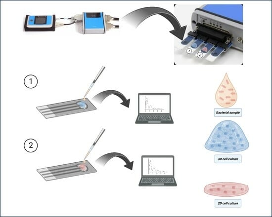
Graphical Abstract

1. Introduction

During the last fifteen years, the number of reported bioassays and biosensor-related approaches in food safety applications based on the use of mammalian cells has steadily increased [1,2,3,4,5]. In many of the published studies, human intestinal cell lines such as Caco-2 and HT-29, both derived from colon adenocarcinomas, have been used to assess the degree of attachment and invasion of key food pathogens, such as Salmonella sp., Bacillus sp., and Listeria sp. [6,7,8]. Caco-2 cells, in particular, are quite useful for modeling bacteria-intestinal epithelium interactions in vitro, since they differentiate into polarized monolayers with a high resemblance to enterocytes in vivo [8,9,10,11,12,13]. Beyond the standard methods for determining cell viability and apoptosis, indirect adhesion assays [14], gentamicin-protective invasion assays [15], and ELISA-type assays [16], bioelectric approaches are being increasingly employed thanks to their usually non-invasive nature, real-time monitoring capability and lack of requirement for reagents and labels. For example, the measurement of transepithelial electrical resistance/impedance (TER/TEI) [17] has been a favorite tool for measuring the reduction of tight junctions in cultured epithelial cells following CaCo-2 (as well as other cell lines) invasion by Salmonella [6,18,19].
Bioelectric impedance spectroscopy (BIS) can be used as a powerful tool to fingerprint cell-to-compound and cell-to-cell interactions in a non-invasive manner, while scanning responses over a wide range of current frequencies provide unique insight into the nature of said interactions, for example, whether cytotoxic effects occur on the cell membrane or intracellularly. Different current frequencies correspond to different phase shifts, which in turn are dependent on the magnitude of the real (i.e., the energy dissipation of the current) and imaginary (i.e., the dielectric capacitance) components of the impedance of the assayed system. For cell assays, applied frequencies usually range between 10 Hz and 100 kHz [20]. It is conditionally assumed that contrary to the cytoplasm, the cell membrane with its lipid bilayer presents a minimum permeability and conductance. As soon as cells attach to the modified electrode’s surface, proliferation begins, and as a result of limited conductivity at low frequencies, leading to high values of the measured impedance directly proportional to the area of the electrode that is covered with cells. Upon cell apoptotic events, however, increased membrane porosity (due to reduced bilayer integrity and loss of tight junctions) will increase conductivity to cytoplasm levels. This simple principle is the basis upon which bioimpedance toxicity assays are being developed. While the measured impedance at the lower frequency range generally reflects the whole cell resistance to current (where membrane contribution to resistance is dominant), responses recorded at higher frequencies can reveal cell membrane-associated interactions (e.g., receptor binding, cellular adhesion, migration) due to the breakdown of dielectric capacitance [21]. That said, researchers have also reported the use of higher frequencies as an alternative approach for electromagnetic assays, i.e., based on the utilization of the electromagnetic spectrum (e.g., microwave emission) [22,23].
Our research group has previously employed this approach to characterize interactions between breast cancer cells and different chemotherapeutical agents [24,25] as well as neuronal cells and neurotransmitters [26]. In the present study, we compared the Salmonella infection progression in the human colon cancer cell line Caco-2 and the mucus-secreting HT29-MTX-E12, after treatment with a different bacterial multiplicity of infection (MOI) by assessing the alterations of frequencies recordings with impedance spectroscopy measurements. In addition, bacterial adhesion and invasion were also evaluated. Finally, the data obtained from impedance spectroscopy recordings were compared to cell viability data from live cell analysis assays.

2. Materials and Methods

2.1. Growth Conditions of Bacterial Strain and Preparation of MOI

The bacterial strain, Salmonella typhimurium (ATCC 14028) was grown in LB broth in a rotary shaker at 37 °C. An overnight culture of bacteria used for cell infection was centrifuged at 3600 g for 10 min at 4 °C, washed twice with phosphate buffer saline (PBS), and grown at 37 °C to reach a bacterial cell density of 108 colony-forming units (cfu) per mL. Then, bacterial cells were diluted in DMEM to yield MOI (multiplicity of infection) ranging from 0.1 to 10.

2.2. Intestinal Human Cell Culture

HT29-MTX-E12 cells (ECACC) were purchased at Sigma-Aldrich (Darmstadt, Germany) and Caco-2 (ATCC HTB-37) were obtained from LGC Standards (Wesel, Germany). Both cell lines were cultured in Dulbecco’s Modified Eagle Medium high glucose (L0103), supplemented with 10% fetal bovine serum and 1% penicillin/streptomycin. When cells reached 80–90% confluency, were subcultured by incubating with 0.25% trypsin and 0.2% EDTA in Ca2+- and Mg2+-free phosphate buffered saline (PBS, Sigma-Aldrich). Cultures were incubated at 37 °C in a humidified atmosphere of 95% air and 5% CO2.

2.3. Adhesion and Invasion Assay

Invasive and attachment capabilities of S. typhimurium were tested in Caco-2 and HT29-MTX-E12 cell lines. Cells were seeded in 96-plate (25,000 cells/well) to reach a monolayer and incubated with 50 μL S. typhimurium different MOI for 1 h at 37 °C. For adhesion assay, cells were washed trice with PBS and detached with trypsin. Adherent bacteria were seeded in LB agar plates and incubated for 24 h at 37 °C and colony-forming units were counted. For invasion assay, the same protocol was followed for cell infection and then cells were incubated with gentamycin 100 μg/mL for 1 h at 37 °C to kill extracellular bacteria. Intestinal cells were washed trice and then disrupted with 0.1% Triton X-100. Cell suspension was plated in LB agar plates and incubated for 24 h at 37 °C for colony-forming enumeration.

2.4. Cell Viability Assessment

Caco-2 and HT29-MTX-E12 cell viability was evaluated using 3-(4,5-dimethylthiazol-2-yl)-2,5-diphenyltetrazolium bromide (MTT) uptake, a marker for cell viability. In brief, Caco-2 cells were incubated with the tested MOI of S. typhimurium for 1 h at 37 °C in DMEM. After washing the cells with PBS, DMEM containing gentamicin at 100 μg/mL was added to kill the adherent bacteria. After 1 h of incubation, cells were washed with PBS, and MTT at 5 mg/mL was added for 1 h at 37 °C. Supernatants were then removed and DMSO was added to dissolve the formazan crystals. The absorbance of the reaction solution was measured at 570 nm using microplate reader (infinite 200M Pro, Tecan, Männedorf, Switzerland). Measurements were performed and % cell viability was determined as follows:
Cell viability (%) = Mean OD/Control OD × 100%.

2.5. Monitoring Mitochondrial Membrane Potential (ΔΨm) in Live Cells

Caco-2 and HT29-MTX-E12 cells were seeded at a density of 25,000 cells in 96-well plates containing complete DMEM medium without antibiotics and incubated at 37 °C in 5% CO2 for 24 h. After the removal of the medium, the cells were infected with the S. typhimurium at the tested MOI (0.1–10) suspended in complete DMEM without antibiotics for 1 h at 37 °C. After incubation, DMEM containing gentamycin at 200 μg/mL was added to each well, and cells were further incubated for 30 min. Cells were then washed twice with PBS and stained with 10 μΜ MitoTracker™ Red (Thermo Fisher Scientific, Waltham, MA, USA) and 10μΜ MitoTracker™ Green (Thermo Fisher Scientific, USA) for 30 min at 37 °C, following PBS washing and then imaged utilizing IncuCyte Live-Cell Imaging System (Sartorius, Göttingen, Germany). For the evaluation of intensity of fluorescence of MitoTracker™ Red/MitoTracker™ Green, four images were taken per well at phase contrast, Red and Green filters, and were analyzed with the IncuCyte ZOOM software (v2019B).

2.6. Electrode Preparation

In this study, in order to investigate the best cell adhesion option, cells were cultured on poly-L-lysine (PLL) pre-coated electrodes (220AT, Metrohm DropSens, Asturias, Spain), as well as in a collagen-based 3D culture biomatrix.
In order to achieve cell adherence on the electrode surface, the gold screen-printed electrodes were initially cleaned with distilled water that was evaporated with nitrogen gas flow. Then, the electrodes were incubated under UV light for 20 min in a laminar flow bench. Subsequently, 10 μL of 50 μg/mL PLL solution was added onto the working electrode surface and left on the laminar flow bench for 30 min. The electrodes were rinsed with sterilized distilled water and were allowed to dry for 1 h into the laminar flow bench.
(A) After this step, a drop of 25,000 cell suspension was added onto the gold working electrode’s surface to form cell monolayer and was incubated for 2 h in the cell culture chamber to obtain cell attachment on the PLL surface. Additional cell culture medium was supplemented, and the electrodes were left in the incubator until measurement (Figure 1A).
(B) For the preparation of the electrodes bearing 3D cell cultures, a stock solution containing 12.5% PBS, 1.25% NaOH 1M, 75% cell suspension in DMEM, and 50% of a collagen-based biomatrix in acetic acid, was used (collagen matrix formulation is subjected to patent submission). Onto each working electrode, 100 μL of the collagen formula was added containing (220AT, Metrohm DropSens, Asturias, Spain) 25,000 cells. The electrodes with the 3D cell cultures were incubated overnight at 37 °C and 5% CO2 (Figure 1B).

2.7. Impedance Measurements

For the measurements’ recordings, the PalmSens 4 potentiostat with the MUX8-R2 extension that allows the use of 8 different electrodes simultaneously was utilized. Five different bacterial MOI (0.1, 0.5, 1, 5, 10) were subjected to frequency scanning with impedance spectroscopy at 3 time intervals (0, 30, and 60 min) for the evaluation of the differential effects during the advancement of infection. The parameters that were used for each measurement are the following: t equilibration 4 s, E dc 0.5 V, t run 1.0 s, t interval 0.01 s, E ac 0.01 V, frequency type scan, number of frequencies 31 = 10/Dec, max–min frequency 10,000–10 Hz.
To test the response of the sensor, plain electrodes were measured. In addition, electrodes with only the different bacterial MOI concentrations, as well as electrodes bearing only cells in 2D and 3D formations served as control. All measurements were conducted with three biological replicates.
All graphic presentations were designed by SigmaPlot 12.0 software (Systat Software). The statistical analysis was conducted using the STATISTICA 12.0 software package (StatSoft Inc., Tulsa, OK, USA). Statistical significance was evaluated by one-way ANOVA at a 95% level of significance (p < 0.05).

3. Results

3.1. S. typhimurium Attachment and Invasion Effect on Caco-2 and HT29-MTX-E12 Cells

S. typhimurium attachment and invasion capacity were tested against Caco-2 and HT29-MTX-E12 cells after 1 h incubation with different MOI. MOI 10 showed a higher ability to adhere and invade both Caco-2 and HT29-MTX-E12 cells. Bacterial attached cell colonies were similar 1 h after cell infection for both colon cell lines following a linear increase according to different MOI (Figure 2A). Bacterial colonies occurring from invaded cells were highly increased upon higher MOI bacterial infection revealing statistically significantly greater invasion capacity against Caco-2 cells (Figure 2B).

3.2. Non-Cytotoxic Effect of S. typhimurium Infection on Caco-2 and HT29-MTX Cells

Caco-2 and HT29-MTX-E12 cell viability studies were performed to assess the potential cytotoxic effect of S. typhimurium infection with a range of different MOIs. None of the tested MOIs had any impact on the viability of Caco-2 and HT29-MTX-E12 cells after 1 h of the bacterial infection (Figure 3).

3.3. Effect of S. typhimurium on Mitochondrial Potential on Caco-2 and HT29-MTX Cells

To further determine the functional mitochondria of Caco-2 and HT29-MTX cells pre-treated with a range of MOIs of S. typhimurium, the ratio of the MitoTracker Red to MitoTracker Green fluorescence intensity was used (Figure 4). In comparison with the control Caco-2 cells, MOI of 0.1 and 0.5 of S. typhimurium exhibited notably decreased mitochondrial membrane potential (ΔΨm), whereas MOI of 1, 5, and 10 presented no statistical differences. Regarding HT29-MTX-E12 cells, none of the tested MOIs of the bacterial strain revealed statistically significant differences compared to the control cells.

3.4. Impedance Comparative Studies on the Effects of S. typhimurium on 2D and 3D Cell Cultures

During this evaluation stage, two experimental procedures have been followed. At the first experimental approach, the impedance analysis was recorded from cells that have been cultured onto the gold screen printed electrodes’ surfaces with PLL for enhancing cell attachment (2D cultures). In the second experimental approach, the cells were immobilized in a collagen-based gel matrix (3D cultures). Figure 5 depicts the mean impedance (n = 3) of the immobilized cells treated with five bacterial MOI (0.1, 0.5, 1, 5, and 10) for each frequency provided by the device. In all measurements, the notation (control) in the graphs indicates the nutrient medium. In the case of Caco-2 2D cultures (Figure 5A,B), a better resolution was obtained at the impedance values after the application of the frequency scan 30 min after treatment with the bacterial MOI. In the case of the 3D cultures (Figure 5C,D), a different pattern at the impedance values was obtained but the differential response was clearer 30 min post-treatment.
On the other hand, the 2D cultures of the HT29-MTX-E12 cells (Figure 6A,B) presented significantly higher impedance values in comparison with the control at both time intervals. At the 30 min treatment, a differential response was observed. The same pattern was observed with the 3D cultures (Figure 6C,D) but the differentiation between different concentrations was not very successful.

4. Discussion

Salmonella typhimurium infects both humans and animals by binding to host cells. In this detailed study, we tried to characterize S. typhimurium interactions with both the Caco-2 epithelial and the mucus-secreting HT29-MTX-E12 2D and 3D cell cultures through impedance recordings. The first cell line derives from human colon adenocarcinoma tissue and closely mimics the structural and functional characteristics of mature enterocytes in vitro [27]. The HT29-MTX-E12 model, also derived from a human colon adenocarcinoma, exhibits structural and functional similarities to mature human enterocytes. However, when cultured in the presence of glucose, it maintains a constant proliferation rate with practically no further differentiation [28]. Initially, no cytotoxicity of S. typhimurium was observed after 1 h treatment with five MOIs, showing that this concentration had no effect on mammalian cells. Thus, we proceeded to investigate bacterial adhesion and invasion.
The adhesion of pathogenic bacteria to the surfaces of host cells plays a crucial role in their survival and replication. This step is pivotal in pathogenesis, as it determines tissue tropism by binding to specific surface receptors and enhances resistance to physical removal from extracellular fluids at mucosal sites [29]. In this study, we observed that S. typhimurium adhered to both host cell surfaces, in a dose MOI concentration-dependent manner. Once attached to the host cell surface, bacteria can enter the cells either by binding to surface receptors or by directly transporting bacterial proteins into the host cell. The invasion of S. typhimurium into host cells is critical for its survival and establishment of infection in the host. Our findings indicate that the invasion efficiency of Salmonella both in Caco-2 and HT29 cells is related to the MOI concentration utilized.
Modern biosensor technologies offer a rapid means of detecting bacteria adhesion, biofilm formation, as well as the effects of antibiotics. Furthermore, these sensor technologies have wide detection limits, making them suitable for studying biological processes at the molecular scale. Usually, these measurements can be performed in real time without labeling. The development of novel sensors provides the potential to alter dramatically fundamental and applied research in biological sciences. Mechanical, optical, and electrical sensing technologies with specific capabilities can be utilized in bacteria monitoring. Electrochemical biosensors, and especially impedimetric sensors, have the potential to lead in the field of biomedical research and monitoring, but there are several important steps, including optimization, miniaturization, and clinical trials, that must be undertaken to ensure their readiness for market deployment and practical use in healthcare and research settings [30].
Impedance-based techniques are considered some of the most trustworthy and reproducible techniques for characterizing electrochemical systems and providing information about diffusive impedance, double-layer capacitance, etc. [31]. Impedance is convenient for system characterization such as fuel cells, semiconductors, batteries, and biosensors. Impedance (Z) measurements are recorded basically by the application of small voltage perturbations to a system and monitoring the respective current response. Hence, impedance is the simple expression of the quotient of the voltage–time function and the current–time function. Furthermore, the measurement of the current–voltage ratio can be easily performed by the electrochemical impedance spectroscopy (EIS) technique, in which the frequency of the applied sinusoidal voltage varies. In addition, electrochemical impedance spectroscopy (EIS), stands for the recording of the current as a function of an applied sinusoidal voltage of a given frequency. It is used very frequently for impedimetric analysis in biosensors because vast amounts of information can be acquired. An additional advantage of this technique is that it offers the ability to repeat a specific measurement over a variety of frequencies, indicating that the resulting data can be utilized for the investigation of several electrochemical phenomena [32]. The resulting data from EIS recordings can be plotted in several manners to extrapolate many parameters relevant to the system.
So far, in studies involving infections with S. typhimurium and similar enteric pathogens, there has been a historical focus on TEER measurements at later time points, possibly due to technological limitations that hindered the measurement of TEER changes during the initial stages of infection. This implies the need for advancements in technology or methods to better understand the early events in these infections and their impact on epithelial barrier function [33]. One exception is a study where MDCK I cells were infected with S. typhimurium and conductance was recorded in an Ussing chamber-type apparatus, [33]. Measurements of transepithelial electrical resistance (TEER) are a standard technical approach to assessing barrier properties and dynamics of various in vitro cell culture models. The existing systems for monitoring the barrier function present the disadvantage of low throughput. In addition, these systems are fabricated to utilize up to 24 membrane inserts which is definitely not enough for high-throughput screenings. Given that the impedance of a system depends on the applied frequency, generally a range of frequencies are scanned by the use of EIS.
We present the first example of an impedance study on the effect of S. typhimurium infection on human intestinal cells. Live-cell-based techniques provide more specific and accurate results, particularly in differentiating live pathogens from dead bacteria or background flora, which can be challenging with other diagnostic methods [34]. The infection of S. typhimurium in Caco-2 2D cultures gave a clearer differential response of the impedance values 30 min post-treatment in comparison with the 3D cultures. The behavior of the HT29-MTX-E12 in 2D cultures was different from Caco-2 cells with higher impedance values in comparison with the control at both time intervals with differential response observed at 30 min. The 3D cultures presented a similar pattern. Bacterial adhesion is considered an initial step in the infection process and suggests that this adhesion process has a significant impact on the rate at which bacteria induce changes in the surface properties of host cells. The data from the study apparently supports this hypothesis.
Originally, impedance spectroscopic methods were used for the investigation of biological phenomena of cell cultures either in suspensions or in conventional 2D cultures. Three-dimensional cultures provide a physiologically relevant microenvironment to the cell [35], leading researchers to numerous attempts to adapt IS to three-dimensional cultures. The conduction of typical in vitro tests in 3D cell models is more difficult and time-consuming than in 2D. More specifically, 3D cultures lead to scattering effects that limit the use of conventional optical techniques. On the contrary, impedance spectroscopy allows relatively non-invasive, real-time data into 3D cell cultures. Thus, a 3D cell morphology could partially explain the differential responses in the impedance values that we observed between 2D and 3D cultures.

5. Conclusions

In this work, human colon cancer cell cultures (2D and 3D) were applied onto gold screen-printed electrodes for impedance analysis. It must be emphasized that our study represents just the first step towards implementing impedance spectroscopy as a reliable way to study bacteria x intestinal cell interactions. Further research should focus on expanding the scope of cell types and bacteria species (possibly including members of the host microbiome) as well as establishing a set-up for long-term study of bacterial infection with the aid of bespoke microfluidics and additional measurement of infection-associated biomarkers. The final goal is the creation of a gut-like organ-on-chip system coupled with the dynamic capabilities of impedance spectroscopy as shown in the present study.

Author Contributions

Conceptualization, S.M. (Sofia Mavrikou), E.F. and S.K.; methodology, S.M. (Sofia Marka), M.-E.Z. and G.-M.P.; formal analysis, S.M. (Sofia Marka), S.M. (Sofia Mavrikou), M.-E.Z. and G.-M.P.; investigation, S.M. (Sofia Marka), M.-E.Z. and G.-M.P.; data curation, S.M. (Sofia Marka), M.-E.Z. and G.-M.P.; writing—original draft preparation, S.M. (Sofia Marka), S.M. (Sofia Mavrikou), M.-E.Z. and G.-M.P.; writing—review and editing, S.M. (Sofia Mavrikou) and S.K.; supervision, S.M. (Sofia Mavrikou), E.F. and S.K. All authors have read and agreed to the published version of the manuscript.

Funding

This research received no external funding.

Institutional Review Board Statement

Not applicable.

Informed Consent Statement

Not applicable.

Data Availability Statement

The data presented in this study are available on request from the corresponding author.

Conflicts of Interest

The authors declare no conflict of interest.

References

  1. Banerjee, P.; Bhunia, A.K. Mammalian Cell-Based Biosensors for Pathogens and Toxins. Trends Biotechnol. 2009, 27, 179–188. [Google Scholar] [CrossRef] [PubMed]
  2. Reverté, L.; Soliño, L.; Carnicer, O.; Diogène, J.; Campàs, M. Alternative Methods for the Detection of Emerging Marine Toxins: Biosensors, Biochemical Assays and Cell-Based Assays. Mar. Drugs 2014, 12, 5719–5763. [Google Scholar] [CrossRef] [PubMed]
  3. Xia, S.; Zhu, P.; Pi, F.; Zhang, Y.; Li, Y.; Wang, J.; Sun, X. Development of a Simple and Convenient Cell-Based Electrochemical Biosensor for Evaluating the Individual and Combined Toxicity of Don, Zen, and Afb1. Biosens. Bioelectron. 2017, 97, 345–351. [Google Scholar] [CrossRef] [PubMed]
  4. Jiang, D.; Ge, P.; Wang, L.; Jiang, H.; Yang, M.; Yuan, L.; Ge, Q.; Fang, W.; Ju, X. A Novel Electrochemical Mast Cell-Based Paper Biosensor for the Rapid Detection of Milk Allergen Casein. Biosens. Bioelectron. 2019, 130, 299–306. [Google Scholar] [CrossRef]
  5. Yazgan Karacaglar, N.N.; Topcu, A.; Dudak, F.C.; Boyaci, I.H. Development of a Green Fluorescence Protein (Gfp)-Based Bioassay for Detection of Antibiotics and Its Application in Milk. J. Food Sci. 2020, 85, 500–509. [Google Scholar] [CrossRef]
  6. Gagnon, M.; Zihler Berner, A.; Chervet, N.; Chassard, C.; Lacroix, C. Comparison of the Caco-2, HT-29 and the Mucus-Secreting HT29-MTX Intestinal Cell Models to Investigate Salmonella Adhesion and Invasion. J. Microbiol. Methods 2013, 94, 274–279. [Google Scholar] [CrossRef]
  7. Zihler, A.; Gagnon, M.; Chassard, C.; Lacroix, C. Protective Effect of Probiotics on Salmonella Infectivity Assessed with Combined in Vitro Gut Fermentation-Cellular Models. BMC Microbiol. 2011, 11, 264. [Google Scholar] [CrossRef]
  8. Ostovan, R.; Pourmontaseri, M.; Hosseinzadeh, S.; Shekarforoush, S.S. Interaction between the Probiotic Bacillus subtilis and Salmonella typhimurium in Caco-2 Cell Culture. Iran. J. Microbiol. 2021, 13, 91–97. [Google Scholar] [CrossRef]
  9. Rousset, M. The Humancolon Carcinoma Cell Lines HT-29 and Caco-2: Two in Vitro Models for the Study of Intestinal Differentiation. Biochimie 1986, 68, 1035–1040. [Google Scholar] [CrossRef]
  10. Reddy, S.; Austin, F. Adhesion and Invasion Assay Procedure Using Caco-2 Cells for Listeria monocytogenes. Bio-Protocol 2017, 7, e2267. [Google Scholar] [CrossRef]
  11. Giannasca, K.T.; Giannasca, P.J.; Neutra, M.R. Adherence of Salmonella typhimurium to Caco-2 Cells: Identification of a Glycoconjugate Receptor. Infect. Immun. 1996, 64, 135–145. [Google Scholar] [CrossRef] [PubMed]
  12. Jankowska, A.; Laubitz, D.; Antushevich, H.; Zabielski, R.; Grzesiuk, E. Competition of Lactobacillus paracasei with Salmonella enterica for Adhesion to Caco-2 cells. J. Biomed. Biotechnol. 2008, 2008, 357964. [Google Scholar] [CrossRef] [PubMed]
  13. Finlay, B.B.; Brumell, J.H. Salmonella Interactions with Host Cells: In Vitro to In Vivo. Philos. Trans. R. Soc. Lond. B Biol. Sci. 2000, 355, 623–631. [Google Scholar] [CrossRef] [PubMed]
  14. Mellor, G.E.; Goulter, R.M.; Raymond Chia, T.W.; Dykes, G.A. Comparative Attachment of Shiga Toxigenic Escherichia coli and Salmonella to Cultured HT-29 and Caco2 Cell Lines. Appl. Environ. Microbiol. 2009, 75, 1796–1799. [Google Scholar] [CrossRef]
  15. Steele-Mortimer, O. Infection of Epithelial Cells with Salmonella enterica. Methods Mol. Biol. 2008, 431, 201–211. [Google Scholar]
  16. Le Blay, G.; Fliss, I.; Lacroix, C. Comparative Detection of Bacterial Adhesion to Caco-2 Cells with ELISA, Radioactivity and Plate Count Methods. J. Microbiol. Methods 2004, 59, 211–221. [Google Scholar] [CrossRef]
  17. Cacopardo, L.; Costa, J.; Giusti, S.; Buoncompagni, L.; Meucci, S.; Corti, A.; Mattei, G.; Ahluwalia, A. Real-Time Cellular Impedance Monitoring and Imaging of Biological Barriers in a Dual-Flow Membrane Bioreactor. Biosens. Bioelectron. 2019, 140, 111340. [Google Scholar] [CrossRef]
  18. Fajdiga, S.; Koninkx, J.F.; Tooten, P.C.; Marinsek-Logar, R. Interference of Salmonella enteritidis and Lactobacillus spp. with IL-8 Levels and Transepithelial Electrical Resistance of Enterocyte-like Caco-2 Cells. Folia Microbiol. 2006, 51, 268–272. [Google Scholar] [CrossRef]
  19. Smith, I.M.; Baker, A.; Arneborg, N.; Jespersen, L. Non-Saccharomyces Yeasts Protect against Epithelial Cell Barrier Disruption Induced by Salmonella enterica subsp. enterica serovar typhimurium. Lett. Appl. Microbiol. 2015, 61, 491–497. [Google Scholar] [CrossRef]
  20. Hassan, Q.; Ahmadi, S.; Kerman, K. Recent Advances in Monitoring Cell Behavior Using Cell-Based Impedance Spectroscopy. Micromachines 2020, 11, 590. [Google Scholar] [CrossRef]
  21. Benson, K.; Cramer, S.; Galla, H.-J. Impedance-Based Cell Monitoring: Barrier Properties and Beyond. Fluids Barriers CNS 2013, 10, 1–11. [Google Scholar] [CrossRef] [PubMed]
  22. Kazemi, N.; Abdolrazzaghi, M.; Light, P.; Musilek, P. In–Human Testing of a Non-Invasive Continuous Low–Energy Microwave Glucose Sensor with Advanced Machine Learning Capabilities. Biosens. Bioelectron. 2023, 241, 115668. [Google Scholar] [CrossRef] [PubMed]
  23. Abdolrazzaghi, M.; Kazemi, N.; Nayyeri, V.; Martin, F. AI-Assisted Ultra-High-Sensitivity/Resolution Active-Coupled CSRR-Based Sensor with Embedded Selectivity. Sensors 2023, 23, 6236. [Google Scholar] [CrossRef] [PubMed]
  24. Paivana, G.; Barmpakos, D.; Mavrikou, S.; Kallergis, A.; Tsakiridis, O.; Kaltsas, G.; Kintzios, S. Evaluation of Cancer Cell Lines by Four-Point Probe Technique, by Impedance Measurements in Various Frequencies. Biosensors 2021, 11, 345. [Google Scholar] [CrossRef] [PubMed]
  25. Paivana, G.; Mavrikou, S.; Kaltsas, G.; Kintzios, S. Bioelectrical Analysis of Various Cancer Cell Types Immobilized in 3D Matrix and Cultured in 3D-Printed Well. Biosensors 2019, 9, 136. [Google Scholar] [CrossRef] [PubMed]
  26. Paivana, G.; Apostolou, T.; Mavrikou, S.; Barmpakos, D.; Kaltsas, G.; Kintzios, S. Impedance Study of Dopamine Effects after Application on 2D and 3D Neuroblastoma Cell Cultures Developed on a 3D-Printed Well. Chemosensors 2019, 7, 6. [Google Scholar] [CrossRef]
  27. Pinto, M.; Robine-Leon, S.; Appay, M.D.; Kedinger, M.; Triadou, N.; Dussaulx, E.; Lacroix, B.; Simon-Assman, P.; Haffen, K.; Fogh, J.; et al. Enterocyte-like Differentiation and Polarization of the Human Colon Carcinoma Cell Line Caco-2 in Culture. Biol. Cell 1983, 47, 323–330. [Google Scholar]
  28. Zweibaum, A.; Pinto, M.; Chevalier, G.; Dussaulx, E.; Triadou, N.; Lacroix, B.; Haffen, K.; Brun, J.L.; Rousset, M. Enterocytic Differentiation of a Subpopulation of the Human Colon Tumor Cell Line HT-29 Selected for Growth in Sugar-Free Medium and Its Inhibition by Glucose. J. Cell. Physiol. 1985, 122, 21–29. [Google Scholar] [CrossRef]
  29. Connell, H.; Hedlund, M.; Agace, W.; Svanborg, C. Bacterial Attachment to Uro-Epithelial Cells: Mechanisms and Consequences. Adv. Dent. Res. 1997, 11, 50–58. [Google Scholar] [CrossRef]
  30. Ahmed, A.; Rushworth, J.V.; Hirst, N.A.; Millner, P.A. Biosensors for Whole-Cell Bacterial Detection. Clin. Microbiol. Rev. 2014, 27, 631–646. [Google Scholar] [CrossRef]
  31. Cencic, A.; Langerholc, T. Functional Cell Models of the Gut and Their Applications in Food Microbiology—A Review. Int. J. Food Microbiol. 2010, 141, S4–S14. [Google Scholar] [CrossRef] [PubMed]
  32. Macdonald, J.R. Impedance Spectroscopy. Ann. Biomed. Eng. 1992, 20, 289–305. [Google Scholar] [CrossRef] [PubMed]
  33. Ates, M. Review Study of Electrochemical Impedance Spectroscopy and Equivalent Electrical Circuits of Conducting Polymers on Carbon Surfaces. Prog. Org. Coat. 2011, 71, 1–10. [Google Scholar] [CrossRef]
  34. Singh, J.; Batish, V.K.; Grover, S. Simultaneous Detection of Listeria monocytogenes and Salmonella spp. in Dairy Products Using Real Time PCR-Melt Curve Analysis. J. Food Sci. Technol. 2012, 49, 234–239. [Google Scholar] [CrossRef] [PubMed]
  35. Rivnay, J.; Ramuz, M.; Leleux, P.; Hama, A.; Huerta, M.; Owens, R.M. Organic Electrochemical Transistors for Cell-Based Impedance Sensing. Appl. Phys. Lett. 2015, 106, 043301. [Google Scholar] [CrossRef]
Figure 1. Graphical representation of (A) 2D and (B) 3D cell culture onto the screen-printed electrode surface.
Chemosensors 11 00534 g001
Figure 2. Adhesion (A) and invasion (B) of different MOI (0.1–10) of Salmonella typhimurium to Caco-2 and HT29-MTX-E12 cells. For adhesion assay (A) cells were detached after 1 h infection and the cell suspension was added. For the invasion assay (B), at 1 h post-infection, cells were incubated for further 1 h with 100 μg/mL gentamycin, washed thrice, and then disrupted with 0.1% Triton X-100 prior to plating of cell suspension. Results are shown as the mean ± SD; n = 4. Significant differences among treatments are indicated by different letters according to Duncan’s range test (p < 0.05).
Chemosensors 11 00534 g002
Figure 3. Relative viability of Caco-2 and HT29-MTX-E12 cells after 1 h treatment with different MOIs (0.1–10) of S. typhimurium by the MTT Assay. All values represent the mean ± SD; n = 5. No significant differences were observed among treatments according to Duncan’s range test (p < 0.05).
Chemosensors 11 00534 g003
Figure 4. Evaluation of mitochondrial membrane potential of Caco-2 and HT29-MTX-E12 cells after 1 h incubation with MOI (0.1–10) of S. typhimurium. Cells were stained with MitoTracker Red and Green fluorescent dyes. Control cell monolayers received no treatment (control). Values are mean ± SEM (n = 4). Values followed by the same letter (a, b, c,) are not significantly different (p < 0.05) by Duncan’s multiple range test.
Chemosensors 11 00534 g004
Figure 5. Bode diagram of the Caco-2 cells impedance responses (A) 2D cultures 30 min after bacteria application, (B) 2D cultures 60 min after bacteria application, (C) 3D cultures 30 min after bacteria application, (D) 3D cultures 60 min after bacteria application. Values are mean of three biological replicates (n = 3).
Chemosensors 11 00534 g005
Figure 6. Bode diagram of the HT29-MTX-E12 cells impedance responses (A) 2D cultures 30 min after bacteria application, (B) 2D cultures 60 min after bacteria application, (C) 3D cultures 30 min after bacteria application, (D) 3D cultures 60 min after bacteria application. Values are mean of three biological replicates (n = 3).
Chemosensors 11 00534 g006
Disclaimer/Publisher’s Note: The statements, opinions and data contained in all publications are solely those of the individual author(s) and contributor(s) and not of MDPI and/or the editor(s). MDPI and/or the editor(s) disclaim responsibility for any injury to people or property resulting from any ideas, methods, instructions or products referred to in the content.