Article

Dilute-and-Shoot-Liquid Chromatography-Quadrupole Time of Flight-Mass Spectrometry for Pteridine Profiling in Human Urine and Its Association with Different Pathologies

by
Claudia Giménez-Campillo
1,
Marta Pastor-Belda
1,
Natalia Arroyo-Manzanares
1,
Natalia Campillo
1,
Blanca del Val Oliver
2,
José Zarauz-García
3,
Luis Sáenz
3 and
Pilar Viñas
1,*
1
Department of Analytical Chemistry, Faculty of Chemistry, University of Murcia, Regional Campus of International Excellence “Campus Mare Nostrum”, E-30071 Murcia, Spain
2
Internal Medicine Service—Gastroenterology and Hepatology Section, Hospital General Universitario Rafael Méndez, 30813 Lorca, Spain
3
Laboratory Medicine Department, Hospital General Universitario Rafael Méndez, 30813 Lorca, Spain
*
Author to whom correspondence should be addressed.
Chemosensors 2023, 11(6), 324; https://doi.org/10.3390/chemosensors11060324
Submission received: 30 March 2023 / Revised: 15 May 2023 / Accepted: 23 May 2023 / Published: 1 June 2023

Abstract

:
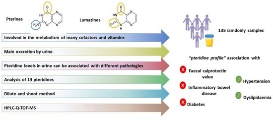
Pteridines are a group of compounds synthesised by many living organisms that are involved in the metabolism of many cofactors and vitamins. Their concentration in biological fluids may be altered by various pathologies such as cancer or inflammatory bowel disease, urine being the main route of excretion. In this study, three lumazines and ten pterins were analysed in their native oxidation state using high-performance liquid chromatography coupled to quadrupole time-of-flight mass spectrometry. Their high concentration in urine samples and their good ionisation behaviour allow the use of the dilute-and-shoot method by simple filtration of the urine prior to chromatographic analysis. The final method offers excellent linearity, sensitivity and precision parameters, and a total of 135 urine samples were analysed from patients with some relevant information such as faecal calprotectin (FCP) levels, common diseases such as diabetes, hypertension and dyslipidaemia and immunological diseases such as inflammatory bowel disease (IBD). The pteridine profile was related to FCP levels without showing any correlation. In addition, pteridine levels were compared between healthy subjects and IBD, diabetic, hypertensive and dyslipidaemic patients, and significant differences were found between the two groups for some of the pteridines.

Graphical Abstract

1. Introduction

Pteridines are low molecular weight organic compounds consisting of fused pyrazine and pyrimidine rings [1]. Pteridines can be divided into two groups based on their structure: pterins and lumazines. Pterins have an amino group at C-2 and a carbonyl group at C-4, while lumazines have carbonyl groups at C-2 and C-4. These compounds can be synthesised by many living organisms and form part of pigments, act as enzymatic cofactors and can activate the immune system. Their biological concentrations in biological fluids, such as urine, serum and cerebrospinal fluid, can be altered by various pathophysiological processes [2]. This fact has led to the consideration of pteridines as molecular biomarkers capable of detecting and predicting disease outcomes, and even assisting in treatment selection and monitoring therapeutic response [2,3].
Pteridines are mainly excreted via urine, which has been the most studied biological fluid because its collection is non-invasive, and no complicated pre-treatment is required to eliminate possible interferences. Several clinical studies have been conducted analysing urine to research the relationship between pteridines and several diseases, using pteridine fingerprinting or “pterinomics” (urinary pteridine profile) methodology [2]. Neopterin is the most studied pteridine, and it is considered a biochemical indicator of immune activity; its level plays a major role in the monocyte activity of the inflammatory response. It has been detected in urine, serum, saliva, synovial fluid, ascitic fluid, cerebrospinal fluid, bile, gastric juice and pancreatic juice [4], and its concentration is of great clinical importance in the monitoring of different diseases. The levels in serum and urine have been related to the size and severity of burns [5], with the detection of rheumatoid arthritis [6], various types of cancer [7,8,9,10], viral or bacterial infections [11], diabetes [12], cardiovascular disease [13] and autoimmune diseases such as inflammatory bowel disease (IBD). Neopterin levels in Chron’s disease and ulcerative colitis have been extensively studied, with conflicting results. Some authors conclude that there is no association [14], others propose a positive association using faecal levels [15]. In contrast, other authors point out that, since it is not a specific marker, other clinical parameters should be assessed to monitor the disease [16]. A positive correlation has been described with clinical parameters such as C-reactive protein, alpha-1-glycoprotein and interleukin-1 antagonist receptor in serum [17,18] or the urinary neopterin concentration concerning the duration of illness, patient body mass and the occurrence of abdominal tumour [19]. Faecal calprotectin (FCP) levels are a widely used clinical indicator of possible IBD due to their relationship with bowel inflammation [20], and to date, no author has established an association with pteridine levels.
The determination of pteridine in urine samples has been performed by enzyme-linked immunosorbent assay (ELISA) [15,21], capillary electrophoresis (CE) [22] and high-performance liquid chromatography (HPLC) [23,24,25,26,27,28,29,30,31,32,33], the latter being the most widely used separation technique coupled with different detection systems. The fluorescence detection (FD) [23,24,25,26,27,29,31] and mass spectrometry (MS) [28,30,31,32,33] are the most widely used detectors, the latter becoming increasingly important in the study of pteridines due to their sensitivity and selectivity. The coupling of HPLC to high-resolution MS (HRMS), specifically quadrupole-time of flight (QTOF-MS), has been applied to the determination of pteridines in cancer cell cultures [34]. Meanwhile, the determination of these compounds in urine samples using this type of analyser has not been found in the literature.
Due to the different natural oxidation states of pteridines (aromatic, dihydro (half-reduced) and tetrahydro (totally reduced)) and the different stability of each form, an oxidative or antioxidative pre-treatment is necessary to fully oxidise or reduce the pteridine compounds to a unique oxidation state using, among others, triiodide, permanganate, manganese dioxide, hydrogen peroxide and UV irradiation [35]. Another possibility is using dithiothreitol as a stabilising agent that preserves the pteridines in their native oxidation state [32].
The total analysis time is critically determined by the sample preparation time because it is usually a laborious step where different chemicals and consumables are used. Therefore, if the sample treatment is reduced to a simple dilution and/or filtration of the sample, this is an environmentally friendly analytical technique called dilute-and-shoot (DS). Its combination with MS could be proposed as a good alternative if the compounds studied have a good ionisation behaviour and if the analytes are present in the samples in concentrations high enough to be detected without the need to concentrate [36]. These two requirements are fulfilled to analyse pteridines in urine, so this technique was chosen together with HRMS for the analysis. This eliminates the need for complicated method development. However, it may present problems such as eliminating the matrix effect and, depending on the MS analyser used, poor measurement robustness could result when analysing a large number of compounds [37].
The major benefits of the DS procedure are simplicity, minimal loss of analytes, high sample throughput and a wide range of applications. This method has been widely used in the areas of analytical toxicology and doping-control analysis for the determination of a wide variety of substances in urine, such as prescription drugs, anabolic steroids and sports drugs [38,39,40,41,42].
This work deals with the determination of 10 pterins and 3 lumanizes in their native oxidation state, such as pterin (PT), 7,8-dihydroxanthopterin (7,8-DHXAN), 6,7-dimethylpterin (6,7-DMPT), 6-hydroxymethylpterin (6-OHMPT), leucopterin (LEU), 6-biopterin (6-BIO), 7,8-dihydro-L-biopterin (7,8-DHBIO), l-monapterin (MON), d-(+)-neopterin (NEO), 7,8-dihydroneopterin (7,8-DHNEO), lumazine (LU), 6-hydroxylumazine (6-HLU) and 7-hydroxylumazine (7-HLU), in human urine by DS combined with HPLC-Q-TOF-MS methodology. Several statistical studies were applied to the concentrations calculated in the 135 urine samples analysed to find relationships with different clinical parameters such as FCP.

2. Materials and Methods

2.1. Reagents

Pteridine standards were supplied by Schircks Laboratories (Jona, Switzerland): Pterins, with purities between 98.4 and 99.5%, were: PT, 7,8-DHXAN, 6,7-DMPT, 6-OHMPT, LEU, 6-BIO, 7,8-DHBIO, MON, NEO, 7,8-DHNEO and 7-methylpterin, which was used as internal standard (IS). Lumazines, such as LU, 6-HLU and 7-HLU, were obtained with purities higher than 99.5% from Shircks Laboratories. Figure 1 shows the chemical structure of each of the analysed pteridines.
The dissolution of pteridins is a major challenge due to their low solubility in water and organic solvents. The use of acidic pH to dissolve pteridines could also cause the cleavage of the double ring, giving rise to other compounds, whereas a basic pH allows the dissolution of the pteridines without affecting their chemical structure. Stabilisers such as dithiothreitol (DTT) and ascorbic acid (AA) were used to slow down the oxidation reaction of pteridines in the presence of air [27,28,35,43]. Individual solutions of the different pterins were prepared at 500 mg L−1 using 2 M NaOH as solvent and DTT and AA were added to each solution at 1 and 4 g L−1, respectively. Both DTT and AA, added as stabilising agents to preserve the pterins in their native oxidation state [2,27,28,32,43], were supplied by Sigma (St. Louis, MO, USA). Standard solutions of lumazines were prepared at 1000 mg L−1 in Milli-Q water with a few drops of 1 M NaOH. The standards were kept in the dark at −20 °C and stability studies were performed by injecting different dilutions of the stock solutions at different times (during the first 7 days, after 15 days and after 1 month). The compounds remained stable for the first 4 days. After this time significant degradation of the compounds begins, with approximately 50% of pteridine degradation occurring within one month of preparing the stock solution. Therefore, the diluted standards solutions used for calibration must be prepared and used within the next 4 days.
The chromatographic grade organic solvent was acetonitrile (ACN) from Chem-Lab (Zedelgem, Belgium). The water used as a solvent in the mobile phase was purified with a Milli-Q system (Millipore, Bedford, MA, USA). Formic acid and sodium hydroxide were purchased from Sigma and Fisher Scientific (Loughborough, UK), respectively.

2.2. Instrumentation

The analysis was performed on an Agilent 1290 Infinity II Series HPLC system (Agilent Technologies, Santa Clara, CA, USA), with a high-speed binary pump. A Discovery RP Amide C16 column (15 cm × 4.6 mm, 5 µm, Merck, Darmstadt, Germany), kept at 25 °C in a thermostatic compartment, was used. The mobile phase was a 99:1 mixture of solvent A (0.1% v/v formic acid) and B (ACN with 0.1% v/v formic acid) in isocratic mode. The analytes eluted with retention times between 5.13 and 13.20 min, corresponding to 7,8-DHNEO and 7-HLU, respectively. Standards and samples were kept at 4 °C in a sampler tray until injection (20 μL) using an autosampler.
The LC system was coupled to an Agilent 6550 Q-TOF mass spectrometer (Agilent Technologies, Santa Clara, CA, USA) with an Agilent Jet Stream Dual (AJS-Dual ESI) electrospray source running in positive ionisation mode with the following parameters: capillary voltage, 4000 V; nebuliser gas pressure, 40 psi; drying gas flow, 16 L min−1; drying gas temperature, 150 °C; fragmentor voltage, 350 V; nozzle voltage, 1000 V and 1 RF Vpp octapole, 750 V. The parameters for HPLC-Q-TOF-MS were obtained from the MassHunter Workstation data acquisition software (Agilent Technologies, Santa Clara, CA, USA, Rev. B.08. 00). Profile data were acquired in the 50–500 m/z range for MS scans in 2 GHz extended dynamic range mode with 3 spectra/s, 333.3 ms/spectrum and 1999 transitions/spectrum. All ions mode was used for non-targeted data acquisition. In each cycle the following collision energies were used 0, 10 and 40 V. MS calibration was carried out introducing into the system a reference mass of 121.0509 for mass correction during the analysis.
Data analysis was performed using MassHunter Qualitative Analysis Navigator software, version B.08.00 (Agilent Technologies, Santa Clara, CA, USA).
Compounds were monitored using the extracted ion chromatogram (EIC) of the protonated molecules, with a 0.01 ppm window and subtracting the background. The exact formula-based theoretical masses were calculated using the Molecular Mass Calculator tool of the MassHunter software.
Centrifugation of the samples was performed in an EBA 20 centrifuge (Hettich, Tuttlingen, Germany) at 600× g and filtration was carried out using 0.2 μm PTFE vial filters without needle (Agilent Uni-Prep, Santa Clara, CA, USA).

2.3. Samples and Analytical Procedure

Urine samples were supplied by Hospital Universitario Rafael Méndez (Lorca, Murcia, Spain) according to hospital guidelines, collected in 15 mL sterile tubes containing DDT and AA and stored immediately frozen at −20 °C until analysis. Ethical report ID: 2908/2020 was obtained from a committee of the University of Murcia for the development of this study. All patients signed an informed consent form.
A total of 135 randomly selected urine samples were analysed, finding patients with very different characteristics. The age range covered is very wide, with samples from patients aged between 18 and 82 years. Regarding sex, the samples were very evenly distributed, with 54% of the urine samples coming from women and the rest from men. The FCP levels in the samples analysed ranged from less than 15 to more than 3000, with only 22 samples having FCP levels above 200. Regarding other common diseases, 35 of the 135 patients had hypertension, 16 had diabetes and 32 had dyslipidaemia. In terms of consumption habits, only nine patients (five men and four women) admitted to being smokers, while only four men reported continuous alcohol consumption. Finally, after reviewing the patients’ medical records, it is confirmed that 10 patients were diagnosed with IBD after sample collection.
After thawing the urine samples, a centrifugation step (2 min at 600× g) was necessary to remove solid particles present in the samples. The DS methodology for urine sample analysis consisted of adding 5 μL of IS (10 mg L−1) to 495 μL of the centrifugated urine and the mixture was filtered with a 0.22 μm syringe less nylon filter prior to HPLC-Q-TOF-MS analysis (20 μL).

3. Results and Discussion

3.1. Optimization of Separation and Detection Conditions

The high polarity and low solubility in water and organic solvents make the chromatographic separation of pteridines an interesting challenge. For this purpose, other authors have used HPLC in different modalities such as reversed phase (RP), hydrophilic interaction (HILIC) and ion exchange, among others [23,24,26,27,28,30,33,44]. HILIC-HPLC was tested with an Agilent InfinityLab Poroshell 120 HILIC-Zwitterionic (100 × 2.1 mm, 2.7 μm) with a 5:95 mobile phase of 0.1% v/v formic acid (solvent A) and ACN (solvent B) in isocratic mode, without obtaining good resolution parameters. All pterins eluted in less than 2 min, so the percentage of organic solvent (B) was increased to 99%. However, good results were not obtained because, although the analytes were better retained, they still coeluted and showed poor peak resolution. Reversed-phase liquid chromatography was tested using different columns with 0.1% formic acid (solvent A) and ACN (solvent B) mobile phase in an isocratic mode to improve chromatographic parameters. The Zorbax RRHD Eclipse Plus C18 column (2.1 × 100 mm, 1.8 μm, Agilent Technologies) was tested from 85 to 100% solvent A. Unsatisfactory results were obtained, as all eluted compounds overlapped at low retention times, except 6,7-DMPT, which eluted after 18 min. Overlapping peaks were also found using the Discovery HS PEG-3 column (polyethene glycol, 4 mm × 100 mm, 3 μm, Sigma Aldrich, St. Louis, MO, USA) with 100% water containing formic acid at 0.1% as mobile phase, which was discarded. Even with minimal eluent strength, in the absence of organic solvent, chromatographic peaks were not delayed. Finally, the Discovery RP Amide C16 column (4.6 × 150 mm, 5 μm, Merk) was tested with different mobile phase compositions from 95 to 100% solvent A in isocratic mode. Considering that ESI systems improve their ionisation efficiency in the presence of organic solvents, the 99:1 ratio (solvent A: solvent B) was finally adopted in the mobile phase for the HPLC-Q-TOF-MS system. Under these conditions, all compounds elute between 5.13–13.20 min corresponding to 7,8-DHNEO and 7-HLU, respectively. The MON/NEO and 6-HLU/7-HLU pairs, despite being diastereoisomers, achieved acceptable chromatographic resolution. Only 7,8-DHXAN and LU eluted with slight differences at 10.45 and 10.59 min, respectively. The retention time for the IS was 16.35 min.
Positive and negative ESI ionisation modes were compared, with positive ionisation giving the best results for all compounds. Therefore, the exact mass of each pteridine was obtained by Q-TOF-MS for the protonated molecule, [M + H]+. For quantification, peak areas were obtained from the EIC for each compound. Table 1 shows the retention time, molecular formula, theoretical and experimental m/z, and the error (expressed in ppm) associated with the m/z values for each analyte.
The highly polar nature of this group of compounds makes the urine extraction process very complicated. This, together with the fact that the concentrations of pteridines in the urine were high enough for direct analysis and the good ionisation behaviour of pteridines, indicate that the best procedure is the analysis by direct injection of filtered urine, using the so-called DS method, which is widely used in doping control and toxicology.

3.2. Method Validation

International guidelines on the performance of analytical methods [45] state that the method must be validated to establish linearity, sensitivity (limits of detection (LOD) and quantification (LOQ)), selectivity and trueness.
Calibration curves were obtained using ten concentration levels, by applying the standard internal method (7-methylpterin, IS) in the absence and presence of a matrix to investigate the matrix effect and to state the linearity. A concentration range of 5–1000 ng mL−1 was used for most compounds, except for 6,7-DMPT, 7-HLU (25–1000 ng mL−1), 7,8-DHXAN and 6-HLU (50–1000 ng mL−1) in urine. In all cases, the results obtained were adjusted by least squares with good regression values (R2 > 0.99) and the slopes obtained were compared by ANOVA test (95% confidence). The ANOVA test showed significant differences with a p-value < 0.05 in most cases for the slope values in the absence and in the presence of a matrix, therefore the matrix-matched method was chosen for quantification. The ionisation yield was considered when assessing the matrix effect and it was concluded that ionisation suppression exists for urine samples. Table S1 shows the slope for each analyte in the presence and absence of the matrix, the regression coefficients, the standard error of estimate (Sy/x), and the error matrix factor (MF) according to the guideline for the validation of bioanalytical methods [46]. The sensitivity of the DS method was assessed by estimating LODs and LOQs using the signal-to-noise (S/N) ratio criteria of 3 and 10, respectively. LOQs ranging from 0.54 to 43 ng mL−1 were found, with the lowest value corresponding to PT and the highest to 6-HLU. Table 2 shows the LODs and LOQs for all compounds in urine samples.
For precision and accuracy experiments, three quality control (QC) levels were established at different concentrations from those used to establish the calibration lines. These levels are 45, 300 and 600 ng mL−1, corresponding to a level close to the upper LOQ, 30% of the range of the calibration curve and 60% of the range of the calibration curve. Table S2 shows the mean concentration found for each sample at each QC level and its 95% confidence interval.
The precision of the DS method was evaluated by repeatability and intermediate precision studies. Four urine aliquots fortified at each QC level were prepared on the same day and injected in triplicate (n = 12). The same procedure was repeated for three consecutive days to assess intermediate precision (n = 36). The values are presented in Table 2 and expressed as relative standard deviations (RSD) in percentage, showing an intra-day analytical precision of less than 8.6% and an inter-day precision value of less than 11.6% at both concentration levels.
To investigate the accuracy of the DS method, recovery studies were performed by fortifying four urine samples at the three QC levels. The recovery values (n = 156) varied between 82.3 and 118.8%, depending on the compound and fortification level.
Table 3 compares different methods proposed by other authors for analysing pteridines in urine samples using different detectors and urine pre-treatments. The DS method presented in this work compared to the others, represents the simplest sample treatment, analysing the highest number of pteridines simultaneously.
Figure 2 shows the EICs obtained for each pteridine for a standard solution mixture (50 ng mL−1) and for a non-fortified urine sample.

3.3. Analysis of Urine Samples and Occurrence Study

The occurrence of pteridines was studied by analysing 135 urine samples using the method developed by HPLC-Q-TOF-MS. The peaks of the thirteen pteridines and IS in each sample were integrated using the qualitative analysis programme MassHunter Workstation, and the concentration of each pterin and lumazine was calculated using least squares regression by the IS method.
Pteridine concentrations were corrected to urinary creatinine to account for concentration-dilution of the urine [2]. Therefore, the final concentration was expressed as ng pteridine per mg creatinine. Urine creatinine was determined by a colourimetric method using picric acid [47] and values were in the 0.014–3.2 mg mL−1 range for all urine analysed.
All pteridines except 6,7-DMPT and 7,8-DHXAN were quantified with a high occurrence in the samples, while in the case of the lumazine group, only 7-HLU was found in 60% of the samples. As Table 4 shows, the concentration range for a given pteridine is very wide finding high standard deviations of the mean values. The highest mean value concentrations were found for 7,8-DHBIO, 6-BIO and 7,8-DHNEO.
Figure 3 shows the box plot where the statistical data are summarized. As can be seen, 7,8-DHBIO shows the widest interquartile range, while for NEO, LEU and 7-HLU there is hardly any difference. For most pteridines, positive asymmetry is also observed, as the part of the box above the median is longer, indicating that the data are concentrated in the lower part of the distribution. In addition, in all cases, some values out of range were found beyond the lower or upper limits. In all cases, there is much more variation of the data at the upper limits, which justifies the mean being higher than the median in all cases.
The use of pteridine profiling in urine samples is booming in the early detection of disease [2] and could be used as a biomarker for IBD, as preliminary studies have found that faecal samples from patients with IBD contain higher levels of pteridines [15]. Currently, the most comprehensive test for IBD is a colonoscopy. Still, a screening method using FCP levels is widely used in medicine because of the possible relationship between FCP levels and the degree of inflammation in the bowel. In general, FCP concentrations between 10 and 50 μg mg−1 are considered normal values for the European population. Values above 200 μg mg−1 have a greater positive diagnostic value for disease, and values of 500 to 600 μg mg−1 almost ensure a pathological finding [20]. Although this parameter is now widely used, FCP determination is not conclusive, as many cases are detected where there is no correlation between the data [48].
Therefore, the possibility of using the pteridine profile as a potential urinary biomarker of IBD was investigated, classifying individuals as diseased or healthy based on their FCP level. For this purpose, chemometric studies were carried out with SIMCA software version 14.1 (Umetrics, Sartorius Stedim Biotech AS, Umea, Sweden) based on orthogonal partial least squares discriminant analysis (OPLS-DA) studies. A data matrix was created using the concentration values of each pteridine in the columns and 41 samples from patients with low FCP (<15 μg mg−1), and 22 samples from patients with high FCP (>200 μg mg−1) in the rows, who could be patients with a high probability of developing the disease. This matrix was divided into a calibration set (80% of patients with low and high FCP values) and a validation set (the remaining 20%) to calibrate and validate, respectively, the designed model. Since data is not normally distributed, OPLS-DA were constructed using a logarithmic transformation of the data and UV scale (Figure 4A), obtaining a classification rate of 80.79% and a validation rate of 75%, with R2X(cum) = 0.569, R2Y(cum) = 0.294 and Q2 = 0.123, indicating a poor classification of patients with low and high FCP with their urinary pteridine level. The relationship between these two variables (pteridine concentration and FCP value) was also studied using the Spearman correlation test, with no significant correlation between these two variables.
Since the FCP value is not a conclusive biomarker for IBD, an OPLS-DA model was constructed using only data from patients with an actual diagnosis of IBD using other techniques (10 patients) and data from healthy patients (Figure 4B). In this case, classification and validation rates of 93.75 and 75%, respectively, were obtained, with R2X(cum) = 0.569, R2Y(cum) = 0.294 and Q2 = −0.132. Although the classification rate obtained is high, the Q2 value indicates that the model is not adequate, and a classification based on the pteridine profile is not possible. Therefore, statistical studies were performed comparing the values obtained for each pteridine in healthy and IBD patients, finding significant differences between the levels in healthy and diseased patients only for 6-BIO (p-value = 0.026), with significantly higher concentrations found for IBD patients.
The clinical history of the patients included in this study was used to correlate pteridine levels with some common diseases, such as hypertension, diabetes and dyslipidaemia. The concentration found for each pteridine in healthy and diseased subjects was compared using a non-parametric t-test. Concerning diabetes, no significant differences were found in the two groups for any pteridine, as previously established by other authors [12] for NEO and 6-BIO. In relation to cardiovascular diseases, a previous study showed that NEO level is higher in hypertensive patients [13], which agrees with the results obtained for NEO levels in this study (p-value = 0.006). In addition to the results obtained for NEO, studies were carried out for the other pteridines, and significant differences were also found for 7-HLU (p-value = 0.015), LEU (p-value < 0.001) and MON (p-value = 0.010). In this case, higher concentrations of the pterins LEU and MON were found in hypertensive patients while in the case of 7-HLU, the higher concentrations were found in healthy subjects. A similar study was conducted for healthy and dyslipidaemic subjects and only LEU showed significant differences (p-value = 0.009) between the groups, with the diseased group presenting higher concentrations.

4. Conclusions

The dilute-and-shoot (DS) method proposed involves simple centrifugation and filtration of urine prior to determination of pteridines by HPLC-Q-TOF-MS, due to the good ionisation behaviour of these compounds and their high concentration in urine, which is their main route of excretion. Excellent analytical parameters with good sensitivity and precision were obtained. The analysis of a large number of random samples with known levels of FCP led to the conclusion that there is no correlation or relationship between the pteridine profile and inflammatory processes. Moreover, pteridine levels were compared between healthy patients and those with common diseases, such as hypertension, diabetes and dyslipidaemia, and significant differences were found in the content of some pteridines, e.g., hypertensive subjects have higher NEO levels.

Supplementary Materials

The following supporting information can be downloaded at: https://www.mdpi.com/article/10.3390/chemosensors11060324/s1, Table S1: Calibration lines data obtained in the presence and absence of matrix; Table S2: Mean concentration values found for each QC level with their 95% confi-dence intervals.

Author Contributions

Conceptualization, P.V., N.A.-M. and N.C.; methodology, P.V. and N.C. and N.A.-M.; software, N.A.-M.; validation, C.G.-C. and M.P.-B.; formal analysis, L.S., B.d.V.O. and J.Z.-G.; investigation, N.A.-M., N.C. and M.P.-B.; resources, L.S., B.d.V.O. and J.Z.-G.; data curation, C.G.-C. and M.P.-B.; writing—original draft preparation, C.G.-C. and M.P.-B.; writing—review and editing, P.V., N.A.-M. and N.C.; visualization, N.A.-M.; supervision, P.V. and N.C.; project administration, P.V.; funding acquisition, P.V. All authors have read and agreed to the published version of the manuscript.

Funding

This research was funded by Spanish MCIN (Project PID2021-123201NB-I00 financed by MCIN/AEI/10.13039/501100011033/FEDER, UE).

Institutional Review Board Statement

The study was conducted in accordance with the Declaration of Helsinki and approved by the Institutional Review Board (or Ethics Committee) of the UNIVERSITY OF MURCIA (Favourable Report ID:2908/2020, 15 May 2020) and HOSPITAL UNIVERSITARIO RAFAEL MENDEZ DE LORCA (13 April 2020).

Informed Consent Statement

Informed consent was obtained from all subjects involved in the study.

Data Availability Statement

The data presented in this study are available within this article.

Acknowledgments

C.G.-C. acknowledges a fellowship from the Spanish Ministry of Science and Innovation.

Conflicts of Interest

The authors declare no conflict of interest.

References

  1. Daniels, B.J.; Li, F.F.; Furkert, D.P.; Brimble, M.A. Naturally Occurring Lumazines. J. Nat. Prod. 2019, 82, 2054–2065. [Google Scholar] [CrossRef]
  2. Burton, C.; Ma, Y. The role of urinary pteridines as disease biomarkers. Pteridines 2017, 28, 1–21. [Google Scholar] [CrossRef]
  3. Carmona-Martínez, V.; Ruiz-Alcaraz, A.J.; Vera, M.; Guirado, A.; Martínez-Esparza, M.; García-Peñarrubia, P. Therapeutic potential of pteridine derivatives: A comprehensive review. Med. Res. Rev. 2019, 39, 461–516. [Google Scholar] [CrossRef] [PubMed]
  4. Plata-Nazar, K.; Jankowska, A. Clinical usefulness of determining the concentration of neopterin. Pteridines 2011, 22, 77–89. [Google Scholar] [CrossRef]
  5. Scholl-Buergi, S.; Neurauter, G.; Karall, D.; Fuchs, D. Serum phenylalanine concentrations in patients post trauma and burn correlate to neopterin concentrations. J. Inherit. Metab. Dis. 2009, 32, 587–588. [Google Scholar] [CrossRef]
  6. Ozkan, Y.; Mete, G.; Sepici-Dincel, A.; Sepici, V.; Simsek, B. Tryptophan degradation and neopterin levels in treated rheumatoid arthritis patients. Clin. Rheumatol. 2012, 31, 29–34. [Google Scholar] [CrossRef] [PubMed]
  7. Ma, Y.; Burton, C. Pteridine detection in urine: The future of cancer diagnostics? Biomark. Med. 2013, 7, 679–681. [Google Scholar] [CrossRef] [PubMed]
  8. Murr, C.; Widner, B.; Wirleitner, B.; Fuchs, D. Neopterin as a Marker for Immune System Activation. Curr. Drug Metab. 2005, 3, 175–187. [Google Scholar] [CrossRef]
  9. Kośliński, P.; Daghir-Wojtkowiak, E.; Szatkowska-Wandas, P.; Markuszewski, M.; Markuszewski, M.J. The metabolic profiles of pterin compounds as potential biomarkers of bladder cancer—Integration of analytical-based approach with biostatistical methodology. J. Pharm. Biomed. Anal. 2016, 127, 256–262. [Google Scholar] [CrossRef]
  10. Burton, C.; Shi, H.; Ma, Y. Normalization of urinary pteridines by urine specific gravity for early cancer detection. Clin. Chim. Acta 2014, 435, 42–47. [Google Scholar] [CrossRef]
  11. Saw, A.C. Serum C-reactive protein and neopterin concentrations in patients with viral or bacterial infections. J. Clin. Pathol. 1991, 44, 596–599. [Google Scholar] [CrossRef] [PubMed]
  12. Gürcü, S.; Girgin, G.; Yorulmaz, G.; Kılıçarslan, B.; Efe, B.; Baydar, T. Neopterin and biopterin levels and tryptophan degradation in patients with diabetes. Sci. Rep. 2020, 10, 17025. [Google Scholar] [CrossRef] [PubMed]
  13. Cieślewicz, A.; Korzeniowska, K.; Bogdański, P.; Jabłecka, A. Increased neopterin concentration in patients with primary arterial hypertension. J. Med. Sci. 2015, 4, 213–217. [Google Scholar] [CrossRef]
  14. Nielsen, O.H.; Vainer, B.; Madsen, S.M.; Seidelin, J.B.; Heegaard, N.H.H. Established and emerging biological activity markers of inflammatory bowel disease. Am. J. Gastroenterol. 2000, 95, 359–367. [Google Scholar] [CrossRef]
  15. Husain, N.; Tokoro, K.; Popov, J.M.; Naides, S.J.; Kwasny, M.J.; Buchman, A.L. Neopterin concentration as an index of disease activity in Crohn’s disease and ulcerative colitis. J. Clin. Gastroenterol. 2013, 47, 246–251. [Google Scholar] [CrossRef]
  16. Forrest, C.M.; Youd, P.; Kennedy, A.; Gould, S.R.; Darlington, L.G.; Stone, T.W. Purine, kynurenine, neopterin and lipid peroxidation levels in inflammatory bowel disease. J. Biomed. Sci. 2002, 9, 436–442. [Google Scholar] [CrossRef]
  17. Propst, A.; Propst, T.; Herold, M.; Vogel, W.; Judmaier, G. Interleukin-1 receptor antagonist in differential diagnosis of inflammatory bowel diseases. Eur. J. Gastroenterol. Hepatol. 1995, 7, 1031–1036. [Google Scholar] [CrossRef]
  18. Duclos, B.; Reimund, J.M.; Lang, J.M.; Coumaros, G.; Chamouard, P.; Lehr, L.; Baumann, R.; Koehl, C.; Weill, J.P. Mononuclear cell activation in Crohn’s disease. Evaluation using serum assay of neopterin and interleukin-2 soluble receptors. Gastroenterol. Clin. Biol. 1990, 14, 22–27. [Google Scholar]
  19. Prior, C.; Bollbach, R.; Fuchs, D.; Hausen, A.; Judmaier, G.; Niederwieser, D.; Reibnegger, G.; Rotthauwe, H.W.; Werner, E.R.; Wachter, H. Urinary neopterin, a marker of clinical activity in patients with Crohn’s disease. Clin. Chim. Acta 1986, 155, 11–21. [Google Scholar] [CrossRef]
  20. Bjarnason, I. The use of fecal calprotectin in inflammatory bowel disease. Gastroenterol. Hepatol. 2017, 13, 53–56. [Google Scholar]
  21. Sacco, A.J.; Mayhew, J.A.; Watsa, M.; Erkenswick, G.; Binder, A.K. Detection of neopterin in the urine of captive and wild platyrrhines. BMC Zool. 2020, 5, 1–8. [Google Scholar] [CrossRef]
  22. Gibbons, S.E.; Stayton, I.; Ma, Y. Optimization of urinary pteridine analysis conditions by CE-LIF for clinical use in early cancer detection. Electrophoresis 2009, 30, 3591–3597. [Google Scholar] [CrossRef] [PubMed]
  23. Durán Merás, I.; Espinosa Mansilla, A.; Rodríguez Gómez, M.J. Determination of methotrexate, several pteridines, and creatinine in human urine, previous oxidation with potassium permanganate, using HPLC with photometric and fluorimetric serial detection. Anal. Biochem. 2005, 346, 201–209. [Google Scholar] [CrossRef] [PubMed]
  24. Culzoni, M.J.; Mancha De Llanos, A.; De Zan, M.M.; Espinosa-Mansilla, A.; Cañada-Cañada, F.; Muñoz De La Peña, A.; Goicoechea, H.C. Enhanced MCR-ALS modeling of HPLC with fast scan fluorimetric detection second-order data for quantitation of metabolic disorder marker pteridines in urine. Talanta 2011, 85, 2368–2374. [Google Scholar] [CrossRef]
  25. Ormazabal, A.; García-Cazorla, A.; Fernández, Y.; Fernández-Álvarez, E.; Campistol, J.; Artuch, R. HPLC with electrochemical and fluorescence detection procedures for the diagnosis of inborn errors of biogenic amines and pterins. J. Neurosci. Methods 2005, 142, 153–158. [Google Scholar] [CrossRef]
  26. De Llanos, A.M.; Espinosa-Mansilla, A.; Cañada-Cañada, F.; De La Peńa, A.M. Separation and determination of 11 marker pteridines in human urine by liquid chromatography and fluorimetric detection. J. Sep. Sci. 2011, 34, 1283–1292. [Google Scholar] [CrossRef]
  27. Tomšíková, H.; Solich, P.; Nováková, L. Sample preparation and UHPLC-FD analysis of pteridines in human urine. J. Pharm. Biomed. Anal. 2014, 95, 265–272. [Google Scholar] [CrossRef]
  28. Xiong, X.; Zhang, Y.; Zhang, W. Simultaneous determination of twelve polar pteridines including dihydro- and tetrahydropteridine in human urine by hydrophilic interaction liquid chromatography with tandem mass spectrometry. Biomed. Chromatogr. 2018, 32, e4244. [Google Scholar] [CrossRef]
  29. Tomandl, J.; Tallova, J.; Tomandlova, M.; Palyza, V. Determination of total oncopterin, neopterin and biopterin in human urine by high performance liquid chromatography with solid phase extraction. J. Sep. Sci. 2003, 26, 674–678. [Google Scholar] [CrossRef]
  30. Burton, C.; Shi, H.; Ma, Y. Development of a High-Performance Liquid Chromatography—Tandem Mass Spectrometry Urinary Pterinomics Workflow. Anal. Chim. Acta 2016, 927, 72–81. [Google Scholar] [CrossRef]
  31. Lindsay, A.; Healy, J.; Mills, W.; Lewis, J.; Gill, N.; Draper, N.; Gieseg, S.P. Impact-induced muscle damage and urinary pterins in professional rugby: 7,8-dihydroneopterin oxidation by myoglobin. Scand. J. Med. Sci. Sports 2016, 26, 329–337. [Google Scholar] [CrossRef] [PubMed]
  32. Jiménez Girón, A.; Martín-Tornero, E.; Hurtado Sánchez, M.C.; Durán Merás, I.; Espinosa Mansilla, A. A simple HPLC-ESI-MS method for the direct determination of ten pteridinic biomarkers in human urine. Talanta 2012, 101, 465–472. [Google Scholar] [CrossRef] [PubMed]
  33. Burton, C.; Shi, H.; Ma, Y. Simultaneous detection of six urinary pteridines and creatinine by high-performance liquid chromatography-tandem mass spectrometry for clinical breast cancer detection. Anal. Chem. 2013, 85, 11137–11145. [Google Scholar] [CrossRef] [PubMed]
  34. Burton, C.; Weng, R.; Yang, L.; Bai, Y.; Liu, H.; Ma, Y. High-throughput intracellular pteridinic profiling by liquid chromatography-quadrupole time-of-flight mass spectrometry. Anal. Chim. Acta 2015, 853, 442–450. [Google Scholar] [CrossRef]
  35. Tomšíková, H.; Tomšík, P.; Solich, P.; Nováková, L. Determination of pteridines in biological samples with an emphasis on their stability. Bioanalysis 2013, 5, 2307–2326. [Google Scholar] [CrossRef]
  36. Deventer, K.; Pozo, O.J.; Verstraete, A.G.; Van Eenoo, P. Dilute-and-shoot-liquid chromatography-mass spectrometry for urine analysis in doping control and analytical toxicology. TrAC Trends Anal. Chem. 2014, 55, 1–13. [Google Scholar] [CrossRef]
  37. Greer, B.; Chevallier, O.; Quinn, B.; Botana, L.M.; Elliott, C.T. Redefining dilute and shoot: The evolution of the technique and its application in the analysis of foods and biological matrices by liquid chromatography mass spectrometry. TrAC Trends Anal. Chem. 2021, 141, 116284. [Google Scholar] [CrossRef]
  38. Dahlin, J.L.; Palte, M.J.; LaMacchia, J.; Petrides, A.K. A rapid dilute-and-shoot UPLC-MS/MS assay to simultaneously measure 37 drugs and related metabolites in human urine for use in clinical pain management. J. Appl. Lab. Med. 2019, 3, 974–992. [Google Scholar] [CrossRef]
  39. Tudela, E.; Deventer, K.; Geldof, L.; Van Eenoo, P. Urinary detection of conjugated and unconjugated anabolic steroids by dilute-and-shoot liquid chromatography-high resolution mass spectrometry. Drug Test. Anal. 2015, 7, 95–108. [Google Scholar] [CrossRef]
  40. Görgens, C.; Guddat, S.; Orlovius, A.K.; Sigmund, G.; Thomas, A.; Thevis, M.; Schänzer, W. “Dilute-and-inject” multi-target screening assay for highly polar doping agents using hydrophilic interaction liquid chromatography high resolution/high accuracy mass spectrometry for sports drug testing. Anal. Bioanal. Chem. 2015, 407, 5365–5379. [Google Scholar] [CrossRef]
  41. Kwok, W.H.; Choi, T.L.S.; Kwok, K.Y.; Chan, G.H.M.; Wong, J.K.Y.; Wan, T.S.M. Doping control analysis of 46 polar drugs in horse plasma and urine using a “dilute-and-shoot” ultra high performance liquid chromatography-high resolution mass spectrometry approach. J. Chromatogr. 2016, 1451, 41–49. [Google Scholar] [CrossRef] [PubMed]
  42. Alcántara-Durán, J.; Moreno-González, D.; Beneito-Cambra, M.; García-Reyes, J.F. Dilute-and-shoot coupled to nanoflow liquid chromatography high resolution mass spectrometry for the determination of drugs of abuse and sport drugs in human urine. Talanta 2018, 182, 218–224. [Google Scholar] [CrossRef] [PubMed]
  43. Espinosa-Mansilla, A.; Durán-Merás, I. Pteridine determination in human serum with special emphasis on HPLC methods with fluorimetric detection. Pteridines 2017, 28, 67–81. [Google Scholar] [CrossRef]
  44. Kozlík, P.; Krajíček, J.; Kalíková, K.; Tesařová, E.; Čabala, R.; Exnerová, A.; Štys, P.; Bosáková, Z. Hydrophilic interaction liquid chromatography with tandem mass spectrometric detection applied for analysis of pteridines in two Graphosoma species (Insecta: Heteroptera). J. Chromatogr. B Anal. Technol. Biomed. Life Sci. 2013, 930, 82–89. [Google Scholar] [CrossRef]
  45. Commission Decision 2002/657/EC Commission Decision 2002/657/EC of 12 August 2002 implementing Council Directive 96/23/EC concerning the performance of analytical methods and the interpretation of results. Off. J. Eur. Union 2002, L221, 8–36.
  46. European Medicines Agency. Guideline on Bioanalytical Method Validation; Committee for Medicinal Products for Human Use: Amsterdam, The Netherlands, 2009; pp. 1–26. [Google Scholar]
  47. Beckman Coulter Ireland Inc. Cretinine Instructions for Use; Beckman Coulter, Inc.: Brea, CA, USA, 2021. [Google Scholar]
  48. Fengming, Y.; Jianbing, W. Biomarkers of inflammatory bowel disease. Dis. Markers 2014, 2014, 710915. [Google Scholar] [CrossRef]
Figure 1. Chemical structure of the pterins, lumazines and the IS analysed.
Chemosensors 11 00324 g001
Figure 2. EICs for a standard solution mixture of pteridines (50 ng mL−1) in the blue line and a non-fortified urine sample in the black line.
Chemosensors 11 00324 g002
Figure 3. Box plot for the data showing the interquartile range, mean (line blue), median (line black), standard deviation and outliers.
Chemosensors 11 00324 g003
Figure 4. Score plot obtained of OPLS-DA binary model using UV scaling and logarithmic transformation of the data: (A) to discriminate between patients with low and high FCP, and (B) to discriminate between healthy and IBD patients.
Chemosensors 11 00324 g004
Table 1. Chromatographic and detection characteristics of pteridines.
CompoundtR (min)Formulam/z
Theoretical
m/z
Experimental
Error
(ppm)
7,8-DHNEO5.13C9H13N5O4256.1040256.10472.73
MON5.69C9H11N5O4254.0884254.08902.36
NEO6.45C9H11N5O4254.0884254.08891.97
6,7-DMPT6.77C8H9N5O192.0880192.08831.56
7,8-DHBIO8.04C9H13N5O3240.1091240.10962.08
6-BIO9.10C9H11N5O3238.0935238.09412.52
LEU9.41C6H5N5O3196.0465196.04723.57
PT9.58C6H5N5O164.0568164.05712.44
6-OHMPT10.28C7H7N5O2194.0673194.06772.06
7,8-DHXAN10.45C6H7N5O2182.0673182.06814.39
LU10.59C6H4N4O2165.0407165.04123.03
6-HLU11.31C6H4N4O3181.0356181.03591.66
7-HLU13.19C6H4N4O3181.0356181.03621.56
IS16.35C7H7N5O178.0723178.07272.25
Table 2. Analytical characteristics of the method.
SensitivityPrecision
CompoundLOD a,
(ng mL−1)
LOQ b,
(ng mL−1)
Repeatability c,
(%)
Intermediate
Precision d, (%)
QC1QC2QC3QC1QC2QC3
7,8-DHNEO0.401.35.95.04.38.37.46.3
MON0.451.56.75.44.68.57.86.8
NEO0.331.07.24.94.49.68.37.3
6,7-DMPT3.3116.95.54.88.97.76.5
7,8-DHBIO1.03.38.67.35.410.58.87.1
6-BIO0.702.37.56.45.19.88.47.4
LEU0.752.57.26.65.210.18.56.9
PT0.160.547.75.94.79.57.46.5
6-OHMPT0.812.78.26.14.69.27.56.2
7,8-DHXAN9.0308.45.84.311.39.17.9
LU1.34.47.96.34.710.48.77.2
6-HLU13438.36.65.411.69.68.0
7-HLU3.2117.56.24.910.69.47.7
a S/N = 3; b S/N = 10; c,d n = 12 and n = 36 for each level, corresponding to QC1 = 45 ng mL−1, QC2 = 300 ng mL−1 and QC3 = 600 ng mL−1; respectively.
Table 3. Comparison of different methods for determination of pteridines in urine.
Number of
Pteridines
Sample
Preparation
Instrumental
Analysis
LOQ,
(ng mL−1)
Reference
8Oxidation I2/I and dilutionHPLC-MS/MS0.08–1.7[33]
10DSHPLC-ESI-MS5.7–166[32]
4SPEUHPLC-FD1.0–25[27]
8Oxidation I2/I and dilutionCE-LIF0.13–0.33[22]
13Oxidation I2/I or KMnO4 and dilutionHPLC-FD0.67–20.3[26]
13DSUHPLC-Q-TOF-MS0.54–43This work
LIF: Laser induced fluorescence.
Table 4. Statistical data related to the concentration of pteridines in urine samples.
CompoundRange
(ng mL−1) a
Range
(ng mg−1) b
Mean ± SD
(ng mg−1) b
Occurrence
(%)
Median (ng mg−1) b
7,8-DHNEO5.4–45510.7–279 88.8 ± 51.510078.0
MON14.2–2108.8–213 55.1 ± 38.910043.7
NEO1.0–77.70.54–33.34.4 ± 4.71003.0
6,7-DMPTNDND---
7,8-DHBIO15.4–68610.7–388119 ± 82.5100103
6-BIO8.6–723.512.8–46989.8 ± 80.310062.8
LEUND–14.6ND–19.82.3 ± 3.579.30.90
PTND–80.0ND–66.716.4 ± 12.299.313.7
6-OHMPTND–90.1ND–59.713.8 ± 10.393.39.7
7,8-DHXANNDND---
LUNDND---
6-HLUNDND---
7-HLUND-20.6ND-18.45.2 ± 3.8604.1
a concentration expressed as ng per mL urine; b concentration expressed as ng per mg creatinine; ND: not detected; SD: standard deviation.
Disclaimer/Publisher’s Note: The statements, opinions and data contained in all publications are solely those of the individual author(s) and contributor(s) and not of MDPI and/or the editor(s). MDPI and/or the editor(s) disclaim responsibility for any injury to people or property resulting from any ideas, methods, instructions or products referred to in the content.