Article

Development of a Methodology for Estimating the Ergosterol in Meat Product-Borne Toxigenic Moulds to Evaluate Antifungal Agents

1
Food Hygiene and Safety, Meat and Meat Products Research Institute, Faculty of Veterinary Science, University of Extremadura, Avda. de las Ciencias, s/n. 10003 Cáceres, Spain
2
Food Quality and Microbiology, University Institute for the Research in Agrifood Resources, School of Agricultural Engineering, University of Extremadura, Avda. Adolfo Suárez, s/n. 06007 Badajoz, Spain
*
Author to whom correspondence should be addressed.
Foods 2021, 10(2), 438; https://doi.org/10.3390/foods10020438
Submission received: 23 December 2020 / Revised: 10 February 2021 / Accepted: 13 February 2021 / Published: 17 February 2021
(This article belongs to the Special Issue Meat Quality and Safety and Microbial Analysis)

Abstract

:
Antifungal agents are commonly used in the meat industry to prevent the growth of unwanted moulds, such as toxigenic ones, on dry-cured meat products. For enhancing the application of antifungals, their mode of action must be evaluated. Their effect on the mould ergosterol content is one of the most studied ones, since it is the target site of some commercialised antifungals or of those that are in development. The aim of this study was to develop a methodology for determining how the antifungal agents used in the meat industry work. A method for analysing ergosterol was firstly developed using high-performance liquid chromatography with fluorescence detection coupled to a diode array detector (HPLC-FLD/DAD). The chromatographically optimised conditions (gradient and mobile phases) allowed us to reduce the time per analysis with respect to previously published methods up to 22 min. Withing the six checked extraction methods, method 5, showing the best mean recovery values (99.51%), the shortest retention time (15.8 min), and the lowest standard deviation values (9.92) and working temperature (60 °C), was selected. The limit of detection and limit of quantification were 0.03 and 0.1 µg/mL, respectively. All the validation parameters corroborated the method’s suitability. Finally, its feasibility for evaluating the effect of a commercial antifungal preparation (AP) and different herbs that are frequently added to meat products on the ergosterol content of several toxigenic moulds was studied. Differences at the strain level were obtained in the presence of AP. Moreover, the addition of herbs significantly reduced the ergosterol content in Penicillium nordicum up to 83.91%. The developed methodology is thus suitable for screening the antifungals’ role in altering mould ergosterol biosynthesis before their application in real meat products.

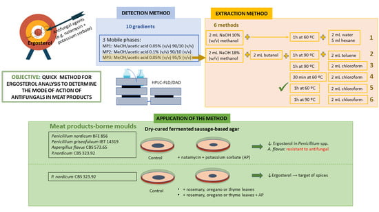
Graphical Abstract

1. Introduction

Toxigenic moulds are frequently found on the surface of a wide range of dry-cured meat products [1,2]. The concern associated with this kind of moulds is linked to their capability to produce mycotoxins like aflatoxins, ochratoxin A (OTA), or cyclopiazonic acid (CPA) [1,3,4]. Examples of their toxicity are the immunosuppression and liver cancer caused by aflatoxins [5], the nephrotoxicity and the carcinogenicity provoked by OTA [6], and the immunotoxicity on human cells due to CPA [7].
The presence of these unwanted moulds has necessitated the development of several methods to avoid mycotoxin production. The most recently described strategies in dry-cured meat products are related to biocontrol agents employing essential oils [8] or microorganisms, such as yeasts, lactic acid bacteria, and non-toxigenic moulds [9,10,11]. Nutritional and environmental factors greatly affect the growth of the protective cultures, which is a critical drawback for their application. Extensive studies of implementation and viability of such cultures in each particular case are thus necessary [11,12]. Due to such difficulties finding a universal biocontrol agent, the meat industry commonly employs legally authorised antifungals, such as benzoic acid, potassium sorbate (PS), and natamycin [13]. The recently developed alternatives to such synthetic preservatives are based on applying natural compounds of plant origin, such as essential oils or spices, in dry-cured fermented sausages [14,15].
To enhance an antifungal treatment, its mode of action has to be studied before its application. Despite the fact that this aspect has not been fully clarified, several reports have proposed ergosterol as the target site of their activity. Ergosterol is a lipid of the cellular membrane of moulds, which is essential for the growth of their mycelium [16]. Natamycin, one of the most applied antifungals in the meat industry, has been described as blocking mould growth by binding specifically to ergosterol [17]. Similarly, essential oils from oregano and cinnamon have been reported to reduce the ergosterol content of Fusarium spp. in maize samples [18]. Eremanthus erythropappus essential oil has been also described as a reducer of ergosterol content in Aspergillus spp. [19]. Moreover, compounds from essential oils, such as thymol and citral, have shown their effect on ergosterol content in Fusarium gramineareum and Alternaria alternata, respectively [20,21]. Nevertheless, there is no data about the targets of antifungals of plant origin when utilised against toxigenic moulds in dry-cured meat products.
Ergosterol is not water soluble, but appropriately dissolves in acetone and ethanol [22]. Accordingly, different kinds of organic solvents like butanol and hexane have been used for ergosterol extraction [23,24]. The analytical methods for its detection have been usually based on high-performance liquid chromatography (HPLC), with methanol as eluent [23,24,25], or gas chromatography (GC) with mass spectrometry detection [26]. Concretely, HPLC coupled with a UV detector is the most used method for analysing ergosterol [27,28], since it is easy to handle and has lower maintenance costs and technical requirements than other detectors or chromatographic techniques [29,30]. Whatever the method chosen, it is necessary to consider that the loss of ergosterol due to decomposition and light sensitivity can occur because of the time required for the different steps of the procedure [31]. Therefore, a reduction of 43% of its content after 24 h has been found under light action [25]. Due to the fact that the use of GC for ergosterol quantification implies previous sample derivatisation [26], such a technique could thus negatively affect the results and collaterally increase the time and the cost per analysis. For all the above-mentioned reasons, it is required that a faster and more convenient method be developed for checking the effect of antifungals on mould ergosterol content.
The aim of this study was to develop a methodology for evaluating the impact of antifungal agents on the ergosterol content of meat product-borne toxigenic moulds. Thus, a method for analysing ergosterol using HPLC with fluorescence detection coupled to a diode array detector (HPLC-FLD/DAD), as well as an extraction procedure based on previously published literature, were firstly optimised. After validating both protocols, their viability to evaluate the effect of a commercial antifungal preparation (AP) and different herbs frequently added to cured meat products on the ergosterol content of several toxigenic moulds was evaluated.

2. Materials and Methods

2.1. Standard and Reagents

All the chemical and chromatographic reagents were HPLC or analytical grade. Acetone, NaOH, 1-butanol, and chloroform were purchased from Scharlab, S.L. (Barcelona, Spain). Hexane, methanol, and acetic acid were obtained from Fisher Scientific S.L. (Hampton, VA, USA). Toluene was purchased from Panreac Química S.A. (Barcelona, Spain), and ergosterol ≥95% HPLC grade from Sigma-Aldrich (San Luis, MO, USA).
The AP containing natamycin and PS was supplemented with 60 g/L of NaCl (Fisher Scientific S.L.), following the manufacturer’s instructions (Manufacturas Taberner, S.A., Valencia, Spain).

2.2. Preparation of Standard Solutions

A stock solution (1 mg/mL) was prepared by dissolving standard ergosterol in acetone following the manufacturer’s recommendations. Working solutions were daily prepared in limited light conditions using amber vials by diluting the standard solution in methanol, since it was the main constituent of the mobile phase (Cosela S.L., Sevilla, Spain). They were filtered through a 0.22 µm pore size nylon membrane (Cosela S.L.).

2.3. HPLC Method

Ergosterol was analysed by a HPLC-FLD/DAD model 1260 Infinity (Agilent Technologies, Santa Clara, CA, USA). The column was a Phenomenex Luna C18, (250 × 4.6 mm, 5 µm particle size; Phenomenex, Macclesfield, United Kingdom). The column temperature was established at 25 °C. The flow rate was set at 1 mL/min, and the injection volume was 20 µL. Ergosterol was detected at λ = 282 nm by the DAD and identified by the absorption spectrum of the standard. Calibration curves were built by diluting the stock solution from 0.1 to 100.0 µg/mL.
To optimise the analytical method for detecting and quantifying ergosterol, the gradients and composition of the mobile phase were assessed using the standard solutions. Firstly, 10 different gradients of mobile phase 1 (MP1), consisting of methanol/acetic acid 0.05% (v/v) 90/10 (v/v) (eluent A [24]) and methanol (eluent B) were evaluated (Table S1, Supplementary Materials).
After selecting the most appropriate gradient, three mobile phases were checked: MP1; mobile phase 2 (MP2), consisting of methanol/acetic acid 0.1% (v/v) 90/10 (v/v); and mobile phase 3 (MP3), composed by methanol/acetic acid 0.05% (v/v) 95/5 (v/v). The selection of the most adequate gradient and mobile phase relied on obtaining a chromatographic peak with a narrow width and symmetric shape, showing the earliest retention time and the shortest run time.
To evaluate the ability of the HPLC-FLD/DAD method to study the effect of antifungal agents, the ergosterol standard (10 µg/mL) was incubated in the presence of three AP concentrations (10%, 50%, and 90%) at different incubation times (0, 8, and 24 h). These concentrations represented different potential quantities of the AP found in meat products: 10% being a low level of the AP, 50% as a medium level, and 90% as a high level. Samples without AP (0%) were used as positive controls.

2.4. Extraction of Ergosterol

A total of six methods for extracting mould ergosterol were evaluated. They consisted of two previously reported ones [23,24] and modifications of such methods (Table 1).
The accuracy of each method was evaluated by the percentages of recovery (absolute recoveries) and the method with the best results (≈100% of recovery) was selected. The neat solvent was spiked by adding 10 µg/mL of ergosterol and then extracted following the above-mentioned methods. The results were compared with the area of a standard at the same concentration (10 µg/mL). The percentage of recovery (RA) was calculated according to the following equation:
R A   ( % ) = average   area   ( samples ) average   area   ( standards ) × 100
All samples were tested in triplicate.

2.5. Validation Assays

The HPLC method was validated by determining the limit of detection (LOD) and limit of quantification (LOQ), linearity, precision, repeatability, within-lab reproducibility, and accuracy [7].
The LOD and LOQ were calculated as the minimum ergosterol concentration level at which the signal exceeded the noise level by a factor of 3 (signal-to-noise) and 10, respectively.
Linearity was determined by applying the coefficient of determination (R2) in the range 0.1–100.0 µg/mL, and the precision by the analysis of three extractions was determined from the same samples under specified conditions. Both parameters were tested in triplicate.
The intra-day repeatability was analysed by three calibration curves in the range 0.1–100.0 µg/mL, conducted by the same operator in duplicate. The inter-day repeatability was checked by three calibration curves performed on three different working days. The within-lab reproducibility was conducted by two different operators, who built three independent calibration curves.
The accuracy was determined by RA, as explained in Section 2.4.

2.6. Mould Assessment

To check the suitability of the developed methodology for extracting and quantifying ergosterol when determining the modes of action of antifungal agents, four toxigenic mould strains were incubated in the presence of AP: the ochratoxigenic Penicillium nordicum CBS 323.92, belonging to Centraalbureau voor Schimmelcultures (The Netherlands), and P. nordicum BFE 856 from the Federal Research Centre for Nutrition and Food (Germany); the aflatoxigenic Aspergillus flavus CBS 573.65 from Centraalbureau voor Schimmelcultures; and the CPA producer Penicillium griseofulvum IBT 14319, supplied by the Type Culture Collection of the Department of Biotechnology from the Technical University of Denmark. In addition, P. nordicum CBS 323.92, one of the main OTA producers in meat products, was also selected to analyse the ergosterol content after applying rosemary, oregano, and thyme, which are herbs recently described as having antifungal activity in dry-cured fermented sausages [15] (Figure 1).
The mould inocula were prepared by growing them on potato dextrose agar (PDA; Scharlab, S.L.) at 25 °C for 7 days. Spores were harvested by scraping the plate surface with a glass rod after adding 3 mL of phosphate-buffered saline (0.32 g of NaH2PO4 (Scharlab, S.L.), 1.09 g of Na2HPO4 (Scharlab, S.L.), 9 g of NaCl (Scharlab, S.L.), and 1 L of distilled water). Spore suspensions were quantified using a Thoma Blaubrand counting chamber (Brand, Germany). A concentration of 106 spores/mL of each mould strain was individually inoculated on a dry-cured fermented sausage-based agar (FS; [15]). A volume of 25 µL of AP was distributed on the surface of the FS before the mould inoculation. To test the herbs, the FS was supplemented with 2 g/kg of rosemary (FS-R), oregano (FS-O), and thyme (FS-T), as described by Álvarez et al. [15]. Fresh rosemary and thyme leaves were used, while oregano leaves were added dried. The herbs were harvested in the region of Extremadura, located in the southwest of Spain. The effect of the herbs on the mould ergosterol content was evaluated alone and in combination with AP. As negative and positive controls, P. nordicum CBS 323.92 grown without herbs and in the presence of AP was used, respectively. The assay was performed in triplicate. After incubating for 14 days at 12 °C, 1 g of the mycelia was collected by scraping the surface with a scalpel and frozen with liquid nitrogen to stop the metabolic activity of the moulds before storing them at −80 °C. For the ergosterol extraction, the samples were firstly homogenised using a mortar and pestle with liquid nitrogen, and the obtained mycelium powder was subjected to the selected extraction method (Figure 1).

2.7. Statistical Analysis

Data analysis was carried out using the SPSS v. 20 software (IBM Corporation, Armonk, NY, USA). The non-parametric Kruskal–Wallis and Mann–Whitney U tests were applied, since the data failed the Shapiro–Wilk and Levene tests. The statistical significance was established at p ≤ 0.05. The standard deviations were determined using Microsoft Excel 365 (Microsoft, Albuquerque, NM, USA).

3. Results and Discussion

3.1. Improvement of the Chromatographic Conditions

A reversed-phase column (C18) was selected for the HPLC-FLD/DAD detection method, due to its good peak resolution [23,27]. Firstly, the selection of the most appropriate mobile phase was performed (Table S1, Supplementary Materials). Gradient 10 was the most suitable, since its retention time was the earliest (17.43 min) and its run time the shortest (22 min). Gradient 8 gave similar results, but it was dismissed because of its longer run time (27 min). The chosen gradient managed to reduce the analysis time to 45% compared to the previously developed methods for ergosterol detection, which employed between 30 and 40 min per analysis [23,24,32]. Such a method would thus allow minimising the potential degradation of ergosterol because of light exposure, with a subsequent decrease in false-negative results.
Regarding the mobile-phase composition, MP3, containing less acetic acid than MP1 and MP2, considerably reduced the retention time without losing precision, as its coefficient of variation value (7.59%) was less than 15% (Table 2). This is considered adequate by different official agencies [33,34]. MP3 also showed the best results for the peak width and asymmetry values (Table 2). A symmetrical, sharp peak with a narrow width (asymmetry value close to 1) has higher precision and is better separated than a more curved peak [35,36]. Such differences between mobile phases are shown in the chromatograms included in Figure 2. The gradient and mobile phase improvement was thus associated with shorter analysis time, enabling increased laboratory productivity because of the increase of the amount of analysis per day and the reduction of reagent costs.
When the ability of the HPLC-FLD/DAD method to study the mode of action of antifungal agents was evaluated in the presence of different amounts of AP, a lack of detection of ergosterol was observed in the presence of the highest amount (Table 3). These findings corroborate those previously reported for both AP’s components (PS and natamycin) when evaluating their effect on mould ergosterol biosynthesis. Therefore, PS has been associated with changes in the lipid composition, decreasing the sterol content [37] with the resultant reduction in ergosterol, the principal sterol in moulds [26]. In contrast, natamycin specifically binds to ergosterol, blocking mycelial growth [17]. In the present work, interference between AP and ergosterol depended on the applied concentration and not on the exposition time, which is of crucial interest for the meat industry, since the use of sub-inhibitory doses of antifungals has been related to undesirable mould growth, the emergence of mould resistance, and mycotoxin production [38,39,40].

3.2. Extraction Method

When searching for reductions in the analysis time, the extraction method is considered to be as important as the detection one. The results obtained when evaluating the six different methods for extracting ergosterol are shown in Table 1. When testing the two previously reported methods (Methods 1 and 2), the lowest recoveries values were obtained. In addition, Method 2 presented a high standard deviation, which could be due to the fact that this method was originally designed to be used in yeast [23] instead of filamentous fungi that have a more complex cell wall. On the contrary, the best mean recovery values, together with the shortest retention times, were shown by Methods 3 and 5. The saponification step, with a higher concentration of NaOH in Methods 2 to 6, seems to be crucial for ergosterol extraction by increasing cell wall permeability and facilitating the entry of alcohols [23]. This feature, as well as the use of methanol instead of 1-butanol for extraction, could be the reason for the low recovery value found in Method 1. This is in accordance with Pastinen et al. [23], who described that alcohols with four carbons, such as 1-butanol, are better solvents for ergosterol extraction than alcohols with fewer carbons like methanol, which was that employed in Method 1.
Furthermore, when comparing Method 2 and those developed from it (Methods 3–6), it appears that the addition of chloroform instead of toluene triggers the improvement of the recoveries. This could be due to the good solubility of ergosterol in chloroform [41], which might also help to increase the permeability of the cell wall. Additionally, the permeability could increase by prolonging the incubation time, as better results were obtained when samples were incubated for 1 h (Method 3) than for 30 min (Method 4).
Within the procedures with the best outcomes, Methods 3 and 5 were the most appropriate. Method 5 was finally selected because of its lower standard deviation values and temperature than Method 3, since the detection of ergosterol can be negatively affected by high temperatures [42]. Consequently, Method 5 has been able to improve the results obtained from the previously reported methods [23,24].

3.3. Validation Parameters

The values for LOD and LOQ were 0.03 and 0.1 µg/mL, respectively, lower than those previously obtained by other authors using HPLC-FLD [24,30,42].
The linearity defined by calibration lines relating to ergosterol concentration and peak area gave an optimum value (Figure 3). The R2 value is considered an indicator of linearity, and values close to the unit are synonymous to a straight line [29,43], while some studies have demonstrated curved relationships and suggest a visual inspection of calibration plots to confirm the linearity [44,45]. In our study, the linear trend of the calibration line is clearly visualised in Figure 3, supporting the good linearity of the method.
The calibration curves for intra-day and inter-day repeatability were similar, and demonstrated no significant differences between them, like the ones used for within-lab reproducibility (p ≤ 0.05) (data not shown). These features support the use of the method independently of the operator.

3.4. Ergosterol Content of Toxigenic Moulds in the Presence of Antifungal Compounds

The extraction and HPLC-FLD/DAD methods were finally used to evaluate the effect of the AP and the selected herbs on the ergosterol content of toxigenic moulds (Table 4 and Table 5). It should be pointed out that the assay was carried out using a meat-based model and not a real meat matrix, since the presence of the native microbial population of the latter having ergosterol would not give feasible results about the effect of the antifungals on the content of such compound. Nonetheless, a full understanding of the targets of antifungal strategies is desirable, since it would allow the establishment of the best conditions for their addition, minimising mycotoxin exposure, and consequently, improving food safety. Accordingly, FS has been selected to perform this assay, due to the fact that this culture medium efficiently simulates the composition of dry-cured, fermented sausages [15].
On the other hand, the novelty of the method applied with moulds has to be highlighted. The method consists of blocking the moulds’ metabolic activity after sampling using liquid nitrogen (Figure 1), thereby reducing ergosterol instability following the mould death.
It has been described that ergosterol production depends on the mould species and colony age, as well as on the substrate [46]. Accordingly, our results show that Penicillium species were able to produce higher ergosterol amounts than A. flavus under control conditions. To our knowledge, there are no studies about mould ergosterol levels in a meat-based substrate, but previous studies have demonstrated a higher production of ergosterol in Penicillia than in Aspergilla in PDA and rice [46,47]. Furthermore, the influence of the AP on the ergosterol content in the tested toxigenic Penicillia moulds was corroborated, because it was significantly reduced in those samples incubated in its presence. The reducing effect of the components of AP on ergosterol content has been previously reported in Penicillia. Thus, PS has been described as significantly reducing the sterol content in Penicillium roqueforti, affecting the lipid bilayer [37], while natamycin has shown a high binding to ergosterol during germination in Penicillium discolor [48]. Additionally, ergosterol has shown to be the target of antifungal drugs used to treat fungal infections, such as azoles, which inhibit ergosterol biosynthesis in Aspergillus fumigatus [49].
On the contrary, the ergosterol content of A. flavus CBS 573.65 was not affected in the presence of the AP in this study. This lack of effect of natamycin on mould ergosterol has been previously described in Aspergillus ochraceus, and has been associated with the existence of resistances due to its continuous use; it even could enhance resistance to other polyene antifungals by cross-tolerance [39].
Regarding the effect of the addition of rosemary, oregano, and thyme leaves against the ochratoxigenic P. nordicum CBS 323.92, all of them significantly reduced the ergosterol content regardless of whether they were in the presence or absence of AP (Table 5). These findings are in accordance with previous studies testing different essential oils against toxigenic moulds. Thus, it has been reported that the essential oil extracted from rosemary decreased in vitro ergosterol content in toxigenic A. flavus and Fusarium verticillioides, although the minimum inhibitory concentration differs depending on the species [50,51]. Regarding oregano and thyme essential oils, some of their main components, such as carvacrol or thymol, have been reported to diminish the ergosterol content in A. flavus [52]. Despite the fact that there are not many studies focused on Penicillia, some essential oils, such as tea tree oil and betel leaf oil, have shown ergosterol reductions in Penicillium expansum [53,54]. The natural compounds cinnamaldehyde and citral from cinnamon and Cymbopogon, respectively, have also shown their effect by decreasing ergosterol in P. expansum [55]. Another study has suggested that the compound antofine from Cynanchum atratum can significantly impair ergosterol biosynthesis in Penicillium digitatum [56].
Therefore, the ergosterol seems to be the target site of the antifungal action of rosemary, oregano, and thyme, which could explain the reduction of OTA presence detected by Álvarez et al. [15] when testing their effects against P. nordicum. Accordingly, the synthesis of ergosterol has been associated with the OTA production in A. ochraceus and P. verrucosum [47,57]. Nevertheless, further studies are necessary to elucidate other modes of action that could be involved in the antifungal activities of rosemary, oregano, and thyme, since synergic effects are generally attributed to strategies against unwanted moulds.

4. Conclusions

A new methodology for the extraction and quantification of ergosterol from toxigenic moulds was developed to be used when evaluating the role of antifungal compounds applied in the meat industry. The method turned out to be precise, fast, and effective for ergosterol quantification in meat product-borne moulds. The method improved the previously developed procedures, due to the reduction of the run time up to 22 min, and consequently, the possible degradation of ergosterol in ergocalciferol. In addition, the methodology proved to be useful for determining the effect of antifungal agents related to dry-cured meat products on the ergosterol content, independent of the toxigenic mould strain. The present method could be thus used as a support tool for improving information about antifungals before their application in meat products, which would enhance their use.

Supplementary Materials

The following are available online at https://www.mdpi.com/2304-8158/10/2/438/s1, Table S1: Mobile phase gradient conditions evaluated in this study for detecting and quantifying ergosterol in meat product-borne toxigenic moulds by HPLC-FLD/DAD.

Author Contributions

Conceptualization: M.J.A. and A.R.; Formal analysis: M.Á.; Funding acquisition: M.J.A.; Investigation: M.Á., E.R.; Methodology: M.Á., M.J.A. and A.R.; Project administration: M.J.A.; Supervision: M.J.A., A.R.; Validation: M.Á., M.J.A. and A.R.; Visualization: M.Á.; Resources: E.B.; Writing—original draft: M.Á. and M.J.A.; Writing—review and Editing: M.Á., M.J.A., A.R., E.B. and E.R. All authors have read and agreed to the published version of the manuscript.

Funding

This research was funded by Junta de Extremadura-Consejería de Economía, Ciencia y Agenda Digital-, Fondo Europeo de Desarrollo Regional: “Una manera de hacer Europa” (IB16045 project and GR18056 grant). M.Á. is the recipient of a fellowship from the Spanish Ministerio de Economía, Industria y Competitividad (BES-2017-081340).

Institutional Review Board Statement

Not applicable.

Informed Consent Statement

Not applicable.

Data Availability Statement

Data is contained within the article or supplementary material.

Acknowledgments

Technical support was provided by the Servicio de Innovación en Productos de Origen Animal (SiPA) of the Servicios de Apoyo a la Investigación de la Universidad de Extremadura (SAIUEx). R. Geisen is acknowledged for the P. nordicum BFE856 donation.

Conflicts of Interest

The authors declare no conflict of interest.

References

  1. Zadravec, M.; Vahčić, N.; Brnić, D.; Markov, K.; Frece, J.; Beck, R.; Lešić, T.; Pleadin, J. A study of surface moulds and mycotoxins in Croatian traditional dry-cured meat products. Int. J. Food Microbiol. 2020, 317, 108459. [Google Scholar] [CrossRef] [PubMed]
  2. Sánchez-Montero, L.; Córdoba, J.J.; Peromingo, B.; Álvarez, M.; Núñez, F. Effects of environmental conditions and substrate on growth and ochratoxin A production by Penicillium verrucosum and Penicillium nordicum: Relative risk assessment of OTA in dry-cured meat products. Food Res. Int. 2019, 121, 604–611. [Google Scholar] [CrossRef] [PubMed]
  3. Baranyi, N.; Kocsubé, S.; Varga, J. Aflatoxins: Climate change and biodegradation. Curr. Opin. Food Sci. 2015, 5, 60–66. [Google Scholar] [CrossRef]
  4. Peromingo, B.; Sulyok, M.; Lemmens, M.; Rodríguez, A.; Rodríguez, M. Diffusion of mycotoxins and secondary metabolites in dry-cured meat products. Food Control. 2019, 101, 144–150. [Google Scholar] [CrossRef]
  5. Payne, G.A. Mycotoxins and Product Safety. In Peanuts; Elsevier BV: Amsterdam, The Netherlands, 2016; pp. 347–361. [Google Scholar]
  6. Pattono, D.; Grosso, A.; Stocco, P.; Pazzi, M.; Zeppa, G. Survey of the presence of patulin and ochratoxin A in traditional semi-hard cheeses. Food Control. 2013, 33, 54–57. [Google Scholar] [CrossRef]
  7. Peromingo, B.; Rodríguez, M.; Núñez, F.; Silva, A.; Rodríguez, A. Sensitive determination of cyclopiazonic acid in dry-cured ham using a QuEChERS method and UHPLC–MS/MS. Food Chem. 2018, 263, 275–282. [Google Scholar] [CrossRef]
  8. Chaves-López, C.; Martin-Sánchez, A.M.; Fuentes-Zaragoza, E.; Viuda-Martos, M.; Fernández-López, J.; Sendra, E.; Sayas, E.; Alvarez, J.A.P. Role of Oregano (Origanum vulgare) Essential Oil as a Surface Fungus Inhibitor on Fermented Sausages: Evaluation of Its Effect on Microbial and Physicochemical Characteristics. J. Food Prot. 2012, 75, 104–111. [Google Scholar] [CrossRef]
  9. Álvarez, M.; Rodríguez, A.; Peromingo, B.; Núñez, F.; Rodríguez, M. Enterococcus faecium: A promising protective culture to control growth of ochratoxigenic moulds and mycotoxin production in dry-fermented sausages. Mycotoxin Res. 2019, 36, 137–145. [Google Scholar] [CrossRef]
  10. Andrade, M.J.; Thorsen, L.; Rodríguez, A.; Córdoba, J.J.; Jespersen, L. Inhibition of ochratoxigenic moulds by Debaryomyces hansenii strains for biopreservation of dry-cured meat products. Int. J. Food Microbiol. 2014, 170, 70–77. [Google Scholar] [CrossRef] [PubMed]
  11. Delgado, J.; Peromingo, B.; Rodríguez, A.; Rodríguez, M. Biocontrol of Penicillium griseofulvum to reduce cyclopiazonic acid contamination in dry-fermented sausages. Int. J. Food Microbiol. 2019, 293, 1–6. [Google Scholar] [CrossRef]
  12. Álvarez, M.; Núñez, F.; Delgado, J.; Andrade, M.J.; Rodríguez, M.; Rodríguez, A. Competitiveness of three biocontrol candidates against ochratoxigenic Penicillium nordicum under dry-cured meat environmental and nutritional conditions. Fungal Biol. 2021, 125, 134–142. [Google Scholar] [CrossRef] [PubMed]
  13. European Commission. Commission Regulation (EU) No 1129/2011 of 11 November 2011 amending Annex II to Regulation (EC) No 1333/2008 of the European Parliament and of the Council by establishing a Union list of food additives. Off. J. Eur. Union. 2011. [Google Scholar] [CrossRef]
  14. Kocić-Tanackov, S.; Dimić, G.; Đerić, N.; Mojović, L.; Tomović, V.; Šojić, B.; Đukić-Vuković, A.; Pejin, J. Growth control of molds isolated from smoked fermented sausages using basil and caraway essential oils, in vitro and in vivo. LWT 2020, 123, 109095. [Google Scholar] [CrossRef]
  15. Álvarez, M.; Rodríguez, A.; Núñez, F.; Silva, A.; Andrade, M.J. In vitro antifungal effects of spices on ochratoxin A production and related gene expression in Penicillium nordicum on a dry-cured fermented sausage medium. Food Control. 2020, 114, 107222. [Google Scholar] [CrossRef]
  16. Vella, F.M.; Laratta, B. UV-based evaluation of ergosterol for monitoring the fungal exposure in Italian buffalo farms. FEMS Microbiol. Lett. 2017, 364, 1–6. [Google Scholar] [CrossRef] [PubMed]
  17. Welscher, Y.M.T.; Napel, H.H.T.; Balagué, M.M.; Souza, C.M.; Riezman, H.; De Kruijff, B.; Breukink, E. Natamycin Blocks Fungal Growth by Binding Specifically to Ergosterol without Permeabilizing the Membrane. J. Biol. Chem. 2008, 283, 6393–6401. [Google Scholar] [CrossRef] [Green Version]
  18. Perczak, A.; Gwiazdowska, D.; Gwiazdowski, R.; Juś, K.; Marchwińska, K.; Waśkiewicz, A. The Inhibitory Potential of Selected Essential Oils on Fusarium spp. Growth and Mycotoxins Biosynthesis in Maize Seeds. Pathogens 2019, 9, 23. [Google Scholar] [CrossRef] [PubMed] [Green Version]
  19. Brandão, R.M.; Ferreira, V.R.F.; Batista, L.R.; Alves, E.; Lira, N.D.A.; Bellete, B.S.; Scolforo, J.R.S.; Cardoso, M.D.G. Antifungal and antimycotoxigenic effect of the essential oil of Eremanthus erythropappus on three different Aspergillus species. Flavour Fragr. J. 2020, 35, 524–533. [Google Scholar] [CrossRef]
  20. Gao, T.; Zhou, H.; Zhou, W.; Hu, L.; Chen, J.; Shi, Z. The Fungicidal Activity of Thymol against Fusarium graminearum via Inducing Lipid Peroxidation and Disrupting Ergosterol Biosynthesis. Molecules 2016, 21, 770. [Google Scholar] [CrossRef] [Green Version]
  21. Wang, L.; Jiang, N.; Wang, D.; Wang, M. Effects of Essential Oil Citral on the Growth, Mycotoxin Biosynthesis and Transcriptomic Profile of Alternaria alternata. Toxins 2019, 11, 553. [Google Scholar] [CrossRef] [Green Version]
  22. Kadakal, Ç.; Tepe, T.K. Is ergosterol a new microbiological quality parameter in foods or not? Food Rev. Int. 2019, 35, 155–165. [Google Scholar] [CrossRef]
  23. Pastinen, O.; Nyyssölä, A.; Pihlajaniemi, V.; Sipponen, M.H. Fractionation process for the protective isolation of ergosterol and trehalose from microbial biomass. Process. Biochem. 2017, 58, 217–223. [Google Scholar] [CrossRef] [Green Version]
  24. Neuhof, T.; Koch, M.; Rasenko, T.; Nehls, I. Distribution of Trichothecenes, Zearalenone, and Ergosterol in a Fractionated Wheat Harvest Lot. J. Agric. Food Chem. 2008, 56, 7566–7571. [Google Scholar] [CrossRef]
  25. Mille-Lindblom, C.; Von Wachenfeldt, E.; Tranvik, L.J. Ergosterol as a measure of living fungal biomass: Persistence in environmental samples after fungal death. J. Microbiol. Methods 2004, 59, 253–262. [Google Scholar] [CrossRef]
  26. Dong, Y.; Steffenson, B.J.; Mirocha, C.J. Analysis of Ergosterol in Single Kernel and Ground Grain by Gas Chromatography−Mass Spectrometry. J. Agric. Food Chem. 2006, 54, 4121–4125. [Google Scholar] [CrossRef] [PubMed]
  27. Barreira, J.C.M.; Oliveira, M.B.P.P.; Ferreira, I.C.F.R. Development of a Novel Methodology for the Analysis of Ergosterol in Mushrooms. Food Anal. Methods 2014, 7, 217–223. [Google Scholar] [CrossRef]
  28. Steel, C.; Schwarz, L.; Qiu, Y.; Schueuermann, C.; Blackman, J.; Clark, A.; Schmidtke, L. Thresholds for Botrytis bunch rot contamination of Chardonnay grapes based on the measurement of the fungal sterol, ergosterol. Aust. J. Grape Wine Res. 2019, 26, 79–89. [Google Scholar] [CrossRef]
  29. Contreras, M.D.M.; Morales-Soto, A.; Segura-Carretero, A.; Valverde, J. Potential of RP-UHPLC-DAD-MS for the qualitative and quantitative analysis of sofosbuvir in film coated tablets and profiling degradants. J. Pharm. Anal. 2017, 7, 208–213. [Google Scholar] [CrossRef]
  30. Lohr, D.; Woeck, C.; Meinken, E. Use of ergosterol as an indicator for colonization of peat-based growing media by saprophytic fungi. Eur. J. Hortic. Sci. 2017, 82, 3–11. [Google Scholar] [CrossRef]
  31. Parkinson, D.-R.; Warren, J.; Pawliszyn, J. Analysis of ergosterol for the detection of mold in soils by automated on-fiber derivatization headspace extraction–SPME-GC/MS. Anal. Chim. Acta 2010, 661, 181–187. [Google Scholar] [CrossRef]
  32. Porep, J.U.; Mrugala, S.; Pour Nikfardjam, M.S.; Carle, R. Online Determination of Ergosterol in Naturally Contaminated Grape Mashes Under Industrial Conditions at Wineries. Food Bioprocess Technol. 2015, 8, 1455–1464. [Google Scholar] [CrossRef]
  33. Food and Drug Administration. Bioanalytical Method Validation: Guidance for Industry. In Rockville, MD: CDER; FDA: Beltsville, MD, USA, 2018; Volume FDA-2013-D-1020. [Google Scholar]
  34. ICH Guideline M10 on Bioanalytical Method Validation. Available online: https://www.ema.europa.eu/en/documents/scientific-guideline/draft-ich-guideline-m10-bioanalytical-method-validation-step-2b_en.pdf (accessed on 4 November 2020).
  35. Hayash, Y.; Matsuda, R. Deductive Prediction of Measurement Precision from Signal and Noise in Photomultiplier and Photodiode Detector of Liquid Chromatography. Anal. Sci. 1994, 10, 725–730. [Google Scholar] [CrossRef] [Green Version]
  36. Snyder, L.R.; Kirkland, J.J.; Dolan, J.W. Introduction to Modern Liquid Chromatography; Wiley: Hoboken, NJ, USA, 2009. [Google Scholar]
  37. Sergeeva, Y.E.; Galanina, L.A.; Kochkina, G.A.; Feofilova, E.P. The effect of the preservative sorbic acid on the lipid composition of the ascomycete fungus Penicillium roqueforti Thom. Microbiology 2009, 78, 630–635. [Google Scholar] [CrossRef]
  38. Deising, H.B.; Reimann, S.; Pascholati, S.F. Mechanisms and significance of fungicide resistance. Braz. J. Microbiol. 2008, 39, 286–295. [Google Scholar] [CrossRef] [PubMed] [Green Version]
  39. Streekstra, H.; Verkennis, A.E.; Jacobs, R.; Dekker, A.; Stark, J.; Dijksterhuis, J. Fungal strains and the development of tolerance against natamycin. Int. J. Food Microbiol. 2016, 238, 15–22. [Google Scholar] [CrossRef] [PubMed]
  40. Arroyo, M.; Aldred, D.; Magan, N. Environmental factors and weak organic acid interactions have differential effects on control of growth and ochratoxin A production by Penicillium verrucosum isolates in bread. Int. J. Food Microbiol. 2005, 98, 223–231. [Google Scholar] [CrossRef] [PubMed]
  41. Bills, C.E. Vitamin D Group. In The Vitamins Chemistry, Physiology and Pathology; Sebrel, W.H., Harris, R.S., Eds.; Academic Press: New York, NY, USA, 1954; pp. 131–266. [Google Scholar]
  42. Newell, S.Y.; Arsuffi, T.L.; Fallon, R.D. Fundamental procedures for determining ergosterol content of decaying plant ma-terial by liquid chromatography. Appl. Environ. Microbiol. 1988, 54, 1876–1879. [Google Scholar] [CrossRef] [Green Version]
  43. Kaale, E.; Shewiyo, D.H.; Jenkins, D. Statistics in validation of quantitative chromatographic methods. In Chemometrics in Chromatography; Komsta, Ł., Vander Heyden, Y., Sherma, J., Eds.; CRC Press, Taylor and Francis: Boca Raton, FL, USA, 2018. [Google Scholar]
  44. ICH Validation of Analytical Procedures: Text and Methodology. Available online: https://www.ema.europa.eu/en/documents/scientific-guideline/ich-q-2-r1-validation-analytical-procedures-text-methodology-step-5_en.pdf (accessed on 4 November 2020).
  45. Raposo, F. Evaluation of analytical calibration based on least-squares linear regression for instrumental techniques: A tutorial review. TrAC Trends Anal. Chem. 2016, 77, 167–185. [Google Scholar] [CrossRef]
  46. Taniwaki, M.H.; Pitt, J.I.; Hocking, A.D.; Fleet, G.H. Comparison of hyphal length, ergosterol, mycelium dry weight, and colony diameter for quantifying growth of fungi from foods. Adv. Exp. Med. Biol. 2006, 571, 49–67. [Google Scholar] [CrossRef]
  47. Saxena, J.; Munimbazi, C.; Bullerman, L.B. Relationship of mould count, ergosterol and ochratoxin A production. Int. J. Food Microbiol. 2001, 71, 29–34. [Google Scholar] [CrossRef]
  48. Van Leeuwen, M.; Smant, W.; De Boer, W.; Dijksterhuis, J. Filipin is a reliable in situ marker of ergosterol in the plasma membrane of germinating conidia (spores) of Penicillium discolor and stains intensively at the site of germ tube formation. J. Microbiol. Methods 2008, 74, 64–73. [Google Scholar] [CrossRef]
  49. Geißel, B.; Loiko, V.; Klugherz, I.; Zhu, Z.; Wagener, N.; Kurzai, O.; Hondel, C.A.M.J.J.V.D.; Wagener, J. Azole-induced cell wall carbohydrate patches kill Aspergillus fumigatus. Nat. Commun. 2018, 9, 1–13. [Google Scholar] [CrossRef] [Green Version]
  50. Bomfim, N.D.S.; Kohiyama, C.Y.; Nakasugi, L.P.; Nerilo, S.B.; Mossini, S.A.G.; Romoli, J.C.Z.; Mikcha, J.M.G.; Filho, B.A.D.A.; Machinski, M., Jr. Antifungal and antiaflatoxigenic activity of rosemary essential oil (Rosmarinus officinalis L.) against Aspergillus flavus. Food Addit. Contam. Part A 2019, 37, 153–161. [Google Scholar] [CrossRef]
  51. Bomfim, N.D.S.; Nakassugi, L.P.; Oliveira, J.F.P.; Kohiyama, C.Y.; Mossini, S.A.G.; Grespan, R.; Nerilo, S.B.; Mallmann, C.A.; Filho, B.A.A.; Machinski, M. Antifungal activity and inhibition of fumonisin production by Rosmarinus officinalis L. essential oil in Fusarium verticillioides (Sacc.) Nirenberg. Food Chem. 2015, 166, 330–336. [Google Scholar] [CrossRef] [PubMed] [Green Version]
  52. Tian, F.; Woo, S.Y.; Lee, S.Y.; Chun, H.S. p-Cymene and its derivatives exhibit antiaflatoxigenic activities against Aspergillus flavus through multiple modes of action. Appl. Biol. Chem. 2018, 61, 489–497. [Google Scholar] [CrossRef] [Green Version]
  53. Li, Y.; Shao, X.; Xu, J.; Wei, Y.; Xu, F.; Wang, H. Effects and possible mechanism of tea tree oil against Botrytis cinerea and Penicillium expansum in vitro and in vivo test. Can. J. Microbiol. 2017, 63, 219–227. [Google Scholar] [CrossRef] [PubMed]
  54. Basak, S.; Guha, P. Use of predictive model to describe sporicidal and cell viability efficacy of betel leaf (Piper betle L.) essential oil on Aspergillus flavus and Penicillium expansum and its antifungal activity in raw apple juice. LWT 2017, 80, 510–516. [Google Scholar] [CrossRef]
  55. Wang, Y.; Feng, K.; Yang, H.; Yuan, Y.; Yue, T. Antifungal mechanism of cinnamaldehyde and citral combination against Penicillium expansum based on FT-IR fingerprint, plasma membrane, oxidative stress and volatile profile. RSC Adv. 2018, 8, 5806–5815. [Google Scholar] [CrossRef] [Green Version]
  56. Xin, Z.; Ouyang, Q.; Wan, C.; Che, J.; Li, L.; Chen, J.; Tao, N. Isolation of antofine from Cynanchum atratum BUNGE (Asclepiadaceae) and its antifungal activity against Penicillium digitatum. Postharvest Biol. Technol. 2019, 157, 110961. [Google Scholar] [CrossRef]
  57. Hua, H.; Xing, F.; Selvaraj, J.N.; Wang, Y.; Zhao, Y.; Zhou, L.; Liu, X.; Liu, Y. Inhibitory Effect of Essential Oils on Aspergillus ochraceus Growth and Ochratoxin A Production. PLoS ONE 2014, 9, e108285. [Google Scholar] [CrossRef] [PubMed] [Green Version]
Figure 1. Flow chart showing the assay to check the suitability of the developed methodology for extracting and quantifying ergosterol when determining the effect of antifungal agents against meat product-borne toxigenic moulds. Mobile phase was composed of methanol/acetic acid 0.05% (v/v) 95/5 (v/v).
Foods 10 00438 g001
Figure 2. High-performance liquid chromatography with fluorescence detection coupled to a diode array detector (HPLC-FLD/DAD) chromatograms using an ergosterol standard (10 µg/mL) and the three different mobile phases tested in the present work. (A) Mobile phase 1, composed of methanol/acetic acid 0.05% (v/v) 90/10 (v/v); retention time: 17.43 min. (B) Mobile phase 2, composed of methanol/acetic acid 0.1% (v/v) 90/10 (v/v); retention time: 17.33 min. (C) Mobile phase 3, composed of methanol/acetic acid 0.05% (v/v) 95/5 (v/v); retention time: 15.44 min. Eluent A was composed of the corresponding mobile phase, and Eluent B of methanol. The gradient consisted of 0–5 min 10% B, 5–8 min linear increase from 10% to 100% B, 8–12 min 100% B, 12–22 min linear decrease from 100% to 10% B, with methanol being used as eluent B.
Foods 10 00438 g002
Figure 3. Standard calibration curve built in the range 0.1–100.0 µg/mL of ergosterol using the optimised HPLC-FLD/DAD method.
Foods 10 00438 g003
Table 1. Main procedure steps and results of the methods for extraction of mould ergosterol evaluated in this study.
MethodsMain Steps Involved in the ProcedureRecovery (%)Standard Deviation (%) 2Retention Time (min)References
Method 12 mL NaOH 10% (w/v) in methanol + vortex 30 s + 1 h at 60 °C + 2 mL distilled water + 5 mL hexane + evaporation of the hexane extract + resuspension in 1 mL MP 19.11 *±6.9716.7[24]
Method 22 mL NaOH 18% (w/v) in distilled water + 2 mL 1-butanol + vortex 30 s + 1 h at 90 °C + 2 mL toluene + centrifugation (5000 rpm for 5 min) + evaporation of the organic phase + resuspension in 1 mL MP47.45 *±73.6916.4[23]
Method 32 mL NaOH 18% (w/v) in distilled water + 2 mL 1-butanol + vortex 30 s + 1 h at 90 °C + 2 mL chloroform + centrifugation (5000 rpm for 5 min) + evaporation of the organic phase + resuspension in 1 mL MP103.10±26.9715.8This study
Method 42 mL NaOH 18% (w/v) in distilled water + 2 mL 1-butanol + vortex 30 s + 30 min at 90 °C + 2 mL chloroform + centrifugation (5000 rpm for 5 min) + evaporation of the organic phase + resuspension in 1 mL MP82.12±80.3715.9This study
Method 52 mL NaOH 18% (w/v) in distilled water + 2 mL 1-butanol + vortex 30 s + 1 h at 60 °C + 2 mL chloroform + centrifugation (5000 rpm for 5 min) + evaporation of the organic phase + resuspension in 1 mL MP99.51±9.9215.8This study
Method 62 mL NaOH 18% (w/v) in distilled water + 2 mL 1-butanol + vortex 30 s + 1 h at 90 °C + 3 mL chloroform + centrifugation (5000 rpm for 5 min) + evaporation of the organic phase + resuspension in 1 mL MP81.09 *±21.6315.9This study
1 MP: mobile phase, composed of methanol/acetic acid 0.05% (v/v) 95/5 (v/v). 2 The experiment was performed in triplicate. * Significant differences regarding 100% recovery (p ≤ 0.05). Ergosterol solutions were prepared from the standard to achieve a final concentration of 10 µg/mL. Different extraction solutions, reagent concentrations, and temperature and incubation times were checked. The extraction was always performed under limited light conditions.
Table 2. Results from the evaluation of three mobile phases (MP1–3) for detecting ergosterol (10 µg/mL) with Gradient 10 (0–5 min 10% B 1, 5–8 min linear increase from 10% to 100% B, 8–12 min 100% B, and 12–22 min linear decrease from 100% to 10% B).
Mobile Phases 2Retention Time (min)Coefficient of Variations (%) 3Width of the Peaks (min)Asymmetry
MP117.4312.750.240.90
MP217.338.790.230.91
MP315.447.590.180.97
1 Eluent A was composed of the corresponding MP and Eluent B by methanol. 2 MP1: methanol/acetic acid 0.05% (v/v) 90/10 (v/v); MP2: methanol/acetic acid 0.1% (v/v) 90/10 (v/v); MP3: methanol/acetic acid 0.05% (v/v) 95/5 (v/v). 3 The experiment was performed in triplicate.
Table 3. The concentration of ergosterol (µg/mL) when co-inoculated (10 µg/mL) with different amounts of antifungal preparation (AP) at different sampling times.
AP (%, v/v)Concentration of Ergosterol
0 h8 h24 h
08.74 ± 1.28 18.44 ± 1.609.11 ± 0.72
109.28 ± 0.718.85 ± 0.569.03 ± 0.69
500.50 ± 0.43 *n.d *n.d *
90n.d 2*n.d *n.d *
1 The experiment was performed in triplicate. 2 n.d: not detected (<limit of detection) * Significant differences regarding the absence of AP at the same sampling time (p ≤ 0.05).
Table 4. Effect of an antifungal preparation (AP) on the ergosterol content (µg/g mycelium) of four toxigenic mould strains.
Mould Strain
Penicillium nordicum CBS 323.92P. nordicum BFE 856Penicillium griseofulvum IBT 14319Aspergillus flavus CBS 573.65
Control731.55 ± 183.54 1365.57 ± 45.961121.61 ± 486.34248.09 ± 174.49
Mould + AP513.59 ± 92.84 *187.15 ± 92.14 *185.89 ± 158.75 *297.22 ± 207.47
1 The experiment was performed in triplicate. * Significant differences regarding the control (p ≤ 0.05).
Table 5. Effect of rosemary, oregano, and thyme on the ergosterol content (µg/g of mycelium) of Penicillium nordicum CBS 323.92.
Treatment 1Ergosterol Content
FS731.55 ± 183.54 2
FS-R184.34 ± 44.50 *
FS-O177.40 ± 17.50 *
FS-T117.70 ± 69.30 *
FS-R + AP132.16 ± 44.50 *
FS-O + AP204.73 ± 50.85 *
FS-T + AP237.57 ± 11.99 *
1 FS: dry-cured, fermented, sausage-based agar (control); FS-R: FS with rosemary; FS-O: FS with oregano; FS-T: FS with thyme; FS-R + AP: FS-R with antifungal preparation; FS-O + AP: FS-O with antifungal preparation; FS-T + AP: FS-T with antifungal preparation. 2 The experiment was performed in triplicate. * Significant differences regarding the control (p ≤ 0.05).
Publisher’s Note: MDPI stays neutral with regard to jurisdictional claims in published maps and institutional affiliations.