Article

Effects of Diamond Nanoparticles Immobilisation on the Surface of Yeast Cells: A Phenomenological Study

1
Institute of Biomedical Engineering and Nanotechnologies, Riga Technical University, Kipsalas Str. 6B, LV-1048 Riga, Latvia
2
Institute of Chemical Physics, University of Latvia, Jelgavas Str. 1, LV-1004 Riga, Latvia
3
Fraunhofer Institute for Ceramic Technologies and Systems IKTS, Maria-Reiche-Straße 2, 01109 Dresden, Germany
4
Laboratory of Cell Biology, Institute of Microbiology and Biotechnology, University of Latvia, Jelgavas Str. 1, LV-1004 Riga, Latvia
*
Author to whom correspondence should be addressed.
Fermentation 2023, 9(2), 162; https://doi.org/10.3390/fermentation9020162
Submission received: 24 December 2022 / Revised: 1 February 2023 / Accepted: 7 February 2023 / Published: 10 February 2023
(This article belongs to the Special Issue Yeast Biotechnology 6.0)

Abstract

:
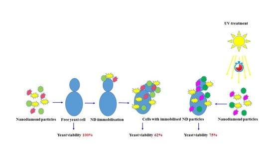
An interesting development of biotechnology has linked microbial cell immobilisation with nanoparticles. The main task of our research was to reveal the possible influences of differently electrically charged diamond nanoparticles upon physiological characteristics of the yeast Saccharomyces cerevisiae. It was revealed that the adverse impact of these nanoparticles can manifest not only against prokaryotes, but also against eukaryotic yeast cells. However, the obtained results also indicate that it is possible to reduce and, most likely, completely eliminate the dangerous effects of nanoparticles to cells by using special physical approaches. Comparison of non-arylated and arylated nanoparticles showed that in terms of changes in the physiological activity of cells, which are important to biotechnology and biomedicine, the selection of certain nanoparticles (non-arylated or arylated) may be necessary in each specific case, depending on the purpose of their use.

Graphical Abstract

1. Introduction

Immobilisation of microorganisms on the surface or inside various carriers is a widely used approach in different biotechnological processes. There are a lot of advantages of immobilised cell application compared to free cells. It was shown that microbial cell immobilisation may lead to enhancement of fermentation productivity, increased stability of cells, as well as decreased overall costs of recovery, regeneration, recycling and downstream processes [1]. Application of immobilised systems allows the possibility of reusing them in the repeated cycles of various biotechnological processes. Besides these advantages, it was shown that, for example, immobilisation of yeast cells may change their physiological characteristics and improve their biotechnological properties, such as ethanol productivity and heavy metal sorption for the purification of the environment, among others [2,3,4,5]. Immobilisation of yeast cells improves their resistance to desiccation, which is important for the storage of industrially important strains in plant collections [6].
An interesting development in this industrial microbiology direction is linked with microbial cell immobilisation with nanoparticles. The systems that are obtained as a result of this process are called either immobilised microbial nanoparticles, microorganisms functionalised by nanoparticles, microorganisms immobilised on nanoparticles or microorganisms decorated with nanoparticles [5,7,8,9,10,11]. We suppose that they may also be referred to as nanoparticles immobilised on the surface of microorganisms or nanoparticles immobilised on or inside the microorganisms. Most of these studies are linked to the application of magnetic nanoparticles for immobilisation and further use of these systems for the biosorption and removal of pollutants and purification of wastewaters, soils and aquatic environments [5]. At the same time, this approach has also been applied to other biotechnological, medical and pharmacological goals over the last few years [11,12,13,14,15]. Another new and important direction of these studies is linked to nanodiamond (ND) particle application in biotechnological and medical fields. It was found that these nanoparticles could enter cells without causing any damage to the cell walls or the cell. This allows for the possibility of using them as efficient potential drug delivery carriers. It is supposed that they are non-toxic and non-reactive [16]. Recent studies revealed the possibility of using nanodiamond particles in treatments for cancers, including brain tumours and breast cancers, autoimmune diseases and cardiovascular affections [16,17,18,19,20].
At the same time, there are different results of studies of microbial sensitivity to diamond nanoparticles. The authors of one study concluded that these nanoparticles have antimicrobial activity in relation to Escherichia coli and Bacillus subtilis. They revealed interaction of diamond nanoparticles with bacterial surfaces and supposed their influence upon permeability of the bacterial cell wall and/or membrane [21]. Other researchers, who used yeast in their studies, have not found any negative effects of diamond nanoparticles on these cells [22,23].
The surface charge of a substrate has a strong influence on immobilisation of cells and their physiological activity [3]. However, the influence of the surface charge value of the nanoparticles on their connection with the cell and the physiological activity of the latter are unknown.
Diamond nanoparticles are unique dielectric materials in which electric charge may be easily induced.
The yeast Saccharomyces cerevisiae have been chosen for the present research. S. cerevisiae cells have a strong cell wall that the nanoparticle likely may not deform and, because of this, they influence cell physiology.
The main task of our research was to reveal possible influences of differently electrically charged diamond nanoparticles on physiological characteristics of S. cerevisiae. We took into account, on the one hand, the practical importance of yeast in various areas of the food industry and biotechnology and that, on the other hand, their cells are unique and a perfect model of any eukaryotic cell that allows them to be used in various molecular biology, medical and pharmacological studies.

2. Materials and Methods

2.1. Nanoparticles and Their Functionalisation

Commercial native Raw Detonation Nanodiamonds (nanoparticles manufactured Adamasnano, Raleigh, NC, USA, average particle size 4–5 nm, aggregated in ~170 nm particles, 2% ash content) were used. To functionalise nanoparticles (NP), physical and chemical approaches were employed.
Physically, the electrical functionalisation of NP (deposition of the electric charge) was provided with ultraviolet radiation (UV). The UV lamp Lightningcure LC5 (Hamamatsu, Japan) was used. The intensity of the ultraviolet light lamp was 3500 W/cm2 with an output wavelength of 365 nm. Following the manufacturer’s certificate of the lamp, the radiation stability was within ±5%. The distance between the light source and the substrates surface was 30 cm. The exposure time was 30 and 90 min. It was verified that the substrate was not heated in such conditions.
To identify the deposited surface electric charge, near-threshold photoelectron emission spectroscopy was employed [24]. The electron work function (ϕ) is directly proportional to the electric charge density (I) at the surface emitting electrons. To measure the electron work function, the NP were radiated with UV light and the energy of the photons was varied from 4.2–6.0 eV. Alongside electron emission current was measured. The electron work function was identified at the energy of the photon when I (hυ) = 0 [24]. To achieve this, the I (hv) line was extrapolated to I = 0. Measurements were taken in vacuum conditions ~10−5 Torr.
To functionalise ND particles chemically, arylation functionalisation was used. First, 1 M borane–tetrahydrofuran (BH3•THF) (Sigma-Aldrich Chemie GmbH, Taufkirchen, Germany), dry tetrahydrofuran (THF) (Life Technologies GmbH, Darmstadt, Germany) and 4-aminobenzoic acid (Sigma-Aldrich Chemie GmbH, Taufkirchen, Germany) were employed.
Arylation of ND was performed by adding 10 g of aminobenzoic acid and 5 mL of amyl nitrite to 10 mL of a 150 mg/mL aqueous ND suspension under stirring at 80 °C for 15 h. Amyl nitrite was used for the in situ generation of diazonium salts [25]. It was conjugated with the NDs via the click chemistry method [26]. Five washing cycles in ddH20 and 15 in acetone were required to obtain a transparent supernatant after centrifugation and to ensure that excess chemicals were removed. All ND samples were dried overnight using a vacuum desiccator after surface modification.
Fourier transmission infrared spectroscopy (FTIR) (FTIR, VERTEX 70v, Bruker) was used to characterise the ND surface. The infrared absorption spectra were recorded in the range from 450 cm−1 to 4000 cm−1 with four scan cycles and a resolution of 2 cm−1. Three measurements were performed for each sample and the mean value was determined for further analysis.
Nanoparticle tracking analysis (NTA) (ZetaView PMX120, Particle Metrix GmbH, Inning am Ammersee, Germany) was used to determine the particle size distribution. Then, 1 mg of ND was mixed with ddH2O and sufficiently diluted. Measurements were recorded at RT and a neutral pH = 7. The wavelength of the laser was 488 nm. Prior to the measurement, nanodiamond suspensions were sonicated using ultrasound bath for 10 min. The final ND suspension was stable for at least 10 h.
Depending on the interplay of electrostatic and van der Waals interactions, ND can aggregate in aqueous media or exist as homogeneous dispersions [27]. The stability of aggregates was tested in water. The mean diameter of arylated nanodiamonds particles was ~134 nm.

2.2. Strains, Nutrient Medium and Culture Conditions

Yeast strain Saccharomyces cerevisiae 77 from Microbial Strain Collection of Latvia was used in this investigation. The strain was maintained on YPD agar medium (gL−1): yeast extract—10, peptone—20, glucose—20 and agar—20 at 4 °C. The yeast was cultivated at 30 °C in a nutrient-rich, complex YPD liquid medium (gL−1): yeast extract—10, peptone—20 and glucose—20. Culture density was monitored at a wavelength of 595 nm using a Tecan’s Sunrise Absorbance Microplate Reader (Switzerland). Mean values of optical density were plotted against incubation times for each sample, as well as the control. At least three independent cultivation experiments were performed during this study.

2.3. Preparation of Yeast Suspension and Immobilisation Procedure

Yeast biomass was collected from 10 mL suspension of S. cerevisiae 77 after 24 h of growth by centrifugation. The biomass was washed with distilled water. The supernatant was discarded after centrifugation, and the washed biomass was diluted with distilled water up to 80 mL in a 100 mL volumetric cylinder to obtain an optical density of OD595 ≈ 0.5 using a Tecan’s Sunrise Absorbance Microplate Reader (Switzerland). If necessary, the suspension was additionally diluted with distilled water until the optimum suspension density was reached. The prepared yeast suspension was added to a certain amount of aqueous suspension of nanodiamond particles in a ratio of 1:1. The samples were placed on an orbital shaker and rotated at 50 rpm for 1 h at 25 °C.

2.4. Immobilisation Evaluation

The nanoparticles’ connection with the cells was evaluated using visual observation under the microscope and the segregation pace of the particle–cell aggregates.
The visual observation was performed in phase-contrast mode, which allowed for the possibility of evaluating the nanoparticle immobilisation level rather quickly. Images of the cells with immobilised nanoparticles were obtained with a computer-controlled Olympus BX51 fluorescence microscope equipped with a 100-W mercury lamp, attached to a Peltier-cooled CCD camera DP71 (Olympus, Tokio, Japan). Microscope magnification 1000 was used to visualise cells in phase-contrast mode. Image acquisition was performed using Cell^F imaging software (Olympus, Tokio, Japan).
The segregation pace of the particle–cell aggregates was indemnified using spectrophotometry (UV-VIS Thermo Spectronic Helios Gamma, Zagreb, Croatia).
The NPs were mixed with cells in the distilled water. The optical absorbance of the solutions at 595 nm was around 1. The absorbance (A) time-dependent relaxation was measured. The time (t) constant (τ) (Equation (1)) of relaxation was identified at the beginning of relaxation when the latter had the highest pace (t < 4 min).
1 τ ~ ln ( A ) t ,
Influence of UV exposure to native and chemically processed NP-cell aggregates on time constant was evaluated.

2.5. Cell Viability Determination

The classical method of colony forming units (CFU) and fluorescent microscopy with the application of fluorochrome primuline (mol. wt. 475.5) [28] were used for viability determination. Fluorescent images were taken after 500 ms of exposure with a U-MNIBA2 mirror unit (470–490 nm excitation/510–550 nm emission). About 1000 cells were counted in microscope for the determination of the viability of each sample using the fluorescent method. At least 3 parallel experiments were evaluated for each sample.

2.6. Determinations of Lag Phase

Next, 10 μL of suspension of the immobilised system of nanodiamond particles on and in yeast cells was taken in 10–12 parallel repetitions from each sample. These aliquots were used to inoculate YPG media (190 μL) in a 96-well microplate. Cells were grown aerobically with shaking under optimal temperature conditions (30 °C). The optical density at a wavelength of 595 nm was then monitored in a Tecan’s Sunrise Absorbance Microplate Reader (Switzerland) at 30 °C for 24 h. The mean values of optical density were plotted against incubation times for each treated sample, as well as for the control.

2.7. Statistical Methods

In microbiological studies, at least 3 independent experiments (each in triplicate or 10–12 parallel repetitions for growth curves) were performed. Results were expressed as the mean ± standard deviation. The results were analysed through a one-way analysis of variance (ANOVA test). In this way, significant differences among samples and sample groups were calculated. A p-value < 0.05 was considered statistically significant.

3. Results and Discussion

3.1. ND Surface Characterisation and Charge Deposition

Following the near-threshold photoelectron emission measurements (Figure 1), the native and chemically functionalised particles were characterised with the values of ϕ (5.10 ± 0.10 and 4.90 ± 0.07 eV, respectively).
The result demonstrates that chemical functionalisation tended to deposit more positive charge on the ND particles surface. This could be because the native ND particles are coated with a layer of atmospheric water consisting of O-H dipoles; however, the chemically functionalised particles covered with polymeric film have C-H dipoles (Figure 2). Because the electron emits via the electric field created by the dipole, the latter has an influence on ϕ. The electric field of O-H is around four times stronger compared to the field created by C-H, because their dipole moments are equal to 1.500 D and 0.339 D, respectively [29,30].
FTIR analyses demonstrated the presence of C-H (wavenumber = 3300 cm−1) against O-H (wavenumber = 3420 cm−1) couples on the chemically processed NPs (Figure 2).
To explore the possibility of controlling ϕ of the chemically processed ND particles, they were irradiated with UV alongside the native particles. Figure 3 demonstrates the increment (Δϕ) of ϕ under UV.
Following the results in Figure 3, the UV irradiation increases the ϕ of both native and chemically processed ND particles. This means the UV deposits the negative charge onto the ND particles.

3.2. Viability of Yeast Cells with Immobilised Nanodiamond Particles

The ND particles were immobilised on/in the yeast cells using a traditional method of joint incubation under non-growing conditions. In this case, the water suspension of ND particles was mixed for a certain time (1 h in this study) with the suspension of the yeast taken from the stationary growth phase of the culture in water. Figure 4 shows the results of visual observation of the quality of native ND particles immobilisation on/in yeast cells using light microscopy.
As it seen in Figure 4, yeast cells with immobilised non-treated and UV-treated native ND particles show a tendency to aggregate. It should be noted here that the aqueous suspension of these ND particles is unstable. Irregular conglomerates formed quite quickly, and they remained after sufficiently hard shaking of the suspension at Vortex. Another situation was observed for yeast cells with chemically processed ND (ArND) immobilised on their surface. In this case, the shape and size of nanodiamond agglomerates were more regular, and the formation of cell clusters was not observed (Figure 5).
Spectrophotometric measurements were applied to evaluate NP-cell aggregates segregation pace. Figure 6 demonstrates that time constant τ decreases with UV exposure in both native and chemically processed NP-cell aggregates.
Figure 6 gives the sense that NP-cell aggregates segregate faster if UV exposure increases. Because of this, one might assume that the size (i.e., weight) of the NP-cell aggregates is controlled by UV: heavier aggregates “fall down” faster.
At the beginning of the study of the physiological state of yeast, after immobilisation on/in diamond nanoparticles, we determined the viability of cells. Two different methodological approaches were used. The first one was based on the use of fluorescent microscopy and fluorochrome primulin (mol. wt. 475.5). This fluorochrome does not penetrate inside viable cells and the only wall of the fluorochrome-stained cell fluoresces. In severely damaged dead cells, this fluorochrome binds to cytoplasm, resulting in a bright yellow-green fluorescence of the whole yeast cell. These experiments showed that immobilisation of ND particles on/in yeast cells led to the loss of viability of some part of the cell population. Viability of the cells with immobilised ND particles that were non-treated with UV was 62 ± 3%. A short pretreatment (for 30 min) of these particles with UV mitigated the negative effect of their immobilisation, and the viability of cells in this case was 75 ± 4%. All these differences are statistically significant (p < 0.05). However, as previously noted, these particles in aqueous suspension of yeast cells have irregular shapes and sizes and have a tendency to form conglomerates that make the determination of cell viability via the fluorescence method more difficult. Therefore, these results were also checked using the classical method of colony forming units (CFU) (Figure 7).
In this case, similar results were obtained, as shown above. Viability determinations using the fluorescent microscopy method were also performed for arylated ND particles. The obtained results were somewhat better than those obtained for the yeast cells with non-arylated ND particles. The viability of the cells with immobilised ArND particles that were non-treated with UV was 72 ± 2%. In this case, a short pretreatment of ArND particles with UV also mitigated the negative effect of their immobilisation, and the viability of cells was 82 ± 3%. All these differences are statistically significant (p < 0.05). We suppose that first of all, these data evidence that the immobilisation of diamond nanoparticles on the surface or in yeast cells is at least not always non-damaging. At the same time, the highest part of the cell’s population still maintains viability. Besides that, these results evidence that arylation of ND particles before their immobilisation on/in yeast reduces the decrease in cell viability compared with non-arylated ND particles. It is very interesting and important that pretreatment of these nanoparticles with UV significantly mitigates their negative effect on yeast cells. We suppose that this means that it would most likely be possible to determine the conditions for the additional or complete mitigation of these negative effects of diamond nanoparticles on yeast cells, if necessary.

3.3. Growth of Yeast Cells with Immobilised Nanodiamond Particles

In further experiments, we checked the growth of yeast with immobilised nanoparticles, comparing it with growth of free cells taken as a control (Figure 8 and Figure 9). As can be seen from Figure 8, immobilisation of ND particles without UV treatment or with short UV-treatment (30 min) does not significantly change the duration of the growth lag phase compared with the culture of free cells. In all these cases, its duration is around 5 h. At the same time, longer exposure of ND particles by UV (90 min) before their immobilisation on/in the cells leads to an essential decrease in yeast culture with immobilised ND lag-phase (2.5–3 h). During the lag phase, the cell metabolism aims to increase the amount of proteins, RNA and the synthesis of enzymes for cell multiplication in a specific environment. Therefore, such results may indicate that this UV treatment not only mitigates some negative effects of ND immobilisation, but somehow activates these cells’ physiological state. Such a conclusion can also be drawn when comparing the biomass yield after 20 h of cultivation of yeast cell cultures with immobilised particles non-treated with UV and treated with ND. In this case, it can be seen that immobilisation of non-treated with UV NDs decreases the yield of accumulated biomass, comparing it to both the control-free cell cultures and the cell cultures with immobilised ND particles treated with UV. Figure 8 reveals the differences between the effects of immobilisation on/in the cells of native ND particles and chemically processed NP.
In the latter case, cells with immobilised ArND particles demonstrated worse results compared to the free cells. The duration of the lag phase of any culture with immobilised ArND particles was essentially longer than for the free cells, for which it was about 5 h. Additionally, in these experiments, preliminary (to immobilisation) UV treatment of ArND particles decreased the duration of a yeast culture’s lag phase. It changed from 9 h (for the yeast culture non-treated with UV particles) to about 7 h (for the cultures that were immobilised with ArND particles that were UV treated for 90 min). When discussing these results, it is necessary to mention that lag phase duration can be indicative, or independent, of cellular stress [31]. Additionally, the total yield of biomass after 20 h of cultivation in these experiments was the highest for the free cell cultures.
Summarising these results, we can conclude that immobilisation on/in yeast cells of non-arylated ND particles is preferential if we require a shorter lag phase duration and higher yield of the biomass. At the same time, a lower yield of the biomass may be linked to higher synthesis of some valuable compounds, which are the targets of biotechnological processes. Additionally, in the case of various biomedical applications of ND particles, it is not clear if the lag phase duration and higher cells proliferation are always better for the achievements of the goals of ND particles use. So, we may conclude that the results of this study showed that the selection of certain ND particles (non-arylated or arylated) may be necessary in each specific case, depending on the purpose of their use.

4. Conclusions

As previously mentioned, one of the important directions of current microbiological studies may be linked to nanodiamond particle application in biotechnological and medical fields. The surface of industrially produced nanodiamonds is polyfunctional [32]. There are various chemically active functional groups (carboxylic, hydroxyl, carbonyl, etc.) on the surface of the nanosized particles. Such properties of nanodiamonds are formed during their production, the technology of which is associated with a directed explosion and subsequent chemical purification of impurities. The physicochemical properties of this material and the chemically active surface of the nanoparticles determine their high sorption properties with respect to biological macromolecules and various chemical compounds. Thus, nanodiamonds find practical application in different fields of biology and for medical purposes. Various applications of ND particles for biomedical and biotechnological goals were described during the last few years. In medicine, nanodiamonds may be used as tissue scaffolds and surgical implants, as fluorescent biomarkers, for photoacoustic microscopy and for MRI applications [33]. The possibility of great success in cancer therapy may be associated with the use of nanodiamonds as carriers for drug delivery. Such drug delivery systems may combine therapeutic and diagnostic agents on a single platform [34]. ND particles may also be used for antibacterial and antiviral treatments, as well as for the construction of medical devices such as nanorobots. ND particles may be used also for biotechnological purposes; for example, for nanocatalysis, nanoseparation and as nanosensors [35,36]. Additionally, there is another important problem linked with nanoparticles. The rapid development of nanotechnology leads to a wide distribution of nanoparticles in the environment. Correspondingly, they may enter inside various tissues of humans and animals through their inhalation and direct contact with the skin [37]. However, there is currently no unambiguous assessment of the results of the use of nanomaterials; the mechanisms of their possible toxicity and potential health risks have not been sufficiently studied. Additionally, in the case of microorganisms, there is no clarity on the possible damaging effects or safety of nanoparticles, including nanodiamond particles, as previously mentioned [21,22,23].
One of the objectives of this study was to analyse the possibility of using the native and electrically charged uncoated and arylated nanodiamonds for their immobilisation on/in yeast cells and to obtain the first phenomenological results regarding the physiological activity of yeast cells with immobilised nanoparticles for their possible use in biotechnology and biomedicine.
Our work confirmed the ambiguity of the results in relation to the possible damaging effects of nanodiamond particles on microorganisms. It revealed that some adverse impact of these nanoparticles can manifest not only against prokaryotes, but also against eukaryotic yeast cells. However, the obtained results also indicate that it is possible to reduce and, most likely, completely eliminate the damage of nanoparticles to cells using special physical approaches. In our case, this is a change in the electric charge of the surface of nanoparticles as a result of their exposure to UV. At the same time, it has been shown that in terms of changes in the physiological activity of cells, which are important for the further possible use of these effects in biotechnology and biomedicine, the use of arylated nanoparticles for this purpose is of greater interest. It should also be noted that our work aimed only to detect the first phenomenological characteristics of the effect of immobilisation of diamond nanoparticles on yeast cells; in subsequent studies, it will be necessary to explore the mechanisms of the detected phenomena.

Author Contributions

Conceptualisation, Y.D., N.B. and A.R.; experimental work, methodology G.K., D.A., P.T., L.A., S.B., J.O., H.S. and A.S.; result evaluation, G.K., P.T., A.R., S.B., A.S. and Y.D.; writing—original draft, G.K., A.R., A.S. and Y.D.; writing—review and editing, A.R., S.B., N.B., J.O. and Y.D.; supervision, Y.D., S.B., N.B., J.O. and A.R.; funding acquisition, N.B. and Y.D.; project administration, Y.D. and N.B. All authors have read and agreed to the published version of the manuscript.

Funding

This research was funded by HORIZON, ERA-NET RUS Plus project #RUS_ST2019-126 BIOACTIVE CARDIOVASCULAR STENT FOR ANTIATHEROSCLEROSIS TREATMENT AND REDUCED RESTENOSIS” (German project part 01DJ21012 was funded by Federal Ministry of Education and Research (BMBF). Latvian project part was funded by State Education Development Agency of the Republic of Latvia, Project No. ES RTD/2021/4).

Institutional Review Board Statement

Not applicable.

Informed Consent Statement

Not applicable.

Data Availability Statement

The data supporting the results in this study are available on request from the corresponding author.

Conflicts of Interest

The authors declare no conflict of interest.

References

  1. Kourkoutas, Y.; Bekatorou, A.; Banat, I.M.; Marchant, R.; Koutinas, A.A. Immobilization technologies and support materials suitable in alcohol beverages production: A review. Food Microbiol. 2004, 21, 377–397. [Google Scholar] [CrossRef]
  2. Peng, Q.; Liu, Y.; Zeng, G.; Xu, W.; Yang, C.; Zhang, J. Biosorption of copper (II) by immobilizing Saccharomyces cerevisiae on the surface of chitosan-coated magnetic nanoparticles from aqueous solution. J. Haz. Mat. 2010, 177, 676–682. [Google Scholar] [CrossRef]
  3. Rapoport, A.; Borovikova, D.; Kokina, A.; Patmalnieks, A.; Polyak, N.; Pavlovska, I.; Mezinskis, G.; Dekhtyar, Y. Immobilisation of yeast cells on the surface of hydroxyapatite ceramics. Proc. Biochem. 2011, 46, 665–670. [Google Scholar] [CrossRef]
  4. Borovikova, D.; Scherbaka, R.; Patmalnieks, A.; Rapoport, A. Effects of yeast immobilisation on bioethanol production. Biotechnol. Appl. Biochem. 2014, 61, 33–39. [Google Scholar] [CrossRef] [PubMed]
  5. Giese, E.C.; Silva, D.D.V.; Costa, A.F.M.; Almeida, S.G.C.; Dussán, K.J. Immobilized microbial nanoparticles for biosorption. Crit. Rev. Biotechnol. 2020, 40, 653–666. [Google Scholar] [CrossRef] [PubMed]
  6. Borovikova, D.; Rozenfelde, L.; Pavlovska, I.; Rapoport, A. Immobilisation increases yeast cells resistance to dehydration-rehydration treatment. J. Biotechnol. 2014, 184, 169–171. [Google Scholar] [CrossRef]
  7. Dotto, G.L.; Lima, E.C.; Pinto, L.A. Biosorption of food dyes onto Spirulina platensis nanoparticles: Equilibrium isotherm and thermodynamic analysis. Biores. Technol. 2012, 103, 123–130. [Google Scholar] [CrossRef]
  8. Azeez, R.A.; Al-Zuhairi, F.K.I. Biosorption of dye by immobilized yeast cells on the surface of magnetic nanoparticles. Alexandr. Engineer. J. 2022, 61, 5213–5222. [Google Scholar] [CrossRef]
  9. Li, X.; Ming, Q.Y.; Cai, R.; Yue, T.; Yuan, Y.; Gao, Z.; Wang, Z. Biosorption of Cd2+ and Pb2+ from apple juice by the magnetic nanoparticles functionalized lactic acid bacteria cells. Food Control. 2020, 109, 106916. [Google Scholar] [CrossRef]
  10. Khatoon, H.; Rai, J. Augmentation of atrazine biodegradation by two Bacilli immobilized on a-Fe2O3 magnetic nanoparticles. Sci. Rep. 2018, 8, 17831. [Google Scholar] [CrossRef] [Green Version]
  11. Taghizadeh, S.-M.; Ebrahiminezhad, A.; Ghoshoon, M.B.; Dehshahri, A.; Berenjian, A.; Ghasemi, Y. Magnetic immobilization of Pichia pastoris cells for the production of recombinant human serum albumin. Nanomaterials 2020, 10, 111. [Google Scholar] [CrossRef] [PubMed]
  12. Ebrahiminezhad, A.; Varma, V.; Yang, S.; Berenjian, A. Magnetic immobilization of Bacillus subtilis natto cells for menaquinone-7 fermentation. Appl. Microbiol. Biotechnol. 2016, 100, 173–180. [Google Scholar] [CrossRef] [PubMed]
  13. Ebrahiminezhad, A.; Varma, V.; Yang, S.; Ghasemi, Y.; Berenjian, A. Synthesis and application of amine functionalized iron oxide nanoparticles on menaquinone-7 fermentation: A step towards process intensification. Nanomaterials 2015, 6, 1. [Google Scholar] [CrossRef]
  14. Raee, M.J.; Ebrahiminezhad, A.; Gholami, A.; Ghoshoon, M.B.; Ghasemi, Y. Magnetic immobilization of recombinant E. coli producing extracellular asparaginase: An effective way to intensify downstream process. Sep. Sci. Technol. 2018, 53, 1397–1404. [Google Scholar] [CrossRef]
  15. Taghizadeh, S.-M.; Jafari, S.; Ahmad-Kiadaliri, T.; Mobasher, M.A.; Lal, N.; Raee, M.J.; Berenjian, A.; Ghasemi, Y.; Ebrahiminezhad, A. Magnetic immobilisation as a promising approach against bacteriophage infection. Mater. Res. Express 2020, 6, 1250a8. [Google Scholar] [CrossRef]
  16. Gupta, C.; Prakash, D.; Gupta, S. Cancer treatment with nano-diamonds. Front. Biosci.-Schol. 2017, 9, 62–70. [Google Scholar] [CrossRef]
  17. Wang, M.; Thanou, M. Targeting nanoparticles to cancer. Pharmacol. Res. 2010, 62, 90–99. [Google Scholar] [CrossRef]
  18. Li, C. A targeted approach to cancer imaging and therapy. Nat. Mater. 2014, 13, 110–115. [Google Scholar] [CrossRef]
  19. Getts, D.R.; Terry, R.L.; Getts, M.T.; Deffrasnes, C.; Müller, M.; van Vreden, C.; Ashhurst, T.M.; Chami, B.; McCarthy, D.; Wu, H.; et al. Therapeutic inflammatory monocyte modulation using immune-modifying microparticles. Sci. Transl. Med. 2014, 6, 219ra7. [Google Scholar] [CrossRef]
  20. Gupta, A.S. Nanomedicine approaches in vascular disease: A review. Nanomed. Nanotechnol. Biol. Med. 2011, 7, 763–779. [Google Scholar] [CrossRef]
  21. Beranova, J.; Seydlova, G.; Kozak, H.; Benada, O.; Fiser, R.; Artemenko, A.; Konopasek, I.; Kromka, A. Sensitivity of bacteria to diamond nanoparticles of various size differs in gram-positive and gram-negative cells. FEMS Microbiol. Lett. 2014, 351, 179–186. [Google Scholar] [CrossRef] [PubMed]
  22. Hemelaar, S.R.; van der Laan, K.J.; Hinterding, S.R.; Koot, M.V.; Ellermann, E.; Perona-Martinez, F.P.; Roig, D.; Hommelet, S.; Novarina, D.; Takahashi, H.; et al. Generally applicable transformation protocols for fluorescent nanodiamond internalization into cells. Sci. Rep. 2017, 7, 5862. [Google Scholar] [CrossRef] [PubMed]
  23. Morita, A.; Hamoh, T.; Sigaeva, A.; Norouzi, N.; Nagl, M.A.; van der Laan, K.J.; Evans, E.P.P.; Schirhagl, R. Targeting nanodiamonds to the nucleus in yeast cells. Nanomaterials 2020, 10, 1962. [Google Scholar] [CrossRef]
  24. Baltacis, K.; Bystrov, V.; Bystrova, A.; Dekhtyar, Y.; Freivalds, T.; Raines, J.; Rozenberga, K.; Sorokins, H.; Zeidaks, M. Physical Fundamentals of Biomaterials Surface Electrical Functionalization. Materials 2020, 13, 4575. [Google Scholar] [CrossRef] [PubMed]
  25. Liang, X.; Jacobs, R.; Martens, W.; Hu, Y.; Adriaensens, P.; Quirynen, M.; Lambrichts, I. Macro- and micro-anatomical, histological and computed tomography scan characterization of the nasopalatine canal. J Clin. Periodontol. 2009, 36, 598–603. [Google Scholar] [CrossRef]
  26. Poonthiyil, V.; Lindhorst, T.K.; Golovko, V.B.; Fairbanks, A.J. Recent applications of click chemistry for the functionalization of gold nanoparticles and their conversion to glyco-gold nanoparticles. Beilstein. J. Org. Chem. 2018, 3, 11–24. [Google Scholar] [CrossRef] [PubMed]
  27. Thess, A.; Lee, R.; Nikolaev, P.; Dai, H.; Petit, P.; Robert, J.; Xu, C.; Lee, Y.H.; Kim, S.G.; Rinzler, A.G.; et al. Crystalline Ropes of Metallic Carbon Nanotubes. Science 1996, 26, 483–487. [Google Scholar] [CrossRef] [PubMed]
  28. Rapoport, A.I.; Meysel, M.N. Survival rates of yeast organisms after dehydration as determined by fluorescence microscopy. Microbiology 1985, 54, 53–55. [Google Scholar]
  29. Chemistry LibreTexts. Available online: https://chem.libretexts.org/Bookshelves/Physical_and_Theoretical_Chemistry_Textbook_Maps/Supplemental_Modules_(Physical_and_Theoretical_Chemistry)/Physical_Properties_of_Matter/Atomic_and_Molecular_Properties/Dipole_Moments (accessed on 5 December 2022).
  30. Lazzeretti, P.; Zanasi, R. On the CH bond dipole moment in alkanes. J. Chem. Phys. 1987, 87, 1681. [Google Scholar] [CrossRef]
  31. Hamill, P.G.; Stevenson, A.; McMullan, P.E.; Williams, J.P.; Lewis, A.D.R.; Sudharsan, S.; Stevenson, K.E.; Farnsworth, K.D.; Khroustalyova, G.; Takemoto, J.Y.; et al. Microbial lag phase can be indicative of, or independent from, cellular stress. Sci. Rep. 2020, 10, 5948. [Google Scholar] [CrossRef]
  32. Schrand, A.M.; Ciftan Hens, S.A.; Shenderova, O.A. Nanodiamond particles: Properties and perspectives for bioapplications. Crit. Rev. Solid State Mater. Sci. 2009, 34, 18–74. [Google Scholar] [CrossRef]
  33. Garifo, S.; Stanicki, D.; Ayata, G.; Muller, R.N.; Laurent, S. Nanodiamonds as nanomaterial for biomedical field. Front. Mater. Sci. 2021, 15, 334–351. [Google Scholar] [CrossRef]
  34. Lai, H.; Stenzel, M.H.; Xiao, P. Surface engineering and applications of nanodiamonds in cancer treatment and imaging. Int. Mater. Rev. 2020, 65, 189–225. [Google Scholar] [CrossRef]
  35. Yadav, A.K.; Shukla, R.; Flora, S.J.S. Nanodiamonds: A Versatile Drug-Delivery System in the Recent Therapeutics Scenario. Critic. Rev. Therap. Drug Carrier Syst. 2021, 38, 39–78. [Google Scholar] [CrossRef]
  36. Bilal, M.; Cheng, H.; Gonzalez-Gonzalez, R.B.; Parra-Saldıvar, R.; Iqbal, H.M.N. Bio-applications and biotechnological applications of nanodiamonds. J. Mater. Res. Technol. 2021, 15, 6175–6189. [Google Scholar] [CrossRef]
  37. Oberdörster, G.; Oberdörster, E.; Oberdörster, J. Nanotoxicology: An emerging discipline evolving from studies of ultrafine particles. Environ. Health Perspect. 2005, 113, 823–839. [Google Scholar] [CrossRef]
Figure 1. Photoemission spectra (I—electric charge density, electron/sec; hυ—photon energy, eV).
Fermentation 09 00162 g001
Figure 2. FTIR spectra of the native and chemically processed ND particles.
Fermentation 09 00162 g002
Figure 3. Influence of UV exposure on increment (Δϕ) of electron work function ϕ: (a) native ND particles; (b) chemically processed ND particles.
Fermentation 09 00162 g003
Figure 4. Phase-contrast microscopy images of UV-non-pretreated and UV- pretreated native ND particles immobilised on/in the S. cerevisiae 77 cells. The total magnification—1000, scale bar 20 μm: (a) free yeast cells; (b) yeast cells with ND particles without UV treatment; (c) yeast cells with ND particles that were treated with UV during 90 min.
Fermentation 09 00162 g004
Figure 5. Phase-contrast microscopy images of UV-pretreated chemically processed ND (ArND immobilised by the Saccharomyces cerevisiae 77 yeast cells. The total magnification—1000, scale bar 20 μm: (a) yeast cells with ArND particles without UV treatment; (b) yeast cells with ArND that were treated with UV during 30 min; (c) yeast cells with ArND that were treated with UV during 90 min.
Fermentation 09 00162 g005
Figure 6. Influence of UV on 1/τ.
Fermentation 09 00162 g006
Figure 7. Viability of yeast cells with immobilised on them ND particles without UV treatment or after UV treatment for 30 min.
Fermentation 09 00162 g007
Figure 8. Growth kinetics of S. cerevisiae 77 with immobilised native ND particles in YPG medium.
Fermentation 09 00162 g008
Figure 9. Growth kinetics of S. cerevisiae 77 with immobilised ArND particles in YPG media.
Fermentation 09 00162 g009
Disclaimer/Publisher’s Note: The statements, opinions and data contained in all publications are solely those of the individual author(s) and contributor(s) and not of MDPI and/or the editor(s). MDPI and/or the editor(s) disclaim responsibility for any injury to people or property resulting from any ideas, methods, instructions or products referred to in the content.