Advances in Metal Binding Proteins

A topical collection in Biomolecules (ISSN 2218-273X). This collection belongs to the section "Cellular Biochemistry".

Viewed by 34818

Editor


E-Mail Website
Collection Editor
Institute for Biological Instrumentation, Russian Academy of Sciences, 142290 Pushchino, Russia
Interests: protein physics; luminescence (fluorescence, phosphorescence) spectroscopy of proteins; scanning and titration calorimetry of proteins; metal binding proteins; calcium binding proteins
Special Issues, Collections and Topics in MDPI journals

Topical Collection Information

Dear Colleagues,

Everyone knows that the functioning of any biological system is impossible without metal ions. The ‘metals of life’ include sodium, potassium, magnesium, calcium, manganese, iron, cobalt, zinc, nickel, vanadium, molybdenum, and tungsten. Any biological system contains special proteins specifically interacting with metal ions. Metal ions play several major roles in proteins: structural, regulatory, and enzymatic. Strong binding of some metal ions increases the stability of proteins or protein domains. Some metal ions can regulate various cell processes being first, second, or third messengers. Calcium is the most universal carrier of signals to cells. It regulates all important aspects of cell activity, beginning with fertilization and ending with apoptotic suicide at the end of the life cycle. Metal ions are an essential part of many enzymes and are indispensable in many catalytic reactions.

Despite the apparent good knowledge of the structure and properties of many metal-binding proteins, many aspects of their structure and, in particular, mechanisms of their functioning are still insufficiently studied. For example, although the three-dimensional structure of many calcium-binding sites in proteins is well known, a closer look at their environment reveals common structural features in these proteins that were not previously noticed. Studying the interactions of metal binding proteins with proteins of other classes allows us to reach a better understanding of their physiological functions. Modern methods of genetic engineering and knowledge of the three-dimensional structure of metal-binding proteins allow us to study their structure and functions at a new level. The obtained fundamental knowledge can already be used in applied science. Comprehensive reviews or original research articles are most welcome.

Dr. Eugene Permyakov
Collection Editor

Manuscript Submission Information

Manuscripts should be submitted online at www.mdpi.com by registering and logging in to this website. Once you are registered, click here to go to the submission form. Manuscripts can be submitted until the deadline. All submissions that pass pre-check are peer-reviewed. Accepted papers will be published continuously in the journal (as soon as accepted) and will be listed together on the collection website. Research articles, review articles as well as short communications are invited. For planned papers, a title and short abstract (about 100 words) can be sent to the Editorial Office for announcement on this website.

Submitted manuscripts should not have been published previously, nor be under consideration for publication elsewhere (except conference proceedings papers). All manuscripts are thoroughly refereed through a single-blind peer-review process. A guide for authors and other relevant information for submission of manuscripts is available on the Instructions for Authors page. Biomolecules is an international peer-reviewed open access monthly journal published by MDPI.

Please visit the Instructions for Authors page before submitting a manuscript. The Article Processing Charge (APC) for publication in this open access journal is 2700 CHF (Swiss Francs). Submitted papers should be well formatted and use good English. Authors may use MDPI's English editing service prior to publication or during author revisions.

Keywords

  • metal ions
  • metal binding proteins
  • regulation
  • enzymes
  • structure
  • function

Published Papers (11 papers)

2023

Jump to: 2022, 2021

13 pages, 2681 KiB  
Article
Cu+/Ag+ Competition in Type I Copper Proteins (T1Cu)
by Nikoleta Kircheva, Silvia Angelova, Stefan Dobrev, Vladislava Petkova, Valya Nikolova and Todor Dudev
Biomolecules 2023, 13(4), 681; https://doi.org/10.3390/biom13040681 - 17 Apr 2023
Cited by 4 | Viewed by 1610
Abstract
Due to the similarity in the basic coordination behavior of their mono-charged cations, silver biochemistry is known to be linked to that of copper in biological systems. Still, Cu+/2+ is an essential micronutrient in many organisms, while no known biological [...] Read more.
Due to the similarity in the basic coordination behavior of their mono-charged cations, silver biochemistry is known to be linked to that of copper in biological systems. Still, Cu+/2+ is an essential micronutrient in many organisms, while no known biological process requires silver. In human cells, copper regulation and trafficking is strictly controlled by complex systems including many cytosolic copper chaperones, whereas some bacteria exploit the so-called “blue copper” proteins. Therefore, evaluating the controlling factors of the competition between these two metal cations is of enormous interest. By employing the tools of computational chemistry, we aim to delineate the extent to which Ag+ might be able to compete with the endogenous copper in its Type I (T1Cu) proteins, and where and if, alternatively, it is handled uniquely. The effect of the surrounding media (dielectric constant) and the type, number, and composition of amino acid residues are taken into account when modelling the reactions in the present study. The obtained results clearly indicate the susceptibility of the T1Cu proteins to a silver attack due to the favorable composition and geometry of the metal-binding centers, along with the similarity between the Ag+/Cu+-containing structures. Furthermore, by exploring intriguing questions of both metals’ coordination chemistry, an important background for understanding the metabolism and biotransformation of silver in organisms is provided. Full article
Show Figures

Graphical abstract

2022

Jump to: 2023, 2021

20 pages, 2520 KiB  
Article
Zinc Modulation of Neuronal Calcium Sensor Proteins: Three Modes of Interaction with Different Structural Outcomes
by Viktoriia E. Baksheeva, Philipp O. Tsvetkov, Arthur O. Zalevsky, Vasiliy I. Vladimirov, Neonila V. Gorokhovets, Dmitry V. Zinchenko, Sergei E. Permyakov, François Devred and Evgeni Yu. Zernii
Biomolecules 2022, 12(7), 956; https://doi.org/10.3390/biom12070956 - 8 Jul 2022
Cited by 7 | Viewed by 3321
Abstract
Neuronal calcium sensors (NCSs) are the family of EF-hand proteins mediating Ca2+-dependent signaling pathways in healthy neurons and neurodegenerative diseases. It was hypothesized that the calcium sensor activity of NCSs can be complemented by sensing fluctuation of intracellular zinc, which could [...] Read more.
Neuronal calcium sensors (NCSs) are the family of EF-hand proteins mediating Ca2+-dependent signaling pathways in healthy neurons and neurodegenerative diseases. It was hypothesized that the calcium sensor activity of NCSs can be complemented by sensing fluctuation of intracellular zinc, which could further diversify their function. Here, using a set of biophysical techniques, we analyzed the Zn2+-binding properties of five proteins belonging to three different subgroups of the NCS family, namely, VILIP1 and neurocalcin-δ/NCLD (subgroup B), recoverin (subgroup C), as well as GCAP1 and GCAP2 (subgroup D). We demonstrate that each of these proteins is capable of coordinating Zn2+ with a different affinity, stoichiometry, and structural outcome. In the absence of calcium, recoverin and VILIP1 bind two zinc ions with submicromolar affinity, and the binding induces pronounced conformational changes and regulates the dimeric state of these proteins without significant destabilization of their structure. In the presence of calcium, recoverin binds zinc with slightly decreased affinity and moderate conformational outcome, whereas VILIP1 becomes insensitive to Zn2+. NCALD binds Zn2+ with micromolar affinity, but the binding induces dramatic destabilization and aggregation of the protein. In contrast, both GCAPs demonstrate low-affinity binding of zinc independent of calcium, remaining relatively stable even at submillimolar Zn2+ concentrations. Based on these data, and the results of structural bioinformatics analysis, NCSs can be divided into three categories: (1) physiological Ca2+/Zn2+ sensor proteins capable of binding exchangeable (signaling) zinc (recoverin and VILIP1), (2) pathological Ca2+/Zn2+ sensors responding only to aberrantly high free zinc concentrations by denaturation and aggregation (NCALD), and (3) Zn2+-resistant, Ca2+ sensor proteins (GCAP1, GCAP2). We suggest that NCS proteins may therefore govern the interconnection between Ca2+-dependent and Zn2+-dependent signaling pathways in healthy neurons and zinc cytotoxicity-related neurodegenerative diseases, such as Alzheimer’s disease and glaucoma. Full article
Show Figures

Graphical abstract

11 pages, 2537 KiB  
Article
Heterologous Expression of Full-Length and Truncated Human ZIP4 Zinc Transporter in Saccharomyces cerevisiae
by Yuting Liu, Elizabeth M. Bafaro and Robert E. Dempski
Biomolecules 2022, 12(5), 726; https://doi.org/10.3390/biom12050726 - 21 May 2022
Cited by 1 | Viewed by 2323
Abstract
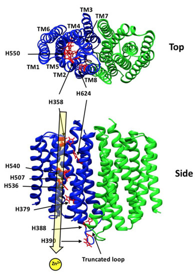
The human (h) transporter hZIP4 is the primary Zn2+ importer in the intestine. hZIP4 is also expressed in a variety of organs such as the pancreas and brain. Dysfunction of hZIP4 can result in the Zn2+ deficiency disease acrodermatitis enteropathica (AE). [...] Read more.
The human (h) transporter hZIP4 is the primary Zn2+ importer in the intestine. hZIP4 is also expressed in a variety of organs such as the pancreas and brain. Dysfunction of hZIP4 can result in the Zn2+ deficiency disease acrodermatitis enteropathica (AE). AE can disrupt digestive and immune system homeostasis. A limited number of hZIP4 expression strategies have hindered increasing knowledge about this essential transmembrane protein. Here, we report the heterologous expression of hZIP4 in Saccharomyces cerevisiae. Both a wild-type and a mutant S. cerevisiae strain, in which the endogenous Zn2+ transporters were deleted, were used to test the expression and localization of an hZIP4–GFP fusion protein. A full-length hZIP4–GFP and a truncated membrane-domain-only (mhZIP4–GFP) protein were observed to be present in the plasma membrane in yeast. Full article
Show Figures

Figure 1

19 pages, 3186 KiB  
Article
Pseudomonas aeruginosa Bacterioferritin Is Assembled from FtnA and BfrB Subunits with the Relative Proportions Dependent on the Environmental Oxygen Availability
by Huili Yao, Anabel Soldano, Leo Fontenot, Fabrizio Donnarumma, Scott Lovell, Josephine R. Chandler and Mario Rivera
Biomolecules 2022, 12(3), 366; https://doi.org/10.3390/biom12030366 - 25 Feb 2022
Cited by 19 | Viewed by 3616
Abstract
Ferritins are iron storage proteins assembled from 24 subunits into a spherical and hollow structure. The genomes of many bacteria harbor genes encoding two types of ferritin-like proteins, the bacterial ferritins (Ftn) and the bacterioferritins (Bfr), which bind heme. The genome of P. [...] Read more.
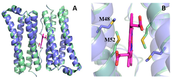
Ferritins are iron storage proteins assembled from 24 subunits into a spherical and hollow structure. The genomes of many bacteria harbor genes encoding two types of ferritin-like proteins, the bacterial ferritins (Ftn) and the bacterioferritins (Bfr), which bind heme. The genome of P. aeruginosa PAO1 (like the genomes of many bacteria) contains genes coding for two different types of ferritin-like molecules, ftnA (PA4235) and bfrB (PA3531). The reasons for requiring the presence of two distinct types of iron storage protein in bacterial cells have remained largely unexplained. Attempts to understand this issue in P. aeruginosa through the recombinant expression of the ftnA and bfrB genes in E. coli host cells, coupled to the biochemical and structural characterization of the recombinant 24-mer FtnA and 24-mer BfrB molecules, have shown that each of the recombinant molecules can form an Fe3+-mineral core. These observations led to the suggestion that 24-mer FtnA and 24-mer BfrB molecules coexist in P. aeruginosa cells where they share iron storage responsibilities. Herein, we demonstrate that P. aeruginosa utilizes a single heterooligomeric 24-mer Bfr assembled from FtnA and BfrB subunits. The relative content of the FtnA and BfrB subunits in Bfr depends on the O2 availability during cell culture, such that Bfr isolated from aerobically cultured P. aeruginosa is assembled from a majority of BfrB subunits. In contrast, when the cells are cultured in O2-limiting conditions, the proportion of FtnA subunits in the isolated Bfr increases significantly and can become the most abundant subunit. Despite the variability in the subunit composition of Bfr, the 24-mer assembly is consistently arranged from FtnA subunit dimers devoid of heme and BfrB subunit dimers each containing a heme molecule. Full article
Show Figures

Figure 1

26 pages, 3482 KiB  
Review
Mass Spectrometry-Based Structural Proteomics for Metal Ion/Protein Binding Studies
by Yanchun Lin and Michael L. Gross
Biomolecules 2022, 12(1), 135; https://doi.org/10.3390/biom12010135 - 15 Jan 2022
Cited by 6 | Viewed by 4727
Abstract
Metal ions are critical for the biological and physiological functions of many proteins. Mass spectrometry (MS)-based structural proteomics is an ever-growing field that has been adopted to study protein and metal ion interactions. Native MS offers information on metal binding and its stoichiometry. [...] Read more.
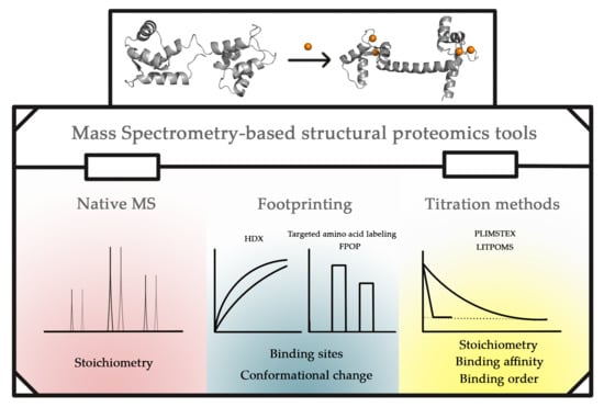
Metal ions are critical for the biological and physiological functions of many proteins. Mass spectrometry (MS)-based structural proteomics is an ever-growing field that has been adopted to study protein and metal ion interactions. Native MS offers information on metal binding and its stoichiometry. Footprinting approaches coupled with MS, including hydrogen/deuterium exchange (HDX), “fast photochemical oxidation of proteins” (FPOP) and targeted amino-acid labeling, identify binding sites and regions undergoing conformational changes. MS-based titration methods, including “protein–ligand interactions by mass spectrometry, titration and HD exchange” (PLIMSTEX) and “ligand titration, fast photochemical oxidation of proteins and mass spectrometry” (LITPOMS), afford binding stoichiometry, binding affinity, and binding order. These MS-based structural proteomics approaches, their applications to answer questions regarding metal ion protein interactions, their limitations, and recent and potential improvements are discussed here. This review serves as a demonstration of the capabilities of these tools and as an introduction to wider applications to solve other questions. Full article
Show Figures

Graphical abstract

17 pages, 2404 KiB  
Article
Erythropoietin Interacts with Specific S100 Proteins
by Alexey S. Kazakov, Evgenia I. Deryusheva, Andrey S. Sokolov, Maria E. Permyakova, Ekaterina A. Litus, Victoria A. Rastrygina, Vladimir N. Uversky, Eugene A. Permyakov and Sergei E. Permyakov
Biomolecules 2022, 12(1), 120; https://doi.org/10.3390/biom12010120 - 12 Jan 2022
Cited by 9 | Viewed by 3160
Abstract
Erythropoietin (EPO) is a clinically significant four-helical cytokine, exhibiting erythropoietic, cytoprotective, immunomodulatory, and cancer-promoting activities. Despite vast knowledge on its signaling pathways and physiological effects, extracellular factors regulating EPO activity remain underexplored. Here we show by surface plasmon resonance spectroscopy, that among eighteen [...] Read more.
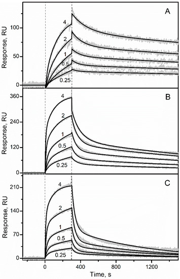
Erythropoietin (EPO) is a clinically significant four-helical cytokine, exhibiting erythropoietic, cytoprotective, immunomodulatory, and cancer-promoting activities. Despite vast knowledge on its signaling pathways and physiological effects, extracellular factors regulating EPO activity remain underexplored. Here we show by surface plasmon resonance spectroscopy, that among eighteen members of Ca2+-binding proteins of the S100 protein family studied, only S100A2, S100A6 and S100P proteins specifically recognize EPO with equilibrium dissociation constants ranging from 81 nM to 0.5 µM. The interactions occur exclusively under calcium excess. Bioinformatics analysis showed that the EPO-S100 interactions could be relevant to progression of neoplastic diseases, including cancer, and other diseases. The detailed knowledge of distinct physiological effects of the EPO-S100 interactions could favor development of more efficient clinical implications of EPO. Summing up our data with previous findings, we conclude that S100 proteins are potentially able to directly affect functional activities of specific members of all families of four-helical cytokines, and cytokines of other structural superfamilies. Full article
Show Figures

Figure 1

2021

Jump to: 2023, 2022

11 pages, 406 KiB  
Article
In Vitro Anti-SARS-CoV-2 Activity of Selected Metal Compounds and Potential Molecular Basis for Their Actions Based on Computational Study
by Damiano Cirri, Tiziano Marzo, Iogann Tolbatov, Alessandro Marrone, Francesco Saladini, Ilaria Vicenti, Filippo Dragoni, Adele Boccuto and Luigi Messori
Biomolecules 2021, 11(12), 1858; https://doi.org/10.3390/biom11121858 - 10 Dec 2021
Cited by 14 | Viewed by 2967
Abstract
Metal-based drugs represent a rich source of chemical substances of potential interest for the treatment of COVID-19. To this end, we have developed a small but representative panel of nine metal compounds, including both synthesized and commercially available complexes, suitable for medical application [...] Read more.
Metal-based drugs represent a rich source of chemical substances of potential interest for the treatment of COVID-19. To this end, we have developed a small but representative panel of nine metal compounds, including both synthesized and commercially available complexes, suitable for medical application and tested them in vitro against the SARS-CoV-2 virus. The screening revealed that three compounds from the panel, i.e., the organogold(III) compound Aubipyc, the ruthenium(III) complex KP1019, and antimony trichloride (SbCl3), are endowed with notable antiviral properties and an acceptable cytotoxicity profile. These initial findings prompted us to perform a computational study to unveil the likely molecular basis of their antiviral actions. Calculations evidenced that the metalation of nucleophile sites in SARS-CoV-2 proteins or nucleobase strands, induced by Aubipyc, SbCl3, and KP1019, is likely to occur. Remarkably, we found that only the deprotonated forms of Cys and Sec residues can react favorably with these metallodrugs. The mechanistic implications of these findings are discussed. Full article
Show Figures

Figure 1

17 pages, 2442 KiB  
Article
Mechanism of Zn2+ and Ca2+ Binding to Human S100A1
by Viktoriia E. Baksheeva, Andrei Yu. Roman, Claude Villard, François Devred, Deborah Byrne, Dahbia Yatoui, Arthur O. Zalevsky, Alisa A. Vologzhannikova, Andrey S. Sokolov, Sergei E. Permyakov, Andrey V. Golovin, Gary S. Shaw, Philipp O. Tsvetkov and Evgeni Yu. Zernii
Biomolecules 2021, 11(12), 1823; https://doi.org/10.3390/biom11121823 - 3 Dec 2021
Cited by 3 | Viewed by 3461
Abstract
S100A1 is a member of the S100 family of small ubiquitous Ca2+-binding proteins, which participates in the regulation of cell differentiation, motility, and survival. It exists as homo- or heterodimers. S100A1 has also been shown to bind Zn2+, but [...] Read more.
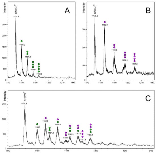
S100A1 is a member of the S100 family of small ubiquitous Ca2+-binding proteins, which participates in the regulation of cell differentiation, motility, and survival. It exists as homo- or heterodimers. S100A1 has also been shown to bind Zn2+, but the molecular mechanisms of this binding are not yet known. In this work, using ESI-MS and ITC, we demonstrate that S100A1 can coordinate 4 zinc ions per monomer, with two high affinity (KD~4 and 770 nm) and two low affinity sites. Using competitive binding experiments between Ca2+ and Zn2+ and QM/MM molecular modeling we conclude that Zn2+ high affinity sites are located in the EF-hand motifs of S100A1. In addition, two lower affinity sites can bind Zn2+ even when the EF-hands are saturated by Ca2+, resulting in a 2Ca2+:S100A1:2Zn2+ conformer. Finally, we show that, in contrast to calcium, an excess of Zn2+ produces a destabilizing effect on S100A1 structure and leads to its aggregation. We also determined a higher affinity to Ca2+ (KD~0.16 and 24 μm) than was previously reported for S100A1, which would allow this protein to function as a Ca2+/Zn2+-sensor both inside and outside cells, participating in diverse signaling pathways under normal and pathological conditions. Full article
Show Figures

Figure 1

10 pages, 1581 KiB  
Review
L-Type Ca2+ Channel Regulation by Calmodulin and CaBP1
by James B. Ames
Biomolecules 2021, 11(12), 1811; https://doi.org/10.3390/biom11121811 - 2 Dec 2021
Cited by 17 | Viewed by 2929
Abstract
L-type voltage-gated Ca2+ channels (CaV1.2 and CaV1.3, called CaV) interact with the Ca2+ sensor proteins, calmodulin (CaM) and Ca2+ binding Protein 1 (CaBP1), that oppositely control Ca2+-dependent channel activity. CaM and CaBP1 can each bind to the IQ-motif [...] Read more.
L-type voltage-gated Ca2+ channels (CaV1.2 and CaV1.3, called CaV) interact with the Ca2+ sensor proteins, calmodulin (CaM) and Ca2+ binding Protein 1 (CaBP1), that oppositely control Ca2+-dependent channel activity. CaM and CaBP1 can each bind to the IQ-motif within the C-terminal cytosolic domain of CaV, which promotes increased channel open probability under basal conditions. At elevated cytosolic Ca2+ levels (caused by CaV channel opening), Ca2+-bound CaM binding to CaV is essential for promoting rapid Ca2+-dependent channel inactivation (CDI). By contrast, CaV binding to CaBP1 prevents CDI and promotes Ca2+-induced channel opening (called CDF). In this review, I provide an overview of the known structures of CaM and CaBP1 and their structural interactions with the IQ-motif to help understand how CaM promotes CDI, whereas CaBP1 prevents CDI and instead promotes CDF. Previous electrophysiology studies suggest that Ca2+-free forms of CaM and CaBP1 may pre-associate with CaV under basal conditions. However, previous Ca2+ binding data suggest that CaM and CaBP1 are both calculated to bind to Ca2+ with an apparent dissociation constant of ~100 nM when CaM or CaBP1 is bound to the IQ-motif. Since the neuronal basal cytosolic Ca2+ concentration is ~100 nM, nearly half of the neuronal CaV channels are suggested to be bound to Ca2+-bound forms of either CaM or CaBP1 under basal conditions. The pre-association of CaV with calcified forms of CaM or CaBP1 are predicted here to have functional implications. The Ca2+-bound form of CaBP1 is proposed to bind to CaV under basal conditions to block CaV binding to CaM, which could explain how CaBP1 might prevent CDI. Full article
Show Figures

Figure 1

10 pages, 858 KiB  
Communication
Ca2+/Sr2+ Selectivity in Calcium-Sensing Receptor (CaSR): Implications for Strontium’s Anti-Osteoporosis Effect
by Diana Cheshmedzhieva, Sonia Ilieva, Eugene A. Permyakov, Sergei E. Permyakov and Todor Dudev
Biomolecules 2021, 11(11), 1576; https://doi.org/10.3390/biom11111576 - 24 Oct 2021
Cited by 26 | Viewed by 2757
Abstract
The extracellular calcium-sensing receptor (CaSR) controls vital bone cell functions such as cell growth, differentiation and apoptosis. The binding of the native agonist (Ca2+) to CaSR activates the receptor, which undergoes structural changes that trigger a cascade of events along the [...] Read more.
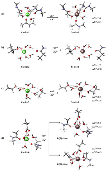
The extracellular calcium-sensing receptor (CaSR) controls vital bone cell functions such as cell growth, differentiation and apoptosis. The binding of the native agonist (Ca2+) to CaSR activates the receptor, which undergoes structural changes that trigger a cascade of events along the cellular signaling pathways. Strontium (in the form of soluble salts) has been found to also be a CaSR agonist. The activation of the receptor by Sr2+ is considered to be the major mechanism through which strontium exerts its anti-osteoporosis effect, mostly in postmenopausal women. Strontium-activated CaSR initiates a series of signal transduction events resulting in both osteoclast apoptosis and osteoblast differentiation, thus strengthening the bone tissue. The intimate mechanism of Sr2+ activation of CaSR is still enigmatic. Herewith, by employing a combination of density functional theory (DFT) calculations and polarizable continuum model (PCM) computations, we have found that the Ca2+ binding sites 1, 3, and 4 in the activated CaSR, although possessing a different number and type of protein ligands, overall structure and charge state, are all selective for Ca2+ over Sr2+. The three binding sites, regardless of their structural differences, exhibit almost equal metal selectivity if they are flexible and have no geometrical constraints on the incoming Sr2+. In contrast to Ca2+ and Sr2+, Mg2+ constructs, when allowed to fully relax during the optimization process, adopt their stringent six-coordinated octahedral structure at the expense of detaching a one-backbone carbonyl ligand and shifting it to the second coordination layer of the metal. The binding of Mg2+ and Sr2+ to a rigid/inflexible calcium-designed binding pocket requires an additional energy penalty for the binding ion; however, the price for doing so (to be paid by Sr2+) is much less than that of Mg2+. The results obtained delineate the key factors controlling the competition between metal cations for the receptor and shed light on some aspects of strontium’s therapeutic effects. Full article
Show Figures

Figure 1

21 pages, 4294 KiB  
Article
Strontium Binding to α-Parvalbumin, a Canonical Calcium-Binding Protein of the “EF-Hand” Family
by Alisa A. Vologzhannikova, Marina P. Shevelyova, Alexey S. Kazakov, Andrey S. Sokolov, Nadezhda I. Borisova, Eugene A. Permyakov, Nikoleta Kircheva, Valya Nikolova, Todor Dudev and Sergei E. Permyakov
Biomolecules 2021, 11(8), 1158; https://doi.org/10.3390/biom11081158 - 5 Aug 2021
Cited by 13 | Viewed by 2713
Abstract
Strontium salts are used for treatment of osteoporosis and bone cancer, but their impact on calcium-mediated physiological processes remains obscure. To explore Sr2+ interference with Ca2+ binding to proteins of the EF-hand family, we studied Sr2+/Ca2+ interaction with [...] Read more.
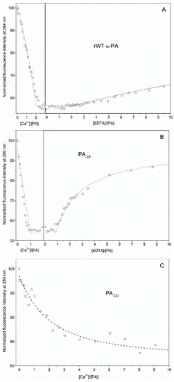
Strontium salts are used for treatment of osteoporosis and bone cancer, but their impact on calcium-mediated physiological processes remains obscure. To explore Sr2+ interference with Ca2+ binding to proteins of the EF-hand family, we studied Sr2+/Ca2+ interaction with a canonical EF-hand protein, α-parvalbumin (α-PA). Evaluation of the equilibrium metal association constants for the active Ca2+ binding sites of recombinant human α-PA (‘CD’ and ‘EF’ sites) from fluorimetric titration experiments and isothermal titration calorimetry data gave 4 × 10⁹ M−1 and 4 × 10⁹ M−1 for Ca2+, and 2 × 10⁷ M−1 and 2 × 10⁶ M−1 for Sr2+. Inactivation of the EF site by homologous substitution of the Ca2+-coordinating Glu in position 12 of the EF-loop by Gln decreased Ca2+/Sr2+ affinity of the protein by an order of magnitude, whereas the analogous inactivation of the CD site induced much deeper suppression of the Ca2+/Sr2+ affinity. These results suggest that Sr2+ and Ca2+ bind to CD/EF sites of α-PA and the Ca2+/Sr2+ binding are sequential processes with the CD site being occupied first. Spectrofluorimetric Sr2+ titration of the Ca2+-loaded α-PA revealed presence of secondary Sr2+ binding site(s) with an apparent equilibrium association constant of 4 × 10⁵ M−1. Fourier-transform infrared spectroscopy data evidence that Ca2+/Sr2+-loaded forms of α-PA exhibit similar states of their COO groups. Near-UV circular dichroism (CD) data show that Ca2+/Sr2+ binding to α-PA induce similar changes in symmetry of microenvironment of its Phe residues. Far-UV CD experiments reveal that Ca2+/Sr2+ binding are accompanied by nearly identical changes in secondary structure of α-PA. Meanwhile, scanning calorimetry measurements show markedly lower Sr2+-induced increase in stability of tertiary structure of α-PA, compared to the Ca2+-induced effect. Theoretical modeling using Density Functional Theory computations with Polarizable Continuum Model calculations confirms that Ca2+-binding sites of α-PA are well protected against exchange of Ca2+ for Sr2+ regardless of coordination number of Sr2+, solvent exposure or rigidity of sites. The latter appears to be a key determinant of the Ca2+/Sr2+ selectivity. Overall, despite lowered affinity of α-PA to Sr2+, the latter competes with Ca2+ for the same EF-hands and induces similar structural rearrangements. The presence of a secondary Sr2+ binding site(s) could be a factor contributing to Sr2+ impact on the functional activity of proteins. Full article
Show Figures

Figure 1

Planned Papers

The below list represents only planned manuscripts. Some of these manuscripts have not been received by the Editorial Office yet. Papers submitted to MDPI journals are subject to peer-review.

Title: Regulation of FYVE domain interactions with membranes
Authors: Cameron Smithers; Troy Kervin; Michael Overduin
Affiliation: University of Alberta, Edmonton, Canada

Title: To be determined
Authors: Svetlana Lutsenko; Jenifer Calvo; Abigael Muchenditsi; et al.
Affiliation: Johns Hopkins University, USA

Title: To be determined
Authors: Artem Bonchuk; et al.
Affiliation: Department of the Control of Genetic Processes, Institute of Gene Biology, Russian Academy of Sciences, 3 4/5 Vavilov St., Moscow, 119334, Russia

Back to TopTop