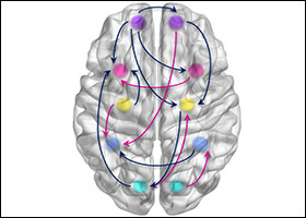
Neural Networks and Connectivity among Brain Regions

A special issue of Brain Sciences (ISSN 2076-3425). This special issue belongs to the section "Computational Neuroscience and Neuroinformatics".

Deadline for manuscript submissions: closed (31 July 2021) | Viewed by 34856

Special Issue Editors


E-Mail Website
Guest Editor
Department of Electrical, Electronic and Information Engineering "Guglielmo Marconi", University of Bologna-Campus of Cesena, Via Dell'Università 50, 47521 Cesena, Italy
Interests: physiological modeling; computational neuroscience; neural networks; multisensory integration; semantic memory; brain rhythms; Parkinson’s disease

E-Mail Website
Co-Guest Editor
Department of Electrical, Electronic, and Information Engineering “Guglielmo Marconi”, University of Bologna, Cesena, Italy
Interests: computational neuroscience; multisensory integration; electroencephalography and brain rhythms; connectivity; biomedical signal processing

E-Mail Website
Co-Guest Editor
Department of Computer, Control and Management Engineering, Sapienza University of Rome, 00185 Rome, Italy
Interests: network medicine; computational biology; bioinformatics
Special Issues, Collections and Topics in MDPI journals

Special Issue Information

Dear Colleagues,

Cognitive phenomena involve the interaction among several mutually interconnected, specialized brain regions. The problem of assessing brain connectivity during different cognitive tasks and of building biologically inspired neural networks of interconnected regions is thus playing a crucial role in neuroscience today.

The aim of the present Special Issue is to provide a general overview of recent signal processing and mathematical modeling techniques useful to assess brain connectivity, and to simulate the behavior of large interconnected brain regions during relevant cognitive problems.

Cutting-edge research topics can include: i) theoretical overviews of advanced techniques for functional or effective connectivity estimation starting from neuroelectric and/or functional neuroimaging data; ii) experimental assessment of brain connectivity networks involved in different cognitive problems (such as semantic and working memory, spatiotemporal episodic memory, multisensory integration, conflict resolution, fear conditioning, emotion and language); iii) neural networks, inspired by neurobiological data, to simulate the behavior of the brain in some of the cognitive problems mentioned above and to study the origin of brain rhythms and the assessment of their role in cognition; v) use of the previous techniques to study the alterations in brain connectivity and their role in important neurological problems (e.g., autism spectrum disorders, schizophrenia, Parkinson’s diseases, semantic dementia and Alzheimer’s, epilepsy).

Both original experimental and theoretical papers on the previous subjects, as well as review papers are solicited.

Prof. Dr. Mauro Ursino
Guest Editor
Dr. Elisa Magosso
Dr. Manuela Petti
Co-Guest Editors

Manuscript Submission Information

Manuscripts should be submitted online at www.mdpi.com by registering and logging in to this website. Once you are registered, click here to go to the submission form. Manuscripts can be submitted until the deadline. All submissions that pass pre-check are peer-reviewed. Accepted papers will be published continuously in the journal (as soon as accepted) and will be listed together on the special issue website. Research articles, review articles as well as short communications are invited. For planned papers, a title and short abstract (about 100 words) can be sent to the Editorial Office for announcement on this website.

Submitted manuscripts should not have been published previously, nor be under consideration for publication elsewhere (except conference proceedings papers). All manuscripts are thoroughly refereed through a single-blind peer-review process. A guide for authors and other relevant information for submission of manuscripts is available on the Instructions for Authors page. Brain Sciences is an international peer-reviewed open access monthly journal published by MDPI.

Please visit the Instructions for Authors page before submitting a manuscript. The Article Processing Charge (APC) for publication in this open access journal is 2200 CHF (Swiss Francs). Submitted papers should be well formatted and use good English. Authors may use MDPI's English editing service prior to publication or during author revisions.

Keywords

  • brain mapping
  • functional connectivity
  • effective connectivity
  • neural networks
  • neurocomputational models
  • cognitive neurodynamics
  • neurodynamical diseases
  • neurodegenerative disorders
  • brain circuits and synapses
  • MRI, fMRI
  • EEG, MEG

Benefits of Publishing in a Special Issue

  • Ease of navigation: Grouping papers by topic helps scholars navigate broad scope journals more efficiently.
  • Greater discoverability: Special Issues support the reach and impact of scientific research. Articles in Special Issues are more discoverable and cited more frequently.
  • Expansion of research network: Special Issues facilitate connections among authors, fostering scientific collaborations.
  • External promotion: Articles in Special Issues are often promoted through the journal's social media, increasing their visibility.
  • e-Book format: Special Issues with more than 10 articles can be published as dedicated e-books, ensuring wide and rapid dissemination.

Further information on MDPI's Special Issue polices can be found here.

Published Papers (10 papers)

Order results
Result details
Select all
Export citation of selected articles as:

Editorial

Jump to: Research

4 pages, 192 KiB  
Editorial
Neural Networks and Connectivity among Brain Regions
by Mauro Ursino, Elisa Magosso and Manuela Petti
Brain Sci. 2022, 12(3), 346; https://doi.org/10.3390/brainsci12030346 - 3 Mar 2022
Cited by 1 | Viewed by 1927
Abstract
As is widely understood, brain functioning depends on the interaction among several neural populations, which are linked via complex connectivity circuits and work together (in antagonistic or synergistic ways) to exchange information, synchronize their activity, adapt plastically to external stimuli or internal requirements, [...] Read more.
As is widely understood, brain functioning depends on the interaction among several neural populations, which are linked via complex connectivity circuits and work together (in antagonistic or synergistic ways) to exchange information, synchronize their activity, adapt plastically to external stimuli or internal requirements, and more generally to participate in solving multifaceted cognitive tasks [...] Full article
(This article belongs to the Special Issue Neural Networks and Connectivity among Brain Regions)

Research

Jump to: Editorial

17 pages, 2093 KiB  
Article
The Directionality of Fronto-Posterior Brain Connectivity Is Associated with the Degree of Individual Autistic Traits
by Luca Tarasi, Elisa Magosso, Giulia Ricci, Mauro Ursino and Vincenzo Romei
Brain Sci. 2021, 11(11), 1443; https://doi.org/10.3390/brainsci11111443 - 29 Oct 2021
Cited by 16 | Viewed by 3642
Abstract
Altered patterns of brain connectivity have been found in autism spectrum disorder (ASD) and associated with specific symptoms and behavioral features. Growing evidence suggests that the autistic peculiarities are not confined to the clinical population but extend along a continuum between healthy and [...] Read more.
Altered patterns of brain connectivity have been found in autism spectrum disorder (ASD) and associated with specific symptoms and behavioral features. Growing evidence suggests that the autistic peculiarities are not confined to the clinical population but extend along a continuum between healthy and maladaptive conditions. The aim of this study was to investigate whether a differentiated connectivity pattern could also be tracked along the continuum of autistic traits in a non-clinical population. A Granger causality analysis conducted on a resting-state EEG recording showed that connectivity along the posterior-frontal gradient is sensitive to the magnitude of individual autistic traits and mostly conveyed through fast oscillatory activity. Specifically, participants with higher autistic traits were characterized by a prevalence of ascending connections starting from posterior regions ramping the cortical hierarchy. These findings point to the presence of a tendency within the neural mapping of individuals with higher autistic features in conveying proportionally more bottom-up information. This pattern of findings mimics those found in clinical forms of autism, supporting the idea of a neurobiological continuum between autistic traits and ASD. Full article
(This article belongs to the Special Issue Neural Networks and Connectivity among Brain Regions)
Show Figures

Figure 1

13 pages, 2227 KiB  
Article
Reduced Effective Connectivity in the Motor Cortex in Parkinson’s Disease
by Emanuela Formaggio, Maria Rubega, Jessica Rupil, Angelo Antonini, Stefano Masiero, Gianna Maria Toffolo and Alessandra Del Felice
Brain Sci. 2021, 11(9), 1200; https://doi.org/10.3390/brainsci11091200 - 12 Sep 2021
Cited by 8 | Viewed by 2793
Abstract
Fast rhythms excess is a hallmark of Parkinson’s Disease (PD). To implement innovative, non-pharmacological, neurostimulation interventions to restore cortical-cortical interactions, we need to understand the neurophysiological mechanisms underlying these phenomena. Here, we investigated effective connectivity on source-level resting-state electroencephalography (EEG) signals in 15 [...] Read more.
Fast rhythms excess is a hallmark of Parkinson’s Disease (PD). To implement innovative, non-pharmacological, neurostimulation interventions to restore cortical-cortical interactions, we need to understand the neurophysiological mechanisms underlying these phenomena. Here, we investigated effective connectivity on source-level resting-state electroencephalography (EEG) signals in 15 PD participants and 10 healthy controls. First, we fitted multivariate auto-regressive models to the EEG source waveforms. Second, we estimated causal connections using Granger Causality, which provide information on connections’ strength and directionality. Lastly, we sought significant differences connectivity patterns between the two populations characterizing the network graph features—i.e., global efficiency and node strength. Causal brain networks in PD show overall poorer and weaker connections compared to controls quantified as a reduction of global efficiency. Motor areas appear almost isolated, with a strongly impoverished information flow particularly from parietal and occipital cortices. This striking isolation of motor areas may reflect an impaired sensory-motor integration in PD. The identification of defective nodes/edges in PD network may be a biomarker of disease and a potential target for future interventional trials. Full article
(This article belongs to the Special Issue Neural Networks and Connectivity among Brain Regions)
Show Figures

Figure 1

13 pages, 1225 KiB  
Article
Changes in Default Mode Network Connectivity in Resting-State fMRI in People with Mild Dementia Receiving Cognitive Stimulation Therapy
by Tianyin Liu, Aimee Spector, Daniel C. Mograbi, Gary Cheung and Gloria H. Y. Wong
Brain Sci. 2021, 11(9), 1137; https://doi.org/10.3390/brainsci11091137 - 27 Aug 2021
Cited by 12 | Viewed by 3311
Abstract
Group cognitive stimulation therapy (CST) is a 7-week activity-based non-pharmacological intervention for people with mild to moderate dementia. Despite consistent evidence of clinical efficacy, the cognitive and brain mechanisms of CST remain unclear. Theoretically, group CST as a person-centred approach may work through [...] Read more.
Group cognitive stimulation therapy (CST) is a 7-week activity-based non-pharmacological intervention for people with mild to moderate dementia. Despite consistent evidence of clinical efficacy, the cognitive and brain mechanisms of CST remain unclear. Theoretically, group CST as a person-centred approach may work through promoting social interaction and personhood, executive function, and language use, especially in people with higher brain/cognitive reserve. To explore these putative mechanisms, structural MRI and resting-state functional MRI data were collected from 16 people with mild dementia before and after receiving CST, and in 13 dementia controls who received treatment as usual (TAU). Voxel-based morphometry (VBM) and resting-state functional connectivity (rs-FC) analyses were performed. Compared with TAU, the CST group maintained the total brain volume/total intracranial volume (TBV/TICV) ratio. Increased rs-FC in the default mode network (DMN) in the posterior cingulate cortex and bilateral parietal cortices nodes was observed in the CST over TAU groups between pre- and post-intervention timepoints. We provided preliminary evidence that CST maintains/enhances brain reserve both structurally and functionally. Considering the role of DMN in episodic memory retrieval and mental self-representation, preservation of personhood may be an important mechanism of CST for further investigation. Full article
(This article belongs to the Special Issue Neural Networks and Connectivity among Brain Regions)
Show Figures

Figure 1

22 pages, 3047 KiB  
Article
Ageing and the Ipsilateral M1 BOLD Response: A Connectivity Study
by Yae Won Tak, Ethan Knights, Richard Henson and Peter Zeidman
Brain Sci. 2021, 11(9), 1130; https://doi.org/10.3390/brainsci11091130 - 26 Aug 2021
Cited by 5 | Viewed by 3332
Abstract
Young people exhibit a negative BOLD response in ipsilateral primary motor cortex (M1) when making unilateral movements, such as button presses. This negative BOLD response becomes more positive as people age. In this study, we investigated why this occurs, in terms of the [...] Read more.
Young people exhibit a negative BOLD response in ipsilateral primary motor cortex (M1) when making unilateral movements, such as button presses. This negative BOLD response becomes more positive as people age. In this study, we investigated why this occurs, in terms of the underlying effective connectivity and haemodynamics. We applied dynamic causal modeling (DCM) to task fMRI data from 635 participants aged 18–88 from the Cam-CAN dataset, who performed a cued button pressing task with their right hand. We found that connectivity from contralateral supplementary motor area (SMA) and dorsal premotor cortex (PMd) to ipsilateral M1 became more positive with age, explaining 44% of the variability across people in ipsilateral M1 responses. In contrast, connectivity from contralateral M1 to ipsilateral M1 was weaker and did not correlate with individual differences in rM1 BOLD. Neurovascular and haemodynamic parameters in the model were not able to explain the age-related shift to positive BOLD. Our results add to a body of evidence implicating neural, rather than vascular factors as the predominant cause of negative BOLD—while emphasising the importance of inter-hemispheric connectivity. This study provides a foundation for investigating the clinical and lifestyle factors that determine the sign and amplitude of the M1 BOLD response in ageing, which could serve as a proxy for neural and vascular health, via the underlying neurovascular mechanisms. Full article
(This article belongs to the Special Issue Neural Networks and Connectivity among Brain Regions)
Show Figures

Figure 1

10 pages, 555 KiB  
Article
Functional Connectivity-Derived Optimal Gestational-Age Cut Points for Fetal Brain Network Maturity
by Josepheen De Asis-Cruz, Scott Douglas Barnett, Jung-Hoon Kim and Catherine Limperopoulos
Brain Sci. 2021, 11(7), 921; https://doi.org/10.3390/brainsci11070921 - 12 Jul 2021
Cited by 9 | Viewed by 3102
Abstract
The architecture of the human connectome changes with brain maturation. Pivotal to understanding these changes is defining developmental periods when transitions in network topology occur. Here, using 110 resting-state functional connectivity data sets from healthy fetuses between 19 and 40 gestational weeks, we [...] Read more.
The architecture of the human connectome changes with brain maturation. Pivotal to understanding these changes is defining developmental periods when transitions in network topology occur. Here, using 110 resting-state functional connectivity data sets from healthy fetuses between 19 and 40 gestational weeks, we estimated optimal gestational-age (GA) cut points for dichotomizing fetuses into ‘young’ and ‘old’ groups based on global network features. We computed the small-world index, normalized clustering and path length, global and local efficiency, and modularity from connectivity matrices comprised 200 regions and their corresponding pairwise connectivity. We modeled the effect of GA at scan on each metric using separate repeated-measures generalized estimating equations. Our modeling strategy involved stratifying fetuses into ‘young’ and ‘old’ based on the scan occurring before or after a selected GA (i.e., 28 to 33). We then used the quasi-likelihood independence criterion statistic to compare model fit between ‘old’ and ‘young’ cohorts and determine optimal cut points for each graph metric. Trends for all metrics, except for global efficiency, decreased with increasing gestational age. Optimal cut points fell within 30–31 weeks for all metrics coinciding with developmental events that include a shift from endogenous neuronal activity to sensory-driven cortical patterns. Full article
(This article belongs to the Special Issue Neural Networks and Connectivity among Brain Regions)
Show Figures

Figure 1

16 pages, 2776 KiB  
Article
Detection of Resting-State Functional Connectivity from High-Density Electroencephalography Data: Impact of Head Modeling Strategies
by Gaia Amaranta Taberna, Jessica Samogin, Marco Marino and Dante Mantini
Brain Sci. 2021, 11(6), 741; https://doi.org/10.3390/brainsci11060741 - 3 Jun 2021
Cited by 10 | Viewed by 4171
Abstract
Recent technological advances have been permitted to use high-density electroencephalography (hdEEG) for the estimation of functional connectivity and the mapping of resting-state networks (RSNs). The reliable estimate of activity and connectivity from hdEEG data relies on the creation of an accurate head model, [...] Read more.
Recent technological advances have been permitted to use high-density electroencephalography (hdEEG) for the estimation of functional connectivity and the mapping of resting-state networks (RSNs). The reliable estimate of activity and connectivity from hdEEG data relies on the creation of an accurate head model, defining how neural currents propagate from the cortex to the sensors placed over the scalp. To the best of our knowledge, no study has been conducted yet to systematically test to what extent head modeling accuracy impacts on EEG-RSN reconstruction. To address this question, we used 256-channel hdEEG data collected in a group of young healthy participants at rest. We first estimated functional connectivity in EEG-RSNs by means of band-limited power envelope correlations, using neural activity estimated with an optimized analysis workflow. Then, we defined a series of head models with different levels of complexity, specifically testing the effect of different electrode positioning techniques and head tissue segmentation methods. We observed that robust EEG-RSNs can be obtained using a realistic head model, and that inaccuracies due to head tissue segmentation impact on RSN reconstruction more than those due to electrode positioning. Additionally, we found that EEG-RSN robustness to head model variations had space and frequency specificity. Overall, our results may contribute to defining a benchmark for assessing the reliability of hdEEG functional connectivity measures. Full article
(This article belongs to the Special Issue Neural Networks and Connectivity among Brain Regions)
Show Figures

Figure 1

16 pages, 5772 KiB  
Article
Time-Frequency Characterization of Resting Brain in Bipolar Disorder during Euthymia—A Preliminary Study
by Adrian Andrzej Chrobak, Bartosz Bohaterewicz, Anna Maria Sobczak, Magdalena Marszał-Wiśniewska, Anna Tereszko, Anna Krupa, Anna Ceglarek, Magdalena Fafrowicz, Amira Bryll, Tadeusz Marek, Dominika Dudek and Marcin Siwek
Brain Sci. 2021, 11(5), 599; https://doi.org/10.3390/brainsci11050599 - 7 May 2021
Cited by 14 | Viewed by 3702
Abstract
The goal of this paper is to investigate the baseline brain activity in euthymic bipolar disorder (BD) patients by comparing it to healthy controls (HC) with the use of a variety of resting state functional magnetic resonance imaging (rs-fMRI) analyses, such as amplitude [...] Read more.
The goal of this paper is to investigate the baseline brain activity in euthymic bipolar disorder (BD) patients by comparing it to healthy controls (HC) with the use of a variety of resting state functional magnetic resonance imaging (rs-fMRI) analyses, such as amplitude of low frequency fluctuations (ALFF), fractional ALFF (f/ALFF), ALFF-based functional connectivity (FC), and r egional homogeneity (ReHo). We hypothesize that above-mentioned techniques will differentiate BD from HC indicating dissimilarities between the groups within different brain structures. Forty-two participants divided into two groups of euthymic BD patients (n = 21) and HC (n = 21) underwent rs-fMRI evaluation. Typical band ALFF, slow-4, slow-5, f/ALFF, as well as ReHo indexes were analyzed. Regions with altered ALFF were chosen as ROI for seed-to-voxel analysis of FC. As opposed to HC, BD patients revealed: increased ALFF in left insula; increased slow-5 in left middle temporal pole; increased f/ALFF in left superior frontal gyrus, left superior temporal gyrus, left middle occipital gyrus, right putamen, and bilateral thalamus. There were no significant differences between BD and HC groups in slow-4 band. Compared to HC, the BD group presented higher ReHo values in the left superior medial frontal gyrus and lower ReHo values in the right supplementary motor area. FC analysis revealed significant hyper-connectivity within the BD group between left insula and bilateral middle frontal gyrus, right superior parietal gyrus, right supramarginal gyrus, left inferior parietal gyrus, left cerebellum, and left supplementary motor area. To our best knowledge, this is the first rs-fMRI study combining ReHo, ALFF, f/ALFF, and subdivided frequency bands (slow-4 and slow-5) in euthymic BD patients. ALFF, f/ALFF, slow-5, as well as REHO analysis revealed significant differences between two studied groups. Although results obtained with the above methods enable to identify group-specific brain structures, no overlap between the brain regions was detected. This indicates that combination of foregoing rs-fMRI methods may complement each other, revealing the bigger picture of the complex resting state abnormalities in BD. Full article
(This article belongs to the Special Issue Neural Networks and Connectivity among Brain Regions)
Show Figures

Figure 1

31 pages, 10049 KiB  
Article
The Relationship between Oscillations in Brain Regions and Functional Connectivity: A Critical Analysis with the Aid of Neural Mass Models
by Giulia Ricci, Elisa Magosso and Mauro Ursino
Brain Sci. 2021, 11(4), 487; https://doi.org/10.3390/brainsci11040487 - 12 Apr 2021
Cited by 11 | Viewed by 3784
Abstract
Propagation of brain rhythms among cortical regions is a relevant aspect of cognitive neuroscience, which is often investigated using functional connectivity (FC) estimation techniques. The aim of this work is to assess the relationship between rhythm propagation, FC and brain functioning using data [...] Read more.
Propagation of brain rhythms among cortical regions is a relevant aspect of cognitive neuroscience, which is often investigated using functional connectivity (FC) estimation techniques. The aim of this work is to assess the relationship between rhythm propagation, FC and brain functioning using data generated from neural mass models of connected Regions of Interest (ROIs). We simulated networks of four interconnected ROIs, each with a different intrinsic rhythm (in θ, α, β and γ ranges). Connectivity was estimated using eight estimators and the relationship between structural connectivity and FC was assessed as a function of the connectivity strength and of the inputs to the ROIs. Results show that the Granger estimation provides the best accuracy, with a good capacity to evaluate the connectivity strength. However, the estimated values strongly depend on the input to the ROIs and hence on nonlinear phenomena. When a population works in the linear region, its capacity to transmit a rhythm increases drastically. Conversely, when it saturates, oscillatory activity becomes strongly affected by rhythms incoming from other regions. Changes in functional connectivity do not always reflect a physical change in the synapses. A unique connectivity network can propagate rhythms in very different ways depending on the specific working conditions. Full article
(This article belongs to the Special Issue Neural Networks and Connectivity among Brain Regions)
Show Figures

Figure 1

22 pages, 2857 KiB  
Article
A Three-Stage Teacher, Student Neural Networks and Sequential Feed Forward Selection-Based Feature Selection Approach for the Classification of Autism Spectrum Disorder
by Naseer Ahmed Khan, Samer Abdulateef Waheeb, Atif Riaz and Xuequn Shang
Brain Sci. 2020, 10(10), 754; https://doi.org/10.3390/brainsci10100754 - 19 Oct 2020
Cited by 14 | Viewed by 3553
Abstract
Autism disorder, generally known as Autism Spectrum Disorder (ASD) is a brain disorder characterized by lack of communication skills, social aloofness and repetitions in the actions in the patients, which is affecting millions of the people across the globe. Accurate identification of autistic [...] Read more.
Autism disorder, generally known as Autism Spectrum Disorder (ASD) is a brain disorder characterized by lack of communication skills, social aloofness and repetitions in the actions in the patients, which is affecting millions of the people across the globe. Accurate identification of autistic patients is considered a challenging task in the domain of brain disorder science. To address this problem, we have proposed a three-stage feature selection approach for the classification of ASD on the preprocessed Autism Brain Imaging Data Exchange (ABIDE) rs-fMRI Dataset. In the first stage, a large neural network which we call a “Teacher ” was trained on the correlation-based connectivity matrix to learn the latent representation of the input. In the second stage an autoencoder which we call a “Student” autoencoder was given the task to learn those trained “Teacher” embeddings using the connectivity matrix input. Lastly, an SFFS-based algorithm was employed to select the subset of most discriminating features between the autistic and healthy controls. On the combined site data across 17 sites, we achieved the maximum 10-fold accuracy of 82% and for the individual site-wise data, based on 5-fold accuracy, our results outperformed other state of the art methods in 13 out of the total 17 site-wise comparisons. Full article
(This article belongs to the Special Issue Neural Networks and Connectivity among Brain Regions)
Show Figures

Figure 1

Back to TopTop