Advances in Musculoskeletal Tissue Regeneration Using Mesenchymal Stromal/Stem Cells, Biomaterials, and Signaling Molecules

A special issue of Cells (ISSN 2073-4409). This special issue belongs to the section "Stem Cells".

Deadline for manuscript submissions: closed (31 January 2024) | Viewed by 16386
Special Issue Editors


E-Mail Website
Guest Editor
Department of Orthopaedics, Division of Sports Medicine, Diabetes Research Institute, Cell Transplant Center, University of Miami, Miller School of Medicine, 1450NW 10th Ave, Room 3012, Miami, FL 33136, USA
Interests: mesenchymal stem cells/multipotential stromal cells (MSCs); MSC trophic and immunomodulatory actions; MSC functionalization ex vivo; inflammation and fibrosis reversal; synovitis; osteoarthritis; regenerative sports medicine; regenerative orthopaedics
Special Issues, Collections and Topics in MDPI journals

E-Mail Website
Guest Editor
Department of Orthopaedics, Division of Sports Medicine, University of Miami, Miller School of Medicine, 5555 Ponce de Leon, Coral Gables, FL 33146, USA
Interests: stem cell research; biomedical engineering; kinesiology; osteoarthritis; regenerative sports medicine; regenerative orthopaedics
Special Issues, Collections and Topics in MDPI journals

Special Issue Information

Dear Colleagues,

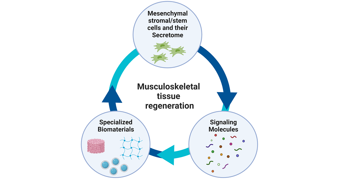
Regeneration of musculoskeletal tissues is a serious clinical challenge given that their injury and disease are leading causes of physical disability worldwide. Approved mesenchymal stromal/stem cells (MSC) and MSC products (i.e., extracellular vesicles) for stem cell-based therapy applications offer huge promise to rejuvenate musculoskeletal tissues, triggering tissue repair and healing. In parallel, the discovery of specialized biomaterials and signaling molecules has opened novel therapeutic avenues as part of tissue engineering approaches. Up-to-date, significant progress has been made in developing and optimizing the components of this therapeutic triad (cells, biomaterials, signaling molecules), but only a limited number of technologies have been successfully transferred into the clinical setting. Currently, efforts are focused on bringing closer basic science and cell-based product manufacturing techniques to potential clinical protocols. This Special Issue aims to present a state-of-the-art update on the use of MSC, biomaterials and signaling molecules for musculoskeletal therapeutic applications.

We are pleased to invite you as a recognized expert in the field to contribute original articles, communications, and reviews covering the entire field of musculoskeletal regeneration research, including (but not limited to):

  • MSC and biomaterial functionalization (e.g., cell phenotype-based purification, cell priming, gene-activated matrices);
  • MSC-derived extracellular vesicle isolation and methods to reinforce their therapeutic signatures;
  • Benefits and limitations of MSC versus MSC-derived extracellular vesicles for musculoskeletal applications;
  • MSC and MSC-derived extracellular vesicle effects on immunomodulation and senescent cell clearance;
  • Potent signaling molecules to promote healing, reduce inflammation, and stimulate cellular communication;
  • Novel specialized biomaterials and scaffolds utilized to regenerate musculoskeletal tissues;
  • Microphysiological systems (organ or tissue chips), are generated according to musculoskeletal tissue engineering principles, as drug testing models.

We look forward to hearing any questions you may have and hopefully receiving your contributions.

Dr. Dimitrios Kouroupis
Dr. Thomas M. Best
Guest Editors

Manuscript Submission Information

Manuscripts should be submitted online at www.mdpi.com by registering and logging in to this website. Once you are registered, click here to go to the submission form. Manuscripts can be submitted until the deadline. All submissions that pass pre-check are peer-reviewed. Accepted papers will be published continuously in the journal (as soon as accepted) and will be listed together on the special issue website. Research articles, review articles as well as short communications are invited. For planned papers, a title and short abstract (about 100 words) can be sent to the Editorial Office for announcement on this website.

Submitted manuscripts should not have been published previously, nor be under consideration for publication elsewhere (except conference proceedings papers). All manuscripts are thoroughly refereed through a single-blind peer-review process. A guide for authors and other relevant information for submission of manuscripts is available on the Instructions for Authors page. Cells is an international peer-reviewed open access semimonthly journal published by MDPI.

Please visit the Instructions for Authors page before submitting a manuscript. The Article Processing Charge (APC) for publication in this open access journal is 2700 CHF (Swiss Francs). Submitted papers should be well formatted and use good English. Authors may use MDPI's English editing service prior to publication or during author revisions.

Keywords

  • mesenchymal stromal/stem cells
  • extracellular vesicles
  • biomaterials
  • signaling molecules
  • microphysiological systems
  • musculoskeletal regeneration
  • immunomodulation
  • cell therapy
  • tissue engineering

Benefits of Publishing in a Special Issue

  • Ease of navigation: Grouping papers by topic helps scholars navigate broad scope journals more efficiently.
  • Greater discoverability: Special Issues support the reach and impact of scientific research. Articles in Special Issues are more discoverable and cited more frequently.
  • Expansion of research network: Special Issues facilitate connections among authors, fostering scientific collaborations.
  • External promotion: Articles in Special Issues are often promoted through the journal's social media, increasing their visibility.
  • e-Book format: Special Issues with more than 10 articles can be published as dedicated e-books, ensuring wide and rapid dissemination.

Further information on MDPI's Special Issue polices can be found here.

Related Special Issue

Published Papers (6 papers)

Order results
Result details
Select all
Export citation of selected articles as:

Research

Jump to: Review

15 pages, 4075 KiB  
Article
Cytokine Receptor-like Factor 1 (CRLF1) and Its Role in Osteochondral Repair
by Fenglin Zhang, Andrew J. Clair, John F. Dankert, You Jin Lee, Kirk A. Campbell and Thorsten Kirsch
Cells 2024, 13(9), 757; https://doi.org/10.3390/cells13090757 - 28 Apr 2024
Viewed by 1222
Abstract
Background: Since cytokine receptor-like factor 1 (CRLF1) has been implicated in tissue regeneration, we hypothesized that CRLF1 released by mesenchymal stem cells can promote the repair of osteochondral defects. Methods: The degree of a femoral osteochondral defect repair in rabbits after intra-articular injections [...] Read more.
Background: Since cytokine receptor-like factor 1 (CRLF1) has been implicated in tissue regeneration, we hypothesized that CRLF1 released by mesenchymal stem cells can promote the repair of osteochondral defects. Methods: The degree of a femoral osteochondral defect repair in rabbits after intra-articular injections of bone marrow-derived mesenchymal stem cells (BMSCs) that were transduced with empty adeno-associated virus (AAV) or AAV containing CRLF1 was determined by morphological, histological, and micro computer tomography (CT) analyses. The effects of CRLF1 on chondrogenic differentiation of BMSCs or catabolic events of interleukin-1beta-treated chondrocyte cell line TC28a2 were determined by alcian blue staining, gene expression levels of cartilage and catabolic marker genes using real-time PCR analysis, and immunoblot analysis of Smad2/3 and STAT3 signaling. Results: Intra-articular injections of BMSCs overexpressing CRLF1 markedly improved repair of a rabbit femoral osteochondral defect. Overexpression of CRLF1 in BMSCs resulted in the release of a homodimeric CRLF1 complex that stimulated chondrogenic differentiation of BMSCs via enhancing Smad2/3 signaling, whereas the suppression of CRLF1 expression inhibited chondrogenic differentiation. In addition, CRLF1 inhibited catabolic events in TC28a2 cells cultured in an inflammatory environment, while a heterodimeric complex of CRLF1 and cardiotrophin-like Cytokine (CLC) stimulated catabolic events via STAT3 activation. Conclusion: A homodimeric CRLF1 complex released by BMSCs enhanced the repair of osteochondral defects via the inhibition of catabolic events in chondrocytes and the stimulation of chondrogenic differentiation of precursor cells. Full article
Show Figures

Graphical abstract

15 pages, 10660 KiB  
Article
Housekeeping Gene Stability in Adipose Mesenchymal Stromal Cells Cultivated in Serum/Xeno-Free Media for Osteoarthritis
by Enrico Ragni, Simona Piccolo, Paola De Luca, Michela Taiana, Giulio Grieco and Laura de Girolamo
Cells 2024, 13(2), 167; https://doi.org/10.3390/cells13020167 - 16 Jan 2024
Viewed by 1513
Abstract
Among the available therapeutics for the conservative treatment of osteoarthritis (OA), mesenchymal stromal cells (MSCs)-based products appear to be the most promising. Alongside minimally manipulated cell-based orthobiologics, where MSCs are the engine of the bioactive properties, cell expansion under good manufacturing practice (GMP) [...] Read more.
Among the available therapeutics for the conservative treatment of osteoarthritis (OA), mesenchymal stromal cells (MSCs)-based products appear to be the most promising. Alongside minimally manipulated cell-based orthobiologics, where MSCs are the engine of the bioactive properties, cell expansion under good manufacturing practice (GMP) settings is actively studied to obtain clinical-grade pure populations able to concentrate the biological activity. One of the main characteristics of GMP protocols is the use of clinical-grade reagents, including the recently released serum-free/xeno-free (SFM/XFM) synthetic media, which differ significantly from the traditional reagents like those based on fetal bovine serum (FBS). As SFM/XFM are still poorly characterized, a main lack is the notion of reliable housekeeping genes (HKGs) for molecular studies, either standalone or in combination with standard conditions. Indeed, the aim of this work was to test the stability of five commonly used HKGs (ACTB, EF1A, GAPDH, RPLP0, and TBP) in adipose-derived MSCs (ASCs) cultivated in two commercially available SFM/XFM and to compare outcomes with those obtained in FBS. Four different applets widely recognized by the scientific community (NormFinder, geNorm, comparative ΔCt method, and BestKeeper) were used and data were merged to obtain a final stability order. The analysis showed that cells cultured in both synthetic media had a similar ranking for HKGs stability (GAPDH being best), albeit divergent from FBS expanded products (EF1A at top). Moreover, it was possible to identify specific HKGs for side by side studies, with EF1A/TBP being the most reliable normalizers for single SFM/XFM vs. FBS cultured cells and TBP the best one for a comprehensive analysis of all samples. In addition, stability of HKGs was donor-dependent. The normalization effect on selected genes coding for factors known to be involved in OA pathology, and whose amount should be carefully considered for the selection of the most appropriate MSC-based treatment, showed how HKGs choice might affect the perceived amount for the different media or donor. Overall, this work confirms the impact of SFM/XFM conditions on HKGs stability performance, which resulted similarly for both synthetic media analyzed in the study. Full article
Show Figures

Figure 1

23 pages, 6265 KiB  
Article
CD10-Bound Human Mesenchymal Stem/Stromal Cell-Derived Small Extracellular Vesicles Possess Immunomodulatory Cargo and Maintain Cartilage Homeostasis under Inflammatory Conditions
by Dimitrios Kouroupis, Lee D. Kaplan, Johnny Huard and Thomas M. Best
Cells 2023, 12(14), 1824; https://doi.org/10.3390/cells12141824 - 11 Jul 2023
Cited by 12 | Viewed by 2078
Abstract
The onset and progression of human inflammatory joint diseases are strongly associated with the activation of resident synovium/infrapatellar fat pad (IFP) pro-inflammatory and pain-transmitting signaling. We recently reported that intra-articularly injected IFP-derived mesenchymal stem/stromal cells (IFP-MSC) acquire a potent immunomodulatory phenotype and actively [...] Read more.
The onset and progression of human inflammatory joint diseases are strongly associated with the activation of resident synovium/infrapatellar fat pad (IFP) pro-inflammatory and pain-transmitting signaling. We recently reported that intra-articularly injected IFP-derived mesenchymal stem/stromal cells (IFP-MSC) acquire a potent immunomodulatory phenotype and actively degrade substance P (SP) via neutral endopeptidase CD10 (neprilysin). Our hypothesis is that IFP-MSC robust immunomodulatory therapeutic effects are largely exerted via their CD10-bound small extracellular vesicles (IFP-MSC sEVs) by attenuating synoviocyte pro-inflammatory activation and articular cartilage degradation. Herein, IFP-MSC sEVs were isolated from CD10High- and CD10Low-expressing IFP-MSC cultures and their sEV miRNA cargo was assessed using multiplex methods. Functionally, we interrogated the effect of CD10High and CD10Low sEVs on stimulated by inflammatory/fibrotic cues synoviocyte monocultures and cocultures with IFP-MSC-derived chondropellets. Finally, CD10High sEVs were tested in vivo for their therapeutic capacity in an animal model of acute synovitis/fat pad fibrosis. Our results showed that CD10High and CD10Low sEVs possess distinct miRNA profiles. Reactome analysis of miRNAs highly present in sEVs showed their involvement in the regulation of six gene groups, particularly those involving the immune system. Stimulated synoviocytes exposed to IFP-MSC sEVs demonstrated significantly reduced proliferation and altered inflammation-related molecular profiles compared to control stimulated synoviocytes. Importantly, CD10High sEV treatment of stimulated chondropellets/synoviocyte cocultures indicated significant chondroprotective effects. Therapeutically, CD10High sEV treatment resulted in robust chondroprotective effects by retaining articular cartilage structure/composition and PRG4 (lubricin)-expressing cartilage cells in the animal model of acute synovitis/IFP fibrosis. Our study suggests that CD10High sEVs possess immunomodulatory miRNA attributes with strong chondroprotective/anabolic effects for articular cartilage in vivo. The results could serve as a foundation for sEV-based therapeutics for the resolution of detrimental aspects of immune-mediated inflammatory joint changes associated with conditions such as osteoarthritis (OA). Full article
Show Figures

Figure 1

12 pages, 2629 KiB  
Communication
Spontaneous Osteogenic Differentiation of Human Mesenchymal Stem Cells by Tuna-Bone-Derived Hydroxyapatite Composites with Green Tea Polyphenol-Reduced Graphene Oxide
by Moon Sung Kang, Rowoon Park, Hyo Jung Jo, Yong Cheol Shin, Chang-Seok Kim, Suong-Hyu Hyon, Suck Won Hong, Junghwan Oh and Dong-Wook Han
Cells 2023, 12(11), 1448; https://doi.org/10.3390/cells12111448 - 23 May 2023
Cited by 7 | Viewed by 2036
Abstract
In recent years, bone tissue engineering (BTE) has made significant progress in promoting the direct and functional connection between bone and graft, including osseointegration and osteoconduction, to facilitate the healing of damaged bone tissues. Herein, we introduce a new, environmentally friendly, and cost-effective [...] Read more.
In recent years, bone tissue engineering (BTE) has made significant progress in promoting the direct and functional connection between bone and graft, including osseointegration and osteoconduction, to facilitate the healing of damaged bone tissues. Herein, we introduce a new, environmentally friendly, and cost-effective method for synthesizing reduced graphene oxide (rGO) and hydroxyapatite (HAp). The method uses epigallocatechin-3-O-gallate (EGCG) as a reducing agent to synthesize rGO (E-rGO), and HAp powder is obtained from Atlantic bluefin tuna (Thunnus thynnus). The physicochemical analysis indicated that the E-rGO/HAp composites had exceptional properties for use as BTE scaffolds, as well as high purity. Moreover, we discovered that E-rGO/HAp composites facilitated not only the proliferation, but also early and late osteogenic differentiation of human mesenchymal stem cells (hMSCs). Our work suggests that E-rGO/HAp composites may play a significant role in promoting the spontaneous osteogenic differentiation of hMSCs, and we envision that E-rGO/HAp composites could serve as promising candidates for BTE scaffolds, stem-cell differentiation stimulators, and implantable device components because of their biocompatible and bioactive properties. Overall, we suggest a new approach for developing cost-effective and environmentally friendly E-rGO/HAp composite materials for BTE application. Full article
Show Figures

Graphical abstract

19 pages, 4548 KiB  
Article
CD146+ Endometrial-Derived Mesenchymal Stem/Stromal Cell Subpopulation Possesses Exosomal Secretomes with Strong Immunomodulatory miRNA Attributes
by Clarissa Leñero, Lee D. Kaplan, Thomas M. Best and Dimitrios Kouroupis
Cells 2022, 11(24), 4002; https://doi.org/10.3390/cells11244002 - 10 Dec 2022
Cited by 19 | Viewed by 2952
Abstract
The perivascular localization of endometrial mesenchymal stem/stromal cells (eMSC) allows them to sense local and distant tissue damage, promoting tissue repair and healing. Our hypothesis is that eMSC therapeutic effects are largely exerted via their exosomal secretome (eMSC EXOs) by targeting the immune [...] Read more.
The perivascular localization of endometrial mesenchymal stem/stromal cells (eMSC) allows them to sense local and distant tissue damage, promoting tissue repair and healing. Our hypothesis is that eMSC therapeutic effects are largely exerted via their exosomal secretome (eMSC EXOs) by targeting the immune system and angiogenic modulation. For this purpose, EXOs isolated from Crude and CD146+ eMSC populations were compared for their miRNA therapeutic signatures and immunomodulatory functionality under inflammatory conditions. eMSC EXOs profiling revealed 121 in Crude and 88 in CD146+ miRNAs, with 82 commonly present in both populations. Reactome and KEGG analysis of miRNAs highly present in eMSC EXOs indicated their involvement among others in immune system regulation. From the commonly present miRNAs, four miRNAs (hsa-miR-320e, hsa-miR-182-3p, hsa-miR-378g, hsa-let-7e-5p) were more enriched in CD146+ eMSC EXOs. These miRNAs are involved in macrophage polarization, T cell activation, and regulation of inflammatory cytokine transcription (i.e., TNF-α, IL-1β, and IL-6). Functionally, stimulated macrophages exposed to eMSC EXOs demonstrated a switch towards an alternate M2 status and reduced phagocytic capacity compared to stimulated alone. However, eMSC EXOs did not suppress stimulated human peripheral blood mononuclear cell proliferation, but significantly reduced secretion of 13 pro-inflammatory molecules compared to stimulated alone. In parallel, two anti-inflammatory proteins, IL-10 and IL-13, showed higher secretion, especially upon CD146+ eMSC EXO exposure. Our study suggests that eMSC, and even more, the CD146+ subpopulation, possess exosomal secretomes with strong immunomodulatory miRNA attributes. The resulting evidence could serve as a foundation for eMSC EXO-based therapeutics for the resolution of detrimental aspects of tissue inflammation. Full article
Show Figures

Graphical abstract

Review

Jump to: Research

25 pages, 1323 KiB  
Review
Therapeutic Perspectives for Inflammation and Senescence in Osteoarthritis Using Mesenchymal Stem Cells, Mesenchymal Stem Cell-Derived Extracellular Vesicles and Senolytic Agents
by Michael G. Rizzo, Thomas M. Best, Johnny Huard, Marc Philippon, Francis Hornicek, Zhenfeng Duan, Anthony J. Griswold, Lee D. Kaplan, Joshua M. Hare and Dimitrios Kouroupis
Cells 2023, 12(10), 1421; https://doi.org/10.3390/cells12101421 - 18 May 2023
Cited by 19 | Viewed by 5167
Abstract
Osteoarthritis (OA) is the most common cause of disability worldwide among the elderly. Alarmingly, the incidence of OA in individuals less than 40 years of age is rising, likely due to the increase in obesity and post-traumatic osteoarthritis (PTOA). In recent years, due [...] Read more.
Osteoarthritis (OA) is the most common cause of disability worldwide among the elderly. Alarmingly, the incidence of OA in individuals less than 40 years of age is rising, likely due to the increase in obesity and post-traumatic osteoarthritis (PTOA). In recent years, due to a better understanding of the underlying pathophysiology of OA, several potential therapeutic approaches targeting specific molecular pathways have been identified. In particular, the role of inflammation and the immune system has been increasingly recognized as important in a variety of musculoskeletal diseases, including OA. Similarly, higher levels of host cellular senescence, characterized by cessation of cell division and the secretion of a senescence-associated secretory phenotype (SASP) within the local tissue microenvironments, have also been linked to OA and its progression. New advances in the field, including stem cell therapies and senolytics, are emerging with the goal of slowing disease progression. Mesenchymal stem/stromal cells (MSCs) are a subset of multipotent adult stem cells that have demonstrated the potential to modulate unchecked inflammation, reverse fibrosis, attenuate pain, and potentially treat patients with OA. Numerous studies have demonstrated the potential of MSC extracellular vesicles (EVs) as cell-free treatments that comply with FDA regulations. EVs, including exosomes and microvesicles, are released by numerous cell types and are increasingly recognized as playing a critical role in cell–cell communication in age-related diseases, including OA. Treatment strategies for OA are being developed that target senescent cells and the paracrine and autocrine secretions of SASP. This article highlights the encouraging potential for MSC or MSC-derived products alone or in combination with senolytics to control patient symptoms and potentially mitigate the progression of OA. We will also explore the application of genomic principles to the study of OA and the potential for the discovery of OA phenotypes that can motivate more precise patient-driven treatments. Full article
Show Figures

Figure 1

Back to TopTop