Tumor Microenvironment: Interaction and Metabolism

A topical collection in Cells (ISSN 2073-4409). This collection belongs to the section "Cellular Metabolism".

Viewed by 9479

Editors


E-Mail Website
Collection Editor
Tumor Immunotherapy and Microenvironment (TIME) group, Department of Oncology, Luxembourg Institute of Health (LIH), Luxembourg City L-1526, Luxembourg
Interests: tumor microenvironment; hypoxia; tumor immune response; natural killer; cytotoxic T lymphocytes; autophagy; cancer immunotherapy; immune checkpoint blockades; tumor immune landscape
Special Issues, Collections and Topics in MDPI journals

E-Mail Website
Collection Editor
Department of Oncology, Faculty of Medicine, University of Aleppo, Aleppo, Syria
Interests: radiation oncology; lymphoma; multiple myeloma; CLL, CML, breast cancer; lung cancer; colon cancer; prostate cancer; bladder cancer

Topical Collection Information

Dear Colleagues,

Following the successful publication of the first Special Issue of “Tumor Microenvironment: Interaction and Metabolism”, we are very pleased to launch the new edition, which is dedicated to highlighting recent developments in the field.

Within a tumor, cancer cells co-exist with other cells (including infiltrated immune cells, endothelial cells of the blood vessels, fibroblasts, and other stromal cells), which together form a niche for cancer development termed the “tumor microenvironment (TME)”. The TME is currently considered an integral part of the tumor organ and key element in shaping the tumor progression and its response to anticancer therapies. Normal cells in the TME are educated by cancer cells to support their own survival benefit through a complex array of interactions and coordinated crosstalks.

Significant progress has been made in our understanding of the different modes of communication within the TME and revealed potential therapeutic targets within the TME. In addition to different cell types, several hallmarks have been identified in the TME, including hypoxia, acidosis, and increased extracellular matrix stiffness. In such a hostile TME, tumor cells rewire their metabolic properties to shape hypoxic stress, resist anticancer therapies, and escape the immune system by turning the physiological roles of different constituents of the TME to ultimately promote tumor progression. Such cancer cell adaptations can be executed autonomously or through interactions with other cells in the tumor microenvironment.

In keeping with this, it has become clear that the TME is a sweeping landscape that should be considered for the development of more effective anticancer therapies based on combining agents targeting key factors in the TME. However, elaborating such combination therapies remains very challenging, since it relies on better knowledge of the interaction between cancer cells and their TME. This Special Issue is dedicated to providing an overview of the accumulating depth of knowledge about the role of the tumor microenvironment in cancer. A special focus is given on efforts undertaken to identify key druggable factors and pathways in the TME that can be manipulated to improve the efficacy of current cancer therapies and provide innovative drugs to the arsenals of cancer therapy.

Dr. Bassam Janji
Dr. Anwar Chammout
Guest Editors

Manuscript Submission Information

Manuscripts should be submitted online at www.mdpi.com by registering and logging in to this website. Once you are registered, click here to go to the submission form. Manuscripts can be submitted until the deadline. All submissions that pass pre-check are peer-reviewed. Accepted papers will be published continuously in the journal (as soon as accepted) and will be listed together on the collection website. Research articles, review articles as well as short communications are invited. For planned papers, a title and short abstract (about 100 words) can be sent to the Editorial Office for announcement on this website.

Submitted manuscripts should not have been published previously, nor be under consideration for publication elsewhere (except conference proceedings papers). All manuscripts are thoroughly refereed through a single-blind peer-review process. A guide for authors and other relevant information for submission of manuscripts is available on the Instructions for Authors page. Cells is an international peer-reviewed open access semimonthly journal published by MDPI.

Please visit the Instructions for Authors page before submitting a manuscript. The Article Processing Charge (APC) for publication in this open access journal is 2700 CHF (Swiss Francs). Submitted papers should be well formatted and use good English. Authors may use MDPI's English editing service prior to publication or during author revisions.

Keywords

  • hypoxia
  • tumor microenvironment
  • cancer cell metabolism
  • stromal cells
  • tumor immune landscape
  • cancer immunotherapy
  • cancer metabolic switch
  • tumor cell plasticity
  • autophagy
  • tumor vasculature

Published Papers (4 papers)

2024

Jump to: 2023, 2022

10 pages, 1214 KiB  
Article
Proteomic Dynamics of Multidrug Resistance Mechanisms in Lucena 1 Cell Line
by Emidio Beraldo-Neto, Fernanda Cardoso Amador, Karolina Rosa Fernandes, Giselle Zenker Justo, José Thalles Lacerda and Maria A. Juliano
Cells 2024, 13(17), 1427; https://doi.org/10.3390/cells13171427 - 26 Aug 2024
Viewed by 820
Abstract
The Lucena 1 cell line, derived from the human chronic myeloid leukemia cell line K562 under selective pressure of vincristine supplementation, exhibits multidrug resistance (MDR). This study aims to explore and elucidate the underlying mechanisms driving MDR in the Lucena 1 cell line. [...] Read more.
The Lucena 1 cell line, derived from the human chronic myeloid leukemia cell line K562 under selective pressure of vincristine supplementation, exhibits multidrug resistance (MDR). This study aims to explore and elucidate the underlying mechanisms driving MDR in the Lucena 1 cell line. A proteomic analysis comparing K562 and Lucena 1 revealed qualitative differences, with a focus on the ATP-dependent efflux pump, Translocase ABCB1, a key contributor to drug resistance. Tubulin analysis identified two unique isoforms, Tubulin beta 8B and alpha chain-like 3, exclusive to Lucena 1, potentially influencing resistance mechanisms. Additionally, the association of Rap1A and Krit1 in cytoskeletal regulation and the presence of STAT1, linked to the urea cycle and tumor development, offered insights into Lucena 1’s distinctive biology. The increased expression of carbonic anhydrase I suggested a role in pH regulation. The discovery of COP9, a tumor suppressor targeting p53, further highlighted the Lucena 1 complex molecular landscape. This study offers new insights into the MDR phenotype and its multifactorial consequences in cellular pathways. Thus, unraveling the mechanisms of MDR holds promise for innovating cancer models and antitumor targeted strategies, since inhibiting the P-glycoprotein (P-gp)/ABCB1 protein is not always an effective approach given the associated treatment toxicity. Full article
Show Figures

Figure 1

19 pages, 6090 KiB  
Article
1H and 31P Magnetic Resonance Spectroscopic Metabolomic Imaging: Assessing Mitogen-Activated Protein Kinase Inhibition in Melanoma
by Pradeep Kumar Gupta, Stepan Orlovskiy, Fernando Arias-Mendoza, David S. Nelson and Kavindra Nath
Cells 2024, 13(14), 1220; https://doi.org/10.3390/cells13141220 - 19 Jul 2024
Viewed by 892
Abstract
The MAPK signaling pathway with BRAF mutations has been shown to drive the pathogenesis of 40–60% of melanomas. Inhibitors of this pathway’s BRAF and MEK components are currently used to treat these malignancies. However, responses to these treatments are not always successful. Therefore, [...] Read more.
The MAPK signaling pathway with BRAF mutations has been shown to drive the pathogenesis of 40–60% of melanomas. Inhibitors of this pathway’s BRAF and MEK components are currently used to treat these malignancies. However, responses to these treatments are not always successful. Therefore, identifying noninvasive biomarkers to predict treatment responses is essential for personalized medicine in melanoma. Using noninvasive 1H magnetic resonance spectroscopy (1H MRS), we previously showed that BRAF inhibition reduces lactate and alanine tumor levels in the early stages of effective therapy and could be considered as metabolic imaging biomarkers for drug response. The present work demonstrates that these metabolic changes observed by 1H MRS and those assessed by 31P MRS are also found in preclinical human melanoma models treated with MEK inhibitors. Apart from 1H and 31P MRS, additional supporting in vitro biochemical analyses are described. Our results indicate significant early metabolic correlations with response levels to MEK inhibition in the melanoma models and are consistent with our previous study of BRAF inhibition. Given these results, our study supports the potential clinical utility of noninvasive MRS to objectively image metabolic biomarkers for the early prediction of melanoma’s response to MEK inhibition. Full article
Show Figures

Figure 1

2023

Jump to: 2024, 2022

21 pages, 2133 KiB  
Review
Lighting Up the Fire in the Microenvironment of Cold Tumors: A Major Challenge to Improve Cancer Immunotherapy
by Alice Benoit, Guillaume Vogin, Caroline Duhem, Guy Berchem and Bassam Janji
Cells 2023, 12(13), 1787; https://doi.org/10.3390/cells12131787 - 5 Jul 2023
Cited by 13 | Viewed by 4195
Abstract
Immunotherapy includes immune checkpoint inhibitors (ICI) such as antibodies targeting cytotoxic T-lymphocyte-associated protein 4 (CTLA-4) or the programmed cell death protein/programmed death ligand 1 (PD-1/PD-L1) axis. Experimental and clinical evidence show that immunotherapy based on immune checkpoint inhibitors (ICI) provides long-term survival benefits [...] Read more.
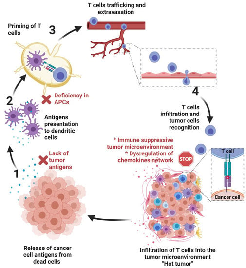
Immunotherapy includes immune checkpoint inhibitors (ICI) such as antibodies targeting cytotoxic T-lymphocyte-associated protein 4 (CTLA-4) or the programmed cell death protein/programmed death ligand 1 (PD-1/PD-L1) axis. Experimental and clinical evidence show that immunotherapy based on immune checkpoint inhibitors (ICI) provides long-term survival benefits to cancer patients in whom other conventional therapies have failed. However, only a minority of patients show high clinical benefits via the use of ICI alone. One of the major factors limiting the clinical benefits to ICI can be attributed to the lack of immune cell infiltration within the tumor microenvironment. Such tumors are classified as “cold/warm” or an immune “desert”; those displaying significant infiltration are considered “hot” or inflamed. This review will provide a brief summary of different tumor properties contributing to the establishment of cold tumors and describe major strategies that could reprogram non-inflamed cold tumors into inflamed hot tumors. More particularly, we will describe how targeting hypoxia can induce metabolic reprogramming that results in improving and extending the benefit of ICI. Full article
Show Figures

Figure 1

2022

Jump to: 2024, 2023

15 pages, 9301 KiB  
Article
The BET Protein Inhibitor JQ1 Decreases Hypoxia and Improves the Therapeutic Benefit of Anti-PD-1 in a High-Risk Neuroblastoma Mouse Model
by Delphine Sauvage, Manon Bosseler, Elodie Viry, Georgia Kanli, Anais Oudin, Guy Berchem, Olivier Keunen and Bassam Janji
Cells 2022, 11(18), 2783; https://doi.org/10.3390/cells11182783 - 6 Sep 2022
Cited by 7 | Viewed by 2746
Abstract
Anti-programmed death 1 (PD-1) is a revolutionary treatment for many cancers. The response to anti-PD-1 relies on several properties of tumor and immune cells, including the expression of PD-L1 and PD-1. Despite the impressive clinical benefit achieved with anti-PD-1 in several cancers in [...] Read more.
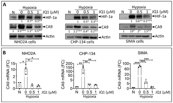
Anti-programmed death 1 (PD-1) is a revolutionary treatment for many cancers. The response to anti-PD-1 relies on several properties of tumor and immune cells, including the expression of PD-L1 and PD-1. Despite the impressive clinical benefit achieved with anti-PD-1 in several cancers in adults, the use of this therapy for high-risk neuroblastoma remains modest. Here, we evaluated the therapeutic benefit of anti-PD-1 in combination with JQ1 in a highly relevant TH-MYCN neuroblastoma transgenic mouse model. JQ1 is a small molecule inhibitor of the extra-terminal domain (BET) family of bromodomain proteins, competitively binding to bromodomains. Using several neuroblastoma cell lines in vitro, we showed that JQ1 inhibited hypoxia-dependent induction of HIF-1α and decreased the expression of the well-known HIF-1α downstream target gene CA9. Using MRI relaxometry performed on TH-MYCN tumor-bearing mice, we showed that JQ1 decreases R2* in tumors, a parameter associated with intra-tumor hypoxia in pre-clinical settings. Decreasing hypoxia by JQ1 was associated with improved blood vessel quality and integrity, as revealed by CD31 and αSMA staining on tumor sections. By analyzing the immune landscape of TH-MYCN tumors in mice, we found that JQ1 had no major impact on infiltrating immune cells into the tumor microenvironment but significantly increased the percentage of CD8+ PD-1+, conventional CD4+ PD-1+, and Treg PD-1+ cells. While anti-PD-1 monotherapy did not affect TH-MYCN tumor growth, we showed that combinatorial therapy associating JQ1 significantly decreased the tumor volume and improved the therapeutic benefit of anti-PD-1. This study provided the pre-clinical proof of concept needed to establish a new combination immunotherapy approach that may create tremendous enthusiasm for treating high-risk childhood neuroblastoma. Full article
Show Figures

Figure 1

Back to TopTop