Beyond Neurotransmission: Emerging Roles of Neurotransmitters in Regulating Morphological, Biochemical, and Functional Properties of Glial and Immune Cells in Healthy and Disease States

A topical collection in Cells (ISSN 2073-4409). This collection belongs to the section "Cells of the Nervous System".

Viewed by 27626

Editor

Topical Collection Information

Dear Colleagues,

For a long time, neurotransmitters have been considered solely as chemical messengers at the forefront of neuronal transmission, being produced by presynaptic neurons and released at synapses to establish neuron-to-neuron communication via post-synaptic receptor binding. However, emerging studies have determined that several neurotransmitters, along with their binding receptors, are also abundantly expressed in glial and immune cells of the central nervous system (CNS), where they produce a barrage of different biological activities, especially in the CNS. Interestingly, several of these activities seem to be pivotal in regulating CNS homeostasis in healthy individuals, and their disruptions are associated with the development of a spectrum of mental illnesses, including mood disorders and neurodegenerative conditions such as multiple sclerosis, Parkinson’s, and Alzheimer’s disease.

In this Topical Collection, our attention will be focused on unveiling the noncanonical roles of neurotransmitters (and their receptors) in CNS glial and immune cells. The primary goal of this collection will be to put together a series of studies that will help to elucidate how neurotransmitter–glia interaction plays an essential role in the development of different mental illnesses and neurodegenerative disorders. The final goal is to shed more light into the complex role of neurotransmitters in the CNS.

Dr. Alessandro Castorina
Collection Editor

Manuscript Submission Information

Manuscripts should be submitted online at www.mdpi.com by registering and logging in to this website. Once you are registered, click here to go to the submission form. Manuscripts can be submitted until the deadline. All submissions that pass pre-check are peer-reviewed. Accepted papers will be published continuously in the journal (as soon as accepted) and will be listed together on the collection website. Research articles, review articles as well as short communications are invited. For planned papers, a title and short abstract (about 100 words) can be sent to the Editorial Office for announcement on this website.

Submitted manuscripts should not have been published previously, nor be under consideration for publication elsewhere (except conference proceedings papers). All manuscripts are thoroughly refereed through a single-blind peer-review process. A guide for authors and other relevant information for submission of manuscripts is available on the Instructions for Authors page. Cells is an international peer-reviewed open access semimonthly journal published by MDPI.

Please visit the Instructions for Authors page before submitting a manuscript. The Article Processing Charge (APC) for publication in this open access journal is 2700 CHF (Swiss Francs). Submitted papers should be well formatted and use good English. Authors may use MDPI's English editing service prior to publication or during author revisions.

Keywords

  • glia
  • neurotransmitters
  • immune cells
  • microglia
  • astrocytes
  • neurodegenerative disease
  • mental illness

Published Papers (6 papers)

2022

Jump to: 2021

17 pages, 1830 KiB  
Article
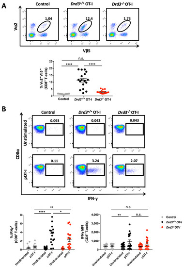
Dopaminergic Signalling Enhances IL-2 Production and Strengthens Anti-Tumour Response Exerted by Cytotoxic T Lymphocytes in a Melanoma Mouse Model
by Ornella Chovar-Vera, Ernesto López, Felipe Gálvez-Cancino, Carolina Prado, Dafne Franz, Diego A. Figueroa, Alexandra Espinoza, Claudio Figueroa, Alvaro Lladser and Rodrigo Pacheco
Cells 2022, 11(22), 3536; https://doi.org/10.3390/cells11223536 - 9 Nov 2022
Cited by 1 | Viewed by 2052
Abstract
Dopamine has emerged as an important regulator of immunity. Recent evidence has shown that signalling through low-affinity dopamine receptors exerts anti-inflammatory effects, whilst stimulation of high-affinity dopamine receptors potentiates immunity in different models. However, the dopaminergic regulation of CD8+ T-cells in anti-tumour [...] Read more.
Dopamine has emerged as an important regulator of immunity. Recent evidence has shown that signalling through low-affinity dopamine receptors exerts anti-inflammatory effects, whilst stimulation of high-affinity dopamine receptors potentiates immunity in different models. However, the dopaminergic regulation of CD8+ T-cells in anti-tumour immunity remains poorly explored. Here, we studied the role of dopamine receptor D3 (DRD3), which displays the highest affinity for dopamine, in the function of CD8+ T-cells and its consequences in the anti-tumour immune response. We observed that the deficiency of Drd3 (the gene encoding DRD3) in CD8+ T-cells limits their in vivo expansion, leading to an impaired anti-tumour response in a mouse melanoma model. Mechanistic analyses suggest that DRD3 stimulation favours the production of interleukin 2 (IL-2) and the surface expression of CD25, the α-chain IL-2 receptor, which are required for expansion and effector differentiation of CD8+ T-cells. Thus, our results provide genetic and pharmacologic evidence indicating that DRD3 favours the production of IL-2 by CD8+ T-cells, which is associated with higher expansion and acquisition of effector function of these cells, promoting a more potent anti-tumour response in a melanoma mouse model. These findings contribute to understanding how dopaminergic signalling affects the cellular immune response and represent an opportunity to improve melanoma therapy. Full article
Show Figures

Figure 1

2021

Jump to: 2022

19 pages, 2145 KiB  
Article
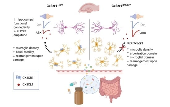
Antibiotics Treatment Modulates Microglia–Synapses Interaction
by Federica Cordella, Caterina Sanchini, Maria Rosito, Laura Ferrucci, Natalia Pediconi, Barbara Cortese, Francesca Guerrieri, Giuseppe Rubens Pascucci, Fabrizio Antonangeli, Giovanna Peruzzi, Maria Giubettini, Bernadette Basilico, Francesca Pagani, Alfonso Grimaldi, Giuseppina D’Alessandro, Cristina Limatola, Davide Ragozzino and Silvia Di Angelantonio
Cells 2021, 10(10), 2648; https://doi.org/10.3390/cells10102648 - 4 Oct 2021
Cited by 19 | Viewed by 4167
Abstract
‘Dysbiosis’ of the adult gut microbiota, in response to challenges such as infection, altered diet, stress, and antibiotics treatment has been recently linked to pathological alteration of brain function and behavior. Moreover, gut microbiota composition constantly controls microglia maturation, as revealed by morphological [...] Read more.
‘Dysbiosis’ of the adult gut microbiota, in response to challenges such as infection, altered diet, stress, and antibiotics treatment has been recently linked to pathological alteration of brain function and behavior. Moreover, gut microbiota composition constantly controls microglia maturation, as revealed by morphological observations and gene expression analysis. However, it is unclear whether microglia functional properties and crosstalk with neurons, known to shape and modulate synaptic development and function, are influenced by the gut microbiota. Here, we investigated how antibiotic-mediated alteration of the gut microbiota influences microglial and neuronal functions in adult mice hippocampus. Hippocampal microglia from adult mice treated with oral antibiotics exhibited increased microglia density, altered basal patrolling activity, and impaired process rearrangement in response to damage. Patch clamp recordings at CA3-CA1 synapses revealed that antibiotics treatment alters neuronal functions, reducing spontaneous postsynaptic glutamatergic currents and decreasing synaptic connectivity, without reducing dendritic spines density. Antibiotics treatment was unable to modulate synaptic function in CX3CR1-deficient mice, pointing to an involvement of microglia–neuron crosstalk through the CX3CL1/CX3CR1 axis in the effect of dysbiosis on neuronal functions. Together, our findings show that antibiotic alteration of gut microbiota impairs synaptic efficacy, suggesting that CX3CL1/CX3CR1 signaling supporting microglia is a major player in in the gut–brain axis, and in particular in the gut microbiota-to-neuron communication pathway. Full article
Show Figures

Graphical abstract

21 pages, 1189 KiB  
Review
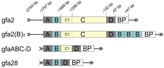
Genetic Constructs for the Control of Astrocytes’ Activity
by Anastasia A. Borodinova, Pavel M. Balaban, Ilya B. Bezprozvanny, Alla B. Salmina and Olga L. Vlasova
Cells 2021, 10(7), 1600; https://doi.org/10.3390/cells10071600 - 25 Jun 2021
Cited by 12 | Viewed by 4493
Abstract
In the current review, we aim to discuss the principles and the perspectives of using the genetic constructs based on AAV vectors to regulate astrocytes’ activity. Practical applications of optogenetic approaches utilizing different genetically encoded opsins to control astroglia activity were evaluated. The [...] Read more.
In the current review, we aim to discuss the principles and the perspectives of using the genetic constructs based on AAV vectors to regulate astrocytes’ activity. Practical applications of optogenetic approaches utilizing different genetically encoded opsins to control astroglia activity were evaluated. The diversity of astrocytic cell-types complicates the rational design of an ideal viral vector for particular experimental goals. Therefore, efficient and sufficient targeting of astrocytes is a multiparametric process that requires a combination of specific AAV serotypes naturally predisposed to transduce astroglia with astrocyte-specific promoters in the AAV cassette. Inadequate combinations may result in off-target neuronal transduction to different degrees. Potentially, these constraints may be bypassed with the latest strategies of generating novel synthetic AAV serotypes with specified properties by rational engineering of AAV capsids or using directed evolution approach by searching within a more specific promoter or its replacement with the unique enhancer sequences characterized using modern molecular techniques (ChIP-seq, scATAC-seq, snATAC-seq) to drive the selective transgene expression in the target population of cells or desired brain regions. Realizing these strategies to restrict expression and to efficiently target astrocytic populations in specific brain regions or across the brain has great potential to enable future studies. Full article
Show Figures

Figure 1

13 pages, 1359 KiB  
Review
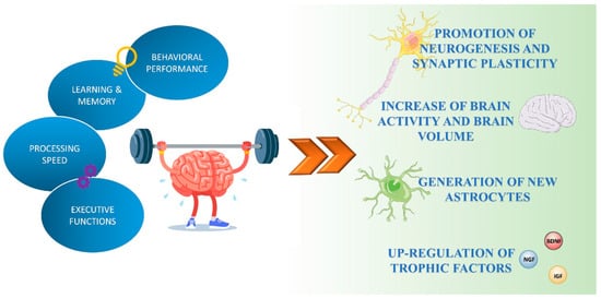
Neuroprotective Effects of Physical Activity via the Adaptation of Astrocytes
by Grazia Maugeri, Velia D’Agata, Benedetta Magrì, Federico Roggio, Alessandro Castorina, Silvia Ravalli, Michelino Di Rosa and Giuseppe Musumeci
Cells 2021, 10(6), 1542; https://doi.org/10.3390/cells10061542 - 18 Jun 2021
Cited by 28 | Viewed by 6475
Abstract
The multifold benefits of regular physical exercise have been largely demonstrated in human and animal models. Several studies have reported the beneficial effects of physical activity, both in peripheral tissues and in the central nervous system (CNS). Regular exercise improves cognition, brain plasticity, [...] Read more.
The multifold benefits of regular physical exercise have been largely demonstrated in human and animal models. Several studies have reported the beneficial effects of physical activity, both in peripheral tissues and in the central nervous system (CNS). Regular exercise improves cognition, brain plasticity, neurogenesis and reduces the symptoms of neurodegenerative diseases, making timeless the principle of “mens sana in corpore sano” (i.e., a healthy mind in a healthy body). Physical exercise promotes morphological and functional changes in the brain, acting not only in neurons but also in astrocytes, which represent the most numerous glial cells in the brain. The multiple effects of exercise on astrocytes comprise the increased number of new astrocytes, the maintenance of basal levels of catecholamine, the increase in glutamate uptake, the major release of trophic factors and better astrocytic coverage of cerebral blood vessels. The purpose of this review is to highlight the effects of exercise on brain function, emphasize the role of astrocytes in the healthy CNS, and provide an update for a better understanding of the effects of physical exercise in the modulation of astrocyte function. Full article
Show Figures

Figure 1

22 pages, 3898 KiB  
Article
Assessing the Anti-Inflammatory Activity of the Anxiolytic Drug Buspirone Using CRISPR-Cas9 Gene Editing in LPS-Stimulated BV-2 Microglial Cells
by Sarah Thomas Broome, Teagan Fisher, Alen Faiz, Kevin A. Keay, Giuseppe Musumeci, Ghaith Al-Badri and Alessandro Castorina
Cells 2021, 10(6), 1312; https://doi.org/10.3390/cells10061312 - 25 May 2021
Cited by 16 | Viewed by 5318
Abstract
Buspirone is an anxiolytic drug with robust serotonin receptor 1A (Htr1a) agonist activities. However, evidence has demonstrated that this drug also targets the dopamine D3 receptor (Drd3), where it acts as a potent antagonist. In vivo, Drd3 blockade is neuroprotective and reduces inflammation [...] Read more.
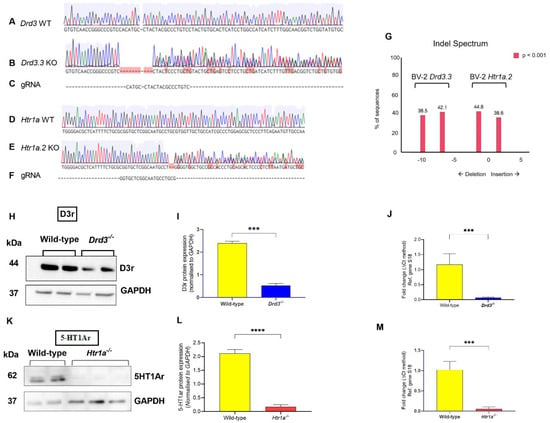
Buspirone is an anxiolytic drug with robust serotonin receptor 1A (Htr1a) agonist activities. However, evidence has demonstrated that this drug also targets the dopamine D3 receptor (Drd3), where it acts as a potent antagonist. In vivo, Drd3 blockade is neuroprotective and reduces inflammation in models of Parkinson’s disease. To test if buspirone also elicited anti-inflammatory activities in vitro, we generated stable Drd3−/− and Htr1a−/− BV2 microglial cell lines using CRISPR-Cas9 technology and then tested the effects of buspirone after lipopolysaccharide (LPS) challenge. We found that LPS exposure had no effect on cell viability, except in Htr1a−/− cells, where viability was reduced (p < 0.001). Drug treatment reduced viability in Drd3−/− cells, but not in WT or Htr1a−/− cells. Buspirone counteracted LPS-induced NO release, NOS2, IL-1β and TNF-α gene expression in WT cells, whereas it exerted limited effects in Drd3−/− or Htr1a−/− microglia. In summary, our findings indicate that buspirone attenuates microglial polarization after LPS challenge. These results also highlight some major effects of Drd3 or Htr1a genetic ablation on microglial biology, raising important questions on the complex role of neurotransmitters in regulating microglia functions. Full article
Show Figures

Figure 1

18 pages, 5105 KiB  
Article
Brain CHID1 Expression Correlates with NRGN and CALB1 in Healthy Subjects and AD Patients
by Paola Castrogiovanni, Cristina Sanfilippo, Rosa Imbesi, Grazia Maugeri, Debora Lo Furno, Daniele Tibullo, Alessandro Castorina, Giuseppe Musumeci and Michelino Di Rosa
Cells 2021, 10(4), 882; https://doi.org/10.3390/cells10040882 - 13 Apr 2021
Cited by 15 | Viewed by 3673
Abstract
Alzheimer’s disease is a progressive, devastating, and irreversible brain disorder that, day by day, destroys memory skills and social behavior. Despite this, the number of known genes suitable for discriminating between AD patients is insufficient. Among the genes potentially involved in the development [...] Read more.
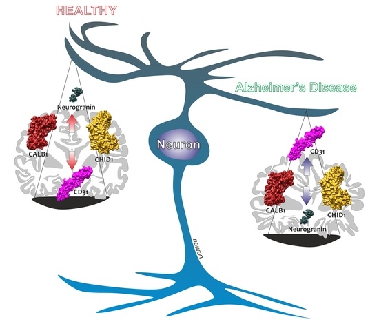
Alzheimer’s disease is a progressive, devastating, and irreversible brain disorder that, day by day, destroys memory skills and social behavior. Despite this, the number of known genes suitable for discriminating between AD patients is insufficient. Among the genes potentially involved in the development of AD, there are the chitinase-like proteins (CLPs) CHI3L1, CHI3L2, and CHID1. The genes of the first two have been extensively investigated while, on the contrary, little information is available on CHID1. In this manuscript, we conducted transcriptome meta-analysis on an extensive sample of brains of healthy control subjects (n = 1849) (NDHC) and brains of AD patients (n = 1170) in order to demonstrate CHID1 involvement. Our analysis revealed an inverse correlation between the brain CHID1 expression levels and the age of NDHC subjects. Significant differences were highlighted comparing CHID1 expression of NDHC subjects and AD patients. Exclusive in AD patients, the CHID1 expression levels were correlated positively to calcium-binding adapter molecule 1 (IBA1) levels. Furthermore, both in NDHC and in AD patient’s brains, the CHID1 expression levels were directly correlated with calbindin 1 (CALB1) and neurogranin (NRGN). According to brain regions, correlation differences were shown between the expression levels of CHID1 in prefrontal, frontal, occipital, cerebellum, temporal, and limbic system. Sex-related differences were only highlighted in NDHC. CHID1 represents a new chitinase potentially involved in the principal processes underlying Alzheimer’s disease. Full article
Show Figures

Graphical abstract

Back to TopTop