Pathology and Parasitic Diseases of Animals

A topical collection in Pathogens (ISSN 2076-0817). This collection belongs to the section "Parasitic Pathogens".

Viewed by 58495

Editors


E-Mail Website
Collection Editor
Department of Veterinary Sciences, University of Pisa, Italy
Interests: parasites (helminths, protozoa, arthropods); animals (domestic, wild, exotic); parasitic diseases; parasites and histopathological findings; interaction between parasites and host animals; parasites evasion strategies of host defense mechanisms; parasites and immunology
Special Issues, Collections and Topics in MDPI journals

E-Mail Website
Collection Editor
School of Biosciences and Veterinary Medicine of the University of Camerino, Italy
Interests: interaction between parasites and host; mutual immunopathological effects and some co-evolutionary benefits; cell-mediated immune response of the host and evolution/fate of parasitic disease; immune cells and parasitic spread; strategies of parasite adaptation to the host immunological mechanisms

Topical Collection Information

Dear Colleagues,

Parasitic diseases are often the cause of growth delay, decrease of physical development and fertility, as well as of serious health problems and death in infected animals. In addition, several parasites may infect both animals and humans, raising public health concerns. The pathological changes that parasites may induce in their animal hosts are caused by a variety of pathogenic mechanisms, depending on the host/parasite relationships. In infected animal hosts, parasites may be responsible for lesions by toxic, obstructive, subtractive, traumatic, allergic, necrotic, inflammatory, and immunosuppressive pathogenic mechanisms. Parasites may also have the ability to cause tumors or tumor-like lesions in affected animals.

Histopathological features of parasitic diseases may play an important role in the diagnosis and knowledge of the severity and complications of parasitic diseases, by allowing the identification of the parasite species involved and the areas of pathological lesions, the detection of histopathological changes, particularly important for the differential diagnosis or to confirm the presumptive diagnosis of a parasitic disease or, again, by highlighting possible bacterial or viral complications and the outlook of treatment. Histopathological examination may also provide insights into the interactions between parasites and animal hosts and their impact on infected animals.

Immune-phenotipization of immune cells participating in the immune response of the animal against parasites is also very important to understand if the response is a positive or negative process for the host. Further, the recent paradigm that permits evaluating host M1 or M2 macrophage response is fundamental to have some provisional information regarding the possibility for the host of improving health conditions or not after parasite infection. 

The postgenomic era has generated unparalleled opportunities for creating and integrating systems biology data (i.e., organism‐ or cellular‐scale data produced through numerous ‐omics or systemwide technologies). This holistic approach is in direct contrast to conventional reductionist methods that ‘reduce’ systems into smaller, more tractable units. Systems‐based methods are particularly useful to study complex biological relationships that are: (1) open, with constant information exchange and a net flow of resources, and (2) stochastic, with spatial, temporal, and population heterogeneity. Host–parasite systems embody all of these defining characteristics. ‐Omics technologies are also much more efficient and economical when comparing the cumulative time, labor, and cost per gene to traditional reductionist strategies. 

This Topic Collection is devoted to collecting original papers and/or review papers dealing with histopathological features of parasitic diseases, immunopathological studies of host–parasite immune-response characterization, and host–parasite systems. It is focused on parasitic diseases caused by different parasite pathogens of veterinary interest responsible for economic losses or that may negatively affect animal and human health and welfare. Parasites affecting vertebrate and invertebrate animal species used for human consumption (e.g., bred or caught fishes, mollusks, and crustaceans) and for producing food for human consumption (e.g., bees) or that can be the vectors or the hosts of important human and animal parasitic diseases (e.g., arthropod species responsible for malaria, Leishmania, Trypanosoma and other parasite transmission; snails involved in transmission of flukes) also represent a relevant field in this Topic Collection.

Prof. Stefania Perrucci
Dr. Livio Galosi
Prof. Dr. Giacomo Rossi
Collection Editors

Manuscript Submission Information

Manuscripts should be submitted online at www.mdpi.com by registering and logging in to this website. Once you are registered, click here to go to the submission form. Manuscripts can be submitted until the deadline. All submissions that pass pre-check are peer-reviewed. Accepted papers will be published continuously in the journal (as soon as accepted) and will be listed together on the collection website. Research articles, review articles as well as short communications are invited. For planned papers, a title and short abstract (about 100 words) can be sent to the Editorial Office for announcement on this website.

Submitted manuscripts should not have been published previously, nor be under consideration for publication elsewhere (except conference proceedings papers). All manuscripts are thoroughly refereed through a single-blind peer-review process. A guide for authors and other relevant information for submission of manuscripts is available on the Instructions for Authors page. Pathogens is an international peer-reviewed open access monthly journal published by MDPI.

Please visit the Instructions for Authors page before submitting a manuscript. The Article Processing Charge (APC) for publication in this open access journal is 2200 CHF (Swiss Francs). Submitted papers should be well formatted and use good English. Authors may use MDPI's English editing service prior to publication or during author revisions.

Keywords

  • parasitic diseases
  • anatomo-histopathological features
  • immunophenotipization
  • immunopathology
  • cell-mediated response
  • parasite strategies
  • co-evolutionary benefits
  • host–parasite systems
  • macrophages
  • animals

Published Papers (15 papers)

2024

Jump to: 2023, 2022, 2021, 2020

30 pages, 7697 KiB  
Article
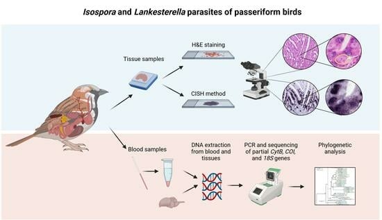
Isospora and Lankesterella Parasites (Eimeriidae, Apicomplexa) of Passeriform Birds in Europe: Infection Rates, Phylogeny, and Pathogenicity
by Carina Keckeisen, Alžbeta Šujanová, Tanja Himmel, Julia Matt, Nora Nedorost, Carolina R. F. Chagas, Herbert Weissenböck and Josef Harl
Pathogens 2024, 13(4), 337; https://doi.org/10.3390/pathogens13040337 - 18 Apr 2024
Viewed by 2108
Abstract
Wild birds are common hosts to numerous intracellular parasites such as single-celled eukaryotes of the family Eimeriidae (order Eucoccidiorida, phylum Apicomplexa). We investigated the infection rates, phylogeny, and pathogenicity of Isospora and Lankesterella parasites in wild and captive passerine birds. Blood and tissue [...] Read more.
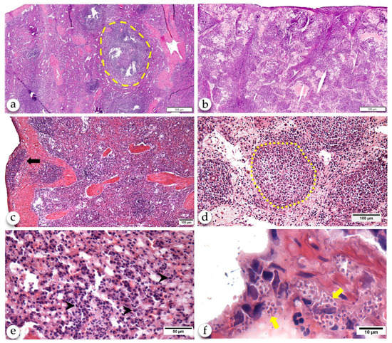
Wild birds are common hosts to numerous intracellular parasites such as single-celled eukaryotes of the family Eimeriidae (order Eucoccidiorida, phylum Apicomplexa). We investigated the infection rates, phylogeny, and pathogenicity of Isospora and Lankesterella parasites in wild and captive passerine birds. Blood and tissue samples of 815 wild and 15 deceased captive birds from Europe were tested using polymerase chain reaction and partial sequencing of the mitochondrial cytochrome b and cytochrome c oxidase I and the nuclear 18S rRNA gene. The infection rate for Lankesterella in wild birds was 10.7% compared to 5.8% for Isospora. Chromogenic in situ hybridization with probes targeting the parasites’ 18S rRNA was employed to identify the parasites’ presence in multiple organs, and hematoxylin–eosin staining was performed to visualize the parasite stages and assess associated lesions. Isospora parasites were mainly identified in the intestine, spleen, and liver. Extraintestinal tissue stages of Isospora were accompanied by predominantly lymphohistiocytic inflammation of varying severity. Lankesterella was most frequently detected in the spleen, lung, and brain; however, infected birds presented only a low parasite burden without associated pathological changes. These findings contribute to our understanding of Isospora and Lankesterella parasites in wild birds. Full article
Show Figures

Graphical abstract

2023

Jump to: 2024, 2022, 2021, 2020

10 pages, 626 KiB  
Article
Use of In-Clinic Diagnostic Kits for the Detection of Seropositivity to Leishmania infantum and Other Major Vector-Borne Pathogens in Healthy Dogs
by Simone Morelli, Anastasia Diakou, Antonio Frangipane di Regalbono, Mariasole Colombo, Giulia Simonato, Angela Di Cesare, Alessandra Passarelli, Carlo Pezzuto, Zoe Tzitzoudi, Alessandra Barlaam, Melissa Beall, Ramaswamy Chandrashekar, Nikola Pantchev and Donato Traversa
Pathogens 2023, 12(5), 696; https://doi.org/10.3390/pathogens12050696 - 11 May 2023
Cited by 4 | Viewed by 2410
Abstract
Canine Vector-Borne Diseases (CVBDs) are widespread in Europe and enzootic in many other countries. Though severe illnesses may occur, dogs living in enzootic areas often show vague or no clinical signs of CVBDs. Undiagnosed infections/co-infections in subclinically infected animals favor the spread of [...] Read more.
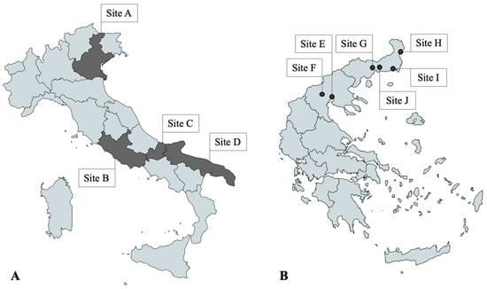
Canine Vector-Borne Diseases (CVBDs) are widespread in Europe and enzootic in many other countries. Though severe illnesses may occur, dogs living in enzootic areas often show vague or no clinical signs of CVBDs. Undiagnosed infections/co-infections in subclinically infected animals favor the spread of CVBDs and increase the risk of transmission to other animals and, in some cases, humans. This study has evaluated the exposure of dogs living in key enzootic countries, i.e., Italy and Greece, to major CVBDs via the use of in-clinic diagnostic kits. Overall, 300 privately owned dogs without/with single mild clinical signs living in different regions of Italy (n. 150) and Greece (n. 150) were included in the study. As part of a clinical examination, a blood sample was collected from each dog and subjected to two serological rapid tests, i.e., the SNAP® 4Dx®Plus (IDEXX Laboratories Inc.) for the detection of antibodies against Ehrlichia spp., Anaplasma spp., Borrelia burgdorferi s.l. and Dirofilaria immitis antigen and the SNAP® Leishmania (IDEXX Laboratories Inc.) for the detection of antibodies against Leishmania infantum. In all, 51 dogs (17%; 95% CI 12.9–21.7) were seropositive to at least 1 pathogen, i.e., 4 in Italy (2.7%; 95% CI 1.4–13.1) and 47 in Greece (31.3%; 95% CI 24–39.4). Dirofilaria immitis antigens were found in 39 dogs (13%; 95% CI 9.4–17.3), while antibodies against Ehrlichia, Anaplasma and Leishmania were detected in 25 (8.3%; 95% CI 5.5–12.1), 8 (2.7%; 95% CI 1.2–5.2) and 5 (1.7%; 95% CI 0.5–3.8) dogs, respectively. None of the dogs tested seropositive for B. burgdorferi s.l. Statistical analyses were performed to evaluate associations between exposure to CVBDs and possible risk factors. The present results indicate that dogs living in enzootic areas may be seropositive for one or more CVBDs in absence of clinical signs. Rapid kits are among first line tools for the detection of CVBDs in clinical settings, as they are cost-effective, straightforward and quick to use. Also, in-clinic tests used herein allowed detection of co-exposure to CVBDs investigated. Full article
Show Figures

Figure 1

10 pages, 2813 KiB  
Article
First Report of Swinepox in a Wild Boar in Italy: Pathologic and Molecular Findings
by Lisa Guardone, Katia Varello, Valeria Listorti, Simone Peletto, Lara Wolfsgruber, Roberto Zoccola, Vittoria Montemurro, Erika Messana, Elena Bozzetta, Pierluigi Acutis, Loretta Masoero and Elisabetta Razzuoli
Pathogens 2023, 12(3), 472; https://doi.org/10.3390/pathogens12030472 - 16 Mar 2023
Cited by 1 | Viewed by 2304
Abstract
Swinepox virus (SWPV) is responsible for sporadic acute poxvirus infections in swine worldwide, causing a pathognomonic eruptive proliferative dermatitis. Beside direct and congenital transmission, the pig louse Haematopinus suis acts as a mechanical vector and favors virus infection through skin lesions. Infections are [...] Read more.
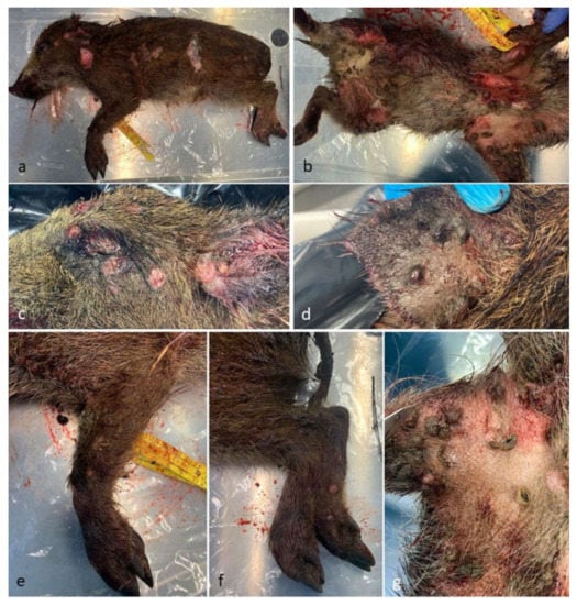
Swinepox virus (SWPV) is responsible for sporadic acute poxvirus infections in swine worldwide, causing a pathognomonic eruptive proliferative dermatitis. Beside direct and congenital transmission, the pig louse Haematopinus suis acts as a mechanical vector and favors virus infection through skin lesions. Infections are generally described in domestic pigs, while only a few cases have been reported in wild boars, in Austria and Germany. In September 2022, SWPV infection was suspected at post-mortem examination of a wild boar piglet with characteristic lesions in Liguria, Northwest Italy. The piglet was heavily parasitized by swine lice (H. suis). SWPV was then confirmed by histological and molecular analyses. Possible viral co-infections were also investigated (African swine fever virus, classical swine fever virus, parvovirus, circovirus, Aujeszky’s disease virus and hepatitis E virus). This article describes gross and histopathologic features of SWPV infection, differential diagnosis, and potential vector-borne transmission to domestic pigs, presenting a brief review of the literature on the topic. SWPV infection is reported in wild boars in Italy for the first time. The finding of SWPV in a wild boar in an area with a very limited pig population may suggest the existence of a “wildlife cycle” in the area. Further investigations are needed to understand the real risk of transmission of SWPV to domestic pigs as well as the role of other arthropod vectors. Full article
Show Figures

Figure 1

2022

Jump to: 2024, 2023, 2021, 2020

14 pages, 4056 KiB  
Article
First Isolation of Yarrowia lipolytica in a Granulomatous Pneumonia of a Spectacled Caiman, Caiman crocodilus Linnaeus, 1758
by Manuela Iurescia, Andrea Santini, Marco Montagnani, Elena Lavinia Diaconu, Fiorentino Stravino, Devid Agnelli, Emanuela Vergari, Gianluca Fichi and Claudia Eleni
Pathogens 2022, 11(11), 1255; https://doi.org/10.3390/pathogens11111255 - 28 Oct 2022
Cited by 1 | Viewed by 2199
Abstract
Contrary to humans, candidiasis is a rare infection in animals. However, in reptiles, candidiasis can cause gastrointestinal, cutaneous, or rarely systemic infections in stressed animals. The infections due to Yarrowia lipolytica have been increasingly described in human medicine, and hundreds of cases are [...] Read more.
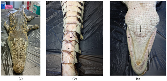
Contrary to humans, candidiasis is a rare infection in animals. However, in reptiles, candidiasis can cause gastrointestinal, cutaneous, or rarely systemic infections in stressed animals. The infections due to Yarrowia lipolytica have been increasingly described in human medicine, and hundreds of cases are reported, comprised of granulomatous lung lesions. Herein, granulomatous pneumonia of a spectacled caiman, Caiman crocodilus, was described, and the presence of Y. lipolytica in the lesion was confirmed through histopathology, microbiologic cultures, and molecular methods. The cause of death of the spectacled caiman was ascribed to bacterial shock septicemia consequentially to a traumatic lesion. However, in the right lung, several nodules containing white exudate were evidenced. At mycological and molecular analyses, Y. lipolytica was evidenced, and the histological finding confirmed the presence of a Candida infection in the lung granulomatous lesions. The comparison of ITS sequences with 11 Yarrowia spp. isolates, recently described in green sea turtles, and with a human strain was conducted, and the whole genome of a strain isolated in the spectacled caiman was sequenced. Even though Y. lipolytica is considered a non-pathogenic yeast and has been rarely described in animals, it seems to cause granulomatous lesions in reptiles as in humans. Full article
Show Figures

Figure 1

2021

Jump to: 2024, 2023, 2022, 2020

11 pages, 2176 KiB  
Article
Anti-Leishmania infantum Antibody-Producing Plasma Cells in the Spleen in Canine Visceral Leishmaniasis
by Jonathan L. M. Fontes, Bianca R. Mesquita, Reginaldo Brito, Juliana C. S. Gomes, Caroline V. B. de Melo and Washington L. C. dos Santos
Pathogens 2021, 10(12), 1635; https://doi.org/10.3390/pathogens10121635 - 17 Dec 2021
Cited by 3 | Viewed by 3279
Abstract
The spleen is involved in visceral leishmaniasis immunopathogenesis, and presents alterations in white-pulp microenvironments that are associated with an increased susceptibility to coinfections and patient death. Plasmacytosis in splenic red pulp (RP) is one observed alteration, but the specificity of antibody-secreting cells and [...] Read more.
The spleen is involved in visceral leishmaniasis immunopathogenesis, and presents alterations in white-pulp microenvironments that are associated with an increased susceptibility to coinfections and patient death. Plasmacytosis in splenic red pulp (RP) is one observed alteration, but the specificity of antibody-secreting cells and the distribution of them has not yet been evaluated. We biotinylated soluble L. infantum membrane antigens (bSLMA) used as probes in modified immunohistochemistry, and detected the presence of anti-L. infantum antibody-secreting cells. Were used spleens from eight dogs from the endemic area for canine visceral leishmaniasis (CanL), and three healthier controls. The spleen sections were cryopreserved, and we performed modified immunohistochemistry. The ratio of plasma cells which were reactive to bSLMA (Anti-Leish-PC) in the spleen RP and periarteriolar lymphatic sheath (PALS) were calculated. Dogs with CanL present hyperglobulinemia and more plasma cells in their RP than the controls. Furthermore, dogs with CanL presented a lower proportion of Anti-Leish-PC in their RP than in PALS. Likewise, dysproteinemia was related to RP and PALS plasmacytosis, and a more severe clinical profile. Full article
Show Figures

Figure 1

25 pages, 4591 KiB  
Article
Parasites, Bacteria, and Associated Pathological Changes in the Digestive System of Diurnal and Nocturnal Raptors in Central Italy
by Giacomo Rossi, Giuliana Terracciano, Riccardo Gherardi, Livio Galosi and Stefania Perrucci
Pathogens 2021, 10(12), 1567; https://doi.org/10.3390/pathogens10121567 - 30 Nov 2021
Cited by 2 | Viewed by 5416
Abstract
The knowledge of raptor pathogens and associated lesions may be extremely important to enhancing raptor conservation efforts and reducing pathogen spillover to humans and domesticated animals and vice versa. Parasite infections of the digestive system and associated bacteria and pathological changes were evaluated [...] Read more.
The knowledge of raptor pathogens and associated lesions may be extremely important to enhancing raptor conservation efforts and reducing pathogen spillover to humans and domesticated animals and vice versa. Parasite infections of the digestive system and associated bacteria and pathological changes were evaluated in deceased diurnal and nocturnal raptors in central Italy. Overall, the prevalence of parasites (nematodes, cestodes, trematodes, acanthocephalans, and protozoa) identified in the examined birds was 72.41%, and most of the positive raptors (71.42%) showed multiple parasite infections. Among bacteria, Salmonella typhimurium, Salmonella enterica subspecies diarizonae, Escherichia coli, Clostridium perfringens, Yersinia enterocolitica, and Pasteurella multocida were identified. The results obtained showed that both parasites and bacteria may cause severe lesions in the digestive system of diurnal and nocturnal raptors; parasites and bacteria may concur in causing these lesions; most severe lesions are caused by the interaction of multiple pathogens, both parasites and bacteria; and the same pathogen taxa are frequently associated with the same pathological changes. This study is the first report of S. typhimurium and S. enterica subspecies diarizonae in Buteo buteo, while Andracantha mergi, Spirocerca spp., Sarcocystis dispersa, Sarcocystis columbae, and Eumonospora spp. were recorded for the first time in Italy. Full article
Show Figures

Figure 1

11 pages, 808 KiB  
Article
Simultaneous Infection of Elaphostrongylus Nematode Species and Parasite Sharing between Sympatrically Occurring Cervids: Moose, Roe Deer, and Red Deer in Poland
by Magdalena Świsłocka, Mirosław Ratkiewicz and Anetta Borkowska
Pathogens 2021, 10(10), 1344; https://doi.org/10.3390/pathogens10101344 - 18 Oct 2021
Cited by 1 | Viewed by 2205
Abstract
It is important to assess the distribution of parasite species across wildlife populations, to design strategies for game management and effective disease control in nature. In this project we quantified the prevalence of Elaphostrongylus species in eight moose populations. We used molecular methods [...] Read more.
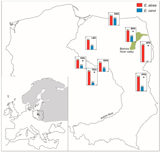
It is important to assess the distribution of parasite species across wildlife populations, to design strategies for game management and effective disease control in nature. In this project we quantified the prevalence of Elaphostrongylus species in eight moose populations. We used molecular methods for identification of parasite species and host individual genotypes from fecal samples. We also demonstrated sharing of parasite species between three cervid hosts sympatrically occurring in the Biebrza River valley, North-Eastern Poland, which is occupied by the largest autochthonous, non-harvested moose population in Central Europe. Nematode species from the genus Elaphostrongylus are ubiquitous in the studied moose populations. The presence of a single parasite species (e.g., E. alces) in moose individuals was more common than simultaneous infection with E. alces and E. cervi. The prevalence of both E. alces and E. cervi was higher in males than females. The distribution of E. alces and E. cervi prevalence in moose, roe deer, and red deer were in accordance with the membership of a host to a subfamily. Simultaneous occurrences of both Elaphostrongylus species were significantly more frequently noted in red deer fecal samples than those collected from moose or roe deer. Thus, we consider red deer to play a dominant role in sharing of those nematodes to other cervids. Our findings promote applications of molecular methods of identifying parasite species and the assessment of the exchange of parasite community between wild ruminant species in management and health monitoring of game animal populations. Full article
Show Figures

Figure 1

18 pages, 2307 KiB  
Article
Dysregulation of Glycerophosphocholines in the Cutaneous Lesion Caused by Leishmania major in Experimental Murine Models
by Adwaita R. Parab, Diane Thomas, Sharon Lostracco-Johnson, Jair L. Siqueira-Neto, James H. McKerrow, Pieter C. Dorrestein and Laura-Isobel McCall
Pathogens 2021, 10(5), 593; https://doi.org/10.3390/pathogens10050593 - 13 May 2021
Cited by 7 | Viewed by 3219
Abstract
Cutaneous leishmaniasis (CL) is the most common disease form caused by a Leishmania parasite infection and considered a neglected tropical disease (NTD), affecting 700,000 to 1.2 million new cases per year in the world. Leishmania major is one of several different species of [...] Read more.
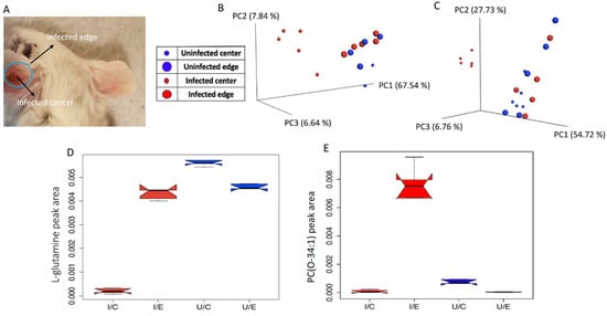
Cutaneous leishmaniasis (CL) is the most common disease form caused by a Leishmania parasite infection and considered a neglected tropical disease (NTD), affecting 700,000 to 1.2 million new cases per year in the world. Leishmania major is one of several different species of the Leishmania genus that can cause CL. Current CL treatments are limited by adverse effects and rising resistance. Studying disease metabolism at the site of infection can provide knowledge of new targets for host-targeted drug development. In this study, tissue samples were collected from mice infected in the ear or footpad with L. major and analyzed by untargeted liquid chromatography-tandem mass spectrometry (LC-MS/MS). Significant differences in overall metabolite profiles were noted in the ear at the site of the lesion. Interestingly, lesion-adjacent, macroscopically healthy sites also showed alterations in specific metabolites, including selected glycerophosphocholines (PCs). Host-derived PCs in the lower m/z range (m/z 200–799) showed an increase with infection in the ear at the lesion site, while those in the higher m/z range (m/z 800–899) were decreased with infection at the lesion site. Overall, our results expanded our understanding of the mechanisms of CL pathogenesis through host metabolism and may lead to new curative measures against infection with Leishmania. Full article
Show Figures

Figure 1

13 pages, 547 KiB  
Article
Exposure to Major Vector-Borne Diseases in Dogs Subjected to Different Preventative Regimens in Endemic Areas of Italy
by Mariasole Colombo, Simone Morelli, Giulia Simonato, Angela Di Cesare, Fabrizia Veronesi, Antonio Frangipane di Regalbono, Laura Grassi, Ilaria Russi, Pietro Giorgio Tiscar, Giulia Morganti, Jasmine Hattab, Valeria Rizzo and Donato Traversa
Pathogens 2021, 10(5), 507; https://doi.org/10.3390/pathogens10050507 - 23 Apr 2021
Cited by 17 | Viewed by 3628
Abstract
Vector-borne diseases (VBDs) are globally widespread arthropod-transmitted diseases with a significant impact on animal and human health. Many drivers have recently spurred the geographic spread of VBDs in dogs. This study has evaluated the exposure to most important VBDs in dogs under different [...] Read more.
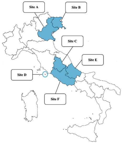
Vector-borne diseases (VBDs) are globally widespread arthropod-transmitted diseases with a significant impact on animal and human health. Many drivers have recently spurred the geographic spread of VBDs in dogs. This study has evaluated the exposure to most important VBDs in dogs under different preventative treatments in different regions of Italy, i.e., Veneto, Friuli Venezia-Giulia, Umbria, Giglio Island (Tuscany), Abruzzo and Latium. Serological analyses were performed to detect antibodies against Leishmania infantum, Babesia canis, Anaplasma phagocytophilum/Anaplasma platys, Ehrlichia canis/Ehrlichia ewingii, Borrelia burgdorferi, Rickettsia conorii and the circulating antigen of Dirofilaria immitis. Dogs were categorized according to the treatment schedule usually received, and the association between seropositivity and possible risk factors was statistically evaluated. Overall, 124/242 (51.2%) dogs tested positive for at least one pathogen, while 34 (14.0%) were exposed to two or more pathogens. The most detected seropositivity was against R. conorii, followed by Anaplasma spp., L. infantum, B. canis, and the other pathogens under study. Significant statistical associations were found according to geographical provenance, history of tick infestation, lifestyle and inadequate prophylactic treatments. Random/irregular treatments have been identified as a clear risk factor. These results show that adequate prophylactic treatment protocols are overlooked by dog owners, despite the availability of several effective products, with possible implications in veterinary medicine and on public health. Full article
Show Figures

Figure 1

11 pages, 2575 KiB  
Article
Pathology of Urinary Bladder in Pearsonema spp. Infected Wildlife from Central Italy
by Claudia Eleni, Alessia Mariacher, Goffredo Grifoni, Elena Cardini, Sara Tonon, Andrea Lombardo, Antonino Barone and Gianluca Fichi
Pathogens 2021, 10(4), 474; https://doi.org/10.3390/pathogens10040474 - 14 Apr 2021
Cited by 4 | Viewed by 3459
Abstract
The genus Pearsonema, in the nematode family Capillariidae, includes several species that parasitize the urinary bladders of wild and domestic carnivores. The infection has been reported worldwide from several wildlife species, including canids, mustelids, and felids, but the pathological aspects have [...] Read more.
The genus Pearsonema, in the nematode family Capillariidae, includes several species that parasitize the urinary bladders of wild and domestic carnivores. The infection has been reported worldwide from several wildlife species, including canids, mustelids, and felids, but the pathological aspects have seldom been investigated. In order to assess the presence and severity of the lesions in Pearsonema-infected wildlife, we performed a parasitological and pathological examination of urinary bladders from 72 animals, belonging to the families Canidae (red fox Vulpes vulpes, n = 28, and wolf Canis lupus, n = 29) and Mustelidae (beech marten Martes foina, n = 3; pine marten Martes martes, n = 2; and European badger Meles meles, n = 10). A greater prevalence of infection for canids (64.91%; 95% confidence interval (95% CI), 52.52–77.30%) than for mustelids (13.33%) (p < 0.001) was recorded. The prevalence of infection in red foxes was 75.0% (95% CI, 58.96–91.04%), in accordance with other reports from European countries, supporting the role of this species as a reservoir for infection. Eosinophilic cystitis was observed in 34 out of the 72 examined animals (47.22%). The influence of Pearsonema sp. infection on the occurrence of eosinophilic cystitis was statistically significant in wolves (p < 0.01), which were also affected by more severe histological lesions compared to foxes. Full article
Show Figures

Figure 1

17 pages, 3252 KiB  
Systematic Review
Skin Lesions in Feline Leishmaniosis: A Systematic Review
by Francesca Abramo, Francesco Albanese, Silvia Gattuso, Alessandra Randone, Ivan Fileccia, Carla Dedola, Fabrizio Ibba, Paola Ottaiano and Emanuele Brianti
Pathogens 2021, 10(4), 472; https://doi.org/10.3390/pathogens10040472 - 13 Apr 2021
Cited by 16 | Viewed by 10080
Abstract
Feline leishmaniosis (FeL) is increasingly reported throughout the world and skin lesions predominate in the clinical picture. There are, however, few evidence-based data on cutaneous feline leishmaniosis and directions are strongly needed for a better management of the disease. In this study, we [...] Read more.
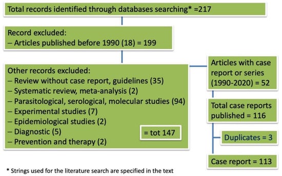
Feline leishmaniosis (FeL) is increasingly reported throughout the world and skin lesions predominate in the clinical picture. There are, however, few evidence-based data on cutaneous feline leishmaniosis and directions are strongly needed for a better management of the disease. In this study, we systematically reviewed what is currently known about the clinical dermatological presentation of FeL through analysis of the literature and, further, by adding unpublished cases managed by Italian veterinary dermatologists. Sixty-six feline cases of cutaneous leishmaniosis published in 33 articles between 1990 and 2020 met the inclusion criteria and were analyzed. Six unpublished cases of cutaneous FeL managed by Italian dermatologists were also reviewed. The majority of cases were reported from South America, followed by Europe and North America. Nodules were the most frequently reported clinical signs and the presence of Leishmania in lesioned skin was assessed mainly by cytology. A total of six Leishmania species have been identified as being responsible for skin lesions. Coinfections by FIV or FeLV were reported in 12.1% and 9.1% of the cases, respectively. Clinical data including treatment have been analyzed and discussed to provide directives for proper management of the disease for which cats may also serve as domestic reservoirs for human infections. Full article
Show Figures

Figure 1

17 pages, 2528 KiB  
Article
Toxocara canis and Toxocara cati Somatic and Excretory-Secretory Antigens Are Recognised by C-Type Lectin Receptors
by Marie-Kristin Raulf, Bernd Lepenies and Christina Strube
Pathogens 2021, 10(3), 321; https://doi.org/10.3390/pathogens10030321 - 9 Mar 2021
Cited by 13 | Viewed by 4056
Abstract
Toxocara canis and Toxocara cati, the worldwide occurring intestinal roundworms of canids and felids, represent an important public health threat due to various disease manifestations in humans. Host recognition of pathogens is mediated by pattern recognition receptors (PRRs). Myeloid C-type lectin receptors [...] Read more.
Toxocara canis and Toxocara cati, the worldwide occurring intestinal roundworms of canids and felids, represent an important public health threat due to various disease manifestations in humans. Host recognition of pathogens is mediated by pattern recognition receptors (PRRs). Myeloid C-type lectin receptors (CLRs) are PRRs and recognise carbohydrate structures of various pathogens. As Toxocara excretory-secretory products (TES) are predominantly composed of glycoconjugates, they represent suitable targets for CLRs. However, the range of host-derived CLRs recognising Toxocara spp. is still unknown. Using a CLR-hFc fusion protein library, T. canis and T. cati L3 somatic antigens (TSOM) were bound by a variety of CLRs in enzyme-linked immunosorbent assay (ELISA), while their TES products interacted with macrophage galactose-type lectin-1 (MGL-1). Two prominent candidate CLRs, MGL-1 and macrophage C-type lectin (MCL), were selected for further binding studies. Immunofluorescence microscopy revealed binding of MGL-1 to the oral aperture of L3. Immunoblot experiments identified distinct protein fractions representing potential ligands for MGL-1 and MCL. To evaluate how these interactions influence the host immune response, bone marrow-derived dendritic cell (BMDC) assays were performed, showing MCL-dependent T. cati-mediated cytokine production. In conclusion, MGL-1 and MCL are promising candidates for immune modulation during Toxocara infection, deserving further investigation in the future. Full article
Show Figures

Graphical abstract

11 pages, 6811 KiB  
Article
An Unusual Case of Mixed Respiratory Capillariosis in a Dog
by Simone Morelli, Giuseppe Marruchella, Alessandra Passarelli, Anastasia Diakou, Angela Di Cesare, Mariasole Colombo, Antonio Frangipane di Regalbono, Alessandro Frate and Donato Traversa
Pathogens 2021, 10(2), 117; https://doi.org/10.3390/pathogens10020117 - 23 Jan 2021
Cited by 5 | Viewed by 3473
Abstract
Nematodes belonging to the genus Capillaria infect a range of domestic and wild animals. Capillaria aerophila and Capillaria boehmi cause respiratory parasitoses in dogs and wild carnivores, e.g., foxes and mustelids, although they are often overlooked in canine clinical practice. The present report [...] Read more.
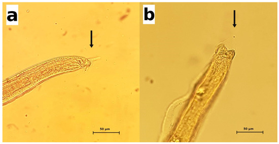
Nematodes belonging to the genus Capillaria infect a range of domestic and wild animals. Capillaria aerophila and Capillaria boehmi cause respiratory parasitoses in dogs and wild carnivores, e.g., foxes and mustelids, although they are often overlooked in canine clinical practice. The present report describes an unusual case of a severe and mixed infection by C. aerophila and C. boehmi in a privately housed dog that showed acute and life-threatening respiratory and neurological signs. Clinic-pathologic and epizootiological implications are described and discussed. Full article
Show Figures

Figure 1

6 pages, 33856 KiB  
Case Report
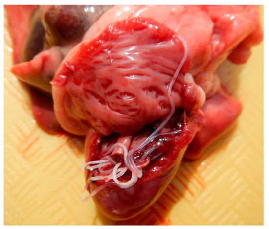
Severe Concomitant Physaloptera sp., Dirofilaria immitis, Toxocara cati, Dipylidium caninum, Ancylostoma sp. and Taenia taeniaeformis Infection in a Cat
by Jose Cesar Menk P. Lima and Fabio Del Piero
Pathogens 2021, 10(2), 109; https://doi.org/10.3390/pathogens10020109 - 22 Jan 2021
Cited by 6 | Viewed by 3660
Abstract
Here we describe an unusual and severe mixed parasitic infection in a cat that died during routine surgery. Gastric Physaloptera sp., cardiac Dirofilaria immitis, and intestinal Toxocara cati, Dipylidium caninum, Ancylostoma sp. and Taenia taeniaeformis were observed. Histologic lesions included [...] Read more.
Here we describe an unusual and severe mixed parasitic infection in a cat that died during routine surgery. Gastric Physaloptera sp., cardiac Dirofilaria immitis, and intestinal Toxocara cati, Dipylidium caninum, Ancylostoma sp. and Taenia taeniaeformis were observed. Histologic lesions included chronic proliferative pulmonary endarteritis, mild increase of mucosal intestinal white cells, and terminal aspiration of gastric content. The severe dirofilariasis may have contributed to this patient death during anesthesia. Full article
Show Figures

Figure 1

2020

Jump to: 2024, 2023, 2022, 2021

14 pages, 1403 KiB  
Article
Drug Resistance in Filarial Parasites Does Not Affect Mosquito Vectorial Capacity
by Erik Neff, Christopher C. Evans, Pablo D. Jimenez Castro, Ray M. Kaplan and Guha Dharmarajan
Pathogens 2021, 10(1), 2; https://doi.org/10.3390/pathogens10010002 - 22 Dec 2020
Cited by 2 | Viewed by 2616
Abstract
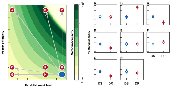
Parasite drug resistance presents a major obstacle to controlling and eliminating vector-borne diseases affecting humans and animals. While vector-borne disease dynamics are affected by factors related to parasite, vertebrate host and vector, research on drug resistance in filarial parasites has primarily focused on [...] Read more.
Parasite drug resistance presents a major obstacle to controlling and eliminating vector-borne diseases affecting humans and animals. While vector-borne disease dynamics are affected by factors related to parasite, vertebrate host and vector, research on drug resistance in filarial parasites has primarily focused on the parasite and vertebrate host, rather than the mosquito. However, we expect that the physiological costs associated with drug resistance would reduce the fitness of drug-resistant vs. drug-susceptible parasites in the mosquito wherein parasites are not exposed to drugs. Here we test this hypothesis using four isolates of the dog heartworm (Dirofilaria immitis)—two drug susceptible and two drug resistant—and two vectors—the yellow fever mosquito (Aedes aegypti) and the Asian tiger mosquito (Ae. albopictus)—as our model system. Our data indicated that while vector species had a significant effect on vectorial capacity, there was no significant difference in the vectorial capacity of mosquitoes infected with drug-resistant vs. drug-susceptible parasites. Consequently, contrary to expectations, our data indicate that drug resistance in D. immitis does not appear to reduce the transmission efficiency of these parasites, and thus the spread of drug-resistant parasites in the vertebrate population is unlikely to be mitigated by reduced fitness in the mosquito vector. Full article
Show Figures

Figure 1

Back to TopTop