Next Issue
Volume 18, November
Previous Issue
Volume 18, September


Mar. Drugs, Volume 18, Issue 10 (October 2020) – 36 articles

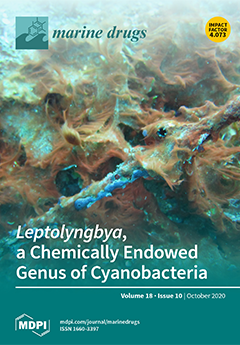
Cover Story (view full-size image): Cyanobacteria of the genus Leptolyngbya are found widely in natural ecosystems and participate in many ecological phenomena, such as nitrogen fixation, photosynthesis, and algal blooms. In this review, we summarize scientific studies to date on the ecology, chemistry, biochemistry, and pharmacology of this genus. The emerging broad view of the genus Leptolyngbya and its capacity for production of bioactive natural products provides new insights and perspectives on its metabolic capacities, as well as its role in the ecology of tropical reef systems. View this paper.
  • Issues are regarded as officially published after their release is announced to the table of contents alert mailing list.
  • You may sign up for e-mail alerts to receive table of contents of newly released issues.
  • PDF is the official format for papers published in both, html and pdf forms. To view the papers in pdf format, click on the "PDF Full-text" link, and use the free Adobe Reader to open them.
Order results
Result details
Section
Select all
Export citation of selected articles as:
12 pages, 1141 KiB  
Article
Antibacterial Activity of Chromomycins from a Marine-Derived Streptomyces microflavus
by Eunji Cho, Oh-Seok Kwon, Beomkoo Chung, Jayho Lee, Jeongyoon Sun, Jongheon Shin and Ki-Bong Oh
Mar. Drugs 2020, 18(10), 522; https://doi.org/10.3390/md18100522 - 21 Oct 2020
Cited by 25 | Viewed by 3539
Abstract
A marine-derived actinomycete (Streptomyces sp. MBTI36) exhibiting antibacterial activities was investigated in the present study. The strain was identified using genetic techniques. The 16S rDNA sequence of the isolate indicated that it was most closely related to Streptomyces microflavus. Furthermore, a [...] Read more.
A marine-derived actinomycete (Streptomyces sp. MBTI36) exhibiting antibacterial activities was investigated in the present study. The strain was identified using genetic techniques. The 16S rDNA sequence of the isolate indicated that it was most closely related to Streptomyces microflavus. Furthermore, a new chromomycin A9 (1), along with chromomycin Ap (2), chromomycin A2 (3), and chromomycin A3 (4), were isolated from the ethyl acetate extract. Their structures were determined using extensive spectroscopic methods including 1D and 2D NMR, and HRMS, as well as comparisons with previously reported data. Compounds 14 showed potent antibacterial activities against Gram-positive bacteria including methicillin-resistant Staphylococcus aureus (MRSA). During a passage experiment, minimum inhibitory concentration (MIC) values for compounds 14 showed no more than a 4-fold increase from the starting MIC value, indicating that no resistance was detected over the 21 passages. Full article
Show Figures

Figure 1

13 pages, 920 KiB  
Article
Induction of Natural Defenses in Tomato Seedlings by Using Alginate and Oligoalginates Derivatives Extracted from Moroccan Brown Algae
by Meriem Aitouguinane, Soukaina Bouissil, Anouar Mouhoub, Halima Rchid, Imen Fendri, Slim Abdelkafi, Mohamed Didi Ould El-Hadj, Zakaria Boual, Pascal Dubessay, Christine Gardarin, Philippe Michaud, Zainab El Alaoui-Talibi, Cherkaoui El Modafar, Guillaume Pierre and Cédric Delattre
Mar. Drugs 2020, 18(10), 521; https://doi.org/10.3390/md18100521 - 19 Oct 2020
Cited by 28 | Viewed by 4527
Abstract
Polysaccharides extracted from marine algae have attracted much attention due to their biotechnological applications, including therapeutics, cosmetics, and mainly in agriculture and horticulture as biostimulants, biofertilizers, and stimulators of the natural defenses of plants. This study aimed to evaluate the ability of alginate [...] Read more.
Polysaccharides extracted from marine algae have attracted much attention due to their biotechnological applications, including therapeutics, cosmetics, and mainly in agriculture and horticulture as biostimulants, biofertilizers, and stimulators of the natural defenses of plants. This study aimed to evaluate the ability of alginate isolated from Bifurcaria bifurcata from the Moroccan coast and oligoalginates derivatives to stimulate the natural defenses of tomato seedlings. Elicitation was carried out by the internodal injection of bioelicitor solutions. The elicitor capacities were evaluated by monitoring the activity of phenylalanine ammonia-lyase (PAL) as well as polyphenols content in the leaves located above the elicitation site for 5 days. Alginate and oligoalginates treatments triggered plant defense responses, which showed their capacity to significantly induce the PAL activity and phenolic compounds accumulation in the leaves of tomato seedlings. Elicitation by alginates and oligoalginates showed an intensive induction of PAL activity, increasing from 12 h of treatment and remaining at high levels throughout the period of treatment. The amount of polyphenols in the leaves was increased rapidly and strongly from 12 h of elicitation by both saccharide solutions, representing peaks value after 24 h of application. Oligoalginates exhibited an effective elicitor capacity in polyphenols accumulation compared to alginate polymers. The alginate and oligosaccharides derivatives revealed a similar elicitor capacity in PAL activity whereas the accumulation of phenolic compounds showed a differential effect. Polysaccharides extracted from the brown seaweed Bifurcaria bifurcate and oligosaccharides derivatives induced significantly the phenylpropanoid metabolism in tomato seedlings. These results contribute to the valorization of marine biomass as a potential bioresource for plant protection against phytopathogens in the context of eco-sustainable green technology. Full article
(This article belongs to the Special Issue Characterization of Bioactive Components in Edible Algae II)
Show Figures

Figure 1

12 pages, 4619 KiB  
Article
Sodium Alginate as a Potential Therapeutic Filler: An In Vivo Study in Rats
by Masanori Mori, Rintaro Asahi, Yoshihiro Yamamoto, Takanobu Mashiko, Kayo Yoshizumi, Natsumi Saito, Takako Shirado, Yunyan Wu and Kotaro Yoshimura
Mar. Drugs 2020, 18(10), 520; https://doi.org/10.3390/md18100520 - 19 Oct 2020
Cited by 11 | Viewed by 3298
Abstract
Filler injection demand is increasing worldwide, but no ideal filler with safety and longevity currently exists. Sodium alginate (SA) is the sodium salt of alginic acid, which is a polymeric polysaccharide obtained by linear polymerization of two types of uronic acid, d-mannuronic [...] Read more.
Filler injection demand is increasing worldwide, but no ideal filler with safety and longevity currently exists. Sodium alginate (SA) is the sodium salt of alginic acid, which is a polymeric polysaccharide obtained by linear polymerization of two types of uronic acid, d-mannuronic acid (M) and l-guluronic acid (G). This study aimed to evaluate the therapeutic value of SA. Nine SA types with different M/G ratios and viscosities were tested and compared with a commercially available sodium hyaluronate (SH) filler. Three injection modes (onto the periosteum, intradermally, or subcutaneously) were used in six rats for each substance, and the animals were sacrificed at 4 or 24 weeks. Changes in the diameter and volume were measured macroscopically and by computed tomography, and histopathological evaluations were performed. SA with a low M/G ratio generally maintained skin uplift. The bulge gradually decreased over time but slightly increased at 4 weeks in some samples. No capsule formation was observed around SA. However, granulomatous reactions, including macrophage recruitment, were observed 4 weeks after SA implantation, although fewer macrophages and granulomatous reactions were observed at 24 weeks. The long-term volumizing effects and degree of granulomatous reactions differed depending on the M/G ratio and viscosity. By contrast, SH showed capsule formation but with minimal granulomatous reactions. The beneficial and adverse effects of SA as a filler differed according to the viscosity or M/G ratio, suggesting a better long-term volumizing effect than SH with relatively low immunogenicity Full article
Show Figures

Figure 1

18 pages, 5450 KiB  
Article
Design, Synthesis and In Vitro Experimental Validation of Novel TRPV4 Antagonists Inspired by Labdane Diterpenes
by Sarah Mazzotta, Gabriele Carullo, Aniello Schiano Moriello, Pietro Amodeo, Vincenzo Di Marzo, Margarita Vega-Holm, Rosa Maria Vitale, Francesca Aiello, Antonella Brizzi and Luciano De Petrocellis
Mar. Drugs 2020, 18(10), 519; https://doi.org/10.3390/md18100519 - 18 Oct 2020
Cited by 13 | Viewed by 4007
Abstract
Labdane diterpenes are widespread classes of natural compounds present in variety of marine and terrestrial organisms and plants. Many of them represents “natural libraries” of compounds with interesting biological activities due to differently functionalized drimane nucleus exploitable for potential pharmacological applications. The transient [...] Read more.
Labdane diterpenes are widespread classes of natural compounds present in variety of marine and terrestrial organisms and plants. Many of them represents “natural libraries” of compounds with interesting biological activities due to differently functionalized drimane nucleus exploitable for potential pharmacological applications. The transient receptor potential channel subfamily V member 4 (TRPV4) channel has recently emerged as a pharmacological target for several respiratory diseases, including the severe acute respiratory syndrome coronavirus 2 (SARS-CoV-2) infection. Inspired by the labdane-like bicyclic core, a series of homodrimane-derived esters and amides was designed and synthesized by modifying the flexible tail in position 1 of (+)-sclareolide, an oxidized derivative of the bioactive labdane-type diterpene sclareol. The potency and selectivity towards rTRPV4 and hTRPV1 receptors were assessed by calcium influx cellular assays. Molecular determinants critical for eliciting TRPV4 antagonism were identified by structure-activity relationships. Among the selective TRPV4 antagonists identified, compound 6 was the most active with an IC50 of 5.3 μM. This study represents the first report of semisynthetic homodrimane TRPV4 antagonists, selective over TRPV1, and potentially useful as pharmacological tools for the development of novel TRPV4 channel modulators. Full article
Show Figures

Graphical abstract

6 pages, 200 KiB  
Editorial
Chemical Defense in Marine Organisms
by Chiara Lauritano and Adrianna Ianora
Mar. Drugs 2020, 18(10), 518; https://doi.org/10.3390/md18100518 - 18 Oct 2020
Cited by 10 | Viewed by 2944
Abstract
Marine organisms are constantly exposed to variations in physical parameters (e [...] Full article
(This article belongs to the Special Issue Chemical Defense in Marine Organisms)
17 pages, 4277 KiB  
Article
Viridicatol Isolated from Deep-Sea Penicillium Griseofulvum Alleviates Anaphylaxis and Repairs the Intestinal Barrier in Mice by Suppressing Mast Cell Activation
by Zhendan Shu, Qingmei Liu, Cuiping Xing, Yafen Zhang, Yu Zhou, Jun Zhang, Hong Liu, Minjie Cao, Xianwen Yang and Guangming Liu
Mar. Drugs 2020, 18(10), 517; https://doi.org/10.3390/md18100517 - 16 Oct 2020
Cited by 16 | Viewed by 3437
Abstract
Viridicatol is a quinoline alkaloid isolated from the deep-sea-derived fungus Penicillium griseofulvum. The structure of viridicatol was unambiguously established by X-ray diffraction analysis. In this study, a mouse model of ovalbumin-induced food allergy and the rat basophil leukemia (RBL)-2H3 cell model were established [...] Read more.
Viridicatol is a quinoline alkaloid isolated from the deep-sea-derived fungus Penicillium griseofulvum. The structure of viridicatol was unambiguously established by X-ray diffraction analysis. In this study, a mouse model of ovalbumin-induced food allergy and the rat basophil leukemia (RBL)-2H3 cell model were established to explore the anti-allergic properties of viridicatol. On the basis of the mouse model, we found viridicatol to alleviate the allergy symptoms; decrease the levels of specific immunoglobulin E, mast cell protease-1, histamine, and tumor necrosis factor-α; and promote the production of interleukin-10 in the serum. The treatment of viridicatol also downregulated the population of B cells and mast cells (MCs), as well as upregulated the population of regulatory T cells in the spleen. Moreover, viridicatol alleviated intestinal villi injury and inhibited the degranulation of intestinal MCs to promote intestinal barrier repair in mice. Furthermore, the accumulation of Ca2+ in RBL-2H3 cells was significantly suppressed by viridicatol, which could block the activation of MCs. Taken together, these data indicated that deep-sea viridicatol may represent a novel therapeutic for allergic diseases. Full article
(This article belongs to the Special Issue Marine Microbial Diversity as Source of Bioactive Compounds)
Show Figures

Graphical abstract

19 pages, 3660 KiB  
Article
N-Docosahexaenoylethanolamine Attenuates Neuroinflammation and Improves Hippocampal Neurogenesis in Rats with Sciatic Nerve Chronic Constriction Injury
by Anna A. Tyrtyshnaia, Evgenia L. Egorova, Anna A. Starinets, Arina I. Ponomarenko, Ekaterina V. Ermolenko and Igor V. Manzhulo
Mar. Drugs 2020, 18(10), 516; https://doi.org/10.3390/md18100516 - 15 Oct 2020
Cited by 23 | Viewed by 3734
Abstract
Chronic neuropathic pain is a condition that causes both sensory disturbances and a variety of functional disorders, indicating the involvement of various brain structures in pain pathogenesis. One of the factors underlying chronic neuropathic pain is neuroinflammation, which is accompanied by microglial activation [...] Read more.
Chronic neuropathic pain is a condition that causes both sensory disturbances and a variety of functional disorders, indicating the involvement of various brain structures in pain pathogenesis. One of the factors underlying chronic neuropathic pain is neuroinflammation, which is accompanied by microglial activation and pro-inflammatory factor release. N-docosahexaenoylethanolamine (DHEA, synaptamide) is an endocannabinoid-like metabolite synthesized endogenously from docosahexaenoic acid. Synaptamide exhibits anti-inflammatory activity and improves neurite outgrowth, neurogenesis, and synaptogenesis within the hippocampus. This study aims to evaluate the effects of synaptamide obtained by the chemical modification of DHA, extracted from the Far Eastern raw material Berryteuthis magister on neuroinflammatory response and hippocampal neurogenesis changes during neuropathic pain. The study of microglial protein and cytokine concentrations was performed using immunohistochemistry and ELISA. The brain lipid analysis was performed using the liquid chromatography-mass spectrometry technique. Behavioral experiments showed that synaptamide prevented neuropathic pain-associated sensory and behavioral changes, such as thermal allodynia, impaired locomotor activity, working and long-term memory, and increased anxiety. Synaptamide attenuated microglial activation, release of proinflammatory cytokines, and decrease in hippocampal neurogenesis. Lipid analysis revealed changes in the brain N-acylethanolamines composition and plasmalogen concentration after synaptamide administration. In conclusion, we show here that synaptamide may have potential for use in preventing or treating neuropathic cognitive pain and emotional effects. Full article
(This article belongs to the Special Issue Marine Compounds from the Far Eastern Organisms)
Show Figures

Figure 1

25 pages, 3572 KiB  
Article
Applying a Chemogeographic Strategy for Natural Product Discovery from the Marine Cyanobacterium Moorena bouillonii
by Christopher A. Leber, C. Benjamin Naman, Lena Keller, Jehad Almaliti, Eduardo J. E. Caro-Diaz, Evgenia Glukhov, Valsamma Joseph, T. P. Sajeevan, Andres Joshua Reyes, Jason S. Biggs, Te Li, Ye Yuan, Shan He, Xiaojun Yan and William H. Gerwick
Mar. Drugs 2020, 18(10), 515; https://doi.org/10.3390/md18100515 - 14 Oct 2020
Cited by 9 | Viewed by 4202
Abstract
The tropical marine cyanobacterium Moorena bouillonii occupies a large geographic range across the Indian and Western Tropical Pacific Oceans and is a prolific producer of structurally unique and biologically active natural products. An ensemble of computational approaches, including the creation of the ORCA [...] Read more.
The tropical marine cyanobacterium Moorena bouillonii occupies a large geographic range across the Indian and Western Tropical Pacific Oceans and is a prolific producer of structurally unique and biologically active natural products. An ensemble of computational approaches, including the creation of the ORCA (Objective Relational Comparative Analysis) pipeline for flexible MS1 feature detection and multivariate analyses, were used to analyze various M. bouillonii samples. The observed chemogeographic patterns suggested the production of regionally specific natural products by M. bouillonii. Analyzing the drivers of these chemogeographic patterns allowed for the identification, targeted isolation, and structure elucidation of a regionally specific natural product, doscadenamide A (1). Analyses of MS2 fragmentation patterns further revealed this natural product to be part of an extensive family of herein annotated, proposed natural structural analogs (doscadenamides B–J, 2–10); the ensemble of structures reflect a combinatorial biosynthesis using nonribosomal peptide synthetase (NRPS) and polyketide synthase (PKS) components. Compound 1 displayed synergistic in vitro cancer cell cytotoxicity when administered with lipopolysaccharide (LPS). These discoveries illustrate the utility in leveraging chemogeographic patterns for prioritizing natural product discovery efforts. Full article
(This article belongs to the Special Issue Natural Product Genomics and Metabolomics of Marine Bacteria)
Show Figures

Graphical abstract

26 pages, 3602 KiB  
Review
Marine Antithrombotics
by Rohini Dwivedi and Vitor H. Pomin
Mar. Drugs 2020, 18(10), 514; https://doi.org/10.3390/md18100514 - 13 Oct 2020
Cited by 20 | Viewed by 5552
Abstract
Thrombosis remains a prime reason of mortality worldwide. With the available antithrombotic drugs, bleeding remains the major downside of current treatments. This raises a clinical concern for all patients undergoing antithrombotic therapy. Novel antithrombotics from marine sources offer a promising therapeutic alternative to [...] Read more.
Thrombosis remains a prime reason of mortality worldwide. With the available antithrombotic drugs, bleeding remains the major downside of current treatments. This raises a clinical concern for all patients undergoing antithrombotic therapy. Novel antithrombotics from marine sources offer a promising therapeutic alternative to this pathology. However, for any potential new molecule to be introduced as a real alternative to existing drugs, the exhibition of comparable anticoagulant potential with minimal off-target effects must be achieved. The relevance of marine antithrombotics, particularly sulfated polysaccharides, is largely due to their unique mechanisms of action and lack of bleeding. There have been many investigations in the field and, in recent years, results have confirmed the role of potential marine molecules as alternative antithrombotics. Nonetheless, further clinical studies are required. This review covers the core of the data available so far regarding the science of marine molecules with potential medical applications to treat thrombosis. After a general discussion about the major biochemical steps involved in this pathology, we discuss the key structural and biomedical aspects of marine molecules of both low and high molecular weight endowed with antithrombotic/anticoagulant properties. Full article
(This article belongs to the Special Issue Marine Molecules for the Treatment of Thrombosis)
Show Figures

Figure 1

10 pages, 763 KiB  
Article
Hatsusamides A and B: Two New Metabolites Produced by the Deep-Sea-Derived Fungal Strain Penicillium steckii FKJ-0213
by Hirotaka Matsuo, Rei Hokari, Aki Ishiyama, Masato Iwatsuki, Mayuka Higo, Kenichi Nonaka, Yuriko Nagano, Yōko Takahashi, Satoshi Ōmura and Takuji Nakashima
Mar. Drugs 2020, 18(10), 513; https://doi.org/10.3390/md18100513 - 12 Oct 2020
Cited by 13 | Viewed by 3322
Abstract
Two new nitrogen-containing metabolites, designated hatsusamide A (1) and B (2), were isolated from a culture broth of Penicilliumsteckii FKJ-0213 together with the known compounds tanzawaic acid B (3) and trichodermamide C (4) by [...] Read more.
Two new nitrogen-containing metabolites, designated hatsusamide A (1) and B (2), were isolated from a culture broth of Penicilliumsteckii FKJ-0213 together with the known compounds tanzawaic acid B (3) and trichodermamide C (4) by physicochemical (PC) screening. The structures of 1 and 2 were determined as a tanzawaic acid B-trichodermamide C hybrid structure and a new analog of aspergillazines, respectively. The absolute configuration of 1 was determined by comparing the values of tanzawaic acid B and trichodermamide C in the literatures, such as 1H-nuclear magnetic resonance (1H-NMR) data and optical rotation, after hydrolysis of 1. Compounds 14 were evaluated for cytotoxicity and anti-malarial activities. Compounds 1 and 3 exhibited weak anti-malarial activity at half-maximal inhibitory concentration (IC50) values of 27.2 and 78.5 µM against the K1 strain, and 27.9 and 79.2 µM against the FCR3 strain of Plasmodium falciparum, respectively. Furthermore, 1 exhibited cytotoxicity against HeLa S3, A549, Panc1, HT29 and H1299 cells, with IC50 values of 15.0, 13.7, 12.9, 6.8, and 18.7 μM, respectively. Full article
(This article belongs to the Special Issue Bioactive Compounds from Marine Sediment Derived Fungi)
Show Figures

Graphical abstract

21 pages, 3419 KiB  
Article
Dipeptide YA is Responsible for the Positive Effect of Oyster Hydrolysates on Alcohol Metabolism in Single Ethanol Binge Rodent Models
by Adrian S. Siregar, Marie Merci Nyiramana, Eun-Jin Kim, Eui-Jung Shin, Min Seok Woo, Jin-Mok Kim, Jung Hwan Kim, Dong Kun Lee, Jong Ryeal Hahm, Hyun Joon Kim, Chang-Woon Kim, Nam-Gil Kim, Si-Hyang Park, Yeung Joon Choi, Sang Soo Kang, Seong-Geun Hong, Jaehee Han and Dawon Kang
Mar. Drugs 2020, 18(10), 512; https://doi.org/10.3390/md18100512 - 11 Oct 2020
Cited by 19 | Viewed by 4570
Abstract
Accumulative alcohol hangovers cause liver damage through oxidative and inflammatory stress. Numerous antioxidant and anti-inflammatory reagents have been developed to reduce alcohol hangovers, but these reagents are still insignificant and have limitations in that they can cause liver toxicity. Oyster hydrolysate (OH), another [...] Read more.
Accumulative alcohol hangovers cause liver damage through oxidative and inflammatory stress. Numerous antioxidant and anti-inflammatory reagents have been developed to reduce alcohol hangovers, but these reagents are still insignificant and have limitations in that they can cause liver toxicity. Oyster hydrolysate (OH), another reagent that has antioxidant and anti-inflammatory activity, is a product extracted through an enzymatic hydrolysis process from oysters (Crassostrea gigas), which can be easily eaten in meals. This study was aimed at determining the effects of OH on alcohol metabolism, using a single high dose of ethanol (EtOH) administered to rodents, by monitoring alcohol metabolic enzymes, oxidative stress signals, and inflammatory mediators. The effect of tyrosine-alanine (YA) peptide, a main component of OH, on EtOH metabolism was also identified. In vitro experiments showed that OH pretreatment inhibited EtOH-induced cell death, oxidative stress, and inflammation in liver cells and macrophages. In vivo experiments showed that OH and YA pre-administration increased alcohol dehydrogenase, aldehyde dehydrogenase, and catalase activity in EtOH binge treatment. In addition, OH pre-administration alleviated CYP2E1 activity, ROS production, apoptotic signals, and inflammatory mediators in liver tissues. These results showed that OH and YA enhanced EtOH metabolism and had a protective effect against acute alcohol liver damage. Our findings offer new insights into a single high dose of EtOH drinking and suggest that OH and YA could be used as potential marine functional foods to prevent acute alcohol-induced liver damage. Full article
Show Figures

Figure 1

16 pages, 4185 KiB  
Article
Type II Collagen from Cartilage of Acipenser baerii Promotes Wound Healing in Human Dermal Fibroblasts and in Mouse Skin
by Ching-Shu Lai, Chun-Wei Tu, Hsing-Chun Kuo, Pei-Pei Sun and Mei-Ling Tsai
Mar. Drugs 2020, 18(10), 511; https://doi.org/10.3390/md18100511 - 11 Oct 2020
Cited by 18 | Viewed by 4747
Abstract
Type II collagen is an important component of cartilage; however, little is known about its effect on skin wound healing. In this study, type II collagen was extracted from the cartilage of Acipenser baerii and its effect on in vitro and in vivo [...] Read more.
Type II collagen is an important component of cartilage; however, little is known about its effect on skin wound healing. In this study, type II collagen was extracted from the cartilage of Acipenser baerii and its effect on in vitro and in vivo wound healing was compared to type I collagen derived from tilapia skin. Sturgeon cartilage collagen (SCC) was composed of α1 chains and with a thermal denaturation (Td) at 22.5 and melting temperature (Tm) at 72.5 °C. Coating SCC potentiated proliferation, migration, and invasion of human dermal fibroblast adult (HDFa) cells. Furthermore, SCC upregulated the gene expression of extracellular matrix (ECM) components (col Iα1, col IIIα1, elastin, and Has2) and epithelial-mesenchymal transition (EMT) molecules (N-cadherin, Snail, and MMP-1) in HDFa. Pretreatment with Akt and mitogen-activated protein kinase (MAPK) inhibitors significantly attenuated the HDFa invasion caused by SCC. In mice, the application of SCC on dorsal wounds effectively facilitated wound healing as evidenced by 40–59% wound contraction, whereas the untreated wounds were 18%. We observed that SCC reduced inflammation, promoted granulation, tissue formation, and ECM deposition, as well as re-epithelialization in skin wounds. In addition, SCC markedly upregulated the production of growth factors in the dermis, and dermal and subcutaneous white adipose tissue; in contrast, the administration of tilapia skin collagen (TSC) characterized by typical type I collagen was mainly expressed in the epidermis. Collectively, these findings indicate SCC accelerated wound healing by targeting fibroblast in vitro and in vivo. Full article
Show Figures

Figure 1

12 pages, 3838 KiB  
Article
Blue Mussel-Derived Peptides PIISVYWK and FSVVPSPK Trigger Wnt/β-Catenin Signaling-Mediated Osteogenesis in Human Bone Marrow Mesenchymal Stem Cells
by Yunok Oh, Chang-Bum Ahn and Jae-Young Je
Mar. Drugs 2020, 18(10), 510; https://doi.org/10.3390/md18100510 - 9 Oct 2020
Cited by 20 | Viewed by 2877
Abstract
Marine-derived bioactive peptides have shown potential bone health promoting effects. Although various marine-derived bioactive peptides have potential nutraceutical or pharmaceutical properties, only a few of them are commercially available. This study presented an osteogenic mechanism of blue mussel-derived peptides PIISVYWK and FSVVPSPK as [...] Read more.
Marine-derived bioactive peptides have shown potential bone health promoting effects. Although various marine-derived bioactive peptides have potential nutraceutical or pharmaceutical properties, only a few of them are commercially available. This study presented an osteogenic mechanism of blue mussel-derived peptides PIISVYWK and FSVVPSPK as potential bone health promoting agents in human bone marrow-derived mesenchymal stem cells (hBMMSCs). Alkaline phosphatase (ALP) activity and mineralization were stimulated using PIISVYWK and FSVVPSPK as early and late markers of osteogenesis in a concentration-dependent manner. Western blot and RT-qPCR results revealed that PIISVYWK and FSVVPSPK increased osteoblast differentiation of hBMMSCs by activating canonical Wnt/β-catenin signaling-related proteins and mRNAs. Immunofluorescence images confirmed nuclear translocation of β-catenin in osteogenic differentiation. Treatment with the pharmacological inhibitor DKK-1 blocked PIISVYWK- and FSVVPSPK-induced ALP activity and mineralization, as well as mRNA expression of the canonical Wnt/β-catenin signaling pathway in hBMMSC differentiation into osteoblasts. These findings suggested that PIISVYWK and FSVVPSPK promoted the canonical Wnt/β-catenin signaling pathway in osteogenesis of hBMMSCs. Blue mussel-derived PIISVYWK and FSVVPSPK might help develop peptide-based therapeutic agents for bone-related diseases. Full article
(This article belongs to the Special Issue Marine Organisms for Bone Regeneration - 2020)
Show Figures

Figure 1

14 pages, 1321 KiB  
Article
Structure of the Polysaccharide Secreted by Vibrio alginolyticus CNCM I-5035 (Epidermist 4.0TM)
by Sophie Drouillard, Rémi Chambon, Isabelle Jeacomine, Laurine Buon, Claire Boisset, Anthony Courtois, Bertrand Thollas, Pierre-Yves Morvan, Romuald Vallée and William Helbert
Mar. Drugs 2020, 18(10), 509; https://doi.org/10.3390/md18100509 - 9 Oct 2020
Cited by 7 | Viewed by 2735
Abstract
Vibrio alginolyticus (CNCM I-5035) secretes an exopolysaccharide used as ingredient in cosmetic industry under the trademark Epidermist 4.0TM. It is appreciated for its ability to improve the physical and chemical barrier functions of the skin by notably increasing the keratinocyte differentiation [...] Read more.
Vibrio alginolyticus (CNCM I-5035) secretes an exopolysaccharide used as ingredient in cosmetic industry under the trademark Epidermist 4.0TM. It is appreciated for its ability to improve the physical and chemical barrier functions of the skin by notably increasing the keratinocyte differentiation and epidermal renewal. Composition analyses and in depth characterization of the polysaccharides as well as oligosaccharides obtained by mild acid hydrolyses revealed that it was composed of a repetition unit of three residues: d-galactose (d-Gal), d-N-acetylglucosamine (GlcNAc) and l-N-acetylguluronic acid, of which 30% (M/M) was acetylated in position 3. The complete structure of the polysaccharide was resolved giving the repetition unit: [→3)-α-d-Gal-(1→4)-α-l-GulNAcA/α-l-3OAc-GulNAcA-(1→4)-β-d-GlcNAc-(1→]. Full article
(This article belongs to the Special Issue Marine Carbohydrate-Based Compounds with Medicinal Properties)
Show Figures

Figure 1

29 pages, 3888 KiB  
Review
The Chemistry, Biochemistry and Pharmacology of Marine Natural Products from Leptolyngbya, a Chemically Endowed Genus of Cyanobacteria
by Yueying Li, C. Benjamin Naman, Kelsey L. Alexander, Huashi Guan and William H. Gerwick
Mar. Drugs 2020, 18(10), 508; https://doi.org/10.3390/md18100508 - 6 Oct 2020
Cited by 19 | Viewed by 5266
Abstract
Leptolyngbya, a well-known genus of cyanobacteria, is found in various ecological habitats including marine, fresh water, swamps, and rice fields. Species of this genus are associated with many ecological phenomena such as nitrogen fixation, primary productivity through photosynthesis and algal blooms. As [...] Read more.
Leptolyngbya, a well-known genus of cyanobacteria, is found in various ecological habitats including marine, fresh water, swamps, and rice fields. Species of this genus are associated with many ecological phenomena such as nitrogen fixation, primary productivity through photosynthesis and algal blooms. As a result, there have been a number of investigations of the ecology, natural product chemistry, and biological characteristics of members of this genus. In general, the secondary metabolites of cyanobacteria are considered to be rich sources for drug discovery and development. In this review, the secondary metabolites reported in marine Leptolyngbya with their associated biological activities or interesting biosynthetic pathways are reviewed, and new insights and perspectives on their metabolic capacities are gained. Full article
(This article belongs to the Special Issue Compounds from Cyanobacteria III)
Show Figures

Graphical abstract

28 pages, 850 KiB  
Review
A Review of Toxins from Cnidaria
by Isabella D’Ambra and Chiara Lauritano
Mar. Drugs 2020, 18(10), 507; https://doi.org/10.3390/md18100507 - 6 Oct 2020
Cited by 55 | Viewed by 9297
Abstract
Cnidarians have been known since ancient times for the painful stings they induce to humans. The effects of the stings range from skin irritation to cardiotoxicity and can result in death of human beings. The noxious effects of cnidarian venoms have stimulated the [...] Read more.
Cnidarians have been known since ancient times for the painful stings they induce to humans. The effects of the stings range from skin irritation to cardiotoxicity and can result in death of human beings. The noxious effects of cnidarian venoms have stimulated the definition of their composition and their activity. Despite this interest, only a limited number of compounds extracted from cnidarian venoms have been identified and defined in detail. Venoms extracted from Anthozoa are likely the most studied, while venoms from Cubozoa attract research interests due to their lethal effects on humans. The investigation of cnidarian venoms has benefited in very recent times by the application of omics approaches. In this review, we propose an updated synopsis of the toxins identified in the venoms of the main classes of Cnidaria (Hydrozoa, Scyphozoa, Cubozoa, Staurozoa and Anthozoa). We have attempted to consider most of the available information, including a summary of the most recent results from omics and biotechnological studies, with the aim to define the state of the art in the field and provide a background for future research. Full article
(This article belongs to the Special Issue Chemical Defense in Marine Organisms)
Show Figures

Figure 1

13 pages, 2876 KiB  
Article
Stereochemistry of Astaxanthin Biosynthesis in the Marine Harpacticoid Copepod Tigriopus Californicus
by Alfonso Prado-Cabrero, Ganjar Saefurahman and John M. Nolan
Mar. Drugs 2020, 18(10), 506; https://doi.org/10.3390/md18100506 - 5 Oct 2020
Cited by 12 | Viewed by 4115
Abstract
The harpacticoid copepod Tigriopus californicus has been recognized as a model organism for the study of marine pollutants. Furthermore, the nutritional profile of this copepod is of interest to the aquafeed industry. Part of this interest lies in the fact that Tigriopus produces [...] Read more.
The harpacticoid copepod Tigriopus californicus has been recognized as a model organism for the study of marine pollutants. Furthermore, the nutritional profile of this copepod is of interest to the aquafeed industry. Part of this interest lies in the fact that Tigriopus produces astaxanthin, an essential carotenoid in salmonid aquaculture. Here, we study for the first time the stereochemistry of the astaxanthin produced by this copepod. We cultured T. californicus with different feeding sources and used chiral high-performance liquid chromatography with diode array detection (HPLC-DAD) to determine that T. californicus synthesizes pure 3S,3’S-astaxanthin. Using meso-zeaxanthin as feed, we found that the putative ketolase enzyme from T. californicus can work with β-rings with either 3R- or 3S-oriented hydroxyl groups. Despite this ability, experiments in the presence of hydroxylated and non-hydroxylated carotenoids suggest that T. californicus prefers to use the latter to produce 3S,3’S-astaxanthin. We suggest that the biochemical tools described in this work can be used to study the mechanistic aspects of the recently identified avian ketolase. Full article
(This article belongs to the Special Issue Marine Carotenoids)
Show Figures

Graphical abstract

21 pages, 3741 KiB  
Article
Mass Spectrometry-Based Characterization of New Spirolides from Alexandrium ostenfeldii (Dinophyceae)
by Joyce A. Nieva, Jan Tebben, Urban Tillmann, Sylke Wohlrab and Bernd Krock
Mar. Drugs 2020, 18(10), 505; https://doi.org/10.3390/md18100505 - 2 Oct 2020
Cited by 6 | Viewed by 2708
Abstract
Spirolides belong to a group of marine phycotoxins produced by the marine planktonic dinophyte Alexandrium ostenfeldii. Composed of an imine moiety and a spiroketal ring system within a macrocylcle, spirolides are highly diverse with toxin types that vary among different strains. This [...] Read more.
Spirolides belong to a group of marine phycotoxins produced by the marine planktonic dinophyte Alexandrium ostenfeldii. Composed of an imine moiety and a spiroketal ring system within a macrocylcle, spirolides are highly diverse with toxin types that vary among different strains. This study aims to characterize the spirolides from clonal A. ostenfeldii strains collected from The Netherlands, Greenland and Norway by mass spectral techniques. The structural characterization of unknown spirolides as inferred from high-resolution mass spectrometry (HR-MS) and collision induced dissociation (CID) spectra revealed the presence of nine novel spirolides that have the pseudo-molecular ions m/z 670 (1), m/z 666 (2), m/z 696 (3), m/z 678 (4), m/z 694 (5), m/z 708 (6), m/z 720 (7), m/z 722 (8) and m/z 738 (9). Of the nine new spirolides proposed in this study, compound 1 was suggested to have a truncated side chain in lieu of the commonly observed butenolide ring in spirolides. Moreover, there is indication that compound 5 might belong to new spirolide subclasses with a trispiroketal ring configuration having a 6:5:6 trispiroketal ring system. On the other hand, the other compounds were proposed as C- and G-type SPX, respectively. Compound 7 is proposed as the first G-type SPX with a 10-hydroxylation as usually observed in C-type SPX. This mass spectrometry-based study thus demonstrates that structural variability of spirolides is larger than previously known and does not only include the presence or absence of certain functional groups but also involves the triketal ring system. Full article
Show Figures

Figure 1

10 pages, 478 KiB  
Review
Epidemiology and Toxicology of Ciguatera Poisoning in the Colombian Caribbean
by Roberto Navarro Quiroz, Juan Carlos Herrera-Usuga, Laura Maria Osorio-Ospina, Katia Margarita Garcia-Pertuz and Elkin Navarro Quiroz
Mar. Drugs 2020, 18(10), 504; https://doi.org/10.3390/md18100504 - 1 Oct 2020
Cited by 4 | Viewed by 3189
Abstract
Ciguatera is a food intoxication caused by the consumption of primarily coral fish; these species exist in large numbers in the seas that surround the Colombian territory. The underreported diagnosis of this clinical entity has been widely highlighted due to multiple factors, such [...] Read more.
Ciguatera is a food intoxication caused by the consumption of primarily coral fish; these species exist in large numbers in the seas that surround the Colombian territory. The underreported diagnosis of this clinical entity has been widely highlighted due to multiple factors, such as, among others, ignorance by the primary care practitioner consulted for this condition as well as clinical similarity to secondary gastroenteric symptoms and common food poisonings of bacterial, parasitic or viral etiology. Eventually, it was found that people affected by ciguatoxins had trips to coastal areas hours before the onset of symptoms. Thanks to multiple studies over the years, it has been possible to identify the relation between toxigenic dinoflagellates and seagrasses, as well as its incorporation into the food chain, starting by fish primarily inhabiting reef ecosystems and culminating in the intake of these by humans. Identifying the epidemiological link, its cardinal symptoms and affected systems, such as gastrointestinal, the peripheral nervous system and, fortunately with a low frequency, the cardiovascular system, leads to a purely clinical diagnostic impression without necessitating further complementary studies; in addition, what would also help fight ciguatera poisoning is performing an adequate treatment of the symptoms right from the start, without underestimating or overlooking any associated complications. Full article
Show Figures

Figure 1

18 pages, 5415 KiB  
Article
Identification of Conomarphin Variants in the Conus eburneus Venom and the Effect of Sequence and PTM Variations on Conomarphin Conformations
by Corazon Ericka Mae M. Itang, Jokent T. Gaza, Dan Jethro M. Masacupan, Dessa Camille R. Batoctoy, Yu-Ju Chen, Ricky B. Nellas and Eizadora T. Yu
Mar. Drugs 2020, 18(10), 503; https://doi.org/10.3390/md18100503 - 1 Oct 2020
Cited by 3 | Viewed by 3551
Abstract
Marine cone snails belonging to the Conidae family make use of neuroactive peptides in their venom to capture prey. Here we report the proteome profile of the venom duct of Conus eburneus, a cone snail belonging to the Tesseliconus clade. Through tandem [...] Read more.
Marine cone snails belonging to the Conidae family make use of neuroactive peptides in their venom to capture prey. Here we report the proteome profile of the venom duct of Conus eburneus, a cone snail belonging to the Tesseliconus clade. Through tandem mass spectrometry and database searching against the C. eburneus transcriptome and the ConoServer database, we identified 24 unique conopeptide sequences in the venom duct. The majority of these peptides belong to the T and M gene superfamilies and are disulfide-bonded, with cysteine frameworks V, XIV, VI/VII, and III being the most abundant. All seven of the Cys-free peptides are conomarphin variants belonging to the M superfamily that eluted out as dominant peaks in the chromatogram. These conomarphins vary not only in amino acid residues in select positions along the backbone but also have one or more post-translational modifications (PTMs) such as proline hydroxylation, C-term amidation, and γ-carboxylation of glutamic acid. Using molecular dynamics simulations, the conomarphin variants were predicted to predominantly have hairpin-like or elongated structures in acidic pH. These two structures were found to have significant differences in electrostatic properties and the inclusion of PTMs seems to complement this disparity. The presence of polar PTMs (hydroxyproline and γ-carboxyglutamic acid) also appear to stabilize hydrogen bond networks in these conformations. Furthermore, these predicted structures are pH sensitive, becoming more spherical and compact at higher pH. The subtle conformational variations observed here might play an important role in the selection and binding of the peptides to their molecular targets. Full article
(This article belongs to the Special Issue Simulation Studies for Marine Drug Discovery)
Show Figures

Figure 1

19 pages, 3025 KiB  
Article
Efficient Extraction and Antioxidant Capacity of Mycosporine-Like Amino Acids from Red Alga Dulse Palmaria palmata in Japan
by Yuki Nishida, Yuya Kumagai, Shunta Michiba, Hajime Yasui and Hideki Kishimura
Mar. Drugs 2020, 18(10), 502; https://doi.org/10.3390/md18100502 - 30 Sep 2020
Cited by 46 | Viewed by 8592
Abstract
Mycosporine-like amino acids (MAAs) are the ultraviolet (UV)-absorbable compounds, which are naturally produced by cyanobacteria and algae. Not only these algae but also marine organisms utilize MAAs to protect their DNA from UV-induced damage. On the other hand, the content of MAAs in [...] Read more.
Mycosporine-like amino acids (MAAs) are the ultraviolet (UV)-absorbable compounds, which are naturally produced by cyanobacteria and algae. Not only these algae but also marine organisms utilize MAAs to protect their DNA from UV-induced damage. On the other hand, the content of MAAs in algae was changed by the environmental condition and season. In addition to the UV-protected function, the antioxidant capacity of MAAs can apply to the cosmetic sunscreen materials and anti-cancer for human health. In this study, we developed the efficient extraction method of MAAs from red alga dulse in Usujiri (Hokkaido, Japan) and investigated the monthly variation. We also evaluated the antioxidant capacity. We employed the successive extraction method of water and then methanol extraction. Spectrophotometric and HPLC analyses revealed that the yield of MAAs by 6 h water extraction was the highest among the tested conditions, and the content of MAAs in the sample of February was the most (6.930 µmol g−1 dry weight) among the sample from January to May in 2019. Antioxidant capacity of MAAs such as crude MAAs, the purified palythine and porphyra-334 were determined by 2,2’-azinobis(3-ethylbenzothiazoline 6-sulfonic acid) (ABTS) radical scavenging and ferrous reducing power assays in various pH conditions, showing that the highest scavenging activity and reducing power were found at alkaline condition (pH 8.0). Full article
(This article belongs to the Special Issue Mycosporine-Like Amino Acids from Marine Resource)
Show Figures

Figure 1

58 pages, 2171 KiB  
Review
Bioactive Properties of Marine Phenolics
by Raquel Mateos, José Ricardo Pérez-Correa and Herminia Domínguez
Mar. Drugs 2020, 18(10), 501; https://doi.org/10.3390/md18100501 - 30 Sep 2020
Cited by 73 | Viewed by 7553
Abstract
Phenolic compounds from marine organisms are far less studied than those from terrestrial sources since their structural diversity and variability require powerful analytical tools. However, both their biological relevance and potential properties make them an attractive group deserving increasing scientific interest. The use [...] Read more.
Phenolic compounds from marine organisms are far less studied than those from terrestrial sources since their structural diversity and variability require powerful analytical tools. However, both their biological relevance and potential properties make them an attractive group deserving increasing scientific interest. The use of efficient extraction and, in some cases, purification techniques can provide novel bioactives useful for food, nutraceutical, cosmeceutical and pharmaceutical applications. The bioactivity of marine phenolics is the consequence of their enzyme inhibitory effect and antimicrobial, antiviral, anticancer, antidiabetic, antioxidant, or anti-inflammatory activities. This review presents a survey of the major types of phenolic compounds found in marine sources, as well as their reputed effect in relation to the occurrence of dietary and lifestyle-related diseases, notably type 2 diabetes mellitus, obesity, metabolic syndrome, cancer and Alzheimer’s disease. In addition, the influence of marine phenolics on gut microbiota and other pathologies is also addressed. Full article
Show Figures

Figure 1

16 pages, 2487 KiB  
Article
Therapeutic Potential of (−)-Agelamide D, a Diterpene Alkaloid from the Marine Sponge Agelas sp., as a Natural Radiosensitizer in Hepatocellular Carcinoma Models
by Changhoon Choi, Yeonwoo Cho, Arang Son, Sung-Won Shin, Yeon-Ju Lee and Hee Chul Park
Mar. Drugs 2020, 18(10), 500; https://doi.org/10.3390/md18100500 - 29 Sep 2020
Cited by 8 | Viewed by 3064
Abstract
Radiation therapy (RT) is an effective local treatment for unresectable hepatocellular carcinoma (HCC), but there are currently no predictive biomarkers to guide treatment decision for RT or adjuvant systemic drugs to be combined with RT for HCC patients. Previously, we reported that extracts [...] Read more.
Radiation therapy (RT) is an effective local treatment for unresectable hepatocellular carcinoma (HCC), but there are currently no predictive biomarkers to guide treatment decision for RT or adjuvant systemic drugs to be combined with RT for HCC patients. Previously, we reported that extracts of the marine sponge Agelas sp. may contain a natural radiosensitizer for HCC treatment. In this study, we isolated (−)-agelamide D from Agelas extract and investigated the mechanism underlying its radiosensitization. (−)-Agelamide D enhanced radiation sensitivity of Hep3B cells with decreased clonogenic survival and increased apoptotic cell death. Furthermore, (−)-agelamide D increased the expression of protein kinase RNA-like endoplasmic reticulum kinase/inositol-requiring enzyme 1α/activating transcription factor 4 (PERK/eIF2α/ATF4), a key pathway of the unfolded protein response (UPR) in multiple HCC cell lines, and augmented radiation-induced UPR signaling. In vivo xenograft experiments confirmed that (−)-agelamide D enhanced tumor growth inhibition by radiation without systemic toxicity. Immunohistochemistry results showed that (−)-agelamide D further increased radiation-induced ATF4 expression and apoptotic cell death, which was consistent with our in vitro finding. Collectively, our results provide preclinical evidence that the use of UPR inducers such as (−)-agelamide D may enhance the efficacy of RT in HCC management. Full article
(This article belongs to the Special Issue Bioactive Compounds from Marine Sponges 2020)
Show Figures

Graphical abstract

13 pages, 2573 KiB  
Article
Characterization of Low Molecular Weight Sulfate Ulva Polysaccharide and its Protective Effect against IBD in Mice
by Yuanyuan Li, Han Ye, Ting Wang, Peng Wang, Ruizhi Liu, Yinping Li, Yingying Tian and Jingliang Zhang
Mar. Drugs 2020, 18(10), 499; https://doi.org/10.3390/md18100499 - 29 Sep 2020
Cited by 31 | Viewed by 3287
Abstract
Inflammatory bowel disease (IBD) has been gradually considered a public health challenge worldwide. Sulfated polysaccharides, extracted from seaweed, have been shown to have an anti-inflammatory effect on the disease. In this study, LMW-ulvan, a unique sulfate Ulva polysaccharide with low molecular weight, was [...] Read more.
Inflammatory bowel disease (IBD) has been gradually considered a public health challenge worldwide. Sulfated polysaccharides, extracted from seaweed, have been shown to have an anti-inflammatory effect on the disease. In this study, LMW-ulvan, a unique sulfate Ulva polysaccharide with low molecular weight, was prepared using the enzymatic method. The structural characterization of LMW-ulvan and its protective effect on colitis induced by dextran sulfate sodium (DSS) were studied. The results showed that LMW-ulvan with molecular weight of 2.56 kDa consists of 57.23% rhamnose (Rha), 28.76% xylose (Xyl), 7.42% glucuronic acid (GlcA), and 1.77% glucose (Glc). Its backbone contains (1→3,4)-linked Rha, (1→4)-linked Xyl, and (1→4)-linked GlcA with small amounts of (1→4)-linked Rha residues; sulfate substitution was at C-3 of Rha. LMW-ulvan was found to reduce DSS-induced disease activity index, colon shortening, and colonic tissue damage, which were associated with decreased oxidative stresses and inflammation, thus improving the expression of tight junction proteins. These results indicate that LMW-ulvan is able to improve colitis and may be a promising application for IBD. Full article
(This article belongs to the Special Issue Marine Carbohydrate-Based Compounds with Medicinal Properties)
Show Figures

Figure 1

27 pages, 17156 KiB  
Article
A Marine Collagen-Based Biomimetic Hydrogel Recapitulates Cancer Stem Cell Niche and Enhances Progression and Chemoresistance in Human Ovarian Cancer
by SooHyeon Moon, YeJin Ok, SeonYeong Hwang, Ye Seon Lim, Hye-Yoon Kim, Yong-Jin Na and Sik Yoon
Mar. Drugs 2020, 18(10), 498; https://doi.org/10.3390/md18100498 - 29 Sep 2020
Cited by 12 | Viewed by 3821
Abstract
Recent attention has focused on the development of an effective three-dimensional (3D) cell culture system enabling the rapid enrichment of cancer stem cells (CSCs) that are resistant to therapies and serving as a useful in vitro tumor model that accurately reflects in vivo [...] Read more.
Recent attention has focused on the development of an effective three-dimensional (3D) cell culture system enabling the rapid enrichment of cancer stem cells (CSCs) that are resistant to therapies and serving as a useful in vitro tumor model that accurately reflects in vivo behaviors of cancer cells. Presently, an effective 3D in vitro model of ovarian cancer (OC) was developed using a marine collagen-based hydrogel. Advantages of the model include simplicity, efficiency, bioactivity, and low cost. Remarkably, OC cells grown in this hydrogel exhibited biochemical and physiological features, including (1) enhanced cell proliferation, migration and invasion, colony formation, and chemoresistance; (2) suppressed apoptosis with altered expression levels of apoptosis-regulating molecules; (3) upregulated expression of crucial multidrug resistance-related genes; (4) accentuated expression of key molecules associated with malignant progression, such as epithelial–mesenchymal transition transcription factors, Notch, and pluripotency biomarkers; and (5) robust enrichment of ovarian CSCs. The findings indicate the potential of our 3D in vitro OC model as an in vitro research platform to study OC and ovarian CSC biology and to screen novel therapies targeting OC and ovarian CSCs. Full article
(This article belongs to the Special Issue Marine Collagen: A Promising Biomaterial for Biomedical Applications)
Show Figures

Figure 1

22 pages, 2365 KiB  
Article
LC-MS/MS Method Development for the Discovery and Identification of Amphidinols Produced by Amphidinium
by Marvin Wellkamp, Francisco García-Camacho, Lorena M. Durán-Riveroll, Jan Tebben, Urban Tillmann and Bernd Krock
Mar. Drugs 2020, 18(10), 497; https://doi.org/10.3390/md18100497 - 29 Sep 2020
Cited by 19 | Viewed by 3704
Abstract
Amphidinols are polyketides produced by dinoflagellates suspected of causing fish kills. Here, we demonstrate a liquid chromatography-tandem mass spectrometry (LC-MS/MS) method for the identification and quantification of amphidinols (AM). Novel AM were detected by neutral loss (NL) scan and then quantified together with [...] Read more.
Amphidinols are polyketides produced by dinoflagellates suspected of causing fish kills. Here, we demonstrate a liquid chromatography-tandem mass spectrometry (LC-MS/MS) method for the identification and quantification of amphidinols (AM). Novel AM were detected by neutral loss (NL) scan and then quantified together with known AM by selection reaction monitoring (SRM). With the new method, AM were detected in four of eight analyzed strains with a maximum of 3680 fg toxin content per cell. In total, sixteen novel AM were detected by NL scan and characterized via their fragmentation patterns. Of these, two substances are glycosylated forms. This is the first detection of glycosylated AM. Full article
Show Figures

Figure 1

15 pages, 4435 KiB  
Article
Hp-s1 Ganglioside Suppresses Proinflammatory Responses by Inhibiting MyD88-Dependent NF-κB and JNK/p38 MAPK Pathways in Lipopolysaccharide-Stimulated Microglial Cells
by Jui-Hu Shih, Yow-Fu Tsai, I-Hsun Li, Ming-Hua Chen and Yuahn-Sieh Huang
Mar. Drugs 2020, 18(10), 496; https://doi.org/10.3390/md18100496 - 29 Sep 2020
Cited by 16 | Viewed by 3908
Abstract
Hp-s1 ganglioside is isolated from the sperm of sea urchin (Hemicentrotus pulcherrimus). In addition to neuritogenic activity, the biological function of Hp-s1 in neuroinflammation is unknown. In this study, we investigated the anti-neuroinflammatory effect of Hp-s1 on lipopolysaccharide (LPS)-stimulated microglial cells. MG6 microglial [...] Read more.
Hp-s1 ganglioside is isolated from the sperm of sea urchin (Hemicentrotus pulcherrimus). In addition to neuritogenic activity, the biological function of Hp-s1 in neuroinflammation is unknown. In this study, we investigated the anti-neuroinflammatory effect of Hp-s1 on lipopolysaccharide (LPS)-stimulated microglial cells. MG6 microglial cells were stimulated with LPS in the presence or absence of different Hp-s1 concentrations. The anti-inflammatory effect and underlying mechanism of Hp-s1 in LPS-activated microglia cells were assessed through a Cell Counting kit-8 assay, Western blot analysis, and immunofluorescence. We found that Hp-s1 suppressed not only the expression of inducible nitric oxide synthase and cyclooxygenase-2 but also the expression of proinflammatory cytokines, such as TNF-α, IL-1β, and IL-6. Hp-s1 inhibited the LPS-induced NF-κB signaling pathway by attenuating the phosphorylation and translocation of NF-κB p65 and by disrupting the degradation and phosphorylation of inhibitor κB-α (IκBα). Moreover, Hp-s1 inhibited the LPS-induced phosphorylation of p38 mitogen-activated protein kinase (MAPK) and c-Jun N-terminal kinase (JNK). Hp-s1 also reduced the expression of myeloid differentiation factor 88 (MyD88) and TNF receptor-associated factors 6 (TRAF6), which are prerequisites for NF-κB and MAPKs activation. These findings indicated that Hp-s1 alleviated LPS-induced proinflammatory responses in microglial cells by downregulating MyD88-mediated NF-κB and JNK/p38 MAPK signaling pathways, suggesting further evaluation as a new anti-neuroinflammatory drug. Full article
Show Figures

Figure 1

17 pages, 5138 KiB  
Article
Astaxanthin Inhibits p70 S6 Kinase 1 Activity to Sensitize Insulin Signaling
by Chunmei Li, Bixia Ma, Junhong Chen, Yoonhwa Jeong and Xiulong Xu
Mar. Drugs 2020, 18(10), 495; https://doi.org/10.3390/md18100495 - 28 Sep 2020
Cited by 5 | Viewed by 2991
Abstract
Astaxanthin (AST) is a carotenoid with therapeutic values on hyperglycemia and diabetic complications. The mechanisms of action of AST remain incompletely understood. p70 S6 kinase 1 (S6K1) is a serine/threonine kinase that phosphorylates insulin receptor substrate 1 (IRS-1)S1101 and desensitizes the insulin [...] Read more.
Astaxanthin (AST) is a carotenoid with therapeutic values on hyperglycemia and diabetic complications. The mechanisms of action of AST remain incompletely understood. p70 S6 kinase 1 (S6K1) is a serine/threonine kinase that phosphorylates insulin receptor substrate 1 (IRS-1)S1101 and desensitizes the insulin receptor (IR). Our present study aims to determine if AST improves glucose metabolisms by targeting S6K1. Western blot analysis revealed that AST inhibited the phosphorylation of two S6K1 substrates, S6S235/236 and IRS-1S1101, but enhanced the phosphorylation of AKTT308, AKTS473, and S6K1T389 by feedback activation of the phosphatidylinositol-3 (PI-3) kinase in 3T3-L1 adipocytes and L6 myotubes. In vitro kinase assays revealed that AST inhibited S6K1 activity with an IC50 value of approximately 13.8 μM. AST increased insulin-induced IR tyrosine phosphorylation and IRS-1 binding to the p85 subunit of PI-3 kinase. Confocal microscopy revealed that AST increased the translocation of the glucose transporter 4 (GLUT4) to the plasma membrane in L6 cells. Glucose uptake assays using a fluorescent dye, 2-NBDG (2-N-(Nitrobenz-2-oxa-1,3-diazol-4-yl)amino)-2-deoxyglucose), revealed that AST increased glucose uptake in 3T3-L1 adipocytes and L6 myotubes under insulin resistance conditions. Our study identifies S6K1 as a previously unrecognized molecular target of AST and provides novel insights into the mechanisms of action of AST on IR sensitization. Full article
Show Figures

Graphical abstract

15 pages, 3308 KiB  
Article
A Marine Alkaloid, Ascomylactam A, Suppresses Lung Tumorigenesis via Inducing Cell Cycle G1/S Arrest through ROS/Akt/Rb Pathway
by Lan Wang, Yun Huang, Cui-hong Huang, Jian-chen Yu, Ying-chun Zheng, Yan Chen, Zhi-gang She and Jie Yuan
Mar. Drugs 2020, 18(10), 494; https://doi.org/10.3390/md18100494 - 27 Sep 2020
Cited by 11 | Viewed by 3445
Abstract
Ascomylactam A was reported for the first time as a new 13-membered-ring macrocyclic alkaloid in 2019 from the mangrove endophytic fungus Didymella sp. CYSK-4 from the South China Sea. The aim of our study was to delineate the effects of ascomylactam A (AsA) [...] Read more.
Ascomylactam A was reported for the first time as a new 13-membered-ring macrocyclic alkaloid in 2019 from the mangrove endophytic fungus Didymella sp. CYSK-4 from the South China Sea. The aim of our study was to delineate the effects of ascomylactam A (AsA) on lung cancer cells and explore the antitumor molecular mechanisms underlying of AsA. In vitro, AsA markedly inhibited the cell proliferation with half-maximal inhibitory concentration (IC50) values from 4 to 8 μM on six lung cancer cell lines, respectively. In vivo, AsA suppressed the tumor growth of A549, NCI-H460 and NCI-H1975 xenografts significantly in mice. Furthermore, by analyses of the soft agar colony formation, 5-ethynyl-20-deoxyuridine (EdU) assay, reactive oxygen species (ROS) imaging, flow cytometry and Western blotting, AsA demonstrated the ability to induce cell cycle arrest in G1 and G1/S phases by increasing ROS generation and decreasing of Akt activity. Conversely, ROS inhibitors and overexpression of Akt could decrease cell growth inhibition and cell cycle arrest induced by AsA. Therefore, we believe that AsA blocks the cell cycle via an ROS-dependent Akt/Cyclin D1/Rb signaling pathway, which consequently leads to the observed antitumor effect both in vitro and in vivo. Our results suggest a novel leading compound for antitumor drug development. Full article
(This article belongs to the Special Issue The Application of Marine Alkaloids and Related Analogues)
Show Figures

Graphical abstract

28 pages, 2454 KiB  
Review
Chemically Diverse and Biologically Active Secondary Metabolites from Marine Phylum chlorophyta
by Sayed Asmat Ali Shah, Syed Shams ul Hassan, Simona Bungau, Yongsheng Si, Haiwei Xu, Md. Habibur Rahman, Tapan Behl, Daniela Gitea, Flavia-Maria Pavel, Raluca Anca Corb Aron, Bianca Pasca and Sebastian Nemeth
Mar. Drugs 2020, 18(10), 493; https://doi.org/10.3390/md18100493 - 26 Sep 2020
Cited by 42 | Viewed by 6850
Abstract
For a long time, algal chemistry from terrestrial to marine or freshwater bodies, especially chlorophytes, has fascinated numerous investigators to develop new drugs in the nutraceutical and pharmaceutical industries. As such, chlorophytes comprise a diverse structural class of secondary metabolites, having functional groups [...] Read more.
For a long time, algal chemistry from terrestrial to marine or freshwater bodies, especially chlorophytes, has fascinated numerous investigators to develop new drugs in the nutraceutical and pharmaceutical industries. As such, chlorophytes comprise a diverse structural class of secondary metabolites, having functional groups that are specific to a particular source. All bioactive compounds of chlorophyte are of great interest due to their supplemental/nutritional/pharmacological activities. In this review, a detailed description of the chemical diversity of compounds encompassing alkaloids, terpenes, steroids, fatty acids and glycerides, their subclasses and their structures are discussed. These promising natural products have efficiency in developing new drugs necessary in the treatment of various deadly pathologies (cancer, HIV, SARS-CoV-2, several inflammations, etc.). Marine chlorophyte, therefore, is portrayed as a pivotal treasure in the case of drugs having marine provenience. It is a domain of research expected to probe novel pharmaceutically or nutraceutically important secondary metabolites resulting from marine Chlorophyta. In this regard, our review aims to compile the isolated secondary metabolites having diverse chemical structures from chlorophytes (like Caulerpa ssp., Ulva ssp., Tydemania ssp., Penicillus ssp., Codium ssp., Capsosiphon ssp., Avrainvillea ssp.), their biological properties, applications and possible mode of action. Full article
(This article belongs to the Special Issue Advances and New Perspectives in Marine Biotechnology 2.0)
Show Figures

Graphical abstract

Previous Issue
Next Issue
Back to TopTop