Next Issue
Volume 9, March
Previous Issue
Volume 9, January


Viruses, Volume 9, Issue 2 (February 2017) – 13 articles

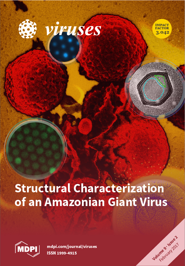
Cover Story (view full-size image): With the initial discovery of a giant virus, mimivirus, the notion of viruses being small and simple was shattered. Since this discovery, there has been an increasing number of unique giant viruses isolated each year. Due to the sheer size, it is extremely challenging to obtain structural information on these giants. Here, we present a structural characterization of Samba Virus, a Mimivirus isolated from the Rio Negro in Brazil, combining data from cryo-EM, cryo-ET, SEM, and fluorescence microscopy. Also, we compare the morphology of Samba Virus and mimivirus virions. Read Full Article
Order results
Result details
Section
Select all
Export citation of selected articles as:
7754 KiB  
Article
Evaluation of Rice Resistance to Southern Rice Black-Streaked Dwarf Virus and Rice Ragged Stunt Virus through Combined Field Tests, Quantitative Real-Time PCR, and Proteome Analysis
by Zhenchao Wang, Lu Yu, Linhong Jin, Wenli Wang, Qi Zhao, Longlu Ran, Xiangyang Li, Zhuo Chen, Rong Guo, Yongtian Wei, Zhongcheng Yang, Enlong Liu, Deyu Hu and Baoan Song
Viruses 2017, 9(2), 37; https://doi.org/10.3390/v9020037 - 22 Feb 2017
Cited by 10 | Viewed by 6376
Abstract
Diseases caused by southern rice black-streaked dwarf virus (SRBSDV) and rice ragged stunt virus (RRSV) considerably decrease grain yield. Therefore, determining rice cultivars with high resistance to SRBSDV and RRSV is necessary. In this study, rice cultivars with high resistance to SRBSDV and [...] Read more.
Diseases caused by southern rice black-streaked dwarf virus (SRBSDV) and rice ragged stunt virus (RRSV) considerably decrease grain yield. Therefore, determining rice cultivars with high resistance to SRBSDV and RRSV is necessary. In this study, rice cultivars with high resistance to SRBSDV and RRSV were evaluated through field trials in Shidian and Mangshi county, Yunnan province, China. SYBR Green I-based quantitative real-time polymerase chain reaction (qRT-PCR) analysis was used to quantitatively detect virus gene expression levels in different rice varieties. The following parameters were applied to evaluate rice resistance: acre yield (A.Y.), incidence of infected plants (I.I.P.), virus load (V.L.), disease index (D.I.), and insect quantity (I.Q.) per 100 clusters. Zhongzheyou1 (Z1) and Liangyou2186 (L2186) were considered the most suitable varieties with integrated higher A.Y., lower I.I.P., V.L., D.I. and I.Q. features. In order to investigate the mechanism of rice resistance, comparative label-free shotgun liquid chromatography tandem-mass spectrometry (LC-MS/MS) proteomic approaches were applied to comprehensively describe the proteomics of rice varieties’ SRBSDV tolerance. Systemic acquired resistance (SAR)-related proteins in Z1 and L2186 may result in the superior resistance of these varieties compared with Fengyouxiangzhan (FYXZ). Full article
(This article belongs to the Section Viruses of Plants, Fungi and Protozoa)
Show Figures

Figure 1

1354 KiB  
Article
A3R Phage and Staphylococcus aureus Lysate Do Not Induce Neutrophil Degranulation
by Jan Borysowski, Ryszard Międzybrodzki, Piotr Wierzbicki, Danuta Kłosowska, Grażyna Korczak-Kowalska, Beata Weber-Dąbrowska and Andrzej Górski
Viruses 2017, 9(2), 36; https://doi.org/10.3390/v9020036 - 21 Feb 2017
Cited by 22 | Viewed by 5406
Abstract
The objective of this study was to evaluate the effects of A3R phage and Staphylococcus aureus lysate obtained after phage infection on neutrophil degranulation. The exocytosis of primary and secondary granules from neutrophils was investigated in vitro in whole blood specimens by flow [...] Read more.
The objective of this study was to evaluate the effects of A3R phage and Staphylococcus aureus lysate obtained after phage infection on neutrophil degranulation. The exocytosis of primary and secondary granules from neutrophils was investigated in vitro in whole blood specimens by flow cytometry based on the expression of specific markers of exocytosis (CD63 for primary granules and CD66b for secondary granules). We found that both A3R and S. aureus lysate had no significant effect on the exocytosis of primary and secondary granules. These data suggest that neither A3R virions nor any products of phage-induced lysis of S. aureus are likely to induce neutrophil degranulation in patients who are treated with phage preparations. Since neutrophil granules contain some potentially toxic proteins, our results provide an important argument for the safety of phage therapy. Moreover, these data indicate that the induction of neutrophil degranulation is not likely to contribute to antibacterial effects of phages. Full article
(This article belongs to the Section Bacterial Viruses)
Show Figures

Figure 1

2417 KiB  
Article
PreC and C Regions of Woodchuck Hepatitis Virus Facilitate Persistent Expression of Surface Antigen of Chimeric WHV-HBV Virus in the Hydrodynamic Injection BALB/c Mouse Model
by Weimin Wu, Yan Liu, Yong Lin, Danzhen Pan, Dongliang Yang, Mengji Lu and Yang Xu
Viruses 2017, 9(2), 35; https://doi.org/10.3390/v9020035 - 21 Feb 2017
Viewed by 4960
Abstract
In the hydrodynamic injection (HI) BALB/c mouse model with the overlength viral genome, we have found that woodchuck hepatitis virus (WHV) could persist for a prolonged period of time (up to 45 weeks), while hepatitis B virus (HBV) was mostly cleared at week [...] Read more.
In the hydrodynamic injection (HI) BALB/c mouse model with the overlength viral genome, we have found that woodchuck hepatitis virus (WHV) could persist for a prolonged period of time (up to 45 weeks), while hepatitis B virus (HBV) was mostly cleared at week four. In this study, we constructed a series of chimeric genomes based on HBV and WHV, in which the individual sequences of a 1.3-fold overlength HBV genome in pBS-HBV1.3 were replaced by their counterparts from WHV. After HI with the WHV-HBV chimeric constructs in BALB/c mice, serum viral antigen, viral DNA (vDNA), and intrahepatic viral antigen expression were analyzed to evaluate the persistence of the chimeric genomes. Interestingly, we found that HI with three chimeric WHV-HBV genomes resulted in persistent antigenemia in mice. All of the persistent chimeric genomes contained the preC region and the part of the C region encoding the N-terminal 1–145 amino acids of the WHV genome. These results indicated that the preC region and the N-terminal part of the C region of the WHV genome may play a role in the persistent antigenemia. The chimeric WHV-HBV genomes were able to stably express viral antigens in the liver and could be further used to express hepadnaviral antigens to study their pathogenic potential. Full article
Show Figures

Figure 1

682 KiB  
Review
Bat Astroviruses: Towards Understanding the Transmission Dynamics of a Neglected Virus Family
by Kerstin Fischer, Vinícius Pinho dos Reis and Anne Balkema-Buschmann
Viruses 2017, 9(2), 34; https://doi.org/10.3390/v9020034 - 21 Feb 2017
Cited by 32 | Viewed by 6762
Abstract
Bats belong to the order Chiroptera that represents the second largest order of mammals with more than 1200 species and an almost global distribution. Environmental changes and deforestation have severely influenced many ecosystems, intensifying the contact between wildlife and humans. In recent years, [...] Read more.
Bats belong to the order Chiroptera that represents the second largest order of mammals with more than 1200 species and an almost global distribution. Environmental changes and deforestation have severely influenced many ecosystems, intensifying the contact between wildlife and humans. In recent years, bats have been found to harbor a number of different viruses with zoonotic potential, as well as a great diversity of astroviruses, for which the question of zoonotic potential remains unanswered to date. Human astroviruses have been identified as the causative agent for diarrhea in children and immunocompromised patients. For a long time, astroviruses have been considered to be strictly species-specific. However, a great genetic diversity has recently been discovered among animal and human astroviruses that might indicate the potential of these viruses to cross species barriers. Furthermore, our knowledge about the tissue tropism of astroviruses has been expanded to some neurotropic strains that have recently been shown to be responsible for encephalitis in humans and livestock. This review gives an overview on what is known about astroviruses in bats, humans and livestock, especially bovines and pigs. Future research activities are suggested to unravel astrovirus infection dynamics in bat populations to further assess the zoonotic potential of these viruses. Full article
(This article belongs to the Special Issue Astroviruses)
Show Figures

Figure 1

2599 KiB  
Review
Epidemiology of Classic and Novel Human Astrovirus: Gastroenteritis and Beyond
by Diem-Lan Vu, Albert Bosch, Rosa M. Pintó and Susana Guix
Viruses 2017, 9(2), 33; https://doi.org/10.3390/v9020033 - 18 Feb 2017
Cited by 140 | Viewed by 12428
Abstract
Since they were identified in 1975, human astroviruses have been considered one of the most important agents of viral acute gastroenteritis in children. However, highly divergent astroviruses infecting humans have been recently discovered and associated with extra-intestinal infections. The report of cases of [...] Read more.
Since they were identified in 1975, human astroviruses have been considered one of the most important agents of viral acute gastroenteritis in children. However, highly divergent astroviruses infecting humans have been recently discovered and associated with extra-intestinal infections. The report of cases of fatal meningitis and encephalitis, especially in immunocompromised individuals, has broadened their disease spectrum. Although zoonotic transmission among animal and human astroviruses has not been clearly recognized, the genetic similarity between some human and animal viruses makes it likely to occur. This review provides an update on the epidemiology of both classic and novel human astroviruses, and a comprehensive view on confirmed or potential association between astrovirus and human disease. Full article
(This article belongs to the Special Issue Astroviruses)
Show Figures

Figure 1

3242 KiB  
Review
HCIV-1 and Other Tailless Icosahedral Internal Membrane-Containing Viruses of the Family Sphaerolipoviridae
by Tatiana A. Demina, Maija K. Pietilä, Julija Svirskaitė, Janne J. Ravantti, Nina S. Atanasova, Dennis H. Bamford and Hanna M. Oksanen
Viruses 2017, 9(2), 32; https://doi.org/10.3390/v9020032 - 18 Feb 2017
Cited by 24 | Viewed by 7540
Abstract
Members of the virus family Sphaerolipoviridae include both archaeal viruses and bacteriophages that possess a tailless icosahedral capsid with an internal membrane. The genera Alpha- and Betasphaerolipovirus comprise viruses that infect halophilic euryarchaea, whereas viruses of thermophilic Thermus bacteria belong to the genus [...] Read more.
Members of the virus family Sphaerolipoviridae include both archaeal viruses and bacteriophages that possess a tailless icosahedral capsid with an internal membrane. The genera Alpha- and Betasphaerolipovirus comprise viruses that infect halophilic euryarchaea, whereas viruses of thermophilic Thermus bacteria belong to the genus Gammasphaerolipovirus. Both sequence-based and structural clustering of the major capsid proteins and ATPases of sphaerolipoviruses yield three distinct clades corresponding to these three genera. Conserved virion architectural principles observed in sphaerolipoviruses suggest that these viruses belong to the PRD1-adenovirus structural lineage. Here we focus on archaeal alphasphaerolipoviruses and their related putative proviruses. The highest sequence similarities among alphasphaerolipoviruses are observed in the core structural elements of their virions: the two major capsid proteins, the major membrane protein, and a putative packaging ATPase. A recently described tailless icosahedral haloarchaeal virus, Haloarcula californiae icosahedral virus 1 (HCIV-1), has a double-stranded DNA genome and an internal membrane lining the capsid. HCIV-1 shares significant similarities with the other tailless icosahedral internal membrane-containing haloarchaeal viruses of the family Sphaerolipoviridae. The proposal to include a new virus species, Haloarcula virus HCIV1, into the genus Alphasphaerolipovirus was submitted to the International Committee on Taxonomy of Viruses (ICTV) in 2016. Full article
(This article belongs to the Special Issue Viruses of Microbes)
Show Figures

Figure 1

3119 KiB  
Article
Genetic Assessment of African Swine Fever Isolates Involved in Outbreaks in the Democratic Republic of Congo between 2005 and 2012 Reveals Co-Circulation of p72 Genotypes I, IX and XIV, Including 19 Variants
by Leopold K. Mulumba–Mfumu, Jenna E. Achenbach, Matthew R. Mauldin, Linda K. Dixon, Curé Georges Tshilenge, Etienne Thiry, Noelia Moreno, Esther Blanco, Claude Saegerman, Charles E. Lamien and Adama Diallo
Viruses 2017, 9(2), 31; https://doi.org/10.3390/v9020031 - 18 Feb 2017
Cited by 52 | Viewed by 8052
Abstract
African swine fever (ASF) is a devastating disease of domestic pigs. It is a socioeconomically important disease, initially described from Kenya, but subsequently reported in most Sub-Saharan countries. ASF spread to Europe, South America and the Caribbean through multiple introductions which were initially [...] Read more.
African swine fever (ASF) is a devastating disease of domestic pigs. It is a socioeconomically important disease, initially described from Kenya, but subsequently reported in most Sub-Saharan countries. ASF spread to Europe, South America and the Caribbean through multiple introductions which were initially eradicated—except for Sardinia—followed by re‑introduction into Europe in 2007. In this study of ASF within the Democratic Republic of the Congo, 62 domestic pig samples, collected between 2005–2012, were examined for viral DNA and sequencing at multiple loci: C-terminus of the B646L gene (p72 protein), central hypervariable region (CVR) of the B602L gene, and the E183L gene (p54 protein). Phylogenetic analyses identified three circulating genotypes: I (64.5% of samples), IX (32.3%), and XIV (3.2%). This is the first evidence of genotypes IX and XIV within this country. Examination of the CVR revealed high levels of intra-genotypic variation, with 19 identified variants. Full article
(This article belongs to the Special Issue Porcine Viruses)
Show Figures

Figure 1

1522 KiB  
Article
Prevalence and Clinical Impact of Human Pegivirus-1 Infection in HIV-1-Infected Individuals in Yunnan, China
by Zhijiang Miao, Li Gao, Yindi Song, Ming Yang, Mi Zhang, Jincheng Lou, Yue Zhao, Xicheng Wang, Yue Feng, Xingqi Dong and Xueshan Xia
Viruses 2017, 9(2), 28; https://doi.org/10.3390/v9020028 - 15 Feb 2017
Cited by 29 | Viewed by 6393
Abstract
Human Pegivirus-1 (HPgV-1) may have a beneficial impact on disease progression in human immunodeficiency virus-1 (HIV-1) infection. However, analysis of the genotypic diversity of HPgV-1 and its relevance to the progression of HIV-1 disease remains limited. A total of 1062 HIV-1-infected individuals were [...] Read more.
Human Pegivirus-1 (HPgV-1) may have a beneficial impact on disease progression in human immunodeficiency virus-1 (HIV-1) infection. However, analysis of the genotypic diversity of HPgV-1 and its relevance to the progression of HIV-1 disease remains limited. A total of 1062 HIV-1-infected individuals were recruited in all sixteen prefectures of Yunnan province, China. The reverse transcription nested polymerase chain reaction (RT-nPCR), phylogenetic analyses, and clinical data analyses were used to detect HPgV-1 infection, determine genotype, and analyze HPgV-1 genotype impact on HIV-1 disease progression. The overall positive rate of HPgV-1 RNA was 23.4% (248/1062), and the frequency of HPgV-1 infection in injecting drug users (IDUs) (28.5%, 131/460) was significantly higher than in heterosexuals (19.4%, 117/602). Multiple genotypes were identified in 212 subjects with successful sequencing for the E2 gene, including genotype 7 (55.7%), genotype 3 (34.9%), genotype 4 (4.7%), genotype 2 (3.3%), and an unclassified group (1.4%). Moreover, genotype 7 predominated in IDUs, whereas genotype 3 was the most common in heterosexuals. Our results revealed that HPgV-1 genotype 7 groups exhibited significantly lower HIV-1 viral load and higher CD4+ cell counts. This finding suggests that HPgV-1 genotype 7 may be associated with a better progression of HIV-1 disease. Full article
(This article belongs to the Special Issue Advances in Flavivirus Research)
Show Figures

Figure 1

61439 KiB  
Article
Microscopic Characterization of the Brazilian Giant Samba Virus
by Jason R. Schrad, Eric J. Young, Jônatas S. Abrahão, Juliana R. Cortines and Kristin N. Parent
Viruses 2017, 9(2), 30; https://doi.org/10.3390/v9020030 - 14 Feb 2017
Cited by 14 | Viewed by 12874
Abstract
Prior to the discovery of the mimivirus in 2003, viruses were thought to be physically small and genetically simple. Mimivirus, with its ~750-nm particle size and its ~1.2-Mbp genome, shattered these notions and changed what it meant to be a virus. Since this [...] Read more.
Prior to the discovery of the mimivirus in 2003, viruses were thought to be physically small and genetically simple. Mimivirus, with its ~750-nm particle size and its ~1.2-Mbp genome, shattered these notions and changed what it meant to be a virus. Since this discovery, the isolation and characterization of giant viruses has exploded. One of the more recently discovered giant viruses, Samba virus, is a Mimivirus that was isolated from the Rio Negro in the Brazilian Amazon. Initial characterization of Samba has revealed some structural information, although the preparation techniques used are prone to the generation of structural artifacts. To generate more native-like structural information for Samba, we analyzed the virus through cryo-electron microscopy, cryo-electron tomography, scanning electron microscopy, and fluorescence microscopy. These microscopy techniques demonstrated that Samba particles have a capsid diameter of ~527 nm and a fiber length of ~155 nm, making Samba the largest Mimivirus yet characterized. We also compared Samba to a fiberless mimivirus variant. Samba particles, unlike those of mimivirus, do not appear to be rigid, and quasi-icosahedral, although the two viruses share many common features, including a multi-layered capsid and an asymmetric nucleocapsid, which may be common amongst the Mimiviruses. Full article
(This article belongs to the Special Issue Viruses of Protozoa)
Show Figures

Figure 1

548 KiB  
Review
A Review of the Strain Diversity and Pathogenesis of Chicken Astrovirus
by Victoria J. Smyth
Viruses 2017, 9(2), 29; https://doi.org/10.3390/v9020029 - 10 Feb 2017
Cited by 57 | Viewed by 7358
Abstract
Although a relatively recently emerged virus, identified only in 2004 as a separate species of avian astrovirus, chicken astrovirus (CAstV) has been associated with poor growth of broiler flocks, enteritis and diarrhea and is a candidate pathogen in cases of runting stunting syndrome. [...] Read more.
Although a relatively recently emerged virus, identified only in 2004 as a separate species of avian astrovirus, chicken astrovirus (CAstV) has been associated with poor growth of broiler flocks, enteritis and diarrhea and is a candidate pathogen in cases of runting stunting syndrome. More recently CAstV has been implicated in cases of two other diseases of broilers as the sole etiological agent, namely severe kidney disease of young broilers with visceral gout and the “White Chicks” hatchery disease. Examination of the strains of CAstV associated with the two latter diseases reveals they are closely related genetically. This review will discuss the pathogenesis of CAstV in relation to strain diversity and the effects of vertical versus horizontal transmission, virus load, co-infections and age of bird at infection, all factors that may impact upon disease severity. Full article
(This article belongs to the Special Issue Astroviruses)
Show Figures

Figure 1

3170 KiB  
Article
Myxoma Virus dsRNA Binding Protein M029 Inhibits the Type I IFN‐Induced Antiviral State in a Highly Species‐Specific Fashion
by Masmudur M. Rahman and Grant McFadden
Viruses 2017, 9(2), 27; https://doi.org/10.3390/v9020027 - 2 Feb 2017
Cited by 10 | Viewed by 5293
Abstract
Myxoma virus (MYXV) is Leporipoxvirus that possesses a specific rabbit‐restricted host tropism but exhibits a much broader cellular host range in cultured cells. MYXV is able to efficiently block all aspects of the type I interferon (IFN)‐induced antiviral state in rabbit cells, partially [...] Read more.
Myxoma virus (MYXV) is Leporipoxvirus that possesses a specific rabbit‐restricted host tropism but exhibits a much broader cellular host range in cultured cells. MYXV is able to efficiently block all aspects of the type I interferon (IFN)‐induced antiviral state in rabbit cells, partially in human cells and very poorly in mouse cells. The mechanism(s) of this species‐specific inhibition of type I IFN‐induced antiviral state is not well understood. Here we demonstrate that MYXV encoded protein M029, a truncated relative of the vaccinia virus (VACV) E3 double‐stranded RNA (dsRNA) binding protein that inhibits protein kinase R (PKR), can also antagonize the type I IFN‐induced antiviral state in a highly species‐specific manner. In cells pre‐treated with type I IFN prior to infection, MYXV exploits M029 to overcome the induced antiviral state completely in rabbit cells, partially in human cells, but not at all in mouse cells. However, in cells pre‐infected with MYXV, IFN‐induced signaling is fully inhibited even in the absence of M029 in cells from all three species, suggesting that other MYXV protein(s) apart from M029 block IFN signaling in a speciesindependent manner. We also show that the antiviral state induced in rabbit, human or mouse cells by type I IFN can inhibit M029‐knockout MYXV even when PKR is genetically knocked‐out, suggesting that M029 targets other host proteins for this antiviral state inhibition. Thus, the MYXV dsRNA binding protein M029 not only antagonizes PKR from multiple species but also blocks the type I IFN antiviral state independently of PKR in a highly species‐specific fashion. Full article
(This article belongs to the Section Animal Viruses)
Show Figures

Figure 1

68762 KiB  
Article
The Characteristics of Herpes Simplex Virus Type 1 Infection in Rhesus Macaques and the Associated Pathological Features
by Shengtao Fan, Hongzhi Cai, Xingli Xu, Min Feng, Lichun Wang, Yun Liao, Ying Zhang, Zhanlong He, Fengmei Yang, Wenhai Yu, Jingjing Wang, Jumin Zhou and Qihan Li
Viruses 2017, 9(2), 26; https://doi.org/10.3390/v9020026 - 30 Jan 2017
Cited by 9 | Viewed by 9357
Abstract
As one of the major pathogens for human herpetic diseases, herpes simplex virus type 1 (HSV1) causes herpes labialis, genital herpes and herpetic encephalitis. Our aim here was to investigate the infectious process of HSV1 in rhesus macaques and the pathological features induced [...] Read more.
As one of the major pathogens for human herpetic diseases, herpes simplex virus type 1 (HSV1) causes herpes labialis, genital herpes and herpetic encephalitis. Our aim here was to investigate the infectious process of HSV1 in rhesus macaques and the pathological features induced during this infection. Clinical symptoms that manifested in the rhesus macaque during HSV1 infection included vesicular lesions and their pathological features. Viral distribution in the nervous tissues and associated pathologic changes indicated the typical systematic pathological processes associated with viral distribution of HSV1.Interestingly, vesicular lesions recurred in oral skin or in mucosa associated with virus shedding in macaques within four to five months post‐infection,and viral latency‐associated transcript (LAT) mRNA was found in the trigeminal ganglia (TG)on day 365 post‐infection. Neutralization testing and enzyme‐linked immunospot (ELISpot) detection of specific T cell responses confirmed the specific immunity induced by HSV1 infection. Thus, rhesus macaques could serve as an infectious model for HSV1 due to their typical clinical symptoms and the pathological recurrence associated with viral latency in nervous tissues. Full article
(This article belongs to the Section Animal Viruses)
Show Figures

Figure 1

2908 KiB  
Article
Echovirus 6 Infects Human Exocrine and Endocrine Pancreatic Cells and Induces Pro-Inflammatory Innate Immune Response
by Luis Sarmiento, Gun Frisk, Mahesh Anagandula, Monika Hodik, Ilaria Barchetta, Eitan Netanyah, Eduardo Cabrera-Rode and Corrado M. Cilio
Viruses 2017, 9(2), 25; https://doi.org/10.3390/v9020025 - 30 Jan 2017
Cited by 9 | Viewed by 8603
Abstract
Human enteroviruses (HEV), especially coxsackievirus serotype B (CVB) and echovirus (E), have been associated with diseases of both the exocrine and endocrine pancreas, but so far evidence on HEV infection in human pancreas has been reported only in islets and ductal cells. This [...] Read more.
Human enteroviruses (HEV), especially coxsackievirus serotype B (CVB) and echovirus (E), have been associated with diseases of both the exocrine and endocrine pancreas, but so far evidence on HEV infection in human pancreas has been reported only in islets and ductal cells. This study aimed to investigate the capability of echovirus strains to infect human exocrine and endocrine pancreatic cells. Infection of explanted human islets and exocrine cells with seven field strains of E6 caused cytopathic effect, virus titer increase and production of HEV protein VP1 in both cell types. Virus particles were found in islets and acinar cells infected with E6. No cytopathic effect or infectious progeny production was observed in exocrine cells exposed to the beta cell-tropic strains of E16 and E30. Endocrine cells responded to E6, E16 and E30 by upregulating the transcription of interferon-induced with helicase C domain 1 (IF1H1), 2'-5'-oligoadenylate synthetase 1 (OAS1), interferon-β (IFN-β), chemokine (C–X–C motif) ligand 10 (CXCL10) and chemokine (C–C motif) ligand 5 (CCL5). Echovirus 6, but not E16 or E30, led to increased transcription of these genes in exocrine cells. These data demonstrate for the first time that human exocrine cells represent a target for E6 infection and suggest that certain HEV serotypes can replicate in human pancreatic exocrine cells, while the pancreatic endocrine cells are permissive to a wider range of HEV. Full article
(This article belongs to the Section Animal Viruses)
Show Figures

Graphical abstract

Previous Issue
Next Issue
Back to TopTop