Next Issue
Volume 15, November-1
Previous Issue
Volume 15, October-1


Cancers, Volume 15, Issue 20 (October-2 2023) – 208 articles

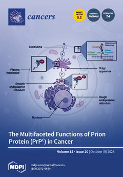
Cover Story (view full-size image): The present review summarizes the multiple roles that the cellular prion protein (PrPC) plays in different cancer settings and highlights its role in the development and progression of epithelial cancers, including gastric, melanoma, breast, colorectal and pancreatic, as well as rare cancers. The review also suggests the consideration of the PrPC protein and its associated proteins as potential targets to treat resistance-associated cancers to conventional treatments. In addition, the review reports the molecular mechanisms by which PrPC triggers cancer development and/or contributes to its aggressiveness. The review strongly suggests that further comprehensive studies, focused on the discovery of PrPC-based therapy, may open up exciting new avenues for cancer research. View this paper
  • Issues are regarded as officially published after their release is announced to the table of contents alert mailing list.
  • You may sign up for e-mail alerts to receive table of contents of newly released issues.
  • PDF is the official format for papers published in both, html and pdf forms. To view the papers in pdf format, click on the "PDF Full-text" link, and use the free Adobe Reader to open them.
Order results
Result details
Section
Select all
Export citation of selected articles as:
16 pages, 5741 KiB  
Article
Endotoxin Tolerance Creates Favourable Conditions for Cancer Development
by Konkonika Roy, Henryk Mikołaj Kozłowski, Tomasz Jędrzejewski, Justyna Sobocińska, Bartosz Maciejewski, Artur Dzialuk and Sylwia Wrotek
Cancers 2023, 15(20), 5113; https://doi.org/10.3390/cancers15205113 - 23 Oct 2023
Cited by 4 | Viewed by 2380
Abstract
Endotoxin tolerance (ET) is an adaptive phenomenon of the immune system that protects the host from clinical complications due to repeated exposure of the body to endotoxins such as lipopolysaccharide (LPS). Since ET is an immunosuppressive mechanism in which a significant reprogramming of [...] Read more.
Endotoxin tolerance (ET) is an adaptive phenomenon of the immune system that protects the host from clinical complications due to repeated exposure of the body to endotoxins such as lipopolysaccharide (LPS). Since ET is an immunosuppressive mechanism in which a significant reprogramming of macrophages is observed, we hypothesized that it could influence cancer development by modifying the tumour environment. This study aimed to explore whether ET influences cancer progression by altering the tumour microenvironment. Endotoxin-tolerant macrophages (MoET) were examined for their impact on breast and colon cancer cells via direct interaction and conditioned media exposure. We characterized cancer cell behaviour by viability, clonogenic potential, motility, scratch assays, and 3D spheroidal assays. MoET-derived factors increased cancer cell viability, motility, and clonogenicity, suggesting a conducive environment for cancer development. Remarkably, despite reduced TNFα and IL-6 levels, MoET exhibited M1 polarization. These findings uncover an ET-associated macrophage reprogramming that fosters a favourable context for cancer progression across diverse tumours. Targeting ET could emerge as a promising avenue for cancer therapy and prevention. Full article
(This article belongs to the Special Issue Macrophages in Cancer Progression, Diagnosis and Treatment)
Show Figures

Figure 1

20 pages, 6735 KiB  
Article
Comparative Analysis of the GNAI Family Genes in Glioblastoma through Transcriptomics and Single-Cell Technologies
by Ahmad Raza, Meng-Chi Yen, Gangga Anuraga, Iram Shahzadi, Muhammad Waqar Mazhar, Hoang Dang Khoa Ta, Do Thi Minh Xuan, Sanskriti Dey, Sachin Kumar, Adrian Wangsawijaya Santoso, Bianca Tobias William and Chih-Yang Wang
Cancers 2023, 15(20), 5112; https://doi.org/10.3390/cancers15205112 - 23 Oct 2023
Cited by 3 | Viewed by 2386
Abstract
Glioblastoma multiforme (GBM) is one of the most aggressive cancers with a low overall survival rate. The treatment of GBM is challenging due to the presence of the blood–brain barrier (BBB), which hinders drug delivery. Invasive procedures alone are not effective at completely [...] Read more.
Glioblastoma multiforme (GBM) is one of the most aggressive cancers with a low overall survival rate. The treatment of GBM is challenging due to the presence of the blood–brain barrier (BBB), which hinders drug delivery. Invasive procedures alone are not effective at completely removing such tumors. Hence, identifying the crucial pathways and biomarkers for the treatment of GBM is of prime importance. We conducted this study to identify the pathways associated with GBM. We used The Cancer Genome Atlas (TCGA) GBM genomic dataset to identify differentially expressed genes (DEGs). We investigated the prognostic values of the guanine nucleotide-binding protein G(i) alpha subunit (GNAI) family of genes in GBM using a Chinese Glioma Genome Atlas (CGGA) dataset. Within this dataset, we observed the association in the tumor microenvironment between the gene expression of GNAI subunit 3 (GNAI3) and a poor prognosis. MetaCore and gene ontology (GO) analyses were conducted to explore the role of GNAI3 in co-expressed genes and associated signaling pathways using a transcript analysis. Notable pathways included “Cytoskeleton remodeling regulation of actin cytoskeleton organization by the kinase effectors of Rho GTPases” and “Immune response B cell antigen receptor (BCR) pathway”. A single-cell analysis was used to assess GNAI3 expression in GBM. The results demonstrated that GNAI family genes, specifically GNAI3, were significantly associated with carcinogenesis and malignancy in GBM patients. Our findings suggest that the GNAI3 gene holds potential as a prognostic biomarker for GBM. Full article
(This article belongs to the Special Issue Invasion in Glioblastoma)
Show Figures

Figure 1

12 pages, 227 KiB  
Review
Cerebral Vasculopathy in Children with Neurofibromatosis Type 1
by Laura L. Lehman and Nicole J. Ullrich
Cancers 2023, 15(20), 5111; https://doi.org/10.3390/cancers15205111 - 23 Oct 2023
Cited by 2 | Viewed by 1358
Abstract
Cerebrovascular abnormalities are a severe and often underrecognized complication of childhood neurofibromatosis type 1 (NF1). There are no prospective studies of cerebral vasculopathy in NF1; thus, the estimated frequency of vasculopathy varies between studies. The data is difficult to interpret due to the [...] Read more.
Cerebrovascular abnormalities are a severe and often underrecognized complication of childhood neurofibromatosis type 1 (NF1). There are no prospective studies of cerebral vasculopathy in NF1; thus, the estimated frequency of vasculopathy varies between studies. The data is difficult to interpret due to the retrospective data collection and variability in whether imaging is done based on screening/surveillance or due to acute neurologic symptoms. The prevalent NF1-associated cerebral vasculopathy is moyamoya syndrome (MMS). Vascular changes can present without symptoms or with acute TIA or stroke-like symptoms or a range of progressive neurologic deficits. Advanced imaging may enhance sensitivity of neuroimaging in children. Medical and/or surgical interventions may prevent short- and long-term complications. Challenges for establishment of a screening protocol for cerebral vasculopathy in children with NF1 include the relatively large number of patients with NF1, the potential need for sedation to achieve quality imaging and the broad age range at time of detection for cerebral vascular changes. The goal of this review is to present the epidemiology, clinical presentation, imaging features and medical/surgical management of cerebral arteriopathies in children with NF1. Full article
(This article belongs to the Special Issue Frontiers in Neurofibromatosis)
3 pages, 166 KiB  
Editorial
Personalized Therapy of Sarcomas
by Ingrid M. E. Desar and Christian Rothermundt
Cancers 2023, 15(20), 5110; https://doi.org/10.3390/cancers15205110 - 23 Oct 2023
Cited by 1 | Viewed by 1036
Abstract
Sarcomas are a group of rare malignant mesenchymal tumors [...] Full article
(This article belongs to the Special Issue Personalized Therapy of Sarcomas)
10 pages, 1595 KiB  
Article
Defining MRI Superiority over CT for Colorectal and Neuroendocrine Liver Metastases
by Marc A. Attiyeh, Gautam K. Malhotra, Daneng Li, Saro B. Manoukian, Pejman M. Motarjem and Gagandeep Singh
Cancers 2023, 15(20), 5109; https://doi.org/10.3390/cancers15205109 - 23 Oct 2023
Viewed by 1654
Abstract
Background: We compared CT and MRI for staging metastatic colorectal or neuroendocrine liver metastases (CRLMs and NELMs, respectively) to assess their impact on tumor burden. Methods: A prospectively maintained database was queried for patients who underwent both imaging modalities within 3 months, with [...] Read more.
Background: We compared CT and MRI for staging metastatic colorectal or neuroendocrine liver metastases (CRLMs and NELMs, respectively) to assess their impact on tumor burden. Methods: A prospectively maintained database was queried for patients who underwent both imaging modalities within 3 months, with two blinded radiologists (R1 and R2) independently assessing the images for liver lesions. To minimize recall bias, studies were grouped by modality, and were randomized and evaluated separately. Results: Our query yielded 76 patients (42 CRLMs; 34 NELMs) with low interrater variability (intraclass correlation coefficients: CT = 0.941, MRI = 0.975). For CRLMs, there were no significant differences in lesion number or size between CT and MRI. However, in NELMs, Eovist®-enhanced MRI detected more lesions (R1: 14.3 vs. 12.1, p = 0.02; R2: 14.4 vs. 12.4, p = 0.01) and smaller lesions (R1: 5.7 vs. 4.4, p = 0.03; R2: 4.8 vs. 2.9, p = 0.02) than CT. Conclusions: CT and MRI are equivalent for CRLMs, but for NELMs, MRI outperforms CT in detecting more and smaller lesions, potentially influencing treatment planning and surgery. Full article
(This article belongs to the Special Issue Neuroendocrine Tumors: Treatment and Management Volume II)
Show Figures

Figure 1

24 pages, 1261 KiB  
Review
Decoding the Impact of Tumor Microenvironment in Osteosarcoma Progression and Metastasis
by Bikesh K. Nirala, Taku Yamamichi, D. Isabel Petrescu, Tasnuva N. Shafin and Jason T. Yustein
Cancers 2023, 15(20), 5108; https://doi.org/10.3390/cancers15205108 - 23 Oct 2023
Cited by 10 | Viewed by 2503
Abstract
Osteosarcoma (OS) is a heterogeneous, highly metastatic bone malignancy in children and adolescents. Despite advancements in multimodal treatment strategies, the prognosis for patients with metastatic or recurrent disease has not improved significantly in the last four decades. OS is a highly heterogeneous tumor; [...] Read more.
Osteosarcoma (OS) is a heterogeneous, highly metastatic bone malignancy in children and adolescents. Despite advancements in multimodal treatment strategies, the prognosis for patients with metastatic or recurrent disease has not improved significantly in the last four decades. OS is a highly heterogeneous tumor; its genetic background and the mechanism of oncogenesis are not well defined. Unfortunately, no effective molecular targeted therapy is currently available for this disease. Understanding osteosarcoma’s tumor microenvironment (TME) has recently gained much interest among scientists hoping to provide valuable insights into tumor heterogeneity, progression, metastasis, and the identification of novel therapeutic avenues. Here, we review the current understanding of the TME of OS, including different cellular and noncellular components, their crosstalk with OS tumor cells, and their involvement in tumor progression and metastasis. We also highlight past/current clinical trials targeting the TME of OS for effective therapies and potential future therapeutic strategies with negligible adverse effects. Full article
(This article belongs to the Special Issue Osteosarcoma Microenvironment)
Show Figures

Figure 1

16 pages, 618 KiB  
Review
Regional Immunotherapy for Peritoneal Carcinomatosis in Gastroesophageal Cancer: Emerging Strategies to Re-Condition a Maladaptive Tumor Environment
by Catherine R. Lewis, Neda Dadgar, Samuel A. Yellin, Vera S. Donnenberg, Albert D. Donnenberg, David L. Bartlett, Casey J. Allen and Patrick L. Wagner
Cancers 2023, 15(20), 5107; https://doi.org/10.3390/cancers15205107 - 23 Oct 2023
Cited by 2 | Viewed by 2335
Abstract
Peritoneal carcinomatosis originating from gastric/gastroesophageal junction cancer (GC-PC) occurs in a defined subset of gastric cancer patients with unique clinical, pathologic, molecular and immunologic characteristics that create significant obstacles to effective treatment with modern therapy. Although systemic chemo- and immuno- therapy have yielded [...] Read more.
Peritoneal carcinomatosis originating from gastric/gastroesophageal junction cancer (GC-PC) occurs in a defined subset of gastric cancer patients with unique clinical, pathologic, molecular and immunologic characteristics that create significant obstacles to effective treatment with modern therapy. Although systemic chemo- and immuno- therapy have yielded disappointing results in GC-PC, recent advances in the characterization of GC-PC and peritoneal immune biology present new opportunities for targeted therapeutics. In this review article, we discuss the distinct properties of GC-PC and the peritoneal immune environment as they pertain to current and investigative treatment strategies. We discuss pre-clinical studies and clinical trials relevant to the modulation of the peritoneal environment as a therapeutic intervention in GC-PC. Finally, we present a road map for future combinatorial strategies based on the conception of the peritoneal cavity as a bioreactor. Within this isolated compartment, prevailing immunosuppressive conditions can be altered through regional interventions toward an adaptive phenotype that would support the effectiveness of regionally delivered cellular therapy products. It is hoped that novel combination strategies would promote efficacy not only in the sequestered peritoneal environment, but also via migration into the circulation of tumor-reactive lymphocytes to produce durable systemic disease control, thereby improving oncologic outcome and quality of life in patients with GC-PC. Full article
Show Figures

Figure 1

22 pages, 3012 KiB  
Article
Combining Mass Cytometry Data by CyTOFmerge Reveals Additional Cell Phenotypes in the Heterogeneous Ovarian Cancer Tumor Microenvironment: A Pilot Study
by Liv Cecilie Vestrheim Thomsen, Katrin Kleinmanns, Shamundeeswari Anandan, Stein-Erik Gullaksen, Tamim Abdelaal, Grete Alrek Iversen, Lars Andreas Akslen, Emmet McCormack and Line Bjørge
Cancers 2023, 15(20), 5106; https://doi.org/10.3390/cancers15205106 - 23 Oct 2023
Cited by 1 | Viewed by 1510
Abstract
The prognosis of high-grade serous ovarian carcinoma (HGSOC) is poor, and treatment selection is challenging. A heterogeneous tumor microenvironment (TME) characterizes HGSOC and influences tumor growth, progression, and therapy response. Better characterization with multidimensional approaches for simultaneous identification and categorization of the various [...] Read more.
The prognosis of high-grade serous ovarian carcinoma (HGSOC) is poor, and treatment selection is challenging. A heterogeneous tumor microenvironment (TME) characterizes HGSOC and influences tumor growth, progression, and therapy response. Better characterization with multidimensional approaches for simultaneous identification and categorization of the various cell populations is needed to map the TME complexity. While mass cytometry allows the simultaneous detection of around 40 proteins, the CyTOFmerge MATLAB algorithm integrates data sets and extends the phenotyping. This pilot study explored the potential of combining two datasets for improved TME phenotyping by profiling single-cell suspensions from ten chemo-naïve HGSOC tumors by mass cytometry. A 35-marker pan-tumor dataset and a 34-marker pan-immune dataset were analyzed separately and combined with the CyTOFmerge, merging 18 shared markers. While the merged analysis confirmed heterogeneity across patients, it also identified a main tumor cell subset, additionally to the nine identified by the pan-tumor panel. Furthermore, the expression of traditional immune cell markers on tumor and stromal cells was revealed, as were marker combinations that have rarely been examined on individual cells. This study demonstrates the potential of merging mass cytometry data to generate new hypotheses on tumor biology and predictive biomarker research in HGSOC that could improve treatment effectiveness. Full article
(This article belongs to the Topic Advances in Tumor Microenvironment)
Show Figures

Graphical abstract

20 pages, 9821 KiB  
Review
An Update on the Role of MRI in Treatment Stratification of Patients with Cervical Cancer
by Amreen Shakur, Janice Yu Ji Lee and Sue Freeman
Cancers 2023, 15(20), 5105; https://doi.org/10.3390/cancers15205105 - 23 Oct 2023
Cited by 6 | Viewed by 4999
Abstract
Cervical cancer is the fourth most common cancer in women worldwide and the most common gynaecological malignancy. The FIGO staging system is the most commonly utilised classification system for cervical cancer worldwide. Prior to the most recent update in the FIGO staging in [...] Read more.
Cervical cancer is the fourth most common cancer in women worldwide and the most common gynaecological malignancy. The FIGO staging system is the most commonly utilised classification system for cervical cancer worldwide. Prior to the most recent update in the FIGO staging in 2018, the staging was dependent upon clinical assessment alone. Concordance between the surgical and clinical FIGO staging decreases rapidly as the tumour becomes more advanced. MRI now plays a central role in patients diagnosed with cervical cancer and enables accurate staging, which is essential to determining the most appropriate treatment. MRI is the best imaging option for the assessment of tumour size, location, and parametrial and sidewall invasion. Notably, the presence of parametrial invasion precludes surgical options, and the patient will be triaged to chemoradiotherapy. As imaging is intrinsic to the new 2018 FIGO staging system, nodal metastases have been included within the classification as stage IIIC disease. The presence of lymph node metastases within the pelvis or abdomen is associated with a poorer prognosis, which previously could not be included in the staging classification as these could not be reliably detected on clinical examination. MRI findings corresponding to the 2018 revised FIGO staging of cervical cancers and their impact on treatment selection will be described. Full article
(This article belongs to the Special Issue Updates on Imaging of Common Urogenital Neoplasms)
Show Figures

Figure 1

22 pages, 4562 KiB  
Article
How Toll-like Receptor 9 Plays a Key Role in the Development of Gastric Cancer and Is Linked to Epstein–Barr Virus Infection
by Marek Majewski, Paulina Mertowska, Sebastian Mertowski, Kamil Torres and Ewelina Grywalska
Cancers 2023, 15(20), 5104; https://doi.org/10.3390/cancers15205104 - 23 Oct 2023
Cited by 2 | Viewed by 1344
Abstract
The relationship between Toll-like receptor 9 (TLR-9) signaling and its involvement with Epstein–Barr virus (EBV) in gastric cancer (GC) is complex and currently under study. This research intended to understand TLR-9’s role in certain T and B lymphocytes and the serum levels of [...] Read more.
The relationship between Toll-like receptor 9 (TLR-9) signaling and its involvement with Epstein–Barr virus (EBV) in gastric cancer (GC) is complex and currently under study. This research intended to understand TLR-9’s role in certain T and B lymphocytes and the serum levels of TLR-9 in GC patients versus healthy subjects. The team explored links between these immune markers and various GC traits, such as histological grade, tumor progression stages, cancer types, and survival rates. Additionally, the research sought to find if EBV genetic material influences these immune reactions. Using flow cytometry, TLR-9 levels in different immune cells were analyzed. At the same time, the amount of TLR-9 in the serum was determined. The results showed GC patients had varied TLR-9 levels compared to healthy subjects, with specific cells showing noticeable changes. When grouped by GC attributes, key relationships emerged between TLR-9 amounts, the histological grade, progression stages, and cancer types. A notable finding was the connection between TLR-9 levels and EBV genetic presence, suggesting possible interactions between TLR-9 responses and EBV-related GC processes. Survival data also hinted at TLR-9’s potential as a predictor linked to clinical traits. Overall, this research emphasizes TLR-9’s complex role in GC’s immune responses, pinpointing its interactions with particular cells, clinical features, and EBV. The study unveils a complex web affecting GC and paves the way for new treatment avenues targeting TLR-9 pathways. Full article
(This article belongs to the Special Issue Immunity and Gastrointestinal Cancer)
Show Figures

Figure 1

24 pages, 1835 KiB  
Review
Cell Membrane Sialome: Sialic Acids as Therapeutic Targets and Regulators of Drug Resistance in Human Cancer Management
by Patrycja Jastrząb, Karolina Narejko, Halina Car and Przemyslaw Wielgat
Cancers 2023, 15(20), 5103; https://doi.org/10.3390/cancers15205103 - 22 Oct 2023
Cited by 4 | Viewed by 3375
Abstract
A cellular sialome is a physiologically active and dynamically changing component of the cell membrane. Sialylation plays a crucial role in tumor progression, and alterations in cellular sialylation patterns have been described as modulators of chemotherapy effectiveness. However, the precise mechanisms through which [...] Read more.
A cellular sialome is a physiologically active and dynamically changing component of the cell membrane. Sialylation plays a crucial role in tumor progression, and alterations in cellular sialylation patterns have been described as modulators of chemotherapy effectiveness. However, the precise mechanisms through which altered sialylation contributes to drug resistance in cancer are not yet fully understood. This review focuses on the intricate interplay between sialylation and cancer treatment. It presents the role of sialic acids in modulating cell–cell interactions, the extracellular matrix (ECM), and the immunosuppressive processes within the context of cancer. The issue of drug resistance is also discussed, and the mechanisms that involve transporters, the tumor microenvironment, and metabolism are analyzed. The review explores drugs and therapeutic approaches that may induce modifications in sialylation processes with a primary focus on their impact on sialyltransferases or sialidases. Despite advancements in cellular glycobiology and glycoengineering, an interdisciplinary effort is required to decipher and comprehend the biological characteristics and consequences of altered sialylation. Additionally, understanding the modulatory role of sialoglycans in drug sensitivity is crucial to applying this knowledge in clinical practice for the benefit of cancer patients. Full article
(This article belongs to the Special Issue Cancer Chemotherapy Resistance)
Show Figures

Figure 1

19 pages, 5171 KiB  
Article
Naringin–Dextrin Nanocomposite Abates Diethylnitrosamine/Acetylaminofluorene-Induced Lung Carcinogenesis by Modulating Oxidative Stress, Inflammation, Apoptosis, and Cell Proliferation
by Eman E. Mohamed, Osama M. Ahmed, Khairy M. A. Zoheir, Ahmed A. G. El-Shahawy, Shadi Tamur, Anwar Shams, Jack T. Burcher, Anupam Bishayee and Adel Abdel-Moneim
Cancers 2023, 15(20), 5102; https://doi.org/10.3390/cancers15205102 - 22 Oct 2023
Cited by 3 | Viewed by 1882
Abstract
Nanotechnology has proven advantageous in numerous scientific applications, one being to enhance the delivery of chemotherapeutic agents. This present study aims to evaluate the mechanisms underlying the chemopreventive action of naringin–dextrin nanocomposites (Nar-Dx-NCs) against diethylnitrosamine (DEN)/2-acetylaminofluorene (2AAF)-induced lung carcinogenesis in male Wistar rats. [...] Read more.
Nanotechnology has proven advantageous in numerous scientific applications, one being to enhance the delivery of chemotherapeutic agents. This present study aims to evaluate the mechanisms underlying the chemopreventive action of naringin–dextrin nanocomposites (Nar-Dx-NCs) against diethylnitrosamine (DEN)/2-acetylaminofluorene (2AAF)-induced lung carcinogenesis in male Wistar rats. DEN was administered intraperitoneally (i.p.) (150 mg/kg/week) for two weeks, followed by the oral administration of 2AAF (20 mg/kg) four times a week for three weeks. Rats receiving DEN/2AAF were concurrently treated with naringin or Nar-Dx-NCs orally at a dose of 10 mg/kg every other day for 24 weeks. Naringin and Nar-Dx-NCs treatments prevented the formation of tumorigenic cells within the alveoli of rats exposed to DEN/2AAF. These findings were associated with a significant decrease in lipid peroxidation, upregulation of antioxidant enzyme (glutathione peroxidase and superoxide dismutase) activity, and enhanced glutathione and nuclear factor erythroid 2–related factor 2 expression in the lungs. Naringin and Nar-Dx-NCs exerted anti-inflammatory actions manifested by a decrease in lung protein expression of tumor necrosis factor-α and interleukin-1β and mRNA expression of interleukin-6, interferon-γ, nuclear factor-κB, and inducible nitric oxide synthase, with a concurrent increase in interleukin-10 expression. The anti-inflammatory effect of Nar-Dx-NCs was more potent than naringin. Regarding the effect on apoptosis, both naringin and Nar-Dx-NCs significantly reduced Bcl-2 and increased Bax and P53 expressions. Moreover, naringin or Nar-Dx-NCs induced a significant decrease in the expression of the proliferator marker, Ki-67, and the effect of Nar-Dx-NCs was more marked. In conclusion, Nar-Dx-NCs improved naringin’s preventive action against DEN/2AAF-induced lung cancer and exerted anticarcinogenic effects by suppressing oxidative stress and inflammation and improving apoptotic signal induction and propagation. Full article
Show Figures

Figure 1

16 pages, 5696 KiB  
Article
The Applications and Pitfalls of Cone-Beam Computed Tomography-Based Synthetic Computed Tomography for Adaptive Evaluation in Pencil-Beam Scanning Proton Therapy
by Pingfang Tsai, Yu-Lun Tseng, Brian Shen, Christopher Ackerman, Huifang A. Zhai, Francis Yu, Charles B. Simone II, J. Isabelle Choi, Nancy Y. Lee, Rafi Kabarriti, Stanislav Lazarev, Casey L. Johnson, Jiayi Liu, Chin-Cheng Chen and Haibo Lin
Cancers 2023, 15(20), 5101; https://doi.org/10.3390/cancers15205101 - 22 Oct 2023
Cited by 1 | Viewed by 1405
Abstract
Purpose: The study evaluates the efficacy of cone-beam computed tomography (CBCT)-based synthetic CTs (sCT) as a potential alternative to verification CT (vCT) for enhanced treatment monitoring and early adaptation in proton therapy. Methods: Seven common treatment sites were studied. Two sets of sCT [...] Read more.
Purpose: The study evaluates the efficacy of cone-beam computed tomography (CBCT)-based synthetic CTs (sCT) as a potential alternative to verification CT (vCT) for enhanced treatment monitoring and early adaptation in proton therapy. Methods: Seven common treatment sites were studied. Two sets of sCT per case were generated: direct-deformed (DD) sCT and image-correction (IC) sCT. The image qualities and dosimetric impact of the sCT were compared to the same-day vCT. Results: The sCT agreed with vCT in regions of homogeneous tissues such as the brain and breast; however, notable discrepancies were observed in the thorax and abdomen. The sCT outliers existed for DD sCT when there was an anatomy change and for IC sCT in low-density regions. The target coverage exhibited less than a 5% variance in most DD and IC sCT cases when compared to vCT. The Dmax of serial organ-at-risk (OAR) in sCT plans shows greater deviation from vCT than small-volume dose metrics (D0.1cc). The parallel OAR volumetric and mean doses remained consistent, with average deviations below 1.5%. Conclusion: The use of sCT enables precise treatment and prompt early adaptation for proton therapy. The quality assurance of sCT is mandatory in the early stage of clinical implementation. Full article
(This article belongs to the Collection Particle Therapy: State-of-the-Art and Future Prospects)
Show Figures

Figure 1

24 pages, 2601 KiB  
Review
Don’t Judge a Book by Its Cover: The Role of Statins in Liver Cancer
by Natalia Piekuś-Słomka, Lavinia Patricia Mocan, Rezarta Shkreli, Cristiana Grapă, Kinga Denkiewicz, Oliwia Wesolowska, Miroslaw Kornek, Zeno Spârchez, Artur Słomka, Rareș Crăciun and Tudor Mocan
Cancers 2023, 15(20), 5100; https://doi.org/10.3390/cancers15205100 - 22 Oct 2023
Cited by 4 | Viewed by 2472
Abstract
Statins, which are inhibitors of 3-hydroxy-3-methyl-glutaryl-coenzyme A (HMG-CoA) reductase, are an effective pharmacological tool for lowering blood cholesterol levels. This property makes statins one of the most popular drugs used primarily to prevent cardiovascular diseases, where hyperlipidemia is a significant risk factor that [...] Read more.
Statins, which are inhibitors of 3-hydroxy-3-methyl-glutaryl-coenzyme A (HMG-CoA) reductase, are an effective pharmacological tool for lowering blood cholesterol levels. This property makes statins one of the most popular drugs used primarily to prevent cardiovascular diseases, where hyperlipidemia is a significant risk factor that increases mortality. Nevertheless, studies conducted mainly in the last decade have shown that statins might prevent and treat liver cancer, one of the leading causes of cancer-related mortality worldwide. This narrative review summarizes the scientific achievements to date regarding the role of statins in liver tumors. Molecular biology tools have revealed that cell growth and proliferation can be inhibited by statins, which further inhibit angiogenesis. Clinical studies, supported by meta-analysis, confirm that statins are highly effective in preventing and treating hepatocellular carcinoma and cholangiocarcinoma. However, this effect may depend on the statin’s type and dose, and more clinical trials are required to evaluate clinical effects. Moreover, their potential hepatotoxicity is a significant caveat for using statins in clinical practice. Nevertheless, this group of drugs, initially developed to prevent cardiovascular diseases, is now a key candidate in hepato-oncology patient management. The description of new drug-statin-like structures, e.g., with low toxicity to liver cells, may bring another clinically significant improvement to current cancer therapies. Full article
(This article belongs to the Special Issue Drug Repurposing and Reformulation for Cancer Treatment)
Show Figures

Figure 1

15 pages, 733 KiB  
Article
Laparoscopic Fertility-Sparing Surgery for Early Ovarian Malignancies
by Julia S. M. Zimmermann, Pauline Ramisch, Marc P. Radosa, Christoph G. Radosa, Askin C. Kaya, Sara Y. Brucker, Florin A. Taran, Uwe A. Ulrich, Andreas Hackethal, Martin Deeken, Marc Sütterlin, Benjamin Tuschy, Erich-Franz Solomayer and Julia C. Radosa
Cancers 2023, 15(20), 5099; https://doi.org/10.3390/cancers15205099 - 22 Oct 2023
Cited by 3 | Viewed by 1532
Abstract
The demand for fertility-sparing surgery (FSS) has increased in the last decade due to increased maternal age, increased incidence of ovarian malignancies in younger patients, and technical advances in surgery. Data on oncological safety and fertility outcomes of patients with ovarian cancer after [...] Read more.
The demand for fertility-sparing surgery (FSS) has increased in the last decade due to increased maternal age, increased incidence of ovarian malignancies in younger patients, and technical advances in surgery. Data on oncological safety and fertility outcomes of patients with ovarian cancer after laparoscopic FSS are sparse, but some retrospective studies have shown that open FSS may be offered to selected patients. We assessed the role of minimally invasive FSS in comparison with radical surgery (RS) in terms of oncological safety and reproductive outcomes after FSS in this multicenter study. Eighty patients with FIGO stage I/II ovarian cancer treated with laparoscopic FSS or RS between 01/2000 and 10/2018 at the participating centers (comprehensive gynecological cancer centers with minimally invasive surgical expertise) were included in this retrospective analysis of prospectively kept data. Case–control (n = 40 each) matching according to the FIGO stage was performed. Progression-free survival [150 (3–150) and 150 (5–150) months; p = 0.61] and overall survival [36 (3–150) and 50 (1–275) months; p = 0.65] did not differ between the FSS and RS groups. Eight (25.8%) women became pregnant after FSS, resulting in seven (22.5%) deliveries; three (37.5%) patients conceived after in vitro fertilization, and five (62.5%) conceived spontaneously. Laparoscopic FSS seems to be applicable and oncologically safe for patients with early-stage ovarian cancer, with adequate fertility outcomes. Full article
(This article belongs to the Special Issue Oncology: State-of-the-Art Research in Germany)
Show Figures

Figure 1

16 pages, 1195 KiB  
Article
Impact of Postoperative Naples Prognostic Score to Predict Survival in Patients with Stage II–III Colorectal Cancer
by Su Hyeong Park, Hye Seung Woo, In Kyung Hong and Eun Jung Park
Cancers 2023, 15(20), 5098; https://doi.org/10.3390/cancers15205098 - 22 Oct 2023
Cited by 1 | Viewed by 1349
Abstract
Background: The Naples prognostic score (NPS) is a scoring system that reflects a patient’s systemic inflammatory and nutritional status. This study aimed to evaluate whether postoperative NPS is effective in assessing the prognosis of stage II–III colorectal cancer (CRC) patients compared with preoperative [...] Read more.
Background: The Naples prognostic score (NPS) is a scoring system that reflects a patient’s systemic inflammatory and nutritional status. This study aimed to evaluate whether postoperative NPS is effective in assessing the prognosis of stage II–III colorectal cancer (CRC) patients compared with preoperative NPS. Methods: Between 2005 and 2012, a total of 164 patients diagnosed with stage II–III CRC, who underwent curative resection followed by adjuvant chemotherapy, were divided into two groups: Group 0–1 (NPS = 0–2) and Group 2 (NPS = 3 or 4). Preoperative NPS was calculated based on the results before surgeries, and postoperative NPS was assessed using the results obtained before adjuvant chemotherapy. Results: The overall survival of Group 0–1 was higher than that of Group 2 in both pre- and postoperative NPS assessments. According to the ROC curve analysis, the Area Under the Curve (AUC) ratio for postoperative NPS was 0.64, compared with 0.57 for preoperative NPS, 0.52 for the preoperative neutrophil–lymphocyte ratio (p = 0.032), and 0.51 for the preoperative platelet–lymphocyte ratio (p = 0.027). Conclusions: Postoperative NPS is effective in predicting the prognosis of stage II–III CRC patients who underwent curative resection followed by adjuvant chemotherapy. The use of NPS could be beneficial in evaluating the prognosis of CRC patients after surgeries. Full article
(This article belongs to the Special Issue The Surgical Management of Colorectal Cancer)
Show Figures

Figure 1

11 pages, 6268 KiB  
Article
Dose Consideration of Lenvatinib’s Anti-Cancer Effect on Hepatocellular Carcinoma and the Potential Benefit of Combined Colchicine Therapy
by Zu-Yau Lin, Ming-Lun Yeh, Po-Cheng Liang, Po-Yao Hsu, Chung-Feng Huang, Jee-Fu Huang, Chia-Yen Dai, Ming-Lung Yu and Wan-Long Chuang
Cancers 2023, 15(20), 5097; https://doi.org/10.3390/cancers15205097 - 22 Oct 2023
Cited by 1 | Viewed by 1511
Abstract
Purpose: The dose-dependent anti-cancer effect of lenvatinib on hepatocellular carcinoma (HCC) cells and the potential benefit of combined colchicine therapy were investigated. Methods: Four primary cultured HCC (S103, S143, S160, S176) cell lines were investigated by differential expressions of genes (11 lenvatinib target [...] Read more.
Purpose: The dose-dependent anti-cancer effect of lenvatinib on hepatocellular carcinoma (HCC) cells and the potential benefit of combined colchicine therapy were investigated. Methods: Four primary cultured HCC (S103, S143, S160, S176) cell lines were investigated by differential expressions of genes (11 lenvatinib target genes and NANOG) and anti-proliferative effect using clinically achievable plasma lenvatinib (250, 350 ng/mL) and colchicine (4 ng/mL) concentrations. Results: Colchicine showed an anti-proliferative effect on all cell lines. Lenvatinib at 250 ng/mL inhibited proliferation in all cell lines, but 350 ng/mL inhibited only three cell lines. For lenvatinib target genes, colchicine down-regulated more genes and up-regulated less genes than lenvatinib did in three cell lines. Lenvatinib up-regulated NANOG in all cell lines. Colchicine down-regulated NANOG in three cell lines but up-regulated NANOG with less magnitude than lenvatinib did in S103. Overall, combined colchicine and 250 ng/mL lenvatinib had the best anti-cancer effects in S143, with similar effects with combined colchicine and 350 ng/mL lenvatinib in S176 but less effects than combined colchicine and 350 ng/mL lenvatinib in S103 and S160. Conclusions: Lenvatinib does not show a dose-dependent anti-cancer effect on HCC. Combined colchicine and lenvatinib can promote the total anti-cancer effects on HCC. Full article
(This article belongs to the Special Issue Systemic Therapy for Hepatocellular Carcinoma)
Show Figures

Graphical abstract

18 pages, 1439 KiB  
Review
Biomarkers in Laryngeal Squamous Cell Carcinoma: The Literature Review
by Barbara Verro, Carmelo Saraniti, Daniela Carlisi, Carlos Chiesa-Estomba, Antonino Maniaci, Jerome R. Lechien, Miguel Mayo, Nicolas Fakhry and Marianna Lauricella
Cancers 2023, 15(20), 5096; https://doi.org/10.3390/cancers15205096 - 22 Oct 2023
Cited by 8 | Viewed by 2257
Abstract
Laryngeal squamous cell carcinoma (LSCC) is the second most common cancer among head and neck cancers. Despite a lower incidence of laryngeal carcinoma, new diagnostic techniques, and more targeted therapies, the overall survival has not changed significantly in the last decades, leading to [...] Read more.
Laryngeal squamous cell carcinoma (LSCC) is the second most common cancer among head and neck cancers. Despite a lower incidence of laryngeal carcinoma, new diagnostic techniques, and more targeted therapies, the overall survival has not changed significantly in the last decades, leading to a negative prognosis in advanced stages. Recently, several studies have focused on the identification of biomarkers that may play a critical role in the pathogenesis of LSCC. Reviewing the literature on the main databases, this study aims to investigate the role of some biomarkers in LSCC that are correlated with oxidative stress and inflammation: heat shock proteins; metallothioneins; nuclear factor erythroid 2-related factor 2; heme oxygenase; cyclooxygenase-2; and micro ribonucleic acids. This review shows that biomarker expression depends on the type, grade of differentiation, stage, and site of carcinoma. In addition, the role of these biomarkers in LSCC is still little-known and little-studied. However, the study of biomarker expression and the detection of a possible correlation with patients’ epidemiological, clinicopathological, and therapeutics data may lead to better awareness and knowledge of the tumor, to the identification of the best therapeutic strategy, and the most proper follow-up protocol tailored for each patient. In conclusion, the achievement of these goals may improve the prognosis of LSCC patients. Full article
(This article belongs to the Special Issue Advanced Research in Oncology in 2023)
Show Figures

Figure 1

15 pages, 956 KiB  
Article
Triage Strategies for Non-16/Non-18 HPV-Positive Women in Primary HPV-Based Cervical Cancer Screening: p16/Ki67 Dual Stain vs. Cytology
by Karolina Mazurec, Martyna Trzeszcz, Maciej Mazurec, Joanna Streb, Agnieszka Halon and Robert Jach
Cancers 2023, 15(20), 5095; https://doi.org/10.3390/cancers15205095 - 21 Oct 2023
Cited by 4 | Viewed by 2054
Abstract
Background: In the context of primary HPV cervical cancer screening, the identification of minor screening abnormalities necessitates triage tests to optimize management and mitigate overtreatment. Currently, reflex cytology and reflex p16/Ki67 dual-stain (DS) are under scrutiny for their applicability in primary HPV-based screening. [...] Read more.
Background: In the context of primary HPV cervical cancer screening, the identification of minor screening abnormalities necessitates triage tests to optimize management and mitigate overtreatment. Currently, reflex cytology and reflex p16/Ki67 dual-stain (DS) are under scrutiny for their applicability in primary HPV-based screening. However, there remains a dearth of comprehensive data for comparing their performance. Methods: Among 30,066 results from liquid-based cervical cancer screening tests, a cohort of 332 cases was meticulously selected based on available high-risk human papillomavirus (HPV) test results, limited genotyping for HPV 16 and 18, liquid-based cytology, DS, and histology outcomes from standardized colposcopy with biopsy. For cases positive for 12 other high-risk HPV genotypes, three retrospective triage approaches were analyzed. We computed the positive predictive value (PPV) for the detection of high-grade squamous intraepithelial lesions or worse (HSIL+). Results: Both triage models employing DS (reflex cytology followed by DS and reflex DS alone in all cases) exhibited significantly higher PPV for HSIL+ compared to the strategy with reflex cytology alone (35.9%/33.3% vs. 18.8%; p < 0.0001). Additionally, these DS-based models showed higher negative predictive values (NPV) (100%/96.2% vs. 69.2%; p = 0.0024/0.0079). In the DS-inclusive models, fewer colposcopies were necessitated (103/102 vs. 154), and fewer cases of HSIL+ were overlooked (0/3 vs. 8). Conclusions: Our findings suggest that p16/Ki67 dual-stain, either as a standalone or combined triage test, holds promise for the effective detection of HSIL+ in patients with minor screening abnormalities in primary HPV-based cervical cancer screening. Full article
Show Figures

Figure 1

14 pages, 11225 KiB  
Article
Establishment of a Prediction Model Based on Preoperative MRI Radiomics for Diffuse Astrocytic Glioma, IDH-Wildtype, with Molecular Features of Glioblastoma
by Peng Du, Xuefan Wu, Xiao Liu, Jiawei Chen, Aihong Cao and Daoying Geng
Cancers 2023, 15(20), 5094; https://doi.org/10.3390/cancers15205094 - 21 Oct 2023
Cited by 3 | Viewed by 1773
Abstract
Purpose: In 2021, the WHO central nervous system (CNS) tumor classification criteria added the diagnosis of diffuse astrocytic glioma, IDH wild-type, with molecular features of glioblastoma, WHO grade 4 (DAG-G). DAG-G may exhibit the aggressiveness and malignancy of glioblastoma (GBM) despite the lower [...] Read more.
Purpose: In 2021, the WHO central nervous system (CNS) tumor classification criteria added the diagnosis of diffuse astrocytic glioma, IDH wild-type, with molecular features of glioblastoma, WHO grade 4 (DAG-G). DAG-G may exhibit the aggressiveness and malignancy of glioblastoma (GBM) despite the lower histological grade, and thus a precise preoperative diagnosis can help neurosurgeons develop more refined individualized treatment plans. This study aimed to establish a predictive model for the non-invasive identification of DAG-G based on preoperative MRI radiomics. Patients and Methods: Patients with pathologically confirmed glioma in Huashan Hospital, Fudan University, between September 2019 and July 2021 were retrospectively analyzed. Furthermore, two external validation datasets from Wuhan Union Hospital and Xuzhou Cancer Hospital were also utilized to verify the reliability and accuracy of the prediction model. Two regions of interest (ROI) were delineated on the preoperative MRI images of the patients using the semi-automatic tool ITK-SNAP (version 4.0.0), which were named the maximum anomaly region (ROI1) and the tumor region (ROI2), and Pyradiomics 3.0 was applied for feature extraction. Feature selection was performed using a least absolute shrinkage and selection operator (LASSO) filter and a Spearman correlation coefficient. Six classifiers, including Gauss naive Bayes (GNB), K-nearest neighbors (KNN), Random forest (RF), Adaptive boosting (AB), and Support vector machine (SVM) with linear kernel and multilayer perceptron (MLP), were used to build the prediction models, and the prediction performance of the six classifiers was evaluated by fivefold cross-validation. Moreover, the performance of prediction models was evaluated using area under the curve (AUC), precision (PRE), and other metrics. Results: According to the inclusion and exclusion criteria, 172 patients with grade 2–3 astrocytoma were finally included in the study, and a total of 44 patients met the diagnosis of DAG-G. In the prediction task of DAG-G, the average AUC of GNB classifier was 0.74 ± 0.07, that of KNN classifier was 0.89 ± 0.04, that of RF classifier was 0.96 ± 0.03, that of AB classifier was 0.97 ± 0.02, that of SVM classifier was 0.88 ± 0.05, and that of MLP classifier was 0.91 ± 0.03, among which, AB classifier achieved the best prediction performance. In addition, the AB classifier achieved AUCs of 0.91 and 0.89 in two external validation datasets obtained from Wuhan Union Hospital and Xuzhou Cancer Hospital, respectively. Conclusions: The prediction model constructed based on preoperative MRI radiomics established in this study can basically realize the prospective, non-invasive, and accurate diagnosis of DAG-G, which is of great significance to help further optimize treatment plans for such patients, including expanding the extent of surgery and actively administering radiotherapy, targeted therapy, or other treatments after surgery, to fundamentally maximize the prognosis of patients. Full article
(This article belongs to the Section Cancer Therapy)
Show Figures

Figure 1

15 pages, 1661 KiB  
Article
Breast Cancer and Bone Mineral Density in a U.S. Cohort of Middle-Aged Women: Associations with Phosphate Toxicity
by Ronald B. Brown, Philip Bigelow and Joel A. Dubin
Cancers 2023, 15(20), 5093; https://doi.org/10.3390/cancers15205093 - 21 Oct 2023
Cited by 3 | Viewed by 2280
Abstract
Breast cancer is associated with phosphate toxicity, the toxic effect from dysregulated phosphate metabolism that can stimulate tumorigenesis. Phosphate toxicity and dysregulated phosphate metabolism are also associated with bone mineral abnormalities, including excessive bone mineral loss and deposition. Based on shared associations with [...] Read more.
Breast cancer is associated with phosphate toxicity, the toxic effect from dysregulated phosphate metabolism that can stimulate tumorigenesis. Phosphate toxicity and dysregulated phosphate metabolism are also associated with bone mineral abnormalities, including excessive bone mineral loss and deposition. Based on shared associations with dysregulated phosphate metabolism and phosphate toxicity, a hypothesis proposed in the present mixed methods–grounded theory study posits that middle-aged women with incidence of breast cancer had a greater magnitude of changes in bone mineral density over time compared with women who remained cancer-free. To test this hypothesis, a mixed-effects model was used to analyze the associations of breast cancer incidence with spinal bone mineral density changes in the U.S. Study of Women’s Health Across the Nation. Compared with women in the cohort who remained cancer-free, women who self-reported breast cancer had higher bone mineral density at baseline, but had more rapid losses in bone mineral density during follow-up visits. These findings agree with the hypothesis that a greater magnitude of changes in bone mineral density over time is associated with breast cancer in a cohort of middle-aged women. The findings also have implications for studies investigating dysregulated phosphate metabolism and phosphate toxicity as causative factors of bone metastasis in metastatic breast cancer. Additionally, the authors previously found increased breast cancer risk associated with high dietary phosphate intake in the same cohort of middle-aged women, and more studies should investigate a low-phosphorus diet to reduce bone mineral abnormalities and tumorigenesis in breast cancer patients. Full article
Show Figures

Graphical abstract

15 pages, 1847 KiB  
Review
Radiosurgery for Brain Metastases: Challenges in Imaging Interpretation after Treatment
by Andrea Romano, Giulia Moltoni, Antonella Blandino, Serena Palizzi, Allegra Romano, Giulia de Rosa, Lara De Blasi Palma, Cristiana Monopoli, Alessia Guarnera, Giuseppe Minniti and Alessandro Bozzao
Cancers 2023, 15(20), 5092; https://doi.org/10.3390/cancers15205092 - 21 Oct 2023
Cited by 3 | Viewed by 2236
Abstract
Stereotactic radiosurgery (SRS) has transformed the management of brain metastases by achieving local tumor control, reducing toxicity, and minimizing the need for whole-brain radiation therapy (WBRT). This review specifically investigates radiation-induced changes in patients treated for metastasis, highlighting the crucial role of magnetic [...] Read more.
Stereotactic radiosurgery (SRS) has transformed the management of brain metastases by achieving local tumor control, reducing toxicity, and minimizing the need for whole-brain radiation therapy (WBRT). This review specifically investigates radiation-induced changes in patients treated for metastasis, highlighting the crucial role of magnetic resonance imaging (MRI) in the evaluation of treatment response, both at very early and late stages. The primary objective of the review is to evaluate the most effective imaging techniques for assessing radiation-induced changes and distinguishing them from tumor growth. The limitations of conventional imaging methods, which rely on size measurements, dimensional criteria, and contrast enhancement patterns, are critically evaluated. In addition, it has been investigated the potential of advanced imaging modalities to offer a more precise and comprehensive evaluation of treatment response. Finally, an overview of the relevant literature concerning the interpretation of brain changes in patients undergoing immunotherapies is provided. Full article
(This article belongs to the Special Issue Advances in Research, Diagnosis and Treatment of Brain Metastases)
Show Figures

Figure 1

19 pages, 9227 KiB  
Article
Computed Tomography-Based Quantitative Texture Analysis and Gut Microbial Community Signatures Predict Survival in Non-Small Cell Lung Cancer
by David Dora, Glen J. Weiss, Zsolt Megyesfalvi, Gabriella Gállfy, Edit Dulka, Anna Kerpel-Fronius, Judit Berta, Judit Moldvay, Balazs Dome and Zoltan Lohinai
Cancers 2023, 15(20), 5091; https://doi.org/10.3390/cancers15205091 - 21 Oct 2023
Cited by 1 | Viewed by 2210
Abstract
This study aims to combine computed tomography (CT)-based texture analysis (QTA) and a microbiome-based biomarker signature to predict the overall survival (OS) of immune checkpoint inhibitor (ICI)-treated non-small cell lung cancer (NSCLC) patients by analyzing their CT scans (n = 129) and [...] Read more.
This study aims to combine computed tomography (CT)-based texture analysis (QTA) and a microbiome-based biomarker signature to predict the overall survival (OS) of immune checkpoint inhibitor (ICI)-treated non-small cell lung cancer (NSCLC) patients by analyzing their CT scans (n = 129) and fecal microbiome (n = 58). One hundred and five continuous CT parameters were obtained, where principal component analysis (PCA) identified seven major components that explained 80% of the data variation. Shotgun metagenomics (MG) and ITS analysis were performed to reveal the abundance of bacterial and fungal species. The relative abundance of Bacteroides dorei and Parabacteroides distasonis was associated with long OS (>6 mo), whereas the bacteria Clostridium perfringens and Enterococcus faecium and the fungal taxa Cortinarius davemallochii, Helotiales, Chaetosphaeriales, and Tremellomycetes were associated with short OS (≤6 mo). Hymenoscyphus immutabilis and Clavulinopsis fusiformis were more abundant in patients with high (≥50%) PD-L1-expressing tumors, whereas Thelephoraceae and Lachnospiraceae bacterium were enriched in patients with ICI-related toxicities. An artificial intelligence (AI) approach based on extreme gradient boosting evaluated the associations between the outcomes and various clinicopathological parameters. AI identified MG signatures for patients with a favorable ICI response and high PD-L1 expression, with 84% and 79% accuracy, respectively. The combination of QTA parameters and MG had a positive predictive value of 90% for both therapeutic response and OS. According to our hypothesis, the QTA parameters and gut microbiome signatures can predict OS, the response to therapy, the PD-L1 expression, and toxicity in NSCLC patients treated with ICI, and a machine learning approach can combine these variables to create a reliable predictive model, as we suggest in this research. Full article
(This article belongs to the Special Issue Precision Immuno-Oncology in NSCLC)
Show Figures

Figure 1

18 pages, 1020 KiB  
Review
Unraveling Resistance to Immunotherapy in MSI-High Colorectal Cancer
by Ronald Heregger, Florian Huemer, Markus Steiner, Alejandra Gonzalez-Martinez, Richard Greil and Lukas Weiss
Cancers 2023, 15(20), 5090; https://doi.org/10.3390/cancers15205090 - 21 Oct 2023
Cited by 7 | Viewed by 4275
Abstract
Colorectal cancer (CRC) is the third most common cancer and the second leading cause of cancer-related deaths. Incidences of early CRC cases are increasing annually in high-income countries, necessitating effective treatment strategies. Immune checkpoint inhibitors (ICIs) have shown significant clinical efficacy in various [...] Read more.
Colorectal cancer (CRC) is the third most common cancer and the second leading cause of cancer-related deaths. Incidences of early CRC cases are increasing annually in high-income countries, necessitating effective treatment strategies. Immune checkpoint inhibitors (ICIs) have shown significant clinical efficacy in various cancers, including CRC. However, their effectiveness in CRC is limited to patients with mismatch-repair-deficient (dMMR)/microsatellite instability high (MSI-H) disease, which accounts for about 15% of all localized CRC cases and only 3% to 5% of metastatic CRC cases. However, the varied response among patients, with some showing resistance or primary tumor progression, highlights the need for a deeper understanding of the underlying mechanisms. Elements involved in shaping the response to ICIs, such as tumor microenvironment, immune cells, genetic changes, and the influence of gut microbiota, are not fully understood thus far. This review aims to explore potential resistance or immune-evasion mechanisms to ICIs in dMMR/MSI-H CRC and the cell types involved, as well as possible pitfalls in the diagnosis of this particular subtype. Full article
(This article belongs to the Special Issue Recent Advances in Basic and Clinical Colorectal Cancer Research)
Show Figures

Figure 1

17 pages, 3197 KiB  
Article
A Combination of Alectinib and DNA-Demethylating Agents Synergistically Inhibits Anaplastic-Lymphoma-Kinase-Positive Anaplastic Large-Cell Lymphoma Cell Proliferation
by Kazunori Kawasoe, Tatsuro Watanabe, Nao Yoshida-Sakai, Yuta Yamamoto, Yuki Kurahashi, Keisuke Kidoguchi, Hiroshi Ureshino, Kazuharu Kamachi, Yuki Fukuda-Kurahashi and Shinya Kimura
Cancers 2023, 15(20), 5089; https://doi.org/10.3390/cancers15205089 - 21 Oct 2023
Viewed by 1917
Abstract
The recent evolution of molecular targeted therapy has improved clinical outcomes in several human malignancies. The translocation of anaplastic lymphoma kinase (ALK) was originally identified in anaplastic large-cell lymphoma (ALCL) and subsequently in non-small cell lung carcinoma (NSCLC). Since ALK fusion gene products [...] Read more.
The recent evolution of molecular targeted therapy has improved clinical outcomes in several human malignancies. The translocation of anaplastic lymphoma kinase (ALK) was originally identified in anaplastic large-cell lymphoma (ALCL) and subsequently in non-small cell lung carcinoma (NSCLC). Since ALK fusion gene products act as a driver of carcinogenesis in both ALCL and NSCLC, several ALK tyrosine kinase inhibitors (TKIs) have been developed. Crizotinib and alectinib are first- and second-generation ALK TKIs, respectively, approved for the treatment of ALK-positive ALCL (ALK+ ALCL) and ALK+ NSCLC. Although most ALK+ NSCLC patients respond to crizotinib and alectinib, they generally relapse after several years of treatment. We previously found that DNA-demethylating agents enhanced the efficacy of ABL TKIs in chronic myeloid leukemia cells. Moreover, aberrant DNA methylation has also been observed in ALCL cells. Thus, to improve the clinical outcomes of ALK+ ALCL therapy, we investigated the synergistic efficacy of the combination of alectinib and the DNA-demethylating agent azacytidine, decitabine, or OR-2100 (an orally bioavailable decitabine derivative). As expected, the combination of alectinib and DNA-demethylating agents synergistically suppressed ALK+ ALCL cell proliferation, concomitant with DNA hypomethylation and a reduction in STAT3 (a downstream target of ALK fusion proteins) phosphorylation. The combination of alectinib and OR-2100 markedly altered gene expression in ALCL cells, including that of genes implicated in apoptotic signaling, which possibly contributed to the synergistic anti-ALCL effects of this drug combination. Therefore, alectinib and OR-2100 combination therapy has the potential to improve the outcomes of patients with ALK+ ALCL. Full article
(This article belongs to the Special Issue Novel Targeted Therapies for T-cell Malignancies)
Show Figures

Figure 1

11 pages, 894 KiB  
Article
Simultaneous 18F-FDG PET/MRI Radiomics and Machine Learning Analysis of the Primary Breast Tumor for the Preoperative Prediction of Axillary Lymph Node Status in Breast Cancer
by Valeria Romeo, Panagiotis Kapetas, Paola Clauser, Sazan Rasul, Renato Cuocolo, Martina Caruso, Thomas H. Helbich, Pascal A. T. Baltzer and Katja Pinker
Cancers 2023, 15(20), 5088; https://doi.org/10.3390/cancers15205088 - 21 Oct 2023
Cited by 6 | Viewed by 1661
Abstract
In this prospective study, 117 female patients (mean age = 53 years) with 127 histologically proven breast cancer lesions (lymph node (LN) positive = 85, LN negative = 42) underwent simultaneous 18F-FDG PET/MRI of the breast. Quantitative parameters were calculated from dynamic contrast-enhanced [...] Read more.
In this prospective study, 117 female patients (mean age = 53 years) with 127 histologically proven breast cancer lesions (lymph node (LN) positive = 85, LN negative = 42) underwent simultaneous 18F-FDG PET/MRI of the breast. Quantitative parameters were calculated from dynamic contrast-enhanced (DCE) imaging (tumor Mean Transit Time, Volume Distribution, Plasma Flow), diffusion-weighted imaging (DWI) (tumor ADCmean), and PET (tumor SUVmax, mean and minimum, SUVmean of ipsilateral breast parenchyma). Manual whole-lesion segmentation was also performed on DCE, T2-weighted, DWI, and PET images, and radiomic features were extracted. The dataset was divided into a training (70%) and a test set (30%). Multi-step feature selection was performed, and a support vector machine classifier was trained and tested for predicting axillary LN status. 13 radiomic features from DCE, DWI, T2-weighted, and PET images were selected for model building. The classifier obtained an accuracy of 79.8 (AUC = 0.798) in the training set and 78.6% (AUC = 0.839), with sensitivity and specificity of 67.9% and 100%, respectively, in the test set. A machine learning-based radiomics model comprising 18F-FDG PET/MRI radiomic features extracted from the primary breast cancer lesions allows high accuracy in non-invasive identification of axillary LN metastasis. Full article
Show Figures

Figure 1

3 pages, 191 KiB  
Editorial
The Different Roles of MET in the Development and Treatment of Cancer
by Jens Mollerup and Jan Trøst Jørgensen
Cancers 2023, 15(20), 5087; https://doi.org/10.3390/cancers15205087 - 21 Oct 2023
Viewed by 1078
Abstract
This Special Issue features contributions from leading international researchers in the field of MET (hepatocyte growth factor (HGF) receptor) biology and therapeutics [...] Full article
(This article belongs to the Special Issue Roles of MET in Cancer Development and Treatment)
16 pages, 2866 KiB  
Article
MicroRNA Expression Patterns Reveal a Role of the TGF-β Family Signaling in AML Chemo-Resistance
by Paula Reichelt, Stephan Bernhart, Franziska Wilke, Sebastian Schwind, Michael Cross, Uwe Platzbecker and Gerhard Behre
Cancers 2023, 15(20), 5086; https://doi.org/10.3390/cancers15205086 - 21 Oct 2023
Cited by 1 | Viewed by 1596
Abstract
Resistance to chemotherapy is ultimately responsible for the majority of AML-related deaths, making the identification of resistance pathways a high priority. Transcriptomics approaches can be used to identify genes regulated at the level of transcription or mRNA stability but miss microRNA-mediated changes in [...] Read more.
Resistance to chemotherapy is ultimately responsible for the majority of AML-related deaths, making the identification of resistance pathways a high priority. Transcriptomics approaches can be used to identify genes regulated at the level of transcription or mRNA stability but miss microRNA-mediated changes in translation, which are known to play a role in chemo-resistance. To address this, we compared miRNA profiles in paired chemo-sensitive and chemo-resistant subclones of HL60 cells and used a bioinformatics approach to predict affected pathways. From a total of 38 KEGG pathways implicated, TGF-β/activin family signaling was selected for further study. Chemo-resistant HL60 cells showed an increased TGF-β response but were not rendered chemo-sensitive by specific inhibitors. Differential pathway expression in primary AML samples was then investigated at the RNA level using publically available gene expression data in the TGCA database and by longitudinal analysis of pre- and post-resistance samples available from a limited number of patients. This confirmed differential expression and activity of the TGF-β family signaling pathway upon relapse and revealed that the expression of TGF-β and activin signaling genes at diagnosis was associated with overall survival. Our focus on a matched pair of cytarabine sensitive and resistant sublines to identify miRNAs that are associated specifically with resistance, coupled with the use of pathway analysis to rank predicted targets, has thus identified the activin/TGF-β signaling cascade as a potential target for overcoming resistance in AML. Full article
(This article belongs to the Special Issue New Approaches to Biology and Treatment of Acute Leukemia)
Show Figures

Graphical abstract

14 pages, 1657 KiB  
Article
Therapeutic Results and Survival of Patients with Myelofibrosis Treated with Ruxolitinib—A Real-Life Longitudinal Study
by Vera Stoeva, Georgi Mihaylov, Konstantin Mitov, Guenka Petrova and Konstantin Tachkov
Cancers 2023, 15(20), 5085; https://doi.org/10.3390/cancers15205085 - 20 Oct 2023
Cited by 1 | Viewed by 1719
Abstract
The aim of this study was to analyze the therapeutic results and survival of patients with myelofibrosis treated with ruxolitinib in comparison with a group on standard therapy. It is a cross-sectional, retrospective, non-interventional, real-life study that was performed between January 2000 and [...] Read more.
The aim of this study was to analyze the therapeutic results and survival of patients with myelofibrosis treated with ruxolitinib in comparison with a group on standard therapy. It is a cross-sectional, retrospective, non-interventional, real-life study that was performed between January 2000 and February 2023. Patients treated between 2000 and 2016, before the introduction of ruxolitinib, constituted the control group (n = 45), while those treated after May 2016, after ruxolitinib inclusion, constituted the active group (n = 66). Demographic characteristics, clinical indicators, the severity of the disease, and survival were explored using Kaplan–Meier survival analyses. Spearman’s correlation, linear regression, and other statistical analyses were performed. According to the Kaplan–Meier analysis, there was a 75.33% reduction in the fatality risk in the sample. On a general-population level, the fatality risk in the group treated with ruxolitinib varied between 7.9% and 77.18% compared to that of the risk in the control group. There was a decrease in blood parameters (leukocytes, hemoglobin, and platelets) and spleen size. During the first six months, the spleen size of the patients on ruxolitinib decreased by 6%, and during the second six months, it decreased by another 9%. This study shows that patients in a real-life clinical setting treated with ruxolitinib exhibited improved clinical signs of the disease, had a lower symptom severity, and survived longer than patients on standard therapy before ruxolitinib’s entrance into the national market. The improvements correlate with those reported in randomized clinical trials. Full article
(This article belongs to the Section Cancer Therapy)
Show Figures

Figure 1

19 pages, 1025 KiB  
Review
An Updated Review of the Biomarkers of Response to Immune Checkpoint Inhibitors in Merkel Cell Carcinoma: Merkel Cell Carcinoma and Immunotherapy
by Adnan Fojnica, Kenana Ljuca, Saghir Akhtar, Zoran Gatalica and Semir Vranic
Cancers 2023, 15(20), 5084; https://doi.org/10.3390/cancers15205084 - 20 Oct 2023
Cited by 5 | Viewed by 2164
Abstract
Merkel cell carcinoma (MCC) is primarily a disease of the elderly Caucasian, with most cases occurring in individuals over 50. Immune checkpoint inhibitors (ICI) treatment has shown promising results in MCC patients. Although ~34% of MCC patients are expected to exhibit at least [...] Read more.
Merkel cell carcinoma (MCC) is primarily a disease of the elderly Caucasian, with most cases occurring in individuals over 50. Immune checkpoint inhibitors (ICI) treatment has shown promising results in MCC patients. Although ~34% of MCC patients are expected to exhibit at least one of the predictive biomarkers (PD-L1, high tumor mutational burden/TMB-H/, and microsatellite instability), their clinical significance in MCC is not fully understood. PD-L1 expression has been variably described in MCC, but its predictive value has not been established yet. Our literature survey indicates conflicting results regarding the predictive value of TMB in ICI therapy for MCC. Avelumab therapy has shown promising results in Merkel cell polyomavirus (MCPyV)-negative MCC patients with TMB-H, while pembrolizumab therapy has shown better response in patients with low TMB. A study evaluating neoadjuvant nivolumab therapy found no significant difference in treatment response between the tumor etiologies and TMB levels. In addition to ICI therapy, other treatments that induce apoptosis, such as milademetan, have demonstrated positive responses in MCPyV-positive MCC, with few somatic mutations and wild-type TP53. This review summarizes current knowledge and discusses emerging and potentially predictive biomarkers for MCC therapy with ICI. Full article
(This article belongs to the Special Issue Merkel Cell Carcinoma: Clinical Challenges and New Developments)
Show Figures

Figure 1

Previous Issue
Back to TopTop