Review

Molecular Theranostic Agents for Photodynamic Therapy (PDT) and Magnetic Resonance Imaging (MRI)

Laboratoire LSAMM, CNRS UMR 7177, Institut de Chimie, Université de Strasbourg, 4, rue Blaise Pascal, 67070 Strasbourg, France
*
Author to whom correspondence should be addressed.
Inorganics 2019, 7(1), 10; https://doi.org/10.3390/inorganics7010010
Submission received: 17 December 2018 / Revised: 8 January 2019 / Accepted: 9 January 2019 / Published: 18 January 2019
(This article belongs to the Special Issue MRI Contrast Agents)

Abstract

:
Magnetic resonance imaging (MRI) is a powerful non-invasive diagnostic tool that can provide important insights for medical treatment monitoring and optimization. Photodynamic therapy (PDT), a minimally invasive treatment for various types of tumors, is drawing increasing interest thanks to its temporal and spatial selectivity. The combination of MRI and PDT offers real-time monitoring of treatment and can give significant information for drug-uptake and light-delivery parameters optimization. In this review we will give an overview of molecular theranostic agents that have been designed for their potential application in MRI and PDT.

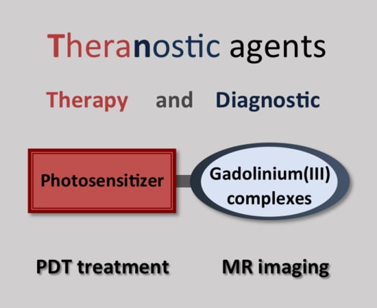
Graphical Abstract

1. Introduction to the Theranostic Approach

Theranostics is an innovative medical treatment research field which incorporates the functions of therapy and diagnostics by imaging and paves a way for personalized medicine [1,2,3,4]. The emergence of this research field has been made possible by tremendous progress in the development of instruments for imaging and treatment. Theranostic agents combine an imaging agent and a therapeutic agent within the same scaffold, both agents being thus delivered at the same time and with the same biodistribution. They give important information for pre-treatment planning, therapy monitoring, and treatment outcome assessment, and, moreover, for the development of new therapeutic agents. Various imaging and therapeutic modalities may be combined, with different assembly strategies, to generate theranostic agents. Here, we review molecular theranostic agents combining magnetic resonance imaging (MRI) and photodynamic therapy (PDT) applications. These multifunctional molecules can be described as low and medium molecular weight compounds that do not self-assemble into bigger systems. Theranostic agents based on nanoparticles belong to another promising and active field and have recently been reviewed [5,6,7].

1.1. PDT Treatment: Strengths and Limitations

PDT is a light-activated treatment modality and has been clinically approved in the treatment of dermatological and ocular disorders and of various cancers. It is a localized treatment which has minimal invasiveness and side effects [8,9,10]. PDT requires the administration of a drug called a photosensitizer (PS) that is usually a porphyrin-type compound (Scheme 1). Light of an appropriate wavelength is then applied on the affected tissue and is absorbed by the photosensitizer. The latter is thus activated and reacts with surrounding oxygen and/or with surrounding molecules to generate cytotoxic species, which induce cellular damage, vascular occlusion, and/or antitumor immune response [11].
Most clinical PSs are activated with excitation wavelengths between 630 and 690 nm, which have limited tissue penetration depth mainly due to light scattering and absorption by endogenous molecules. The weak tissue penetration of light is a major concern for PDT development. Therefore, PSs that can be activated in the optical transparency window of tissues (from 700 nm up to 1000 nm with a two-photon absorption process) are very appealing [8]. In addition to the wavelength range, strong absorption capacity (characterized by a high absorption coefficient value ε for one-photon absorption and by a high two-photon absorption cross-section value σ2 for two-photon absorption) increases the production of cytotoxic species and enhances the treatment efficacy. New PSs are designed and studied in order to respond to these criteria. Two porphyrin-based photosensitizers (Scheme 1c,d), with strong one-photon absorption in the near infrared (NIR), have recently been designed. Padeliporfin (Tookad®WST11), with an excitation wavelength at 763 nm (ε = 100,000 M−1·cm−1), has been approved for the treatment of prostate cancer [12]. Redaporfin (LUZ11), which can be activated at 749 nm (ε = 140,000 M−1·cm−1) has been approved for biliary tract cancer and is also progressing to other clinical trials [13,14].
Some PSs are able to preferentially accumulate in tumor cells. However, the mechanisms of this behavior are not fully understood and tumor selectivity needs to be improved. There is also a request for identifying precisely the time period of maximum drug accumulation in the tumor. Light irradiation during this time period should generate many reactive oxygen species and lead to best PDT efficiency [5]. Drug accumulation and light delivery are specific to each PS and to tumor characteristics. Thus, the optimization of treatment planning is difficult to achieve. With these limitations, it has become evident that there is a need for treatment guidance via imaging that can pilot pre- and post-treatment evaluation [6,15,16,17].

1.2. MRI Guidance of PDT

MRI is a non-invasive visualization tool of soft tissues with both high spatial and time resolution [18]. It has already proven to be powerful for therapy monitoring in oncology and for drug development [19]. The magnetic resonance (MR) signal contrast arises from differences in proton properties such as relaxation times (T1 and T2) or the density of water molecules [20]. These properties are acquired using appropriate MR pulse sequences; they highlight different proton behaviors and allow tissue discrimination. In some cases, the contrast between healthy and diseased tissue is weak and the use of a contrast agent is necessary. Clinically used contrast agents are small gadolinium-based complexes containing octadentate chelators based on macrocyclic (GdDOTA with DOTA = 1,4,7,10-tetraazacyclododecane-1,4,7,10-tetraacetate) or linear (GdDTPA with DTPA = diethylenetriamine pentaacetate) structures (Scheme 2). The efficiency of a contrast agent is measured by its relaxivity, which corresponds to the ability to decrease the relaxation time T1 of water protons in the presence of a paramagnetic gadolinium complex at a concentration of 1 mM.
PDT-induced changes in the tumoral area alter proton behaviors and these changes can be strongly emphasized by the presence of a contrast agent. The image modification can supply essential information in order to estimate the PDT effect. It is then possible to establish relations between this effect and the drug concentration and light application. The drug and light parameters can then be finely tuned in order to increase the efficiency of the treatment [21].
MRI-guided PDT can be realized with repeated contrast agent injection at different time points after PS injection. Depending on the choice of MRI methodology, PDT response can be followed at different time points after the treatment and give different information. Contrast-enhanced MRI has been shown to be very sensitive to PDT-induced vascular occlusion [22,23]. It has been used for the assessment of tumor response to PDT several days and even shortly after treatment [24,25,26].
The optimization of PDT treatment can be further improved using theranostic agents. With this approach, both the imaging and the therapeutic agents have the same biodistribution and bioelimination behaviors. Gadolinium-based contrast agents linked to a porphyrin-based PS are expected to confer increased tumoral residence time; they thus provide a longer time window to monitor the treatment. They also have increased relaxivities compared to classical MRI contrast agents. Therefore, they can be administered at much lower doses than current clinical contrast agents and offer enhanced safety. Different approaches to developing nanoparticules for MRI and PDT applications have attracted increasing attention during the past decade [5,6,7,27]. In spite of the efficient targeting and high payload of some nanotheranostic agents, clinical translation has not yet been possible [6,7,27]. Molecular theranostic agents for PDT and MRI have been developed to a lesser extent, particularly due to their more elaborated and time-consuming synthesis. Nevertheless, these small or medium-sized molecular agents have their own advantages, such as high reproducibility, stability, purity, and good biocompatibility, and this approach continues to draw attention for cancer treatment [28].

2. Porphyrin-Gd-Complexes Conjugates with Potential MRI and PDT Applications

Several porphyrin analogues have been associated with Gd(III) complexes, and their ability to accumulate in cancer cells and/or their relaxivity have been studied. Although their ability to behave as PDT PSs has not been explored, these compounds are potential bifunctional compounds for MRI and PDT applications. At an early stage, two compounds, gadophrin-2 and gadophrin-3 (composed respectively of a free-base and a copper(II) porphyrin linked to two GdDTPA complexes) have been investigated. Studies in mice have shown comparable pharmacological properties and these PSs have been found to accumulate in necrotic areas [29,30,31,32,33]. A 5,10,15,20-tetraphenylporphyrin (TPP) core has been linked to one and four GdDTPA complexes through amide bond formation [34]. Increased relaxivity values have been found. Free-base and copper(II) porphyrins linked to one, two, and four GdDO3A-amide complexes have been developed for potential multimodal MRI/positron emission tomography (PET) applications. Preliminary relaxivity studies have indicated promising contrast enhancement [35,36]. Zinc(II) and copper(II) porphyrazine have been linked to two GdDO3A-amide complexes. The copper-containing compound showed the highest relaxivity and very good cellular internalization, and tumor-bearing mice images indicated necrotic localization [37].

3. Molecular Theranostic Agents with Combined PDT and MRI Studies

In 1993, a pioneering study was realized with two bifunctional compounds containing porphyrin derivatives linked to one or two GdDTPA complexes [38]. The compound TPP-Gd2(DTPA)4, consisting of a tetra-p-aminophenylporphyrin core coupled through amide bonds to four DTPA ligands, and two of which being metalated by Gd(III) ions, was the most promising. The relaxivity value per Gd(III) was found to be twice as high as that of GdDTPA at 20 MHz and the substantial image contrast enhancement of the tumor compared to adjacent normal tissue in tumor-bearing mice evidenced the affinity of the compound for tumor tissue. The photoinduced toxicity studies, realized with irradiation using a multiwavelength laser beam at 488 and 514 nm on two cell lines (HT29 and L1210), showed phototoxicities comparable to that induced by a commercial hematoporphyrin derivative (HPD) PS. This porphyrin-Gd complex conjugate was the first prototype built for MRI and PDT.
More than a decade later, Pandey and collaborators extensively investigated several theranostic agents that combine diagnostic imaging (MR and fluorescence imaging) and PDT treatment properties. The agents investigated are based on different photosensitizers (pyropheophorbide analogues with different lipophilic/hydrophilic chains) linked to one, two, three, or six GdDTPA complexes [39,40,41,42]. In these compounds, the linkage is realized through the C-functionalization of the diethylenetriamine backbone and the stability of the GdDTPA core is preserved by the five anionic carboxylate groups. The theranostic compounds containing one and two Gd complexes required liposomal formulation to resolve the poor water-solubility problem. With the presence of three and six GdDTPA units, the water solubility improved. The compound HPPH-3GdDTPA, which bears three GdDTPA complexes (Figure 1a) was found to be the best candidate with respect to its imaging and treatment results. It showed remarkable MR contrast enhancement of tumors in mice 24 h after injection, with a 10-fold lower dose than the clinical dose used with Magnevist and preferential uptake in tumors compared to muscle (Figure 1b). This important result shows that, with the presence of three Gd(III) complexes appended to one photosensitizer, it is possible to perform MR imaging and PDT treatment at the same low concentration. Fluorescence imaging, resulting from the light emission of the HPPH derivative, also showed maximum intensity 24 h after injection. Finally, this compound also showed an efficient PDT effect after one instance of irradiation at 665 nm (70 J/cm2) 24 h after injection.
Phthalocyanine and porphyrazine are tetrapyrrolic compounds known for their intense electronic absorption in the NIR region; they require addition of peripheric substitution groups to avoid aggregation and favor water solubilization. They have been linked to GdDO3A-amide complexes that are GdDOTA derivatives where one carboxylate arm is replaced by an acetamide group. The amide-bond formation allows rapid access to Gd(III) complex functionalization but it strongly induces reduced thermodynamic stability while maintaining the same kinetic inertness as the GdDOTA complex [43]. The phthalocyanine-based PS has been linked to one particular GdDO3A-amide complex, ZnPht-1Gd (Figure 2a) [44]. This complex has exhibited low relaxivity (1.43 mM−1·s−1 at 128 MHz) and the authors have proposed that this weak value could be due to the presence of the amide function on the arm that could block water access to the metallic center. The compound has shown good ability to produce cytotoxic singlet oxygen under irradiation with a quantum yield of 0.67 (in DMSO). The porphyrazine-based PS has been linked to one, four, and eight GdDO3A-amide complexes to give the bifunctional compounds ZnPz-nGd, with n = 1, 4, 8 (Figure 2b) [45]. These compounds have shown strong relaxivity increases (up to 12.8 mM−1·s−1 for ZnPz-8Gd at 60 MHz and 37 °C) with the number of Gd-complexes attached to the PS. Cellular uptake has been observed only with compounds bearing one Gd complex (ZnPz-1Gd) and a notable phototoxic effect (with 50% cell killing) has been observed after 10 min irradiation with white light (Figure 2c).
Porphyrin derivatives incorporating a Gd(III) ion are at first sight appealing agents due to their simplified structure and synthesis, and to their promising in vitro properties as phosphorescence-based oxygen sensors, PDT photosensitizers, and MRI contrast agents. However, in vivo studies have resulted in disparate conclusions. Koenig [46] and Furmanski [47] have studied gadolinium-incorporated porphyrins as MRI contrast agents and have observed stability problems with ion dissociation from the porphyrin during their studies in plasma and in mice. Recently, two porphyrazine-based compounds incorporating a Gd(III) ion, GdPz1, and GdPz2, which differ in the nature of their peripheral groups, have been obtained (Figure 3a) [48]. A good relaxivity value was obtained at a very high magnetic field (4.67 mM−1·s−1 at 9.4 T) for GdPz1 once solubilized in polymer polyimide brushes. Cellular uptake, dual in vivo fluorescence and MR imaging, and in vivo PDT activity were studied. Significant in vivo tumor accumulation was demonstrated by fluorescence and MR images for both compounds. PDT activity was assessed in cancerous CT26 cells with light irradiation performed at 615–635 nm (10–20 J/cm2, 10−7 to 10−4 M incubation concentration). PDT treatment of CT26 tumor-bearing Balb/c mice (Figure 3b) was realized three hours post-injection by irradiation at 593 nm (30 min, 120 J/cm2). Moderate tumor death was observed and this study indicated the need to optimize different parameters such as drug dose and light application.
A porphyrin-based PS linked to chemotoxic platinum(II) complexes and incorporating a gadolinium(III) ion has been reported for tumor treatment by PDT and chemotherapy and for MR imaging [49]. It has been obtained from a 5,10,15,20-tetra(4-pyridyl)-porphyrin (P1) that is coordinated to four Pt(II) complexes (Pt-P1) and to one Gd(III) ion (Gd/Pt-P1) (Figure 4a). The compound showed nearly doubled relaxivity at 3 T compared to GdDTPA. A phototoxic effect was observed in C6 cells after 10 min irradiation at 630 nm (Figure 4b). A synergetic chemo-photodynamic antitumor effect was observed in cells and in C6 tumor-bearing mice.
In order to obtain contrast agents with high relaxivity at high magnetic fields, the strategy of increasing the number of coordinated water molecules at the Gd(III) center is particularly appealing as this allows for the almost doubling of the r1 relaxivity value independently of the magnetic field. In this case, careful design of hepta- or hexadentate ligands is necessary in order to obtain Gd(III) complexes with good thermodynamic stability and kinetic inertness, and to avoid ternary complex formation with endogenous molecules. Chen et al. have developed a potential theranostic agent (Scheme 3a) consisting of a tetraphenylporphyrin core linked to four GdDTTA complexes [50]. High relaxivity has been measured (14.1 mM−1·s−1 at 0.55 T in Hepes (4-(2-hydroxyethyl)-1-piperazine ethanesulfonic acid) buffer) and this value doubled in the presence of human serum albumin, indicating strong binding of the conjugate to this blood pool protein. The fluorescence of the PS has been evidenced in H1299 lung cancer cells and has shown harmless cellular uptake. Singlet oxygen has been efficiently produced upon irradiation at 650 nm in deuterated water. These studies show the potential of this compound to behave as a contrast agent for multimodal (MR and luminescence) imaging and as a PS for PDT.
The development of a theranostic agent gathering four GdDTTA complexes and a TPP core has also been realized with a design which differs with regards to the nature of the linkers separating the two agents (Scheme 3b). Short and relatively rigid benzyl linkers have been used, which allow for the minimization of rotational flexibility and thus for the optimization of the relaxivity gain brought about by the increase in molecular weight [51]. This water-soluble bifunctional system has the highest relaxivity reported for a medium-sized system with a maximum of 43.7 mM−1·s−1 (per Gd(III) ion at 20 MHz). A 27% relaxivity increase has also been observed in the presence of bovine serum albumin (BSA). Phantom images of cell pellets (Figure 5a) obtained at a high magnetic field (7 T) have evidenced cellular uptake. Inductively coupled plasma mass spectrometry (ICP-MS) measurements have shown that cellular uptake of Gd ions is 60 times more effective with the theranostic agent than with the commercial GdDTPA contrast agent at 10 μM Gd incubation concentration. This result can be explained by the amphiphillic character of the theranostic compound which favors cell internalization. A good PDT effect has been observed in HeLa cells (Figure 5b) upon irradiation at 636 nm (1 h) and this effect was found to increase with light intensity and with the incubation concentration of the theranostic agent.
The theranostic agents reported so far are activated by one-photon absorption with excitation wavelengths below 700 nm. The two-photon excitation process allows for the use of excitation wavelengths in the NIR region [52,53]. This process allows deep treatment and minimal photodamage to healthy tissues. Two-photon irradiation is possible only in a very small area and high spatial treatment precision can be obtained, but the application to bulky tumors is currently limited. The design of new PSs with high two-photon absorption capability requires large pi-electronic delocalization. A one- and two-photon activatable PS based on a diketopyrrolopyrrole-zinc-porphyrin component (DPP-ZnP) and linked to a GdDOTA complex as an imaging probe has been studied (Scheme 4) [54]. The GdDOTA attachment to the PS has been realized with the use of the commercial DOTAGA (1,4,7,10-tetraazacyclododecane-1-glutaric-4,7,10-triacetic acid) ligand which brings local flexibility but keeps the GdDOTA stability intact. Remarkable relaxivity values (r1 = 19.9 mM−1·s−1 at 20 MHz) for a monohydrated and medium-sized system have been obtained. A 20% relaxivity increase in the presence of BSA has also been observed. The compound has strong one-photon absorption ability (εmax = 41,000 M−1·cm−1 at 667 nm in water). Large two-photon absorption capacity quantified by large σ2 values has been evidenced in solution over a broad range of wavelengths with a maximum of 1000 GM between 910 and 940 nm. A high PDT effect evaluated in HeLa cells was observed by one-photon excitation at 660 nm (1 h, 1 μM incubation concentration) and a moderate two-photon PDT effect was observed at 930 nm (300 scans, 1 μM incubation concentration).
A theranostic agent containing a PS with high one- and two-photon absorption capacity in the near infrared region and two stable contrast agents for MR imaging has been studied [55]. The structure of this agent consists of a Zn-porphyrin dimer (ZnP–ZnP) linked to two GdDOTA complexes (Figure 6a). High relaxivity values have been obtained with a maximum of r1 = 14.4 mM−1·s−1 (at 40 MHz in water containing 2% of pyridine to ensure complete water solubility). Due to the presence of the two Gd(III) complexes, the corresponding molecular relaxivities are doubled; this trait is important to realize the imaging and therapeutic studies at the same low concentration required for PDT treatment. It is also interesting to note that the relaxivity value was seen to double in presence of BSA at 20 MHz. This compound has shown strong one-photon absorption capacity in the near infrared region (with a maximum at 746 nm with ε = 105 M−1·cm−1 in DMSO). It has also shown very strong two-photon absorption ability with a maximum between 880 and 930 nm (σ2 ≈ 8000 GM in DMSO). An efficient PDT effect was observed in HeLa cells after one-photon irradiation at 740 nm (30 min, 1 μM incubation concentration). A two-photon PDT effect was observed at 910 nm (300 scans, 2 μM incubation concentration) as a function of light power, and 100% cell death was observed with an average power of 108 mW at the back pupil of the objective (Figure 6b).

4. Conclusions

In this review, the design and study of molecular theranostic agents for potential applications in MR imaging and PDT treatment has been highlighted and discussed.
Compared to the MR imaging properties of small commercial contrast agents, the properties of theranostic agents are superior, due to the presence of the lipophilic PS. Increased cellular uptake and/or tumor accumulation have been observed, and together with the increased relaxivity brought about by the large size of these compounds, the use of such theranostic agents requires much lower doses than those used with clinical contrast agents. To further improve the imaging efficiency of the theranostic agent, the same criteria as those for classical contrast agents need to be considered. In particular, local rigidity and stability are two important parameters to take into account. In addition to MR imaging, the fluorescence properties of the tetrapyrrolic core have also been explored in some cases. They provide a second imaging modality with high sensitivity.
Tetrapyrrolic PSs are often weakly soluble in aqueous media, the presence of hydrophilic Gd(III) complexes brings increased water solubility; it also modulates the in vivo distribution and elimination. A good ratio between the number of Gd(III) complexes and the PS has to be found in order to maintain the cellular uptake ability. The design of PDT sensitizers with strong absorption in the biological transparency window is necessary for increased tissue penetration depth and high cytotoxic species production.
Cellular and animal studies require strong and long experimentation efforts. Studies showing the influence of drug concentration and light application on PDT efficiency should be developed. Finally, a chemical design that allows for better tumoral selectivity will undoubtedly greatly enhance the capacity of these theranostic agents.

Author Contributions

A.S. wrote the review. S.J. has critically and scientifically revised the manuscript and prepared the schemes and the figures. The final revision was made by both authors.

Funding

The authors thank the CNRS (France) and the Ministry of Education and Research. The research realized in the frame of the LSAMM (Laboratoire de synthèse des assemblages moléculaires multifonctionnels) laboratory was funded by the icFRC (http://www.icfrc.fr), LabEx CSC, Region Alsace and Université de Strasbourg.

Acknowledgments

The authors thank the LSAMM coworkers and all the collaborators involved in the work done in the frame of the LSAMM laboratory.

Conflicts of Interest

The authors declare no conflict of interest.

References

  1. Yoon, H.Y.; Jeon, S.; You, D.G.; Park, J.H.; Kwon, I.C.; Koo, H.; Kim, K. Inorganic Nanoparticles for Image-Guided Therapy. Bioconj. Chem. 2017, 28, 124–134. [Google Scholar] [CrossRef] [PubMed]
  2. Crawley, N.; Thompson, M.; Romaschin, A. Theranostics in the Growing Field of Personalized Medicine: An Analytical Chemistry Perspective. Anal. Chem. 2014, 86, 130–160. [Google Scholar] [CrossRef] [PubMed]
  3. Terreno, E.; Uggeri, F.; Aime, S. Image guided therapy: The advent of theranostic agents. J. Control. Release 2012, 161, 328–337. [Google Scholar] [CrossRef] [PubMed]
  4. Kelkar, S.S.; Reineke, T.M. Theranostics: Combining Imaging and Therapy. Bioconj. Chem. 2011, 22, 1879–1903. [Google Scholar] [CrossRef] [PubMed]
  5. Kunjachan, S.; Ehling, J.; Storm, G.; Kiessling, F.; Lammers, T. Noninvasive Imaging of Nanomedicines and Nanotheranostics: Principles, Progress, and Prospects. Chem. Rev. 2015, 115, 10907–10937. [Google Scholar] [CrossRef] [PubMed]
  6. Chen, G.; Roy, I.; Yang, C.; Prasad, P.N. Nanochemistry and Nanomedicine for Nanoparticle-based Diagnostics and Therapy. Chem. Rev. 2016, 116, 2826–2885. [Google Scholar] [CrossRef] [PubMed]
  7. Björnmalm, M.; Thurecht, K.J.; Michael, M.; Scott, A.M.; Caruso, F. Bridging Bio–Nano Science and Cancer Nanomedicine. ACS Nano 2017, 11, 9594–9613. [Google Scholar] [CrossRef]
  8. Fan, W.; Huang, P.; Chen, X. Overcoming the Achilles’ heel of photodynamic therapy. Chem. Soc. Rev. 2016, 45, 6488–6519. [Google Scholar] [CrossRef]
  9. Abrahamse, H.; Hamblin, M.R. New photosensitizers for photodynamic therapy. Biochem. J. 2016, 473, 347–364. [Google Scholar] [CrossRef] [Green Version]
  10. Dąbrowski, J.M.; Arnaut, L.G. Photodynamic therapy (PDT) of cancer: From local to systemic treatment. Photochem. Photobiol. Sci. 2015, 14, 1765–1780. [Google Scholar] [CrossRef]
  11. Mroz, P.; Yaroslavsky, A.; Kharkwal, G.B.; Hamblin, M.R. Cell Death Pathways in Photodynamic Therapy of Cancer. Cancers 2011, 3, 2516–2539. [Google Scholar] [CrossRef] [PubMed] [Green Version]
  12. Azzouzi, A.-R.; Vincendeau, S.; Barret, E.; Cicco, A.; Kleinclauss, F.; van der Poel, H.G.; Stief, C.G.; Rassweiler, J.; Salomon, G.; Solsona, E.; et al. Padeliporfin vascular-targeted photodynamic therapy versus active surveillance in men with low-risk prostate cancer (CLIN1001 PCM301): An open-label, phase 3, randomised controlled trial. Lancet Oncol. 2017, 18, 181–191. [Google Scholar] [CrossRef]
  13. Van Straten, D.; Mashayekhi, V.; de Bruijn, H.; Oliveira, S.; Robinson, D. Oncologic Photodynamic Therapy: Basic Principles, Current Clinical Status and Future Directions. Cancers 2017, 9, 19. [Google Scholar] [CrossRef] [PubMed]
  14. Luz, A.F.S.; Pucelik, B.; Pereira, M.M.; Dąbrowski, J.M.; Arnaut, L.G. Translating phototherapeutic indices from in vitro to in vivo photodynamic therapy with bacteriochlorins. Lasers Surg. Med. 2018, 50, 451–459. [Google Scholar] [CrossRef] [PubMed]
  15. Celli, J.P.; Spring, B.Q.; Rizvi, I.; Evans, C.L.; Samkoe, K.S.; Verma, S.; Pogue, B.W.; Hasan, T. Imaging and Photodynamic Therapy: Mechanisms, Monitoring, and Optimization. Chem. Rev. 2010, 110, 2795–2838. [Google Scholar] [CrossRef] [Green Version]
  16. Rai, P.; Mallidi, S.; Zheng, X.; Rahmanzadeh, R.; Mir, Y.; Elrington, S.; Khurshid, A.; Hasan, T. Development and applications of photo-triggered theranostic agents. Adv. Drug Deliv. Rev. 2010, 62, 1094–1124. [Google Scholar] [CrossRef] [Green Version]
  17. Bhaumik, J.; Mittal, A.K.; Banerjee, A.; Chisti, Y.; Banerjee, U.C. Applications of phototheranostic nanoagents in photodynamic therapy. Nano Res. 2015, 8, 1373–1394. [Google Scholar] [CrossRef]
  18. Pierre, V.C.; Allen, M.J.; Caravan, P. Contrast agents for MRI: 30+ years and where are we going? JBIC J. Biol. Inorg. Chem. 2014, 19, 127–131. [Google Scholar] [CrossRef] [Green Version]
  19. Evelhoch, J.L. In vivo MR in the drug pipeline. J. Magn. Reson. 2018, 292, 117–128. [Google Scholar] [CrossRef]
  20. De León-Rodríguez, L.M.; Martins, A.F.; Pinho, M.C.; Rofsky, N.M.; Sherry, A.D. Basic MR relaxation mechanisms and contrast agent design: MR Relaxation Mechanisms and Contrast Agents. J. Magn. Reson. Imaging 2015, 42, 545–565. [Google Scholar] [CrossRef]
  21. Josefsen, L.B.; Boyle, R.W. Unique Diagnostic and Therapeutic Roles of Porphyrins and Phthalocyanines in Photodynamic Therapy, Imaging and Theranostics. Theranostics 2012, 2, 916–966. [Google Scholar] [CrossRef] [PubMed] [Green Version]
  22. Kennedy, S.D.; Szczepaniak, L.S.; Gibson, S.L.; Hilf, R.; Foster, T.H.; Bryant, R.G. Quantitative MRI of Gd-DTPA uptake in tumors: Response to photo dynamic therapy. Magn. Reson. Med. 1994, 31, 292–301. [Google Scholar] [CrossRef] [PubMed]
  23. Haider, M.A.; Davidson, S.R.H.; Kale, A.V.; Weersink, R.A.; Evans, A.J.; Toi, A.; Gertner, M.R.; Bogaards, A.; Wilson, B.C.; Chin, J.L.; et al. Prostate Gland: MR Imaging Appearance after Vascular Targeted Photodynamic Therapy with Palladium-Bacteriopheophorbide. Radiology 2007, 244, 196–204. [Google Scholar] [CrossRef] [PubMed]
  24. Huang, Z.; Haider, M.A.; Kraft, S.; Chen, Q.; Blanc, D.; Wilson, B.C.; Hetzel, F.W. Magnetic resonance imaging correlated with the histopathological effect of Pd-bacteriopheophorbide (Tookad) photodynamic therapy on the normal canine prostate gland. Lasers Surg. Med. 2006, 38, 672–681. [Google Scholar] [CrossRef] [PubMed] [Green Version]
  25. Schreurs, T.J.L.; Hectors, S.J.; Jacobs, I.; Grüll, H.; Nicolay, K.; Strijkers, G.J. Quantitative Multi-Parametric Magnetic Resonance Imaging of Tumor Response to Photodynamic Therapy. PLoS ONE 2016, 11, e0165759. [Google Scholar] [CrossRef] [PubMed]
  26. Zilberstein, J.; Schreiber, S.; Bloemers, M.C.W.M.; Bendel, P.; Neeman, M.; Schechtman, E.; Kohen, F.; Scherz, A.; Salomon, Y. Antivascular Treatment of Solid Melanoma Tumors with Bacteriochlorophyll-serine-based Photodynamic Therapy. Photochem. Photobiol. 2007, 73, 257–266. [Google Scholar] [CrossRef]
  27. Schleich, N.; Danhier, F.; Préat, V. Iron oxide-loaded nanotheranostics: Major obstacles to in vivo studies and clinical translation. J. Control. Release 2015, 198, 35–54. [Google Scholar] [CrossRef]
  28. Kumar, R.; Shin, W.S.; Sunwoo, K.; Kim, W.Y.; Koo, S.; Bhuniya, S.; Kim, J.S. Small conjugate-based theranostic agents: An encouraging approach for cancer therapy. Chem. Soc. Rev. 2015, 44, 6670–6683. [Google Scholar] [CrossRef]
  29. Hofmann, B.; Bogdanov, A.; Marecos, E.; Ebert, W.; Semmler, W.; Weissleder, R. Mechanism of gadophrin-2 accumulation in tumor necrosis. J. Magn. Reson. Imaging 1999, 9, 336–341. [Google Scholar] [CrossRef] [Green Version]
  30. Barkhausen, J.Ö.; Ebert, W.; Debatin, J.Ö.F.; Weinmann, H.-J. Imaging of myocardial infarction: Comparison of magnevist and gadophrin-3 in rabbits. J. Am. Coll. Cardiol. 2002, 39, 1392–1398. [Google Scholar] [CrossRef]
  31. Daldrup-Link, H.; Rudelius, M.; Metz, S.; Piontek, G.; Pichler, B.; Settles, M.; Heinzmann, U.; Schlegel, J.R.; Oostendorp, R.J.; Rummeny, E. Cell tracking with gadophrin-2: A bifunctional contrast agent for MR imaging, optical imaging, and fluorescence microscopy. Eur. J. Nucl. Med. Mol. Imaging 2004, 31. [Google Scholar] [CrossRef] [PubMed]
  32. Metz, S.; Daldrup-Link, H.E.; Richter, T.; Räth, C.; Ebert, W.; Settles, M.; Rummeny, E.J.; Link, T.M.; Piert, M. Detection and Quantification of Breast Tumor Necrosis with MR Imaging. Acad. Radiol. 2003, 10, 484–490. [Google Scholar] [CrossRef]
  33. Ni, Y. Metalloporphyrins and Functional Analogues as MRI Contrast Agents. Curr. Med. Imaging Rev. 2008, 4, 96–112. [Google Scholar] [CrossRef]
  34. Haroon Ur, R.; Umar, M.N.; Khan, K.; Anjum, M.N.; Yaseen, M. Synthesis and relaxivity measurement of porphyrin-based Magnetic Resonance Imaging (MRI) contrast agents. J. Struct. Chem. 2014, 55, 910–915. [Google Scholar] [CrossRef]
  35. Gros, C.P.; Eggenspiller, A.; Nonat, A.; Barbe, J.-M.; Denat, F. New potential bimodal imaging contrast agents based on DOTA-like and porphyrin macrocycles. Med. Chem. Commun. 2011, 2, 119–125. [Google Scholar] [CrossRef]
  36. Eggenspiller, A.; Michelin, C.; Desbois, N.; Richard, P.; Barbe, J.-M.; Denat, F.; Licona, C.; Gaiddon, C.; Sayeh, A.; Choquet, P.; et al. Design of Porphyrin-dota-Like Scaffolds as All-in-One Multimodal Heterometallic Complexes for Medical Imaging: Porphyrin-dota-Like Scaffolds for Medical Imaging. Eur. J. Org. Chem. 2013, 2013, 6629–6643. [Google Scholar] [CrossRef]
  37. Trivedi, E.R.; Ma, Z.; Waters, E.A.; Macrenaris, K.W.; Subramanian, R.; Barrett, A.G.M.; Meade, T.J.; Hoffman, B.M. Synthesis and characterization of a porphyrazine-Gd(III) MRI contrast agent and in vivo imaging of a breast cancer xenograft model: TUMOR IMAGING. Contrast Media Mol. Imaging 2014, 9, 313–322. [Google Scholar] [CrossRef]
  38. Hindré, F.; Plouzennec, M.L.; de Certaines, J.D.; Foultier, M.T.; Patrice, T.; Simonneaux, G. Tetra-p-aminophenylporphyrin conjugated with Gd-DTPA: Tumor-specific contrast agent for MR imaging. J. Magn. Reson. Imaging 1993, 3, 59–65. [Google Scholar] [CrossRef]
  39. Li, G.; Slansky, A.; Dobhal, M.P.; Goswami, L.N.; Graham, A.; Chen, Y.; Kanter, P.; Alberico, R.A.; Spernyak, J.; Morgan, J.; et al. Chlorophyll-a Analogues Conjugated with Aminobenzyl-DTPA as Potential Bifunctional Agents for Magnetic Resonance Imaging and Photodynamic Therapy. Bioconj. Chem. 2005, 16, 32–42. [Google Scholar] [CrossRef]
  40. Pandey, R.K.; Goswami, L.N.; Chen, Y.; Gryshuk, A.; Missert, J.R.; Oseroff, A.; Dougherty, T.J. Nature: A rich source for developing multifunctional agents. tumor-imaging and photodynamic therapy. Lasers Surg. Med. 2006, 38, 445–467. [Google Scholar] [CrossRef]
  41. Spernyak, J.A.; White, W.H.; Ethirajan, M.; Patel, N.J.; Goswami, L.; Chen, Y.; Turowski, S.; Missert, J.R.; Batt, C.; Mazurchuk, R.; et al. Hexylether Derivative of Pyropheophorbide-a (HPPH) on Conjugating with 3 Gadolinium(III) Aminobenzyldiethylenetriaminepentaacetic Acid Shows Potential for in Vivo Tumor Imaging (MR, Fluorescence) and Photodynamic Therapy. Bioconj. Chem. 2010, 21, 828–835. [Google Scholar] [CrossRef] [PubMed]
  42. Goswami, L.N.; White, W.H.; Spernyak, J.A.; Ethirajan, M.; Chen, Y.; Missert, J.R.; Morgan, J.; Mazurchuk, R.; Pandey, R.K. Synthesis of Tumor-Avid Photosensitizer−Gd(III)DTPA Conjugates: Impact of the Number of Gadolinium Units in T1/T2 Relaxivity, Intracellular localization, and Photosensitizing Efficacy. Bioconj. Chem. 2010, 21, 816–827. [Google Scholar] [CrossRef]
  43. Lattuada, L.; Barge, A.; Cravotto, G.; Giovenzana, G.B.; Tei, L. The synthesis and application of polyamino polycarboxylic bifunctional chelating agents. Chem. Soc. Rev. 2011, 40, 3019. [Google Scholar] [CrossRef] [PubMed]
  44. Aydın Tekdaş, D.; Garifullin, R.; Şentürk, B.; Zorlu, Y.; Gundogdu, U.; Atalar, E.; Tekinay, A.B.; Chernonosov, A.A.; Yerli, Y.; Dumoulin, F.; et al. Design of a Gd-DOTA-Phthalocyanine Conjugate Combining MRI Contrast Imaging and Photosensitization Properties as a Potential Molecular Theranostic. Photochem. Photobiol. 2014, 90, 1376–1386. [Google Scholar] [CrossRef] [PubMed] [Green Version]
  45. Song, Y.; Zong, H.; Trivedi, E.R.; Vesper, B.J.; Waters, E.A.; Barrett, A.G.M.; Radosevich, J.A.; Hoffman, B.M.; Meade, T.J. Synthesis and Characterization of New Porphyrazine-Gd(III) Conjugates as Multimodal MR Contrast Agents. Bioconj. Chem. 2010, 21, 2267–2275. [Google Scholar] [CrossRef] [PubMed] [Green Version]
  46. Lyon, R.C.; Faustino, P.J.; Cohen, J.S.; Katz, A.; Mornex, F.; Colcher, D.; Baglin, C.; Koenig, S.H.; Hambright, P. Tissue distribution and stability of metalloporphyrin MRI contrast agents. Magn. Reson. Med. 1987, 4, 24–33. [Google Scholar] [CrossRef] [PubMed]
  47. Furmanski, P.; Longley, C. Metalloporphyrin Enhancement of Magnetic Resonance Imaging of Human Tumor Xenografts in Nude Mice. Cancer Res. 1988, 48, 4604–4610. [Google Scholar]
  48. Yuzhakova, D.V.; Lermontova, S.A.; Grigoryev, I.S.; Muravieva, M.S.; Gavrina, A.I.; Shirmanova, M.V.; Balalaeva, I.V.; Klapshina, L.G.; Zagaynova, E.V. In vivo multimodal tumor imaging and photodynamic therapy with novel theranostic agents based on the porphyrazine framework-chelated gadolinium (III) cation. Biochim. Biophys. Acta Gen. Subj. 2017, 1861, 3120–3130. [Google Scholar] [CrossRef]
  49. Wu, B.; Li, X.-Q.; Huang, T.; Lu, S.-T.; Wan, B.; Liao, R.-F.; Li, Y.-S.; Baidya, A.; Long, Q.-Y.; Xu, H.-B. MRI-guided tumor chemo-photodynamic therapy with Gd/Pt bifunctionalized porphyrin. Biomater. Sci. 2017, 5, 1746–1750. [Google Scholar] [CrossRef]
  50. Luo, J.; Chen, L.-F.; Hu, P.; Chen, Z.-N. Tetranuclear Gadolinium(III) Porphyrin Complex as a Theranostic Agent for Multimodal Imaging and Photodynamic Therapy. Inorg. Chem. 2014, 53, 4184–4191. [Google Scholar] [CrossRef]
  51. Sour, A.; Jenni, S.; Ortí-Suárez, A.; Schmitt, J.; Heitz, V.; Bolze, F.; Loureiro de Sousa, P.; Po, C.; Bonnet, C.S.; Pallier, A.; et al. Four Gadolinium(III) Complexes Appended to a Porphyrin: A Water-Soluble Molecular Theranostic Agent with Remarkable Relaxivity Suited for MRI Tracking of the Photosensitizer. Inorg. Chem. 2016, 55, 4545–4554. [Google Scholar] [CrossRef] [PubMed]
  52. Bolze, F.; Jenni, S.; Sour, A.; Heitz, V. Molecular photosensitisers for two-photon photodynamic therapy. Chem. Commun. 2017, 53, 12857–12877. [Google Scholar] [CrossRef] [PubMed]
  53. Sun, Z.; Zhang, L.-P.; Wu, F.; Zhao, Y. Photosensitizers for Two-Photon Excited Photodynamic Therapy. Adv. Funct. Mater. 2017, 27, 1704079. [Google Scholar] [CrossRef]
  54. Schmitt, J.; Heitz, V.; Sour, A.; Bolze, F.; Kessler, P.; Flamigni, L.; Ventura, B.; Bonnet, C.S.; Tóth, É. A Theranostic Agent Combining a Two-Photon-Absorbing Photosensitizer for Photodynamic Therapy and a Gadolinium(III) Complex for MRI Detection. Chem. Eur. J. 2016, 22, 2775–2786. [Google Scholar] [CrossRef] [PubMed]
  55. Schmitt, J.; Jenni, S.; Sour, A.; Heitz, V.; Bolze, F.; Pallier, A.; Bonnet, C.S.; Tóth, É.; Ventura, B. A Porphyrin Dimer–GdDOTA Conjugate as a Theranostic Agent for One- and Two-Photon Photodynamic Therapy and MRI. Bioconj. Chem. 2018, 29, 3726–3738. [Google Scholar] [CrossRef] [PubMed]
Scheme 1. Schematic representation of (a) porfimer sodium (Photofrin®), (b) verteporfin (Visudyne®), (c) padeliporfin (Tookad®WST11), and (d) redaporfin (LUZ11).
Inorganics 07 00010 sch001
Scheme 2. Chemical structures of two clinical contrast agents (a) GdDOTA (Dotarem® or Clariscan®) and (b) GdDTPA (Magnevist®).
Inorganics 07 00010 sch002
Figure 1. (a) Chemical structure of the theranostic compound HPPH-3GdDTPA; (b) magnetic resonance (MR) images of a rat (Fischer) bearing Ward colon tumors before (left) and 24 h after (right) injection of HPPH–3-GdDTPA conjugate (dose: 10 μmol/kg). Adapted with permission from [41]. Copyright 2010 American Chemical Society.
Inorganics 07 00010 g001
Figure 2. Chemical structure of (a) ZnPht-1Gd [44] and (b) ZnPz-1Gd [45]; (c) phototoxic effect in WI-38 VA13 cells incubated with Zn-Pz-nGd (n = 1, 4, 8; 50 μM, 24 h) and irradiated by white light for 0 or 10 min. The same protocol was realized with Photofrin as a positive control. Adapted with permission from [45]. Copyright 2010 American Chemical Society.
Inorganics 07 00010 g002
Figure 3. (a) Molecular structure of GdPz1 and GdPz2; (b) tumor volume variation as a function of time. Intravenous injection of the theranostic agent and photodynamic therapy (PDT) treatment three hours after this injection were realized on day 10 after tumor inoculation. Adapted with permission from [48]. Copyright 2017 Elsevier.
Inorganics 07 00010 g003
Figure 4. (a) Molecular representation of Gd/Pt-P1; (b) C6 cell viability as a function of three porphyrin (P1, Pt-P1, and Gd/Pt-P1) concentrations without irradiation (6 h incubation) and 44 h after irradiation at 630 nm (0.2 W/cm, 10 min). Adapted from [49] with permission from the Royal Society of Chemistry.
Inorganics 07 00010 g004
Scheme 3. Chemical structures of the theranostic compounds developed by (a) Chen et al. [50] and (b) Sour et al. [51].
Inorganics 07 00010 sch003
Figure 5. (a) T1 maps of HeLa cell pellets at 7 T which were incubated for 24 h with different concentrations of the theranostic compound; (b) phototoxicity of the theranostic compound following 1 h irradiation at 636 nm after 24 h incubation at 2.5 µM (dark gray) and 6 µM (light gray). Adapted with permission from [51]. Copyright 2016 American Chemical Society.
Inorganics 07 00010 g005
Scheme 4. Chemical structure of the theranostic compound DPP-ZnP-GdDOTA developed by Heitz et al. [54].
Inorganics 07 00010 sch004
Figure 6. (a) Structure of the zinc porphyrin dimer linked to two GdDOTA complexes; (b) phototoxicity induced by a two-photon excitation at 910 nm after 24 h incubation in the absence (left) or presence (right) of the compound. Scale bar is 100 µm. Adapted with permission from [55]. Copyright 2018 American Chemical Society.
Inorganics 07 00010 g006