Next Issue
Volume 9, June
Previous Issue
Volume 9, April


J. Cardiovasc. Dev. Dis., Volume 9, Issue 5 (May 2022) – 44 articles

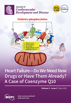
Cover Story (view full-size image): Coenzyme Q10’s main role is participating in the production of energy in the mitochondria, which maintains the proper functioning of all cells. The highest concentration of coenzyme Q10 is found in heart tissue. As the body ages, the concentration of this coenzyme in the heart tissue decreases, which makes it more susceptible to damage by free radicals. In patients with HF, the aggravation of disease symptoms is inversely related to the concentration of coenzyme Q10. Importantly, coenzyme Q10 concentration in patients with HF is an important predictor of the risk of death. Long-term coenzyme Q10 supplementation at a dose of 300 mg/day can significantly improve heart function and prognosis in patients with HF. View this paper
  • Issues are regarded as officially published after their release is announced to the table of contents alert mailing list.
  • You may sign up for e-mail alerts to receive table of contents of newly released issues.
  • PDF is the official format for papers published in both, html and pdf forms. To view the papers in pdf format, click on the "PDF Full-text" link, and use the free Adobe Reader to open them.
Order results
Result details
Section
Select all
Export citation of selected articles as:
18 pages, 751 KiB  
Systematic Review
A Systematic Review of Adherence to Immunosuppression among Pediatric Heart Transplant Patients
by Keira Nassetta, Tasmeen Hussain, Katheryn Gambetta, Kevin Le, Linda C. O’Dwyer and Sherif M. Badawy
J. Cardiovasc. Dev. Dis. 2022, 9(5), 165; https://doi.org/10.3390/jcdd9050165 - 23 May 2022
Cited by 10 | Viewed by 2365
Abstract
After pediatric heart transplant, commitment to lifelong immunosuppression is crucial to maintaining graft health. However, a review of the current literature surrounding adherence to immunosuppression in pediatric heart transplant patients is lacking. This systematic review aims to summarize the current landscape of adherence [...] Read more.
After pediatric heart transplant, commitment to lifelong immunosuppression is crucial to maintaining graft health. However, a review of the current literature surrounding adherence to immunosuppression in pediatric heart transplant patients is lacking. This systematic review aims to summarize the current landscape of adherence to immunosuppression in pediatric heart transplant patients. We conducted searches in PubMed MEDLINE, Embase, CENTRAL register of Controlled Trials (Wiley), and Scopus, from inception to March 2020. Studies were eligible if they outlined an aspect of adherence to immunosuppression and the measurement of adherence was performed with an objective or otherwise validated measure of adherence (e.g., drug levels, adherence questionnaires). The titles/abstracts of 880 articles were reviewed. After initial screening, 106 articles underwent full text review. As such, 14 articles were included in the final review. Baseline adherence estimates varied greatly, with most values between 40% and 70%. Nonadherence to immunosuppression is associated with worse outcomes (rejection, hospitalization, mortality), impaired quality of life, and mental health concerns in pediatric heart transplant patients. As nonadherence to immunosuppression is common and associated with worse outcomes, there is a need for further development and evaluation of interventions in this space. Full article
Show Figures

Figure 1

11 pages, 616 KiB  
Article
Holter Recordings at Initial Assessment for Long QT Syndrome: Relationship to Genotype Status and Cardiac Events
by Kathryn E. Waddell-Smith, Alexandra A. Chaptynova, Jian Li, Jackie R. Crawford, Halina Hinds and Jonathan R. Skinner
J. Cardiovasc. Dev. Dis. 2022, 9(5), 164; https://doi.org/10.3390/jcdd9050164 - 23 May 2022
Cited by 4 | Viewed by 2154
Abstract
Background: The relationship of Holter recordings of repolarization length to outcome in long QT syndrome (LQTS) is unknown. Methods: Holter recordings and initial 12 lead ECG QTc were related to outcome in 101 individuals with LQTS and 28 gene-negative relatives. Mean QTc (mQTc) [...] Read more.
Background: The relationship of Holter recordings of repolarization length to outcome in long QT syndrome (LQTS) is unknown. Methods: Holter recordings and initial 12 lead ECG QTc were related to outcome in 101 individuals with LQTS and 28 gene-negative relatives. Mean QTc (mQTc) and mean RTPc (R-wave to peak T-wave, mRTPc) using Bazett correction were measured, analyzing heart rates 40 to 120 bpm. Previously reported upper limit of normal (ULN) were: women and children (<15 years), mQTc 454, mRTPc 318 ms; men mQTc 446 ms, mRTPc 314 ms. Results: Measurements in LQTS patients were greatly prolonged; children and women mean mQTc 482 ms (range 406–558), mRTPc 351 ms (259–443); males > 15 years mQTc 469 ms (407–531), mRTPc 338 ms (288–388). Ten patients had cardiac arrest (CA), and 24 had arrhythmic syncope before or after the Holter. Holter values were more closely related to genotype status and symptoms than 12 lead QTc, e.g., sensitivity/specificity for genotype positive status, mRTPc > ULN (89%/86%); CA, mRTPc > 30 ms over ULN (48%/100%). Of 34 symptomatic (CA/syncope) patients, only 9 (26%) had 12 lead QTc > 500 ms, whereas 33/34 (94%) had an mRTPc or mQTc above ULN. In 10 with CA, all Holter measurements were > 15 ms above ULN, but only two had 12 lead QTc > 500 m. Conclusions: Holter average repolarization length, particularly mRTPc, reflects definite LQTS status and clinical risk better than the initial 12 lead QTc. Values below ULN indicate both a low risk of having LQTS and a low risk of cardiac events in the small percentage that do. Full article
(This article belongs to the Section Genetics)
Show Figures

Figure 1

9 pages, 780 KiB  
Review
Understanding the Role of SERCA2a Microdomain Remodeling in Heart Failure Induced by Obesity and Type 2 Diabetes
by Ping Lai, Viacheslav O. Nikolaev and Kirstie A. De Jong
J. Cardiovasc. Dev. Dis. 2022, 9(5), 163; https://doi.org/10.3390/jcdd9050163 - 19 May 2022
Cited by 9 | Viewed by 2903
Abstract
Obesity and type 2 diabetes (T2D) are on trend to become a huge burden across all ages. They cause harm to almost every organ, especially the heart. For decades, the incidence of heart failure with impaired diastolic function (or called heart failure with [...] Read more.
Obesity and type 2 diabetes (T2D) are on trend to become a huge burden across all ages. They cause harm to almost every organ, especially the heart. For decades, the incidence of heart failure with impaired diastolic function (or called heart failure with preserved ejection fraction, HFpEF) has increased sharply. More and more studies have uncovered obesity and T2D to be closely associated with HFpEF. The sarcoplasmic/endoplasmic reticulum calcium ATPase2a (SERCA2a) microdomain is a key regulator of calcium reuptake into the sarcoplasmic reticulum (SR) during diastole. 3′,5′-cyclic adenosine monophosphate (cAMP) and its downstream effector cAMP dependent protein kinase (PKA) act locally within the SERCA2a microdomain to regulate the phosphorylation state of the small regulatory protein phospholamban (PLN), which forms a complex with SERCA2a. When phosphorylated, PLN promotes calcium reuptake into the SR and diastolic cardiac relaxation by disinhibiting SERCA2a pump function. In this review, we will discuss previous studies investigating the PLN/SERCA2a microdomain in obesity and T2D in order to gain a greater understanding of the underlying mechanisms behind obesity- and T2D-induced diastolic dysfunction, with the aim to identify the current state of knowledge and future work that is needed to guide further research in the field. Full article
Show Figures

Figure 1

33 pages, 1635 KiB  
Review
Is Obesity a Risk Factor for Carotid Atherosclerotic Disease?—Opportunistic Review
by Joana Ferreira, Pedro Cunha, Alexandre Carneiro, Isabel Vila, Cristina Cunha, Cristina Silva, Adhemar Longatto-Filho, Amílcar Mesquita, Jorge Cotter, Margarida Correia-Neves and Armando Mansilha
J. Cardiovasc. Dev. Dis. 2022, 9(5), 162; https://doi.org/10.3390/jcdd9050162 - 17 May 2022
Cited by 5 | Viewed by 2376
Abstract
Obesity is a risk factor for coronary atherosclerosis. However, the influence of adipose tissue in carotid atherosclerosis is not completely understood. No systematic review/meta-analysis was previously performed to understand if obesity is a risk factor for carotid atherosclerosis. This paper aims to provide [...] Read more.
Obesity is a risk factor for coronary atherosclerosis. However, the influence of adipose tissue in carotid atherosclerosis is not completely understood. No systematic review/meta-analysis was previously performed to understand if obesity is a risk factor for carotid atherosclerosis. This paper aims to provide an opportunistic review of the association between obesity and carotid atherosclerosis and define the role of the different adipose tissue depots in the characteristics of carotid stenosis. The databases PubMed and Cochrane Library were searched on 15–27 April and 19 May 2021. A total of 1750 articles published between 1985 and 2019 were identified, 64 were preselected, and 38 papers (35,339 subjects) were included in the final review. The most frequent methods used to determine obesity were anthropometric measures. Carotid plaque was mostly characterized by ultrasound. Overall obesity and visceral fat were not associated with the presence of carotid plaque when evaluated separately. Waist-hip ratio, however, was a significant anthropometric measure associated with the prevalence of carotid plaques. As it reflected the ratio of visceral and subcutaneous adipose tissue, the balance between these depots could impact the prevalence of carotid plaques. Full article
Show Figures

Figure 1

17 pages, 3775 KiB  
Review
Heart Failure—Do We Need New Drugs or Have Them Already? A Case of Coenzyme Q10
by Krzysztof J. Filipiak, Stanisław Surma, Monika Romańczyk and Bogusław Okopień
J. Cardiovasc. Dev. Dis. 2022, 9(5), 161; https://doi.org/10.3390/jcdd9050161 - 16 May 2022
Cited by 6 | Viewed by 5855
Abstract
Heart failure (HF) is a global epidemic that contributes to the deterioration of quality of life and its shortening in 1–3% of adult people in the world. Pharmacotherapy of HF should rely on highly effective drugs that improve prognosis and prolong life. Currently, [...] Read more.
Heart failure (HF) is a global epidemic that contributes to the deterioration of quality of life and its shortening in 1–3% of adult people in the world. Pharmacotherapy of HF should rely on highly effective drugs that improve prognosis and prolong life. Currently, the ESC guidelines from 2021 indicate that ACEI, ARNI, BB, and SGLT2 inhibitors are the first-line drugs in HF. It is also worth remembering that the use of coenzyme Q10 brought many benefits in patients with HF. Coenzyme Q10 is a very important compound that performs many functions in the human body. The most important function of coenzyme Q10 is participation in the production of energy in the mitochondria, which determines the proper functioning of all cells, tissues, and organs. The highest concentration of coenzyme Q10 is found in the tissue of the heart muscle. As the body ages, the concentration of coenzyme Q10 in the tissue of the heart muscle decreases, which makes it more susceptible to damage by free radicals. It has been shown that in patients with HF, the aggravation of disease symptoms is inversely related to the concentration of coenzyme Q10. Importantly, the concentration of coenzyme Q10 in patients with HF was an important predictor of the risk of death. Long-term coenzyme Q10 supplementation at a dose of 300 mg/day (Q-SYMBIO study) has been shown to significantly improve heart function and prognosis in patients with HF. This article summarizes the latest and most important data on CoQ10 in pathogenesis. Full article
(This article belongs to the Special Issue Therapeutic Strategies for Heart Failure)
Show Figures

Figure 1

11 pages, 1486 KiB  
Article
Thoracoscopic Implantation of Epicardial Left Ventricular Lead for Cardiac Resynchronization Therapy
by Hye Ree Kim, Kyunghee Lim, Seung-Jung Park, Jong-Sung Park, Ju Youn Kim, Suryeun Chung, Dong-Seop Jung, Kyoung-Min Park, Young Keun On and June Soo Kim
J. Cardiovasc. Dev. Dis. 2022, 9(5), 160; https://doi.org/10.3390/jcdd9050160 - 16 May 2022
Cited by 5 | Viewed by 2977
Abstract
(1) Background: Limited data exist on the safety and efficacy of epicardial left ventricular (LV) lead placement using video-assisted thoracoscopic surgery (VATS) for cardiac resynchronization therapy (CRT). (2) Methods: Acute and post-discharge outcomes of CRT were compared between patients with epicardial LV leads [...] Read more.
(1) Background: Limited data exist on the safety and efficacy of epicardial left ventricular (LV) lead placement using video-assisted thoracoscopic surgery (VATS) for cardiac resynchronization therapy (CRT). (2) Methods: Acute and post-discharge outcomes of CRT were compared between patients with epicardial LV leads (Epicardial-LV group, n = 13) and those with endocardial LV leads (Endocardial-LV group, n = 243). (3) Results: Epicardial LV leads were implanted via VATS alone (n = 8) or along with mini-thoracotomy (n = 5), for failed endocardial implantation (n = 11) or recurrent lead dislodgement (n = 2). All epicardial procedures under general anesthesia with one-lung ventilation were successfully completed in 1.0 ± 0.4 h without phrenic nerve stimulation. LV pacing thresholds in the epicardial-LV (1.5 ± 1.0 V) and endocardial-LV (1.3 ± 0.8 V) were comparable (p = 0.651). All patients were discharged alive post-VATS 8.8 ± 3.9 days. During the follow-up (34.3 ± 28.6 months), all patients with epicardial LV leads stayed alive except for one cardiac death post-CRT 14 months and one heart transplantation post-CRT 30 months. All epicardial LV leads maintained stable performance without dislodgement/significant changes in pacing threshold/impedance. LV lead dislodgement occurred only in endocardial-LV (7/243, 2.9%). Efficacy in both groups was comparable in terms of QRS narrowing, increase in LV ejection fraction, and survival free of cardiac death, or heart-failure-related hospitalization. (4) Conclusions: Epicardial LV lead placement using VATS can be a safe and effective alternative to endocardial implantation, with comparable acute and post-discharge outcomes achieved by both approaches. Full article
(This article belongs to the Section Cardiac Surgery)
Show Figures

Figure 1

11 pages, 1880 KiB  
Article
Impact of S-Wave Amplitude in Right Precordial Leads on Improvement in Mitral Regurgitation following Cardiac Resynchronization Therapy
by Naoya Kataoka, Teruhiko Imamura, Takahisa Koi, Shuhei Tanaka, Nobuyuki Fukuda, Hiroshi Ueno and Koichiro Kinugawa
J. Cardiovasc. Dev. Dis. 2022, 9(5), 159; https://doi.org/10.3390/jcdd9050159 - 16 May 2022
Viewed by 2202
Abstract
Background: The therapeutic strategy for mitral regurgitation (MR) in patients with advanced heart failure and wide QRS complex who are indicated for both intervention to MR and cardiac resynchronization therapy (CRT), remains unclear. Objective: We aimed to determine electrocardiogram parameters that associate with [...] Read more.
Background: The therapeutic strategy for mitral regurgitation (MR) in patients with advanced heart failure and wide QRS complex who are indicated for both intervention to MR and cardiac resynchronization therapy (CRT), remains unclear. Objective: We aimed to determine electrocardiogram parameters that associate with MR reduction following CRT implantation. Methods: Among the patients with advanced heart failure and functional MR who intended to receive CRT implantation, baseline QRS morphology, electrical axis, PR interval, QRS duration, and averaged S-wave in right precordial leads (V1 to V3) in surface electrocardiogram were measured. The impact of these parameters on MR reduction following CRT implantation, which was defined as a reduction in MR ≥1 grade six months later, was investigated. Results: In 35 patients (median 71 years old, 18 men), 17 (49%) achieved an MR reduction following CRT implantation. Among baseline characteristics, only the higher S-wave amplitude in right precordial leads was an independent predictor of MR reduction (odds ratio 14.00, 95% confidence interval 1.65–119.00, p = 0.016) with a cutoff of 1.3 mV calculated through the area under the curve. The cutoff significantly stratified the cumulative incidences of heart failure re-admission and percutaneous mitral valve repair following CRT implantation (p = 0.032 and p = 0.011, respectively). Conclusions: In patients with advanced heart failure and functional MR, the baseline higher amplitude of S-wave in the right precordial leads might be a good indicator of MR improvement following CRT. Full article
(This article belongs to the Special Issue Advances in Cardiac Pacing and Defibrillation)
Show Figures

Figure 1

9 pages, 1018 KiB  
Review
Practical Approaches to Build and Sustain a Cardio-Oncology Clinic
by Angeliki Chasouraki, Christos Kourek, Alexandros Sianis, Konstantinos Loritis, Peggy Kostakou, Elias Tsougos, Ioannis Paraskevaidis, Argyrios Ntalianis and Alexandros Briasoulis
J. Cardiovasc. Dev. Dis. 2022, 9(5), 158; https://doi.org/10.3390/jcdd9050158 - 14 May 2022
Cited by 2 | Viewed by 2762
Abstract
The therapeutical advances in recent years in the field of oncology treatment have increased survival rates and improved the quality of life of oncology patients, thus turning cancer into a chronic disease. However, most of the new cancer treatments come at the expense [...] Read more.
The therapeutical advances in recent years in the field of oncology treatment have increased survival rates and improved the quality of life of oncology patients, thus turning cancer into a chronic disease. However, most of the new cancer treatments come at the expense of serious cardiovascular adverse events threatening the success story of these patients. The establishment of multidisciplinary medical teams to prevent, monitor, and treat cardiovascular diseases in cancer-treated patients is needed now more than ever. The aim of this narrative review is to demonstrate the existing knowledge and practical approaches on how to establish and maintain a cardio-oncology program for the rising number of patients who need it. Full article
(This article belongs to the Special Issue Cardiovascular Toxicity Related to Cancer Treatment)
Show Figures

Figure 1

13 pages, 1579 KiB  
Article
Association of Circulating IgE and CML Levels with In-Stent Restenosis in Type 2 Diabetic Patients with Stable Coronary Artery Disease
by Jingmeng Liu, Qiujing Chen, Lin Lu, Qi Jin, Yangyang Bao, Tianyou Ling, Changjian Lin, Fenghua Ding, Xiaoqun Wang, Weifeng Shen, Ying Shen, Yang Dai and Liqun Wu
J. Cardiovasc. Dev. Dis. 2022, 9(5), 157; https://doi.org/10.3390/jcdd9050157 - 13 May 2022
Cited by 1 | Viewed by 2611
Abstract
Background: We investigated whether serum levels of immunoglobin (Ig) E and Nε-carboxymethyl-lysine (CML) are related to in-stent restenosis (ISR) in patients with stable coronary artery disease and type 2 diabetes mellitus (T2DM). Methods: Serum levels of IgE and CML were measured in 196 [...] Read more.
Background: We investigated whether serum levels of immunoglobin (Ig) E and Nε-carboxymethyl-lysine (CML) are related to in-stent restenosis (ISR) in patients with stable coronary artery disease and type 2 diabetes mellitus (T2DM). Methods: Serum levels of IgE and CML were measured in 196 ISR patients and 220 non-ISR patients with stable angina and T2DM who received angiographic follow-up 12 months after percutaneous coronary intervention (PCI) with third-generation drug-eluting stent (DES) implantation for de novo lesions. Multivariate logistic regression analysis was performed to assess the association between IgE or CML and ISR. Results: Both IgE and CML levels were higher in patients with ISR compared with non-ISR patients (IgE: 187.10 (63.75–489.65) vs. 80.25 (30.65–202.50), p < 0.001; CML: 203.26 (164.50–266.84) vs. 174.26 (130.85–215.56), p < 0.001). The rate of ISR increased stepwise with increasing tertiles of IgE and CML levels (p for all trends < 0.001), and IgE correlated significantly with CML. After adjusting for potential confounders, IgE and CML levels remained independently associated with ISR. Moreover, IgE and CML levels improved the predictive capability of traditional risk factors for ISR, and there existed an interaction between IgE and CML in relation to ISR (p for interaction < 0.01). Conclusion: Elevated circulating IgE and CML levels confer an increased risk for ISR after DES-based PCI in type 2 diabetic patients with stable coronary artery disease. Full article
(This article belongs to the Section Cardiovascular Clinical Research)
Show Figures

Figure 1

10 pages, 2426 KiB  
Article
Comparison of Different Laboratory Tests to Identify “Aspirin Resistance” and Risk of Vascular Events among Ischaemic Stroke Patients: A Double-Blind Study
by Narayanaswamy Venketasubramanian, Sherwin Joy Agustin, Jorge L. Padilla, Maricar P. Yumul, Christina Sum, Sze Haur Lee, Kuperan Ponnudurai and Robert N. Gan
J. Cardiovasc. Dev. Dis. 2022, 9(5), 156; https://doi.org/10.3390/jcdd9050156 - 12 May 2022
Cited by 7 | Viewed by 4398
Abstract
“Aspirin resistance” (AR) is associated with increased risk of vascular events. We aimed to compare different platelet function tests used in identifying AR and assess their implications on clinical outcome. We performed platelet aggregation studies on non-cardioembolic ischaemic stroke patients taking aspirin 100 [...] Read more.
“Aspirin resistance” (AR) is associated with increased risk of vascular events. We aimed to compare different platelet function tests used in identifying AR and assess their implications on clinical outcome. We performed platelet aggregation studies on non-cardioembolic ischaemic stroke patients taking aspirin 100 mg/day and 30 non-stroke controls. Data were collected on demographics, vascular risk factors, and concomitant medications. Cut-offs for AR were (1) light transmission aggregometry (LTA) of ≥20% using arachidonic acid (AA), ≥70% using ADP, or ≥60% using collagen; and (2) VerifyNow® assay ≥ 550 ARU. Telephone follow-ups were conducted by study staff blinded to AR status to ascertain the occurrence of vascular outcomes (stroke, myocardial infarction, amputation, death). A total of 113 patients were recruited, mean age 65 ± 8 years, 47% women, 45 ± 15 days from index stroke. 50 (44.3%, 95% CI 34.9–53.9) had AR on at least 1 test. Frequency of AR varied from 0% to 39% depending on method used and first vs. recurrent stroke. There were strong correlations between LTA AA, VerifyNow® and Multiplate® ASPItest (r = 0.7457–0.8893), but fair to poor correlation between LTA collagen and Multiplate® COLtest (r = 0.5887) and between LTA ADP and Multiplate® ADPtest (r = 0.0899). Of 103 patients with a mean follow up of 801 ± 249 days, 10 (9.7%) had vascular outcomes, of which six had AR by LTA-ADP. AR by LTA-ADP is associated with increased risk of vascular outcome (p = 0.034). Identification of AR is not consistent across different platelet function tests. LTA of ≥70% using 10 µM ADP in post-stroke patients taking aspirin is associated with increased risk of vascular outcome. Full article
(This article belongs to the Section Stroke and Cerebrovascular Disease)
Show Figures

Figure 1

13 pages, 3394 KiB  
Review
The Significance of Ventricular Topology in the Analysis of Congenitally Malformed Hearts
by Adrian C. Crucean, Diane E. Spicer and Robert H. Anderson
J. Cardiovasc. Dev. Dis. 2022, 9(5), 155; https://doi.org/10.3390/jcdd9050155 - 12 May 2022
Cited by 2 | Viewed by 2870
Abstract
There are still confusing descriptions of how congenitally malformed hearts should be categorised, even in their simplest forms. Despite repeated attempts toward a unified and simplified analysis, morphologists and clinicians continue to use different nomenclatures. This variability has a profound impact not only [...] Read more.
There are still confusing descriptions of how congenitally malformed hearts should be categorised, even in their simplest forms. Despite repeated attempts toward a unified and simplified analysis, morphologists and clinicians continue to use different nomenclatures. This variability has a profound impact not only on how we communicate with patients but also on how the healthcare professionals produce clinical reports, research papers and educational and training materials, not to mention the impact on other levels such as managerial, administrative, coding, financial and media communications. Moreover, there are influences on how we actually treat patients based on a different understanding of nomenclature. This paper aims to explain a method of analysing the cardiac segments and their connections based on the current understanding of structural development. Full article
(This article belongs to the Special Issue Congenital Heart Defects: Diagnosis, Management, and Treatment)
Show Figures

Figure 1

10 pages, 1182 KiB  
Review
Flow-Mediated Factors in the Pathogenesis of Hypoplastic Left Heart Syndrome
by Anum Rahman, Rajiv R. Chaturvedi and John G. Sled
J. Cardiovasc. Dev. Dis. 2022, 9(5), 154; https://doi.org/10.3390/jcdd9050154 - 12 May 2022
Cited by 8 | Viewed by 3472
Abstract
Hypoplastic left heart syndrome (HLHS) is a life-threatening congenital heart disease that is characterized by severe underdevelopment of left heart structures. Currently, there is no cure, and affected individuals require surgical palliation or cardiac transplantation to survive. Despite these resource-intensive measures, only about [...] Read more.
Hypoplastic left heart syndrome (HLHS) is a life-threatening congenital heart disease that is characterized by severe underdevelopment of left heart structures. Currently, there is no cure, and affected individuals require surgical palliation or cardiac transplantation to survive. Despite these resource-intensive measures, only about half of individuals reach adulthood, often with significant comorbidities such as liver disease and neurodevelopmental disorders. A major barrier in developing effective treatments is that the etiology of HLHS is largely unknown. Here, we discuss how intracardiac blood flow disturbances are an important causal factor in the pathogenesis of impaired left heart growth. Specifically, we highlight results from a recently developed mouse model in which surgically reducing blood flow through the mitral valve after cardiogenesis led to the development of HLHS. In addition, we discuss the role of interventional procedures that are based on improving blood flow through the left heart, such as fetal aortic valvuloplasty. Lastly, using the surgically-induced mouse model, we suggest investigations that can be undertaken to identify the currently unknown biological pathways in left heart growth failure and their associated therapeutic targets. Full article
Show Figures

Figure 1

10 pages, 1257 KiB  
Article
Effect of Early Initiation of Evolocumab on Lipoprotein(a) in Patients with Acute Myocardial Infarction: Sub-Analysis of a Randomized Controlled Trial
by Tomoaki Okada, Toru Miyoshi, Masayuki Doi, Kazumasa Nosaka, Ryu Tsushima, Satoko Ugawa, Wataru Takagi, Masahiro Sogo, Masahiko Takahashi and Hiroshi Ito
J. Cardiovasc. Dev. Dis. 2022, 9(5), 153; https://doi.org/10.3390/jcdd9050153 - 12 May 2022
Cited by 7 | Viewed by 3183
Abstract
Elevated circulating lipoprotein(a) levels are associated with an increased risk of cardiovascular events. We reported that early initiation of evolocumab, a proprotein convertase subtilisin/kexin type 9 inhibitor, in addition to a statin substantially reduced the lipoprotein(a) levels in patients with acute myocardial infarction [...] Read more.
Elevated circulating lipoprotein(a) levels are associated with an increased risk of cardiovascular events. We reported that early initiation of evolocumab, a proprotein convertase subtilisin/kexin type 9 inhibitor, in addition to a statin substantially reduced the lipoprotein(a) levels in patients with acute myocardial infarction (AMI) after primary percutaneous coronary intervention (PCI). This sub-analysis sought to investigate the effect of evolocumab on lipoprotein(a) based on baseline lipoprotein(a) levels and characteristics. This study was a prespecified analysis of a randomized controlled trial that enrolled 102 patients who underwent primary PCI for AMI. Patients received pitavastatin (2 mg/day) alone or pitavastatin and evolocumab 140 mg subcutaneously within 24 h and 2 weeks after the index PCI. The evolocumab group showed significantly suppressed lipoprotein(a) levels in patients with baseline lipoprotein(a) levels of ≤10 mg/dL, 10 < lipoprotein(a) ≤ 20 mg/dL, and >20 mg/dL compared with the control group, as well as similar reductions in lipoprotein(a) levels in all patient subgroups. Among these subgroups, evolocumab tended to show more favorable effects in patients with diabetes mellitus. In AMI patients, early initiation of evolocumab therapy within 24 h of primary PCI suppressed the increase in lipoprotein(a) levels within 4 weeks, regardless of baseline levels and characteristics. Full article
(This article belongs to the Section Cardiovascular Clinical Research)
Show Figures

Graphical abstract

12 pages, 755 KiB  
Perspective
Medical Therapies for Heart Failure in Hypoplastic Left Heart Syndrome
by Angela N. Baybayon-Grandgeorge, Ashley E. Pietra, Shelley D. Miyamoto and Anastacia M. Garcia
J. Cardiovasc. Dev. Dis. 2022, 9(5), 152; https://doi.org/10.3390/jcdd9050152 - 12 May 2022
Cited by 1 | Viewed by 6223
Abstract
Significant surgical and medical advances over the past several decades have resulted in a growing number of infants and children surviving with hypoplastic left heart syndrome (HLHS) and other congenital heart defects associated with a single systemic right ventricle (RV). However, cardiac dysfunction [...] Read more.
Significant surgical and medical advances over the past several decades have resulted in a growing number of infants and children surviving with hypoplastic left heart syndrome (HLHS) and other congenital heart defects associated with a single systemic right ventricle (RV). However, cardiac dysfunction and ultimately heart failure (HF) remain the most common cause of death and indication for transplantation in this population. Moreover, while early recognition and treatment of single ventricle-related complications are essential to improving outcomes, there are no proven therapeutic strategies for single systemic RV HF in the pediatric population. Importantly, prototypical adult HF therapies have been relatively ineffective in mitigating the need for cardiac transplantation in HLHS, likely due to several unique attributes of the failing HLHS myocardium. Here, we discuss the most commonly used medical therapies for the treatment of HF symptoms in HLHS and other single systemic RV patients. Additionally, we provide an overview of potential novel therapies for systemic ventricular failure in the HLHS and related populations based on fundamental science, pre-clinical, clinical, and observational studies in the current literature. Full article
Show Figures

Figure 1

9 pages, 277 KiB  
Article
Effectiveness of Mild to Moderate Hypothermic Cardiopulmonary Bypass on Early Clinical Outcomes
by Adnan Haider, Irfan Azmatullah Khwaja, Abdul Basit Qureshi, Imran Khan, Khalid Abdul Majeed, Muhammad Shahbaz Yousaf, Hafsa Zaneb, Abdul Rehman, Imtiaz Rabbani, Sajid Khan Tahir and Habib Rehman
J. Cardiovasc. Dev. Dis. 2022, 9(5), 151; https://doi.org/10.3390/jcdd9050151 - 9 May 2022
Cited by 4 | Viewed by 2508
Abstract
Background: Intraoperative hypothermia is an integral part of cardiopulmonary bypass (CPB), and a precise degree of hypothermia may improve the early clinical outcomes of cardiac surgery. Presently, there is no agreement on an accurate, advantageous temperature range for routine use in CPB. To [...] Read more.
Background: Intraoperative hypothermia is an integral part of cardiopulmonary bypass (CPB), and a precise degree of hypothermia may improve the early clinical outcomes of cardiac surgery. Presently, there is no agreement on an accurate, advantageous temperature range for routine use in CPB. To address this issue, we conducted a retrospective observational study to compare the effects of different hypothermic temperature ranges on primary (inotropic support, blood loss, and platelet count) and secondary (ventilation support and in-hospital stay) outcomes in patients undergoing elective cardiac surgery. Methods: Data were retrieved from the medical database of the Cardiovascular Surgery Department, King Edward Medical University, Lahore-Pakistan (a tertiary care hospital), dating from February 2015 to December 2017. Patients were divided into mild (34 °C to 36 °C), intermediate (31 °C to 33 °C), or moderate (28 °C to 30 °C) hypothermic groups. Results: Out of 275 patients, 245 (89.09%) fit the inclusion criteria. The cohort with mild hypothermic CPB temperatures presented better clinical outcomes in terms of requiring less inotropic support, less blood loss, fewer blood transfusions, improved platelet counts, shorter in-hospital stays, and required less ventilation support, when compared with other hypothermic groups. Conclusions: Mild hypothermic CPB (34 °C to 36 °C) may produce better clinical outcomes for cardiac surgery and improve the quality of health of cardiac patients. Full article
9 pages, 863 KiB  
Systematic Review
Blood Pressure Increase following COVID-19 Vaccination: A Systematic Overview and Meta-Analysis
by Fabio Angeli, Gianpaolo Reboldi, Monica Trapasso, Gabriella Santilli, Martina Zappa and Paolo Verdecchia
J. Cardiovasc. Dev. Dis. 2022, 9(5), 150; https://doi.org/10.3390/jcdd9050150 - 9 May 2022
Cited by 32 | Viewed by 14410
Abstract
Coronavirus disease 2019 (COVID-19) vaccines proved a strong clinical efficacy against symptomatic or moderate/severe COVID-19 and are considered the most promising approach for curbing the pandemic. However, some questions regarding the safety of COVID-19 vaccines have been recently raised. Among adverse events to [...] Read more.
Coronavirus disease 2019 (COVID-19) vaccines proved a strong clinical efficacy against symptomatic or moderate/severe COVID-19 and are considered the most promising approach for curbing the pandemic. However, some questions regarding the safety of COVID-19 vaccines have been recently raised. Among adverse events to vaccines and despite a lack of signal during phase III clinical trials, an increase in blood pressure (BP) after COVID-19 vaccination has been reported as a potential adverse reaction. We systematically analyze this topic and undertook a meta-analysis of available data to estimate the proportion of patients with abnormal BP or raise in BP after vaccination. Six studies entered the final analysis. Overall, studies accrued 357,387 subjects with 13,444 events of abnormal or increased BP. After exclusion of outlier studies, the pooled estimated proportion of abnormal/increased BP after vaccination was 3.20% (95% CI: 1.62–6.21). Proportions of cases of stage III hypertension or hypertensive urgencies and emergencies was 0.6% (95% CI: 0.1% to 5.1%). In conclusion, abnormal BP is not rare after COVID-19 vaccination, but the basic mechanisms of this phenomenon are still unclear and require further research. Full article
(This article belongs to the Special Issue Recent Advances in the Treatment of Hypertension)
Show Figures

Figure 1

14 pages, 796 KiB  
Article
20 Years of Real-World Data to Estimate the Prevalence of Heart Failure and Its Subtypes in an Unselected Population of Integrated Care Units
by Cristina Gavina, Daniel Seabra Carvalho, Filipa Valente, Filipa Bernardo, Ricardo Jorge Dinis-Oliveira, Carla Santos-Araújo and Tiago Taveira-Gomes
J. Cardiovasc. Dev. Dis. 2022, 9(5), 149; https://doi.org/10.3390/jcdd9050149 - 7 May 2022
Cited by 11 | Viewed by 5414
Abstract
Introduction: Heart failure (HF) is a clinical syndrome caused by structural and functional cardiac abnormalities resulting in the impairment of cardiac function, entailing significant mortality. The prevalence of HF has reached epidemic proportions in the last few decades, mainly in the elderly, but [...] Read more.
Introduction: Heart failure (HF) is a clinical syndrome caused by structural and functional cardiac abnormalities resulting in the impairment of cardiac function, entailing significant mortality. The prevalence of HF has reached epidemic proportions in the last few decades, mainly in the elderly, but recent evidence suggests that its epidemiology may be changing. Objective: Our objective was to estimate the prevalence of HF and its subtypes, and to characterize HF in a population of integrated care users. Material and Methods: A non-interventional cross-sectional study was performed in a healthcare center that provides primary, secondary and tertiary health cares. Echocardiographic parameters (left ventricle ejection fraction (LVEF) and evidence of structural heart disease) and elevated levels of natriuretic peptides were used to define two HF phenotypes: (i) HF with a reduced ejection fraction (HFrEF, LVEF ≤ 40% and either NT-proBNP ≥ 400 pg/mL (≥600 pg/mL if atrial fibrillation (AF)/flutter) or BNP ≥ 100 pg/mL (≥125 pg/mL if AF/flutter)) and (ii) HF with a non-reduced ejection fraction (HFnrEF), which encompasses both HFpEF (LVEF ≥ 50% and either NT-proBNP ≥ 200 pg/mL (≥600 pg/mL if AF/flutter) or BNP ≥ 100 pg/mL (≥125 pg/mL if AF/flutter) in the presence of at least one structural cardiac abnormality) and HF with a mildly reduced fraction (HFmrEF, LVEF within 40–50% and either NT-proBNP ≥ 200 pg/mL (≥600 pg/mL if AF/flutter) or BNP ≥ 100 pg/mL (≥125 pg/mL if AF/flutter) in the presence of at least one structural cardiac abnormality). The significance threshold was set at p ≤ 0.001. Results: We analyzed 126,636 patients with a mean age of 52.2 (SD = 18.3) years, with 57% (n = 72,290) being female. The prevalence of HF was 2.1% (n = 2700). The HF patients’ mean age was 74.0 (SD = 12.1) years, and 51.6% (n = 1394) were female. Regarding HF subtypes, HFpEF accounted for 65.4% (n = 1765); 16.1% (n = 434) had HFmrEF and 16.3% (n = 439) had HFrEF. The patients with HFrEF were younger (p < 0.001) and had a history of myocardial infarction more frequently (p < 0.001) compared to HFnrEF, with no other significant differences between the HF groups. The HFrEF patients were more frequently prescribed CV medications than HFnrEF patients. Type 2 Diabetes Mellitus (T2D) was present in 44.7% (n = 1207) of the HF patients. CKD was more frequently present in T2D vs. non-T2D HF patients at every stage (p < 0.001), as well as stroke, peripheral artery disease, and microvascular disease (p < 0.001). Conclusions: In this cohort, considering a contemporary definition, the prevalence of HF was 2.1%. HFrEF accounted for 16.3% of the cases, with a similar clinical–epidemiological profile having been previously reported in the literature. Our study revealed a high prevalence of patients with HFpEF (65.4%), raising awareness for the increasing prevalence of this entity in cardiology practice. These results may guide local and national health policies and strategies for HF diagnosis and management. Full article
(This article belongs to the Special Issue Models and Methods for Computational Cardiology)
Show Figures

Figure 1

11 pages, 1157 KiB  
Article
Donor-Recipient Weight Match in Pediatric Heart Transplantation: Liberalizing Weight Matching with Caution
by Ming Chen, Li Xu, Wenjing Yu, Xingyu Qian, Zhenqi Rao, Jingrong Tu, Nianguo Dong and Fei Li
J. Cardiovasc. Dev. Dis. 2022, 9(5), 148; https://doi.org/10.3390/jcdd9050148 - 7 May 2022
Cited by 1 | Viewed by 2384
Abstract
(1) Background: To expand the donor pool, greater donor hearts tended to be used in heart transplantation. However, the data about the feasibility of expanding the donor and recipient weight ratios (DRWRs. All donor and recipient weight ratio (DRWR) in this study or [...] Read more.
(1) Background: To expand the donor pool, greater donor hearts tended to be used in heart transplantation. However, the data about the feasibility of expanding the donor and recipient weight ratios (DRWRs. All donor and recipient weight ratio (DRWR) in this study or cited from other articles were converted to the DRWR calculated by ((donor weight-recipient weight)/recipient weight) × 100%.) to >30% was still scant in China’s pediatric heart transplantation (HTx). The potential risk increased along with the further expansion of the appropriate range of DRWR to >30% and its upper limit was still in debate. (2) Methods: Seventy-eight pediatric patients (age < 18 years) undergoing HTx between 2015 and 2020 at our center were divided into two groups based on the DRWR (>30% and ≤30%). Variables were summarized and analyzed via univariate analyses and multivariate analyses. A Kaplan-Meier methodology was used to calculate survival and conditional survival. (3) Results: No significant difference was found in one-year, three-year or five-year survival between the two groups. (4) Conclusions: The expansion of DRWR to >30% was acceptable for China’s pediatric HTx. Notably, continuously liberalizing of the upper DRWR boundary to more than 200% could be used as a stop-loss option but should be applied with caution. Full article
(This article belongs to the Special Issue Outcomes of Heart Transplantation)
Show Figures

Figure 1

9 pages, 1069 KiB  
Article
The Cardiovascular-Mortality-Based Estimate for Normal Range of the Ankle–Brachial Index (ABI)
by Essi Peltonen, Mirjami Laivuori, Damir Vakhitov, Päivi Korhonen, Maarit Venermo and Harri Hakovirta
J. Cardiovasc. Dev. Dis. 2022, 9(5), 147; https://doi.org/10.3390/jcdd9050147 - 5 May 2022
Cited by 6 | Viewed by 4802
Abstract
Background: The ankle–brachial index (ABI) is a first-line examination in cardiovascular risk evaluation. Since cut-off values for normal ABI vary, the aim of the present study was to identify the cardiovascular-mortality-based estimate for the normal range of the ABI. After determining the reference [...] Read more.
Background: The ankle–brachial index (ABI) is a first-line examination in cardiovascular risk evaluation. Since cut-off values for normal ABI vary, the aim of the present study was to identify the cardiovascular-mortality-based estimate for the normal range of the ABI. After determining the reference range for the ABI, the corresponding toe–brachial index (TBI) and toe pressure for normal ABI were analyzed. Methods: All consecutive non-invasive pressure measurements in the vascular laboratory of a large university hospital 2011–2013 inclusive were collected and combined with patient characteristics and official dates and causes of death. Patients with an ABI range of 0.8–1.4 on both lower limbs were included in this study. Results: From 2751 patients, 868 had bilateral ABI values within the inclusion. Both ABI category ranges 0.80–0.89 and 0.90–0.99 had poorer survival compared to ABI categories 1.00–1.29 (p < 0.05). The 1-, 3-, and 5-year cardiovascular-death-free survival for respective ABI categories 0.80–0.99 vs. 1.00–1.29 were 90% vs. 96%, 84% vs. 92%, and 60% vs. 87%. The 1-, 3-, and 5-year overall survival for ABI categories 0.80–0.99 vs. 1.00–1.29 were 85% vs. 92%, 75% vs. 83%, and 42% vs. 74%. Conclusions: Borderline ABI (0.90–0.99) associates with higher overall and cardiovascular mortality compared to ABI values 1.00–1.29. Full article
Show Figures

Figure 1

3 pages, 156 KiB  
Editorial
Losing a Child with HLHS and Creating Brighter Futures for Children and Families
by Jessica M. Lindberg
J. Cardiovasc. Dev. Dis. 2022, 9(5), 146; https://doi.org/10.3390/jcdd9050146 - 5 May 2022
Viewed by 2819
Abstract
It was a lazy Saturday and my new husband, and I sat on the couch in our Chicago loft to watch TV [...] Full article
7 pages, 389 KiB  
Article
Frailty as a Predictor of In-Hospital Outcome in Patients with Myocardial Infarction
by Michał Węgiel, Paweł Kleczyński, Artur Dziewierz, Łukasz Rzeszutko, Andrzej Surdacki, Stanisław Bartuś and Tomasz Rakowski
J. Cardiovasc. Dev. Dis. 2022, 9(5), 145; https://doi.org/10.3390/jcdd9050145 - 5 May 2022
Viewed by 2373
Abstract
(1) Background: Frailty is a condition associated with aging, co-morbidity, and disability. We aimed to investigate the relationship between frailty and in-hospital outcome in patients with myocardial infarction (MI), including the occurrence of delirium, hospital-acquired pneumonia (HAP), and length of hospital stay. (2) [...] Read more.
(1) Background: Frailty is a condition associated with aging, co-morbidity, and disability. We aimed to investigate the relationship between frailty and in-hospital outcome in patients with myocardial infarction (MI), including the occurrence of delirium, hospital-acquired pneumonia (HAP), and length of hospital stay. (2) Methods: We analyzed 55 patients ≥ 75 years old with ST-elevation and non-ST-elevation MI. Assessment with Abbreviated Mental Test Score (AMTS), Activity of Daily Living (ADL), Instrumental Activity of Daily Living (IADL) and Clinical Frailty Scale (CFS) was performed. (3) Results: In ROC analysis, IADL and CFS presented good predictive values for the occurrence of delirium (AUC = 0.81, p = 0.023, and AUC = 0.86, p = 0.009, respectively). For predicting HAP, only AMTS showed a significant value (AUC = 0.69, p = 0.036). In regression analyses, all tests presented significant predictive values for delirium. For predicting HAP, only IADL and CFS presented significant values (in an analysis adjusted for age, gender and type of MI). Frail patients (≥5 points in CFS) had longer hospital stays (10 days IQR: 8–17 vs. 8 days IQR: 7–10; p = 0.03). (4) Conclusions: While recognizing the limitations of our study associated with the relatively low sample size, we believe that our analysis shows that frailty is a predictor of poorer in-hospital outcomes in patients with MI, including higher rates of delirium, HAP and longer hospital stay. Full article
(This article belongs to the Special Issue Interventional Therapies and Management in Coronary Artery Disease)
Show Figures

Graphical abstract

16 pages, 899 KiB  
Perspective
Significance of α-Myosin Heavy Chain (MYH6) Variants in Hypoplastic Left Heart Syndrome and Related Cardiovascular Diseases
by Melissa Anfinson, Robert H. Fitts, John W. Lough, Jeanne M. James, Pippa M. Simpson, Stephanie S. Handler, Michael E. Mitchell and Aoy Tomita-Mitchell
J. Cardiovasc. Dev. Dis. 2022, 9(5), 144; https://doi.org/10.3390/jcdd9050144 - 3 May 2022
Cited by 10 | Viewed by 3888
Abstract
Hypoplastic left heart syndrome (HLHS) is a severe congenital heart disease (CHD) with complex genetic inheritance. HLHS segregates with other left ventricular outflow tract (LVOT) malformations in families, and can present as either an isolated phenotype or as a feature of a larger [...] Read more.
Hypoplastic left heart syndrome (HLHS) is a severe congenital heart disease (CHD) with complex genetic inheritance. HLHS segregates with other left ventricular outflow tract (LVOT) malformations in families, and can present as either an isolated phenotype or as a feature of a larger genetic disorder. The multifactorial etiology of HLHS makes it difficult to interpret the clinical significance of genetic variants. Specific genes have been implicated in HLHS, including rare, predicted damaging MYH6 variants that are present in >10% of HLHS patients, and which have been shown to be associated with decreased transplant-free survival in our previous studies. MYH6 (α-myosin heavy chain, α-MHC) variants have been reported in HLHS and numerous other CHDs, including LVOT malformations, and may provide a genetic link to these disorders. In this paper, we outline the MYH6 variants that have been identified, discuss how bioinformatic and functional studies can inform clinical decision making, and highlight the importance of genetic testing in HLHS. Full article
Show Figures

Figure 1

15 pages, 1423 KiB  
Review
Pediatric Myocarditis: What Have We Learnt So Far?
by Elettra Pomiato, Marco Alfonso Perrone, Rosalinda Palmieri and Maria Giulia Gagliardi
J. Cardiovasc. Dev. Dis. 2022, 9(5), 143; https://doi.org/10.3390/jcdd9050143 - 3 May 2022
Cited by 8 | Viewed by 5432
Abstract
Myocarditis is an inflammatory disease of the myocardium that is troublesome to diagnose and manage, especially in children. Since the introduction of endomyocardial biopsy (EMB), new diagnostic tools have provided useful data. Especially when enhanced with immunohistochemistry and polymerase chain reaction (PCR) studies, [...] Read more.
Myocarditis is an inflammatory disease of the myocardium that is troublesome to diagnose and manage, especially in children. Since the introduction of endomyocardial biopsy (EMB), new diagnostic tools have provided useful data. Especially when enhanced with immunohistochemistry and polymerase chain reaction (PCR) studies, EMB remains the gold standard for the diagnosis. Notably, cardiac magnetic resonance (MRI) is a non-invasive tool that can confirm the diagnosis and has a particular usefulness during the follow-up. The causes of myocarditis are heterogeneous (mostly viral in children). The course and outcome of the illness in the pediatric population represent a complex interaction between etiologic agents and the immune system, which is still not fully understood. The clinical presentation and course of myocarditis vary widely from paucisymptomatic illness to acute heart failure refractory to therapy, arrhythmias, angina-like presentation and sudden cardiac death. In this setting, cardiac biomarkers (i.e., troponins and BNP), although unspecific, can be used to support the diagnosis. Finally, the efficacy of therapeutic strategies is controversial and not confirmed by clinical trials. In this review, we summarized the milestones in diagnosis and provided an overview of the therapeutic options for myocarditis in children. Full article
(This article belongs to the Special Issue Pediatric Cardiomyopathies: From Genotype to Phenotype)
Show Figures

Figure 1

10 pages, 994 KiB  
Article
Does a Vagal Response Indicate Cardiac Autonomic Modulation and Improve the Therapeutic Effect of Pulmonary Vein Isolation in Patients with Paroxysmal Atrial Fibrillation? Insights from Cryoballoon Ablation
by Radoslaw M. Kiedrowicz, Maciej Wielusinski, Marcin Zakrzewski and Jaroslaw Kazmierczak
J. Cardiovasc. Dev. Dis. 2022, 9(5), 142; https://doi.org/10.3390/jcdd9050142 - 2 May 2022
Cited by 1 | Viewed by 2495
Abstract
Background: The incidence and influence of vagal response (VR) observed during cryoballoon-based pulmonary vein isolation (CBA-based PVI) on the cardiac autonomic nervous system (CANS) and ablation outcomes in paroxysmal atrial fibrillation (PAF) remain unknown. Methods: 296 patients were treated with a 28 mm [...] Read more.
Background: The incidence and influence of vagal response (VR) observed during cryoballoon-based pulmonary vein isolation (CBA-based PVI) on the cardiac autonomic nervous system (CANS) and ablation outcomes in paroxysmal atrial fibrillation (PAF) remain unknown. Methods: 296 patients were treated with a 28 mm second-generation cryoballoon (Medtronic). A total of 74 patients without structural heart disease and concomitant diseases were chosen for a detailed CANS assessment with a heart rate variability (HRV) analysis. All patients were screened over a 2-year post-ablation period. Results: VR was detected in 30% of patients and included sinus arrest (64%) or severe sinus bradycardia (46%). The presence of VR was not related to PV ostial dimension, patient clinical characteristics or intraprocedural ablation details. CANS modulation, manifesting as increased median HR and decreased HRV parameters with intact sympatho-vagal balance occurred independently of VR presence or absence and sustained for at least 12 months following ablation. VR was not related with more intensive CANS modulation and did not translate into better ablation outcomes when compared to the non-VR group (74% vs. 71% at 12 months and 69% vs. 65% at 24 months respectively). Conclusions: VR is frequent during CBA-based PVI for PAF and unrelated to any additional clinical benefit. Full article
(This article belongs to the Special Issue Catheter Ablation of Cardiac Arrhythmias: Practices and Outcomes)
Show Figures

Figure 1

14 pages, 2460 KiB  
Review
Clonal Tracing of Heart Regeneration
by Kamal Kolluri, Taline Nazarian and Reza Ardehali
J. Cardiovasc. Dev. Dis. 2022, 9(5), 141; https://doi.org/10.3390/jcdd9050141 - 1 May 2022
Viewed by 3022
Abstract
Cardiomyocytes in the adult mammalian heart have a low turnover during homeostasis. After myocardial injury, there is irreversible loss of cardiomyocytes, which results in subsequent scar formation and cardiac remodeling. In order to better understand and characterize the proliferative capacity of cardiomyocytes, in [...] Read more.
Cardiomyocytes in the adult mammalian heart have a low turnover during homeostasis. After myocardial injury, there is irreversible loss of cardiomyocytes, which results in subsequent scar formation and cardiac remodeling. In order to better understand and characterize the proliferative capacity of cardiomyocytes, in vivo methods have been developed to track their fate during normal development and after injury. Lineage tracing models are of particular interest due to their ability to record cell proliferation events over a long period of time, either during development or in response to a pathological event. This paper reviews two well-studied lineage-tracing, transgenic mouse models—mosaic analysis with double markers and rainbow reporter system. Full article
(This article belongs to the Special Issue Model Systems for Heart Regeneration)
Show Figures

Figure 1

10 pages, 1331 KiB  
Systematic Review
Association of Catheter Ablation and Reduced Incidence of Dementia among Patients with Atrial Fibrillation during Long-Term Follow-Up: A Systematic Review and Meta-Analysis of Observational Studies
by Andrea Saglietto, Andrea Ballatore, Henri Xhakupi, Gaetano Maria De Ferrari and Matteo Anselmino
J. Cardiovasc. Dev. Dis. 2022, 9(5), 140; https://doi.org/10.3390/jcdd9050140 - 30 Apr 2022
Cited by 12 | Viewed by 2835
Abstract
Background: Atrial fibrillation (AF) is independently associated with the onset of cognitive decline/dementia. AF catheter ablation (AFCA) is the most effective treatment strategy in terms of sinus rhythm maintenance, but its effects on dementia prevention remain under investigation. The aim of the present [...] Read more.
Background: Atrial fibrillation (AF) is independently associated with the onset of cognitive decline/dementia. AF catheter ablation (AFCA) is the most effective treatment strategy in terms of sinus rhythm maintenance, but its effects on dementia prevention remain under investigation. The aim of the present study was to perform a systematic review and meta-analysis of the presently available studies exploring the effect of AFCA on dementia occurrence. Methods: PubMed/MEDLINE databases were screened for articles through 14 March 2022 reporting adjusted time-to-event outcome data comparing AFCA and non-AFCA cohorts in terms of de novo dementia occurrence. A random effect meta-analysis was performed to estimate the meta-analytic hazard ratio (HR) of dementia occurrence in AFCA vs. non-AFCA cohorts, as well as the meta-analytic incidence rate of dementia in the non-AFCA cohort. Based on the aforementioned estimates, the number needed to treat (NNT), projected at median follow-up, was derived. Results: Four observational studies were included in the analysis, encompassing 40,146 patients (11,312 in the AFCA cohort; 28,834 in the non-AFCA cohort). AFCA conferred a significant protection to the development of dementia with an overall HR of 0.52 (95% CI 0.35–0.76). The incidence rate of dementia in the non-AFCA group was 1.12 events per 100 person-year (95% CI 0.47–2.67). The derived NNT projected to the median follow-up (4.5 years) was 41. Conclusion: AFCA is associated with a nearly 50% reduction in dementia occurrence during a median 4.5-year follow-up. Future randomized clinical trials are needed to reinforce these findings. Full article
(This article belongs to the Special Issue Catheter Ablation of Cardiac Arrhythmias: Practices and Outcomes)
Show Figures

Figure 1

16 pages, 331 KiB  
Review
Tight Blood Pressure Control in Chronic Kidney Disease
by Giorgio Gentile, Kathryn Mckinney and Gianpaolo Reboldi
J. Cardiovasc. Dev. Dis. 2022, 9(5), 139; https://doi.org/10.3390/jcdd9050139 - 30 Apr 2022
Cited by 4 | Viewed by 3897
Abstract
Hypertension affects over a billion people worldwide and is the leading cause of cardiovascular disease and premature death worldwide, as well as one of the key determinants of chronic kidney disease worldwide. People with chronic kidney disease and hypertension are at very high [...] Read more.
Hypertension affects over a billion people worldwide and is the leading cause of cardiovascular disease and premature death worldwide, as well as one of the key determinants of chronic kidney disease worldwide. People with chronic kidney disease and hypertension are at very high risk of renal outcomes, including progression to end-stage renal disease, and, even more importantly, cardiovascular outcomes. Hence, blood pressure control is crucial in reducing the human and socio-economic burden of renal and cardiovascular outcomes in those patients. However, current guidelines from hypertension and renal societies have issued different and sometimes conflicting recommendations, which risk confusing clinicians and potentially contributing to a less effective prevention of renal and cardiovascular outcomes. In this review, we critically appraise existing evidence and key international guidelines, and we finally formulate our own opinion that clinicians should aim for a blood pressure target lower than 130/80 in all patients with chronic kidney disease and hypertension, unless they are frail or with multiple comorbidities. We also advocate for an even more ambitious systolic blood pressure target lower than 120 mmHg in younger patients with a lower burden of comorbidities, to minimise their risk of renal and cardiovascular events during their lifetime. Full article
(This article belongs to the Special Issue Recent Advances in the Treatment of Hypertension)
12 pages, 3122 KiB  
Article
Serial T2-Weighted Thoracic and Abdominal Lymphatic Imaging in Fontan Patients—New Insights into Dynamics of Lymphatic Abnormalities after Total Cavopulmonary Connection
by Julia Moosmann, Christian Schroeder, Oliver Rompel, Ariawan Purbojo and Sven Dittrich
J. Cardiovasc. Dev. Dis. 2022, 9(5), 138; https://doi.org/10.3390/jcdd9050138 - 29 Apr 2022
Cited by 2 | Viewed by 2105
Abstract
Lymphatic congestion in single-ventricle patients has been associated with increased morbidity and poor outcomes. Little is known about the dynamics of lymphatic abnormalities over time, on their association with clinical presentation or response to catheter interventions. This retrospective, single-center study describes Fontan patients [...] Read more.
Lymphatic congestion in single-ventricle patients has been associated with increased morbidity and poor outcomes. Little is known about the dynamics of lymphatic abnormalities over time, on their association with clinical presentation or response to catheter interventions. This retrospective, single-center study describes Fontan patients who underwent at least two magnetic resonance imaging (MRI) studies. T2-weighted lymphatic imaging was used to classify thoracic and abdominal (para-aortic and portal-venous) lymphatic abnormalities. The relationship between lymphatic congestion and hemodynamic changes after cardiac catheter interventions, clinical presentation and MRI data was analyzed. A total of 33 Fontan patients underwent at least two cardiac MRI studies. Twenty-two patients had two, eight had three and three had four lymphatic imaging studies (total of 80 MRIs studies). No significant changes in lymphatic classification between MRI 1 and 2 were observed for thoracic (p = 0.400), para-aortic (0.670) and portal-venous (p = 0.822) abnormalities. No significant correlation between lymphatic classification and hemodynamic changes after intervention or MRI parameters was found. This study illustrates thoracic and abdominal lymphatic abnormalities in serial T2-weighted imaging after Fontan. Fontan patients did not demonstrate significant changes in their lymphatic perfusion, despite clinical or hemodynamic changes. We assume that lymphatic congestion might develop after total cavopulmonary connection (TCPC) and remain relatively stable, despite further intervention targeting hemodynamic parameters. Full article
(This article belongs to the Section Pediatric Cardiology and Congenital Heart Disease)
Show Figures

Figure 1

11 pages, 1683 KiB  
Article
Three-Dimensional Adaptive Image Compression Concept for Medical Imaging: Application to Computed Tomography Angiography for Peripheral Arteries
by Guillaume Fahrni, David C. Rotzinger, Chiaki Nakajo, Jamshid Dehmeshki and Salah Dine Qanadli
J. Cardiovasc. Dev. Dis. 2022, 9(5), 137; https://doi.org/10.3390/jcdd9050137 - 27 Apr 2022
Cited by 1 | Viewed by 2157
Abstract
Advances in computed tomography (CT) have resulted in a substantial increase in the size of datasets. We built a new concept of medical image compression that provides the best compromise between compression rate and image quality. The method is based on multiple contexts [...] Read more.
Advances in computed tomography (CT) have resulted in a substantial increase in the size of datasets. We built a new concept of medical image compression that provides the best compromise between compression rate and image quality. The method is based on multiple contexts and regions-of-interest (ROI) defined according to the degree of clinical interest. High priority areas (primary ROIs) are assigned a lossless compression. Other areas (secondary ROIs and background) are compressed with moderate or heavy losses. The method is applied to a whole dataset of CT angiography (CTA) of the lower extremity vasculature. It is compared to standard lossy compression techniques in terms of quantitative and qualitative image quality. It is also compared to standard lossless compression techniques in terms of image size reduction and compression ratio. The proposed compression method met quantitative criteria for high-quality encoding. It obtained the highest qualitative image quality rating score, with a statistically significant difference compared to other methods. The average compressed image size was up to 61% lower compared to standard compression techniques, with a 9:1 compression ratio compared with original non-compressed images. Our new adaptive 3D compression method for CT images can save data storage space while preserving clinically relevant information. Full article
(This article belongs to the Special Issue Models and Methods for Computational Cardiology)
Show Figures

Figure 1

18 pages, 1022 KiB  
Review
Mechanisms and Clinical Implications of Endothelial Dysfunction in Arterial Hypertension
by Pasquale Ambrosino, Tiziana Bachetti, Silvestro Ennio D’Anna, Brurya Galloway, Andrea Bianco, Vito D’Agnano, Antimo Papa, Andrea Motta, Fabio Perrotta and Mauro Maniscalco
J. Cardiovasc. Dev. Dis. 2022, 9(5), 136; https://doi.org/10.3390/jcdd9050136 - 27 Apr 2022
Cited by 41 | Viewed by 9002
Abstract
The endothelium is composed of a monolayer of endothelial cells, lining the interior surface of blood and lymphatic vessels. Endothelial cells display important homeostatic functions, since they are able to respond to humoral and hemodynamic stimuli. Thus, endothelial dysfunction has been proposed as [...] Read more.
The endothelium is composed of a monolayer of endothelial cells, lining the interior surface of blood and lymphatic vessels. Endothelial cells display important homeostatic functions, since they are able to respond to humoral and hemodynamic stimuli. Thus, endothelial dysfunction has been proposed as a key and early pathogenic mechanism in many clinical conditions. Given the relevant repercussions on cardiovascular risk, the complex interplay between endothelial dysfunction and systemic arterial hypertension has been a matter of study in recent years. Numerous articles have been published on this issue, all of which contribute to providing an interesting insight into the molecular mechanisms of endothelial dysfunction in arterial hypertension and its role as a biomarker of inflammation, oxidative stress, and vascular disease. The prognostic and therapeutic implications of endothelial dysfunction have also been analyzed in this clinical setting, with interesting new findings and potential applications in clinical practice and future research. The aim of this review is to summarize the pathophysiology of the relationship between endothelial dysfunction and systemic arterial hypertension, with a focus on the personalized pharmacological and rehabilitation strategies targeting endothelial dysfunction while treating hypertension and cardiovascular comorbidities. Full article
(This article belongs to the Special Issue Recent Advances in the Treatment of Hypertension)
Show Figures

Figure 1

Previous Issue
Next Issue
Back to TopTop