Next Issue
Volume 4, December
Previous Issue
Volume 4, June


Prosthesis, Volume 4, Issue 3 (September 2022) – 15 articles

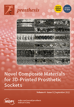
Cover Story (view full-size image): The 3D printing revolution is reshaping the methods by which clinicians build prosthetic sockets for persons with limb loss. However, off-the-shelf 3D printing materials commercially available today are not well-suited for use in structural biomedical devices. This cover story describes a methodical effort to develop new composite materials for prosthetic sockets. Sandwich structure composites were manufactured utilizing 3D-printed face sheets and a vacuum infiltrated resin core. Several of the composites showed improvements in tensile strength and reduced anisotropy compared to their control samples. A proof-of-concept prosthetic socket was manufactured using these novel materials, demonstrating the potential of these composites to be used in practice. View this paper
  • Issues are regarded as officially published after their release is announced to the table of contents alert mailing list.
  • You may sign up for e-mail alerts to receive table of contents of newly released issues.
  • PDF is the official format for papers published in both, html and pdf forms. To view the papers in pdf format, click on the "PDF Full-text" link, and use the free Adobe Reader to open them.
Order results
Result details
Section
Select all
Export citation of selected articles as:
11 pages, 5047 KiB  
Article
Scanning Electron Microscopy Analyses of Dental Implant Abutments Debonded from Monolithic Zirconia Restorations Using Heat Treatment: An In Vitro Study
by Marco Tallarico, Łukasz Zadrożny, Nino Squadrito, Leonardo Colella, Maurizio Gualandri, Daniele Montanari, Gianantonio Zibetti, Simone Santini, Witold Chromiński, Edoardo Baldoni, Silvio Mario Meloni, Aurea Immacolata Lumbau and Milena Pisano
Prosthesis 2022, 4(3), 500-510; https://doi.org/10.3390/prosthesis4030041 - 19 Sep 2022
Viewed by 2258
Abstract
Aim: The aim of this in vitro study is to present a debonding protocol developed to remove a screw-retained, monolithic, zirconia restoration from its titanium-base abutment, and to microscopically evaluate the abutment integrity at both the prosthetic and connection levels. Materials and Methods: [...] Read more.
Aim: The aim of this in vitro study is to present a debonding protocol developed to remove a screw-retained, monolithic, zirconia restoration from its titanium-base abutment, and to microscopically evaluate the abutment integrity at both the prosthetic and connection levels. Materials and Methods: A total of 30 samples were tested. Each sample consisted of a monolithic zirconia restoration bonded on a titanium link abutment. Five different shapes were designed and fabricated. Randomly, one-third of the Ti-link abutments were subjected to an anodizing process. Then, all the zirconia samples were bonded to the Ti-link abutments according to a pre-established protocol. Forty-eight hours later, the samples were debonded according to the experimental protocol. The outcomes were evaluated by a visual inspection with an optical microscope, scanning electron microscopy (SEM), and chemical composition analysis. Results: Thirty samples were collected and visually analyzed. Seven samples were randomly evaluated via scanning electron microscopy. In all the examinations, no relevant changes were reported. Chemical composition analysis also relieved no changes in the chemical structure of the titanium. Conclusions: The titanium-base abutments do not alter the structure and properties of the material, not creating phase changes or the birth of oxides such as to induce fragility. Further clinical studies with longer follow-up periods are needed to confirm these preliminary results. Full article
(This article belongs to the Collection Oral Implantology: Current Aspects and Future Perspectives)
Show Figures

Figure 1

10 pages, 10053 KiB  
Article
Finite Element Analysis of Zirconia Dental Implant
by Luca Fiorillo, Dario Milone, Danilo D’Andrea, Dario Santonocito, Giacomo Risitano, Gabriele Cervino and Marco Cicciù
Prosthesis 2022, 4(3), 490-499; https://doi.org/10.3390/prosthesis4030040 - 8 Sep 2022
Cited by 17 | Viewed by 3144
Abstract
Titanium dental implants have had new competitors in recent years, such as fixtures made of zirconia, which promise better aesthetics. The purpose of this study is to evaluate their mechanical performance in silico (Finite Element Analysis). The investigation was performed on a single [...] Read more.
Titanium dental implants have had new competitors in recent years, such as fixtures made of zirconia, which promise better aesthetics. The purpose of this study is to evaluate their mechanical performance in silico (Finite Element Analysis). The investigation was performed on a single tooth Patent™ Dental Implant (Zircon Medical®, Altendorf, Switzerland) in two configurations: without offset (Test I) and with offset (Test II, 1.5 mm within the cortical bone). The Patent Implant system consists of two components: the implant with integrated abutment and the fibreglass post. The components of the dental implants were tested using a compression load of 400 N along the implant axis. The results showed that the chewing load generates stress distribution on the bone, therefore, the offset configuration should be avoided. Full article
(This article belongs to the Collection Oral Implantology: Current Aspects and Future Perspectives)
Show Figures

Figure 1

10 pages, 1174 KiB  
Article
The Effects of Vitamin E Analogues α-Tocopherol and γ-Tocotrienol on the Human Osteocyte Response to Ultra-High Molecular Weight Polyethylene Wear Particles
by Renee T. Ormsby, Kunihiro Hosaka, Andreas Evdokiou, Andreani Odysseos, David M. Findlay, Lucian B. Solomon and Gerald J. Atkins
Prosthesis 2022, 4(3), 480-489; https://doi.org/10.3390/prosthesis4030039 - 2 Sep 2022
Cited by 2 | Viewed by 1858
Abstract
Polyethylene (PE) liners are a common bearing surface of orthopaedic prostheses. Wear particles of ultra-high molecular weight PE (UHMWPE) contribute to periprosthetic osteolysis, a major cause of aseptic loosening. Vitamin E is added to some PE liners to prevent oxidative degradation. Osteocytes, an [...] Read more.
Polyethylene (PE) liners are a common bearing surface of orthopaedic prostheses. Wear particles of ultra-high molecular weight PE (UHMWPE) contribute to periprosthetic osteolysis, a major cause of aseptic loosening. Vitamin E is added to some PE liners to prevent oxidative degradation. Osteocytes, an important cell type for controlling both bone mineralisation and bone resorption, have been shown to respond UHMWPE particles by upregulating pro-osteoclastogenic and osteocytic osteolysis. Here, we examined the effects of the vitamin E analogues α-tocopherol and γ-tocotrienol alone or in the context of UHMWPE particles on human osteocyte gene expression and mineralisation behaviour. Human osteoblasts differentiated to an osteocyte-like stage were exposed to UHMWPE wear particles in the presence or absence of either α-Tocopherol or γ-Tocotrienol. Both α-Tocopherol and γ-Tocotrienol induced antioxidant-related gene expression. UHMWPE particles independently upregulated antioxidant gene expression, suggesting an effect of wear particles on oxidative stress. Both vitamin E analogues strongly induced OPG mRNA expression and γ-Tocotrienol also inhibited RANKL mRNA expression, resulting in a significantly reduced RANKL:OPG mRNA ratio (p < 0.01) overall. UHMWPE particles reversed the suppressive effect of α-Tocopherol but not of γ-Tocotrienol on this pro-osteoclastogenic index. UHMWPE particles also upregulated osteocytic-osteolysis related gene expression. Vitamin E analogues alone or in combination with UHMWPE particles also resulted in upregulation of these genes. Consistent with this, both vitamin E analogues promoted calcium release from mineralised cultures of osteocyte-like cells. Our findings suggest that while vitamin E may suppress osteocyte support of osteoclastogenesis in the presence of UHMWPE particles, the antioxidant effect may induce osteocytic osteolysis, which could promote periprosthetic osteolysis. It will be important to conduct further studies of vitamin E to determine the long-term effects of its inclusion in prosthetic materials. Full article
Show Figures

Figure 1

12 pages, 15820 KiB  
Review
CAD/CAM Abutments versus Stock Abutments: An Update Review
by Simone Gallo, Maurizio Pascadopoli, Matteo Pellegrini, Federica Pulicari, Mattia Manfredini, Paolo Zampetti, Francesco Spadari, Carlo Maiorana and Andrea Scribante
Prosthesis 2022, 4(3), 468-479; https://doi.org/10.3390/prosthesis4030038 - 16 Aug 2022
Cited by 9 | Viewed by 2969
Abstract
With the evolution of CAD/CAM technology, custom titanium and/or zirconia abutments are increasingly being used, leading to several comparisons in the literature, both mechanical and aesthetic, to evaluate performance differences between these two types of abutments. Therefore, the aim of this comprehensive review [...] Read more.
With the evolution of CAD/CAM technology, custom titanium and/or zirconia abutments are increasingly being used, leading to several comparisons in the literature, both mechanical and aesthetic, to evaluate performance differences between these two types of abutments. Therefore, the aim of this comprehensive review is to present the most recent data on the latest comparisons between CAD/CAM and stock abutment applications. The PICO model was used to perform this review, through a literature search of the PubMed (MEDLINE) and Scopus electronic databases. CAD/CAM abutments allow individualization of abutment parameters with respect to soft tissue, allow increased fracture toughness, predict the failure mode, show no change in the fracture toughness over time, reduce the prosthetic steps, and reduce the functional implant prosthesis score and pain perceived by patients in the early stages. The advantages associated with the use of stock abutments mainly concern the risk of corrosion, time spent, cost, and fit, evaluated in vitro, in the implant–abutment connection. Equal conditions are present regarding the mechanical characteristics during dynamic cycles, screw loss, radiographic fit, and degree of micromotion. Further randomized controlled clinical trials should be conducted to evaluate the advantages reported to date, following in vitro studies about titanium and/or zirconia stock abutments. Full article
(This article belongs to the Collection Oral Implantology: Current Aspects and Future Perspectives)
Show Figures

Figure 1

10 pages, 726 KiB  
Review
The Conometric Connection for the Implant-Supported Fixed Prosthesis: A Narrative Review
by Saturnino Marco Lupi, Claudia Todaro, Dario De Martis, Paola Blasi, Ruggero Rodriguez y Baena and Stefano Storelli
Prosthesis 2022, 4(3), 458-467; https://doi.org/10.3390/prosthesis4030037 - 16 Aug 2022
Cited by 7 | Viewed by 3068
Abstract
Aim: The conometric concept was proposed as a possible connection between the abutment and the prosthetic coping. This research aimed to review the features and possible clinical uses of this connection in an implant-supported fixed prosthesis. Methods: An electronic search was conducted on [...] Read more.
Aim: The conometric concept was proposed as a possible connection between the abutment and the prosthetic coping. This research aimed to review the features and possible clinical uses of this connection in an implant-supported fixed prosthesis. Methods: An electronic search was conducted on an online database for the topic in object; articles published in international literature were considered and the research gave 17 results, and 6 parameters were analyzed. Results: This connection eliminated the possibility of cement residues in the subgingival region, reducing the risk of inflammation of peri-implant soft and hard tissues; not having to remove the cement residues, it is possible to place the margins in more apical portions, improving the aesthetics outcomes of the rehabilitations. It is also known that the retention by means of a screw causes a weakening of the restoration. The retentive force is adequate for fixed rehabilitation even after a high number of insertion–disengagement cycles; in vitro studies have also shown a high bacterial sealing. Implant rehabilitation using preformed components, such as conometric hoods, is helpful for CAD/CAM, so a digital workflow is possible. Several types of prosthesis were presented, all of which demonstrated adequate clinical performance in the follow-up observation. Conclusions: This type of connection seems to be suitable to support fixed implant rehabilitations, but long-term clinical studies are needed to validate this system. Full article
(This article belongs to the Special Issue Feature Review Papers for Prosthesis)
Show Figures

Figure 1

11 pages, 1320 KiB  
Article
Investigating the Cytotoxicity of Dual-Cure Bulk-Fill Resin Materials on L929 Cells
by Safiye Selin Koymen, Nazmiye Donmez, Vildan Betul Yenigun, Fatemeh Bahadori and Abdurrahim Kocyigit
Prosthesis 2022, 4(3), 447-457; https://doi.org/10.3390/prosthesis4030036 - 15 Aug 2022
Cited by 5 | Viewed by 2359
Abstract
The aim of this in vitro study was to investigate cytotoxic effects of dual-cure bulk-fill resin materials polymerized with a third-generation LED light-curing unit (LCU) on L929 fibroblast cells in terms of morphology and viability. Three novel dual-cure, flowable bulk-fill materials (Fill-Up!™), a [...] Read more.
The aim of this in vitro study was to investigate cytotoxic effects of dual-cure bulk-fill resin materials polymerized with a third-generation LED light-curing unit (LCU) on L929 fibroblast cells in terms of morphology and viability. Three novel dual-cure, flowable bulk-fill materials (Fill-Up!™), a bioactive material (ACTIVA™ BioACTIVE-RESTORATIVE™), and a dual-cure bulk-fill composite material (HyperFIL® HAp) polymerized by LED LCU (VALO™ Cordless) were tested. Each material was placed in plastic rings (4 mm × 5 mm) in a single layer. Unpolymerized rings filled with each material were placed in direct contact with cells and then polymerized. After polymerization, the removed medium was readded to wells. In this study, four control groups were performed: the medium-free control group, medium control group, physical control group, and light applied control group. Three samples were prepared from each group. After 24 h, the morphology of cells was examined and a WST-1 test was performed. The percentage of cell viability (PCV) of each group was calculated. The experiment was repeated three times. Data were analyzed by a Kruskal–Wallis Test and a Mann–Whitney U test. p < 0.05 was considered significant. The PCV of all groups were found to be significantly lower than the medium control group (p < 0.05). The lowest PCV was obtained in HyperFIL® Hap, while highest was in the Fill-Up!™. In the morphology of cells related to the experimental groups, it was observed that the spindle structures of cells were disrupted due to cytotoxicity; cells became rounded and intercellular space increased. There were no significant differences between the control groups (p > 0.05). All control groups showed acceptable PCV (>70%) and cells were spindle-like, similar to the original fibroblast cells. It can be suggested that clinicians should pay attention when applying dual-cure bulk-fill materials in deep cavities, or they should use a liner material under these materials. Full article
(This article belongs to the Special Issue Bioactive Materials for Dental and Maxillofacial Repair)
Show Figures

Figure 1

3 pages, 828 KiB  
Editorial
Lessons Learnt from Götz of the Iron Hand
by Andreas Otte
Prosthesis 2022, 4(3), 444-446; https://doi.org/10.3390/prosthesis4030035 - 15 Aug 2022
Cited by 3 | Viewed by 1717
Abstract
Recently, we reported the three-dimensional computer-aided design (3D-CAD) reconstruction of the first “Iron Hand” of the famous Franconian knight, Götz von Berlichingen (1480–1562), who lost his right hand by a cannon ball splinter injury in 1504 in the War of the Succession of [...] Read more.
Recently, we reported the three-dimensional computer-aided design (3D-CAD) reconstruction of the first “Iron Hand” of the famous Franconian knight, Götz von Berlichingen (1480–1562), who lost his right hand by a cannon ball splinter injury in 1504 in the War of the Succession of Landshut [...] Full article
Show Figures

Figure 1

20 pages, 1646 KiB  
Review
Contemporary Tools for the Cure against Pernicious Microorganisms: Micro-/Nanorobots
by Mustafa Sami Ozaydin, Lorin Doganturk, Fulden Ulucan-Karnak, Ozan Akdogan and Pelin Erkoc
Prosthesis 2022, 4(3), 424-443; https://doi.org/10.3390/prosthesis4030034 - 5 Aug 2022
Cited by 9 | Viewed by 4175
Abstract
One of the most pressing concerns to global public health is the emergence of drug-resistant pathogenic microorganisms due to increased unconscious antibiotic usage. With the rising antibiotic resistance, existing antimicrobial agents lose their effectiveness over time. This indicates that newer and more effective [...] Read more.
One of the most pressing concerns to global public health is the emergence of drug-resistant pathogenic microorganisms due to increased unconscious antibiotic usage. With the rising antibiotic resistance, existing antimicrobial agents lose their effectiveness over time. This indicates that newer and more effective antimicrobial agents and methods should be investigated. Many studies have shown that micro-/nanorobots exhibit promise in the treatment of microbial infections with their great properties, such as the intrinsic antimicrobial activities owing to their oxidative stress induction and metal ion release capabilities, and effective and autonomous delivery of antibiotics to the target area. In addition, they have multiple simultaneous mechanisms of action against microbes, which makes them remarkable in antimicrobial activity. This review focuses on the antimicrobial micro-/nanorobots and their strategies to impede biofilm formation, following a brief introduction of the latest advancements in micro-/nanorobots, and their implementations against various bacteria, and other microorganisms. Full article
(This article belongs to the Special Issue Antiviral and Antimicrobial Surface Design Strategies)
Show Figures

Graphical abstract

10 pages, 1796 KiB  
Article
Analysis of CBCT Bone Density Using the Hounsfield Scale
by Leonard Morar, Grigore Băciuț, Mihaela Băciuț, Simion Bran, Horațiu Colosi, Avram Manea, Oana Almășan and Cristian Dinu
Prosthesis 2022, 4(3), 414-423; https://doi.org/10.3390/prosthesis4030033 - 3 Aug 2022
Cited by 19 | Viewed by 9296
Abstract
Assessing bone density in prospective dental implant sites is crucial both for choosing the implant type and for planning a drilling procedure that will ensure the implant’s primary stability and osseointegration. This study aimed to investigate possible differences between the bone densities of [...] Read more.
Assessing bone density in prospective dental implant sites is crucial both for choosing the implant type and for planning a drilling procedure that will ensure the implant’s primary stability and osseointegration. This study aimed to investigate possible differences between the bone densities of various edentulous sites in the maxilla and mandible. The study was conducted on a group of forty partly edentulous patients who underwent radiological examination by scanning the areas of interest using cone beam computed tomography (CBCT). Hounsfield units (HU) were analyzed using dedicated software. Higher HU were observed at the site of mandibular central incisors compared to the site of maxillary central incisors. The HU values in the mandibular first molars region were higher than those of the maxillary first molars. Buccal vs. lingual or palatal cortical HU values did not differ significantly. Within the limitations of this study, it can be stated that an objective assessment of site-specific bone density before the installation of dental implants may provide valuable clinical information for the selection of implant size and the planning of a patient-specific drilling protocol. Full article
(This article belongs to the Collection Oral Implantology: Current Aspects and Future Perspectives)
Show Figures

Figure 1

20 pages, 4937 KiB  
Technical Note
Reimagining Prosthetic Control: A Novel Body-Powered Prosthetic System for Simultaneous Control and Actuation
by Vikranth H. Nagaraja, Jhonatan da Ponte Lopes and Jeroen H. M. Bergmann
Prosthesis 2022, 4(3), 394-413; https://doi.org/10.3390/prosthesis4030032 - 29 Jul 2022
Cited by 9 | Viewed by 15196
Abstract
Globally, the most popular upper-limb prostheses are powered by the human body. For body-powered (BP) upper-limb prostheses, control is provided by changing the tension of (Bowden) cables to open or close the terminal device. This technology has been around for centuries, and very [...] Read more.
Globally, the most popular upper-limb prostheses are powered by the human body. For body-powered (BP) upper-limb prostheses, control is provided by changing the tension of (Bowden) cables to open or close the terminal device. This technology has been around for centuries, and very few BP alternatives have been presented since. This paper introduces a new BP paradigm that can overcome certain limitations of the current cabled systems, such as a restricted operation space and user discomfort caused by the harness to which the cables are attached. A new breathing-powered system is introduced to give the user full control of the hand motion anywhere in space. Users can regulate their breathing, and this controllable airflow is then used to power a small Tesla turbine that can accurately control the prosthetic finger movements. The breathing-powered device provides a novel prosthetic option that can be used without limiting any of the user’s body movements. Here we prove that it is feasible to produce a functional breathing-powered prosthetic hand and show the models behind it along with a preliminary demonstration. This work creates a step-change in the potential BP options available to patients in the future. Full article
Show Figures

Figure 1

11 pages, 756 KiB  
Article
Clinical Evaluation of Cement-Retained Implant-Supported CAD/CAM Monolithic Zirconia Single Crowns in Posterior Areas: Results of a 6-Year Prospective Clinical Study
by Roberto Sorrentino, Gennaro Ruggiero, Eralda Toska, Renato Leone and Fernando Zarone
Prosthesis 2022, 4(3), 383-393; https://doi.org/10.3390/prosthesis4030031 - 27 Jul 2022
Cited by 8 | Viewed by 3243
Abstract
Background: Different technologies and materials can be used for implant-supported restorations in posterior areas. Our study aimed to evaluate the clinical performance of CAD/CAM implant-supported monolithic zirconia single crowns (ZrSCs) cemented onto titanium abutments with a dual-curing resin cement after 6 years of [...] Read more.
Background: Different technologies and materials can be used for implant-supported restorations in posterior areas. Our study aimed to evaluate the clinical performance of CAD/CAM implant-supported monolithic zirconia single crowns (ZrSCs) cemented onto titanium abutments with a dual-curing resin cement after 6 years of use. Methods: Fifty patients in need of one single crown in posterior regions were enrolled. The patients were recalled for a follow-up after 6 months and then yearly for a total observation period of 6 years. The biological and technical complications were examined using criteria set by the United States Public Health Service (USPHS), whereas visual analogue scales (VASs) were used to evaluate the patients’ aesthetic and functional satisfaction. Results: No patient was lost at follow-up; both the survival and success rates were 100%. No fractures, decementation, peri-implantitis, or soft tissue inflammation were recorded. Forty-six restorations were rated Alpha in each evaluated parameter of the USPHS’s criteria. According to the patients’ VASs assessments, the overall function of the ZrSCs was 8.4 (±2.1), whereas the overall aesthetic was 8.7 (±0.7). Conclusions: Monolithic zirconia single crowns cemented with dual-curing resin cements onto titanium abutments can be considered an effective clinical option in posterior regions after 6 years of function. Full article
(This article belongs to the Special Issue Novel Zirconia Materials Applied in Dental Prostheses)
Show Figures

Figure 1

14 pages, 2216 KiB  
Article
Prevalence and Spatial Patterns of Major Limb Loss in the Acholi Sub-Region of Uganda
by Jonathan J. Huck, Pamela Atim, Emmanuel B. Moro and Mahesh Nirmalan
Prosthesis 2022, 4(3), 369-382; https://doi.org/10.3390/prosthesis4030030 - 19 Jul 2022
Cited by 5 | Viewed by 2560
Abstract
There is a widely reported yet poorly understood preponderance of major limb loss in Northern Uganda, caused in large part by prolonged civil war. Access to rehabilitation services is extremely limited, and there is no understanding of how many people have major limb [...] Read more.
There is a widely reported yet poorly understood preponderance of major limb loss in Northern Uganda, caused in large part by prolonged civil war. Access to rehabilitation services is extremely limited, and there is no understanding of how many people have major limb loss, nor how many have had access to medical or rehabilitative services. This research represents the first prevalence study of disability and major limb loss in the Acholi sub-region of Northern Uganda, as well as a novel evaluation of spatial patterns in cases of major limb loss. We conservatively estimate that there are c. 10,117 people with major limb loss in Acholi (c. 0.5% of the population) who require long-term rehabilitation services, as well as c. 150,512 people with other disabilities (c. 8.2% of the population). We also demonstrate that people with major limb loss are spread throughout the region (as opposed to clustered in specific locations) and are disproportionately male, older, and less well educated than the general population. This research demonstrates a clear inadequacy in both the supply and service delivery models of rehabilitation services for those with major limb loss in the study area and provides much-needed insight into the challenges that must be addressed for an effective system to be put in place. Full article
(This article belongs to the Section Orthopedics and Rehabilitation)
Show Figures

Figure 1

16 pages, 20316 KiB  
Case Report
Digital Workflow for Prosthetically Driven Implants Placement and Digital Cross Mounting: A Retrospective Case Series
by Marco Tallarico, Davide Galiffi, Roberto Scrascia, Maurizio Gualandri, Łukasz Zadrożny, Marta Czajkowska, Santo Catapano, Francesco Grande, Edoardo Baldoni, Aurea Immacolata Lumbau, Silvio Mario Meloni and Milena Pisano
Prosthesis 2022, 4(3), 353-368; https://doi.org/10.3390/prosthesis4030029 - 18 Jul 2022
Cited by 9 | Viewed by 4027
Abstract
Fully digital workflow in implant dentistry is ever increasing. Treatment of partial edentulous cases is well-documented; nevertheless, complete edentulous cases are still a challenge. To present several innovations in the treatment of complete edentulous patients using digital solutions, both for implant placement and [...] Read more.
Fully digital workflow in implant dentistry is ever increasing. Treatment of partial edentulous cases is well-documented; nevertheless, complete edentulous cases are still a challenge. To present several innovations in the treatment of complete edentulous patients using digital solutions, both for implant placement and restoration delivery, was the objective of this study. It was designed as a retrospective case series study, aimed to tune further research with larger sample size, and a longer follow-up. Patients requiring complete, implant-supported restoration were asked to participate in this study. Enrolled patients were treated with four implants, immediate loading and a definitive complete arch restoration. Patients were treated using computer-assisted, template-based surgery. Multi-piece surgical templates were used to accurately place the implants, to manage the bone if needed and to make immediate loading procedure quicker and easier. After osseointegration period, definitive, extra-oral, digital impressions were taken using newly developed scan analogs, connected in the patient mouth using temporary cylinders and stabilized by means of the low-shrinkage, flowable, resin composite. Outcomes were implant and prosthesis survival rate, complications, accuracy, and patient satisfaction. Radiographic evaluation performed with a preliminary, radiopaque aluminum try-in, was used to test the accuracy of the digital impressions. Overall, 20 implants were placed in five patients. All the implants osseointegrated without complications. One impression was taken a second time due to inaccuracy of the aluminum tray-in. Finally, all of the patients were completely satisfied with both surgical and prosthetic procedures. Within the limitations of this case series, multi-piece surgical templates showed promising results improving the clinician’s confidence in the case of bone reduction, post-extractive implants and immediate loading. The prosthetic template increased the trueness of the digital impression for complete edentulous patients. Finally, even if an impression was performed again, the scan-analog used for extra-oral chair-side digital impressions seemed to be a promising tool. Continuous improvements and further study are needed to confirm these preliminary results. Full article
(This article belongs to the Collection Oral Implantology: Current Aspects and Future Perspectives)
Show Figures

Figure 1

15 pages, 3642 KiB  
Review
A Review on Risk Management of Coronavirus Disease 19 (COVID-19) Infection in Dental Practice: Focus on Prosthodontics and All-Ceramic Materials
by Roberto Sorrentino, Michele Basilicata, Gennaro Ruggiero, Maria Irene Di Mauro, Renato Leone, Patrizio Bollero and Fernando Zarone
Prosthesis 2022, 4(3), 338-352; https://doi.org/10.3390/prosthesis4030028 - 5 Jul 2022
Cited by 5 | Viewed by 2519
Abstract
Background: A novel β-coronavirus infection (COVID-19) was first detected in Wuhan city, spreading rapidly to other countries and leading to a pandemic. Dental professionals and patients are exposed to a high risk of COVID-19 infection, particularly in the prosthodontic practice, because of the [...] Read more.
Background: A novel β-coronavirus infection (COVID-19) was first detected in Wuhan city, spreading rapidly to other countries and leading to a pandemic. Dental professionals and patients are exposed to a high risk of COVID-19 infection, particularly in the prosthodontic practice, because of the bio-aerosol produced during teeth preparation with dental handpieces and the strict contact with oral fluids during impression making. This paper aimed to provide an overview to limit the risk of transmission of COVID-19 infections during prosthetic procedures in dental offices. Methods: An electronic search was conducted on the electronic databases of PubMed/Medline, Google Scholar, Embase, Scopus, Dynamed, and Open Grey with the following queries: (COVID-19) AND/OR (SARS-CoV-2) AND/OR (Coronavirus) AND/OR (contaminated surface) AND/OR (cross-infection) AND/OR (Prosthodontics) AND/OR (dental ceramic) AND/OR (glass-ceramic). A manual search was performed as well. Results: From the 1023 collected records, 32 papers were included. Conclusions: Dental offices are at high risk of spreading SARS-CoV-2 infection due to the close contact with patients and continuous exposure to saliva during dental procedures. Therefore, pre-check triages via telephone, decontamination, the disinfection of impressions, the sterilization of scanner tips, and the use of specific personal protective equipment, dental high-speed handpieces with dedicated anti-retraction valves, and effective mouthwashes are strongly recommended. Full article
(This article belongs to the Special Issue Feature Review Papers for Prosthesis)
Show Figures

Figure 1

21 pages, 37396 KiB  
Article
Prosthetic Sockets: Tensile Behavior of Vacuum Infiltrated Fused Deposition Modeling Sandwich Structure Composites
by Isaac A. Cabrera, Parker J. Hill, Win-Ying Zhao, Trinity C. Pike, Marc A. Meyers, Ramesh R. Rao and Albert Y. M. Lin
Prosthesis 2022, 4(3), 317-337; https://doi.org/10.3390/prosthesis4030027 - 22 Jun 2022
Cited by 4 | Viewed by 3838
Abstract
The development of novel materials will enable a new generation of prosthetic devices to be built with additive manufacturing (AM). Vacuum infiltrated sandwich structure composites are a promising approach for building prosthetic sockets via AM. In this paper, we test the tensile properties [...] Read more.
The development of novel materials will enable a new generation of prosthetic devices to be built with additive manufacturing (AM). Vacuum infiltrated sandwich structure composites are a promising approach for building prosthetic sockets via AM. In this paper, we test the tensile properties of 18 different composite material configurations using ASTM D638. These composites were manufactured using a custom vacuum infiltration method and had varying filament materials, infiltrated matrix materials, and print directions. Several material-matrix-print composites showed higher ultimate tensile strengths and reduced anisotropy compared to full-infill control samples. However, the mechanical properties of these composites were limited by a large degree of porosity due to the manufacturing method. Still, the results were sufficiently promising to create a proof of concept prosthetic socket via the vacuum infiltration method. Future research should focus on reducing porosity defects and investigating additional material-matrix-print combinations. Full article
(This article belongs to the Special Issue 3D Printing Strategies for Limb Prostheses)
Show Figures

Figure 1

Previous Issue
Next Issue
Back to TopTop