Next Issue
Volume 6, March
Previous Issue
Volume 5, September


Endocrines, Volume 5, Issue 4 (December 2024) – 11 articles

Cover Story (view full-size image): 
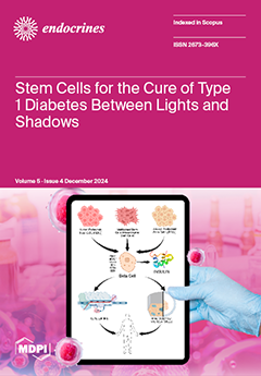
Human pluripotent stem cells—whether derived from embryos (hESC) or adult tissues induced to pluripotency (hiPSC)—and adult multipotent mesenchymal stem cells (MSCs) might represent a possible solution to the restricted availability of insulin-producing cells for T1D cell therapy. However, ethical barriers prevent the wide use of hESC, hiPSC has not yet reached clinical feasibility, and MSCs still need to prove their clinical efficacy. A Vertex Inc. clinical trial for hESC in T1D immunosuppressed patients showed that four patients suspended insulin therapy after several months due to heavy financial burden. Proof of principle for stem cell use has been demonstrated, but time is required to expedite the process, eliminate immunosuppression, and cut back on expenses to validate stem cell types that are universally acceptable. View this paper
  • Issues are regarded as officially published after their release is announced to the table of contents alert mailing list.
  • You may sign up for e-mail alerts to receive table of contents of newly released issues.
  • PDF is the official format for papers published in both, html and pdf forms. To view the papers in pdf format, click on the "PDF Full-text" link, and use the free Adobe Reader to open them.
Order results
Result details
Section
Select all
Export citation of selected articles as:
8 pages, 263 KiB  
Article
Testing for Endothelial Dysfunction in Children with Rare Genetic Variants of Obesity
by Ilham Farhat and Vivian L. Chin
Endocrines 2024, 5(4), 613-620; https://doi.org/10.3390/endocrines5040044 - 10 Dec 2024
Viewed by 612
Abstract
Background: Endothelial dysfunction (ED), an early indicator of atherosclerosis, is a well-established predictor of cardiovascular disease. This study investigates ED in children with rare genetic variants linked to obesity and explores the prevalence of these variants in pediatric obesity. Methods: Under an IRB-approved [...] Read more.
Background: Endothelial dysfunction (ED), an early indicator of atherosclerosis, is a well-established predictor of cardiovascular disease. This study investigates ED in children with rare genetic variants linked to obesity and explores the prevalence of these variants in pediatric obesity. Methods: Under an IRB-approved protocol, 54 pediatric patients with severe obesity (BMI ≥ 97%) were screened using the Rhythm® Genetics Test panel between 2021 and 2024 through the Uncovering Rare Obesity® program. This clinically approved buccal test targets 79 genes and one chromosomal region. ED was measured using EndoPAT® (Itamar Medical Ltd by Zoll US based company) in 24 of these patients with related gene variants and compared to controls. Results: Genetic screening: Among the 54 patients screened, 42 (78%) had positive genetic variants, including 18 males and 24 females. The most common variants were PCNT (n = 9), BBS (n = 9), SEMA3 (n = 8), ALMS1 (n = 6), SDCCAG8 (n = 5) and MC4R (n = 5). Endothelial dysfunction: Included 21 subjects with a mean age of 12 years and a mean BMI of 33.31 kg/m². The mean RHI for patients with the PCNT variant was significantly higher (1.34, p = 0.02) compared to controls, but no significant differences were observed for other variants, including BBS, ALMS1, and SH2B1. Conclusions: In this small pilot study, no significant difference in ED was found between children with or without genetic variants, except for PCNT, which showed a higher RHI. Targeted genetic screening revealed 78% with identified pathogenic variants like MC4R, which can clinically guide therapy. Further research is needed to investigate ED in children with obesity variants. Full article
(This article belongs to the Section Obesity, Diabetes Mellitus and Metabolic Syndrome)
13 pages, 3724 KiB  
Case Report
Multiple Osteoporotic Fractures in Cushing Syndrome: A Case Report and a Review of the Literature
by Ariadni Spyroglou, Panagiota Konstantakou, Konstantinos Iliakopoulos, Vasiliki Themelidi, Dorothea Tsekoura, Denise Kolomodi, Georgios Kyriakopoulos, Pantelis Antonakis, Konstantinos Bramis, Achilles Chatziioannou, George Mastorakos, Manousos M. Konstadoulakis and Krystallenia I. Alexandraki
Endocrines 2024, 5(4), 600-612; https://doi.org/10.3390/endocrines5040043 - 6 Dec 2024
Viewed by 632
Abstract
Background: Osteoporotic fractures are a common clinical sign of Cushing syndrome (CS). However, Cushing diagnosis can occur years after this clinical manifestation. Methods: Herein, we present the case of a 45-year-old woman who was referred to our department for further diagnosis and treatment. [...] Read more.
Background: Osteoporotic fractures are a common clinical sign of Cushing syndrome (CS). However, Cushing diagnosis can occur years after this clinical manifestation. Methods: Herein, we present the case of a 45-year-old woman who was referred to our department for further diagnosis and treatment. Results: The patient was already under treatment for arterial hypertension and osteoporosis and was recently diagnosed with dyslipidemia and type 2 diabetes. She reported several previous fractures starting already 8 years before presentation. An adrenal CS was diagnosed, and the patient was treated with laparoscopic adrenalectomy, with a subsequent complete remission of her hypercortisolism. This case report presenting a particularly long time gap between initial osteoporosis signs and the final diagnosis underlines the need for an investigation into secondary osteoporosis in low-energy fractures also in the peripheral skeleton. In this context, we performed a literature review, including case reports with fragility fractures that were attributed to endogenous CS. Conclusions: In summary, a delayed diagnosis of CS in patients with a previous accumulation of such fractures is a worrisome observation and should be considered in everyday clinical practice in order to improve the timely diagnosis and treatment of CS. Full article
(This article belongs to the Special Issue Feature Papers in Endocrines: 2024)
Show Figures

Figure 1

15 pages, 723 KiB  
Review
Nutritional Studies Evaluating Ketogenic Diets as a Treatment for Obesity and Obesity-Associated Morbidities: Underlying Mechanisms and Potential for Clinical Implementation
by Luciano Pirola, Karolina Górecka, Carol Gois Leandro and Aneta Balcerczyk
Endocrines 2024, 5(4), 585-599; https://doi.org/10.3390/endocrines5040042 - 3 Dec 2024
Viewed by 1772
Abstract
Background: The ketogenic diet (KD), characterized by high-fat content, virtually no carbohydrates, and adequate protein intake, induces a metabolic state resembling fasting, as the absence of carbohydrates forces the body to rely on the energetic supply from hepatically produced ketone bodies using free [...] Read more.
Background: The ketogenic diet (KD), characterized by high-fat content, virtually no carbohydrates, and adequate protein intake, induces a metabolic state resembling fasting, as the absence of carbohydrates forces the body to rely on the energetic supply from hepatically produced ketone bodies using free fatty acids as substrate. While the KD is clinically used in pharmacologically refractory epilepsy and specific genetic conditions such as GLUT1 deficiency, recent research suggests that, due to its “fasting mimicking” properties, the KD may also beneficially affect obesity and obesity-associated metabolic diseases. Results: Here, we present a narrative review discussing completed and ongoing nutritional studies in human volunteers specifically addressing the potential of the ketogenic diet as an anti-obesity approach and, from a larger perspective, as an intervention to ameliorate the metabolic state in conditions such as type 1 and 2 diabetes and polycystic ovary syndrome (PCOS). Published studies as well as ongoing clinical trials will be discussed. Efficacy and safety considerations will be discussed, as well as the potential physiological mechanisms mediating the effects of the KD in humans in the context of the (i) energy balance model (EBM) and (ii) carbohydrate–insulin model (CIM) of body weight control. Conclusion: Ketogenic diets may be beneficial to attenuate obesity and improve obesity-related metabolic disease, and here, we try, based on current evidence, to define the boundaries of the KD’s nutritional and clinical usefulness. Full article
(This article belongs to the Section Obesity, Diabetes Mellitus and Metabolic Syndrome)
Show Figures

Figure 1

19 pages, 350 KiB  
Review
The Latest Trends in the Management of Type 1 and Type 2 Diabetes Mellitus
by Cory DeClue, Matthew Gonzalez, Anna Beth Bradley, Barbara G. Carranza-Leon and Gitanjali Srivastava
Endocrines 2024, 5(4), 566-584; https://doi.org/10.3390/endocrines5040041 - 2 Dec 2024
Viewed by 1255
Abstract
Over the past few years, we have witnessed many advances in the understanding of diabetes and its management. Greater insight into pathogenesis has led to the approval of the first immunopreventative therapy for T1DM. We are using non-insulin agents more for nephro- and [...] Read more.
Over the past few years, we have witnessed many advances in the understanding of diabetes and its management. Greater insight into pathogenesis has led to the approval of the first immunopreventative therapy for T1DM. We are using non-insulin agents more for nephro- and cardioprotection than glucose-lowering effects while leaning on advancing technology to use insulin more safely. We now recognize that over half of T1DM is diagnosed in adulthood, the prevalence of obesity in patients with T1DM matches that of the general population, and rates of pediatric T2DM have dramatically risen amongst marginalized youths in recent years. Diabetes is now considered more of a heterogenous disease state than ever before, and practitioners will need to be familiar with these endotypes as personalized medicine replaces standardized treatment approaches. To this end, this article aims to summarize recent findings in an easily digestible manner so that providers may be more familiar with this ever-growing complex disease state. Full article
(This article belongs to the Special Issue Advances in Diabetes Care)
19 pages, 836 KiB  
Review
Granulosa Cells: Central Regulators of Female Fertility
by Luis Fernando Schütz and Isadora M. Batalha
Endocrines 2024, 5(4), 547-565; https://doi.org/10.3390/endocrines5040040 - 27 Nov 2024
Viewed by 1242
Abstract
Background: Granulosa cells are somatic cells within the ovarian follicle. As the primary site of estradiol production, they are critical regulators of several aspects of female reproduction. This review aims to provide an overview of the physiology of mammalian granulosa cells and their [...] Read more.
Background: Granulosa cells are somatic cells within the ovarian follicle. As the primary site of estradiol production, they are critical regulators of several aspects of female reproduction. This review aims to provide an overview of the physiology of mammalian granulosa cells and their importance for female fertility. Methods: the literature about the function and regulation of granulosa cells was reviewed. Results: a comprehensive summary and discussion of the role of granulosa cells on ovarian steroidogenesis and folliculogenesis, as well as factors that control granulosa cells function, are presented. Conclusion: The functions of granulosa cells are regulated by a plethora of intra- and extra-ovarian factors via autocrine, paracrine, and endocrine pathways, which creates a complex regulatory network. A comprehensive understanding of granulosa cells’ physiology is vital for the development of innovative strategies to enhance reproductive outcomes in several species. Full article
(This article belongs to the Section Female Reproductive System and Pregnancy Endocrinology)
Show Figures

Figure 1

9 pages, 284 KiB  
Article
Exploring the Associations of Neck Circumference, Blood Pressure, CRP, and Insulin Resistance on the Visceral Adiposity Index: Insights from a Cross-Sectional Study
by Lucas Fornari Laurindo, Francine Cruz Camargo, Alessandra Perfeito, Bruno Benedito Ciano, Clara Tainá Coelho, Gleice Assis Apolinário, Isabela do Nascimento Vicentin, Jéssica Cambui Andreasi, Beatriz Leme Boaro, Ricardo José Tofano, Cláudia Rucco Penteado Detregiachi, Jesselina Francisco dos Santos Haber, Sandra Maria Barbalho, Lance Alan Sloan, Kátia Portero Sloan, Antonelly Cassio Alves de Carvalho, Marie Oshiiwa, Patrícia Cincotto dos Santos Bueno, Tereza Laís Menegucci Zutim, Rebeca Maria Siqueira da Silva, Eduardo Federighi Baisi Chagas, Marcelo Dib Bechara and Karina R. Quesadaadd Show full author list remove Hide full author list
Endocrines 2024, 5(4), 538-546; https://doi.org/10.3390/endocrines5040039 - 20 Nov 2024
Viewed by 880
Abstract
Background: The visceral adiposity index (VAI) is a composite marker designed to quantify visceral adiposity and its metabolic implications. It integrates anthropometric (such as waist circumference and BMI) and metabolic parameters (including triglyceride levels and HDL cholesterol), providing a more comprehensive assessment of [...] Read more.
Background: The visceral adiposity index (VAI) is a composite marker designed to quantify visceral adiposity and its metabolic implications. It integrates anthropometric (such as waist circumference and BMI) and metabolic parameters (including triglyceride levels and HDL cholesterol), providing a more comprehensive assessment of visceral fat distribution than traditional measures alone. Higher VAI values are indicative of increased visceral adiposity and have been linked to heightened cardiovascular risk and metabolic disturbances. In recent years, understanding the complex interplay between metabolic factors and cardiovascular health has become increasingly important. Methods: This cross-sectional study delves into the influence of neck circumference (NC), blood pressure (BP), C-reactive protein (CRP), and insulin resistance on the VAI among outpatient cardiology patients, offering insights into sex-specific disparities and the utility of VAI as a diagnostic tool for assessing visceral adiposity and associated cardiovascular risks. Results: The sample comprised 268 outpatient cardiology patients (152 men, 116 women). Men, averaging 55.4 years old (SD = 14.4), exhibited significantly higher VAI values than women, with robust correlations found between VAI and markers of insulin resistance (Insulin: ρ = −0.167, p = 0.006; HOMA-IR: ρ = −0.163, p = 0.007). Analysis across VAI quartiles highlighted distinct patterns, revealing lower NC and elevated systolic blood pressure (SBP) values in higher VAI categories. Despite these associations, multiple linear regression controlling for age and sex demonstrated a limited predictive capacity of NC, BP, CRP, and lipid profiles on VAI (R2 range: 0.001–0.011). Conclusions: These findings underscore sex-specific disparities and suggest that VAI serves as a modest yet valuable tool in assessing visceral adiposity and associated cardiovascular risks. Full article
(This article belongs to the Special Issue Advances in Diabetes Care)
9 pages, 844 KiB  
Review
Precocious Puberty in Boys with NR0B1 Variants
by Atsushi Hattori and Maki Fukami
Endocrines 2024, 5(4), 529-537; https://doi.org/10.3390/endocrines5040038 - 15 Nov 2024
Viewed by 641
Abstract
Precocious puberty (PP) requires appropriate management to prevent short adult height, psychosocial issues, and other adverse outcomes. Genetic diagnosis potentially improves the management of PP. Pathogenic NR0B1 variants, which typically cause X-linked adrenal hypoplasia congenita, can also affect gonadal function. While boys with [...] Read more.
Precocious puberty (PP) requires appropriate management to prevent short adult height, psychosocial issues, and other adverse outcomes. Genetic diagnosis potentially improves the management of PP. Pathogenic NR0B1 variants, which typically cause X-linked adrenal hypoplasia congenita, can also affect gonadal function. While boys with NR0B1 variants usually exhibit hypogonadotropic hypogonadism during adolescence, previous reports have suggested that minipuberty, a physiological transient activation of the hypothalamic–pituitary–gonadal axis during infancy, occurs in these patients and can persist beyond a typical duration. In rare cases, NR0B1 variants cause PP. PP associated with NR0B1 variants has unique features such as early onset and high serum testosterone levels that are often disproportionate to testicular size. Three underlying mechanisms have been proposed for the association between PP and NR0B1 variants: (1) adrenocorticotropic hormone (ACTH)-dependent, (2) gonadotropin-dependent, and (3) ACTH- and gonadotropin-independent mechanisms. The factors contributing to PP vary among cases. Determining the underlying mechanisms is crucial for adopting appropriate therapeutic strategies to control PP. However, as the detailed molecular networks mediating these mechanisms are largely unclear, further research is needed to pave the way for a more effective and personalized management of patients with PP associated with NR0B1 variants. Full article
(This article belongs to the Section Pediatric Endocrinology and Growth Disorders)
Show Figures

Figure 1

13 pages, 1369 KiB  
Article
Analytical Characteristics and Clinical Performance of Anti-Müllerian Hormone Immunoassay on the ADVIA Centaur® System: A Comparison with Other Chemiluminescent Methods
by Jelena Bogdanovic, Kaitlin Freeman, Chadwick Brown, Rachel Singleton, Millie Behera, Jeanne E. O’Brien, Edward Zbella and Robert H. Christenson
Endocrines 2024, 5(4), 516-528; https://doi.org/10.3390/endocrines5040037 - 5 Nov 2024
Viewed by 966
Abstract
Background: Serum anti-Müllerian hormone (AMH) levels and antral follicle count are key in evaluating ovarian reserve (OR) for fertility. The performance of the Siemens Healthineers AMH assay was assessed on the ADVIA Centaur® System. Methods: Analytical characteristics, clinical performance, and method comparison [...] Read more.
Background: Serum anti-Müllerian hormone (AMH) levels and antral follicle count are key in evaluating ovarian reserve (OR) for fertility. The performance of the Siemens Healthineers AMH assay was assessed on the ADVIA Centaur® System. Methods: Analytical characteristics, clinical performance, and method comparison studies were performed in a prospective cohort of 532 women at fertility clinics. Serum AMH levels were determined using ADVIA Centaur, Beckman Access®, and Roche Elecsys® assays. Results: The limit of quantitation for the ADVIA Centaur AMH assay was 0.030 ng/mL. Repeatability was ≤2.9% CV, within-lab repeatability was ≤3.2% CV, and reproducibility was ≤4.4% CV. Results using serum or lithium heparin sample types were equivalent. Diagnostic sensitivity across assays ranged from 77.3% to 90.2% and specificity ranged from 51.0 to 71.0%; corresponding positive and negative predictive values ranged from 66.6% to 74.3% and 74.2% to 83.0%, respectively. Receiver operating characteristic analyses demonstrated that the assays have a high probability for discriminating between diminished–normal and high OR. ADVIA and Beckman assays agreed according to ADVIA = 1.00 × Beckman + 0.014 ng/mL, τ = 0.909, while a more modest correlation of ADVIA = 1.41 × Roche − 0.024 ng/mL, τ = 0.777 was observed with Roche assay. Conclusions: The ADVIA Centaur assay demonstrates acceptable analytical characteristics and clinical performance comparable to the Roche AMH assay and is essentially interchangeable with the Beckman AMH assay for reliable OR assessment. Full article
(This article belongs to the Section Female Reproductive System and Pregnancy Endocrinology)
Show Figures

Figure 1

15 pages, 869 KiB  
Review
The Interplay of the Mammalian Brain and Thyroid Hormones, and the Threat of Endocrine-Disrupting Chemicals
by Nuha Ahmad Dsouki, Bruno Fiorelini Pereira, Roberta Goes da Silva, Vinicius Gonçalves Rodrigues, Rafaella da Silva Brito, Marina Malta Letro Kizys, Maria Izabel Chiamolera, Rui Monteiro Maciel, Caroline Serrano-Nascimento and Gisele Giannocco
Endocrines 2024, 5(4), 501-515; https://doi.org/10.3390/endocrines5040036 - 25 Oct 2024
Viewed by 1409
Abstract
Introduction: During the formation of neural circuits, the developing brain demonstrates extraordinary plasticity, heavily influenced by hormones. These chemical messengers interact with specific receptors to regulate vital physiological functions. The thyroid gland plays a pivotal role in maintaining hormonal balance and guiding brain [...] Read more.
Introduction: During the formation of neural circuits, the developing brain demonstrates extraordinary plasticity, heavily influenced by hormones. These chemical messengers interact with specific receptors to regulate vital physiological functions. The thyroid gland plays a pivotal role in maintaining hormonal balance and guiding brain development. However, emerging threats like endocrine-disrupting chemicals (EDCs) can interfere with this intricate system. EDCs are exogenous substances that can mimic, enhance, or block the actions of endogenous hormones, disrupting hormonal signaling in the brain at various developmental stages. Exposure can impair cognitive function and behavior due to disruptions in thyroid function. Studies indicate that mixtures of EDCs negatively impact brain development, leading to lower IQ and behavioral problems. Reducing EDC exposure through regulations and public awareness is crucial, and further research is needed to elucidate their mechanisms. Conclusions: Protecting vulnerable populations, such as pregnant women and children, is essential through prompt regulatory measures. Full article
(This article belongs to the Special Issue Feature Papers in Endocrines: 2024)
Show Figures

Figure 1

23 pages, 454 KiB  
Review
The Bidirectional Link between Major Depressive Disorder and Type 2 Diabetes: The Role of Inflammation
by Alexandra M. Bodnaruc, Mathilde Roberge, Isabelle Giroux and Céline Aguer
Endocrines 2024, 5(4), 478-500; https://doi.org/10.3390/endocrines5040035 - 9 Oct 2024
Viewed by 1063
Abstract
Background/Objectives: There is a bidirectional relationship between major depressive disorder (MDD) and type 2 diabetes (T2D), as MDD increases the risk of T2D by 38% to 67%, and T2D increases the risk of MDD by 15% to 33%. Many factors contribute to [...] Read more.
Background/Objectives: There is a bidirectional relationship between major depressive disorder (MDD) and type 2 diabetes (T2D), as MDD increases the risk of T2D by 38% to 67%, and T2D increases the risk of MDD by 15% to 33%. Many factors contribute to the occurrence of comorbid MDD and T2D, including converging pathophysiological pathways like inflammation. The objective of this review was to comprehensively summarize available evidence on the relationship between MDD, T2D, and inflammation. Results: Although the precise mechanisms linking T2D and MDD are still not fully understood, shared inflammatory mechanisms likely contributes to the heightened risk of developing this comorbidity. To date, the evidence supports that chronic low-grade inflammation is a feature of both MDD and T2D and has been shown to interact with pathways that are relevant to the development of both chronic disorders, including the hypothalamic–pituitary–adrenal (HPA) axis, neuroplastic processes, gut microbiome, insulin resistance, and adipose tissue dysfunction. Through their impact on inflammation, dietary and physical activity interventions can play a role in the risk and management of MDD and T2D. Conclusions: Deepening our understanding of the mechanisms underlying the augmented inflammatory responses observed in individuals with the MDD and T2D comorbidity is essential for tailoring appropriate therapeutic strategies. Full article
(This article belongs to the Special Issue Feature Papers in Endocrines: 2024)
13 pages, 2005 KiB  
Review
Human Stem Cell Therapy for the Cure of Type 1 Diabetes Mellitus (T1D): A Hurdle Course between Lights and Shadows
by Riccardo Calafiore, Giovanni Luca, Francesco Gaggia and Giuseppe Basta
Endocrines 2024, 5(4), 465-477; https://doi.org/10.3390/endocrines5040034 - 5 Oct 2024
Viewed by 1944
Abstract
Background: T1D is a severe metabolic disorder due to selective autoimmune pancreatic islet β-cell killing, which results in complete abrogation of endogenous insulin secretion. The affected patients, once the disease is clinically overt, must immediately undertake insulin supplementation according to intensive therapy regimens [...] Read more.
Background: T1D is a severe metabolic disorder due to selective autoimmune pancreatic islet β-cell killing, which results in complete abrogation of endogenous insulin secretion. The affected patients, once the disease is clinically overt, must immediately undertake insulin supplementation according to intensive therapy regimens to prevent the onset of acute and chronic complications, some of them potentially lethal. Replacement of the destroyed β-cells with fresh and vital pancreatic endocrine tissue, either of the whole organ or isolated islets transplantation, started a few decades ago with progressively encouraging results, although exogenous insulin withdrawal was obtained in a minor cohort of the treated patients. The restricted availability of donor organs coupled with general immunosuppression treatment of recipients to avoid graft immune rejection may, at least partially, explain the limited success achieved by these procedures. Results: The introduction of pluripotent stem cells (either of human embryonic origin or adult cells genetically induced to pluripotency) that can be differentiated toward insulin secretory β-like cells could provide an indefinite resource for insulin-producing cells (IPCs). Conclusions: Because the use of human embryos may encounter ethical problems, employment of adult multipotent mesenchymal stem cells (MSCs) extracted from several tissues may represent an alternative option. MSCs are associated with strong immunoregulatory properties that can alter early stages of β-cell-directed autoimmunity in T1D, other than holding the potential to differentiate themselves into β-like cells. Lights and shadows of these new strategies for the potential cure of T1D and their advancement state are reviewed. Full article
(This article belongs to the Section Endocrine Immunology, Cytokines and Cell Signaling)
Show Figures

Figure 1

Previous Issue
Next Issue
Back to TopTop