Imaging Biomarker in Oncology

A topical collection in Cancers (ISSN 2072-6694). This collection belongs to the section "Cancer Biomarkers".

Viewed by 68764

Editor


E-Mail Website
Collection Editor
Radiology Unit, Department of Medical Surgical Sciences and Translational Medicine, Sant’Andrea University Hospital, Sapienza-University of Rome, 00100 Rome, Italy
Interests: imaging; oncology; CT; MRI; artificial intelligence; radiomics; response to therapy
Special Issues, Collections and Topics in MDPI journals

Topical Collection Information

Dear Colleagues,

Cancers affect a large percentage of chronic and fragile patients, and their workup and therapeutic options depend on timing of diagnosis, staging, and tumor aggressiveness. In such a scenario, imaging has been acquiring a pivotal role in oncology by providing some relevant non-invasive biomarkers extracted from medical images. Conventional radiological evaluation, based on qualitative and subjective assessment of images, is the recognized imaging approach to study cancer patients. However, the main drawbacks of subjective images assessment are represented by their subjective nature and the difficulty to reproduce the measures. Today, imaging is shifting from a qualitative to a quantitative approach, especially in tumor diagnosis, prognosis prediction, and assessment of response to therapy. In such a scenario, radiomics has been overcoming conventional imaging using dedicated software having the ability to extract high dimensional data having the expectancy to provide objective and quantitative non-invasive imaging biomarkers in cancer patients. Medical images could narrow some quantitative data reflecting microenvironmental tumor heterogeneity, neoplasm phenotypes and heterogeneity, usually correlated with tumor aggressiveness and patient prognosis. Then, radiomic data could be extracted, analyzed, and integrated with clinical data using the strengths of Artificial Intelligence, helpful to overcome the main limitations of traditional tumor management, starting from conventional lesion biopsy, often affected by bias in tumor sampling, lack of repeatability, and possible procedure complications.

In the new era of target therapy, imaging is expected to become a supporting tool for clinicians by providing non-invasive biomarkers. In this manner, imaging could have the ability to outline the profile of tumors based on several features extracted from medical images, assessed with both qualitative and quantitative approaches.

Dr. Damiano Caruso
Collection Editor

Manuscript Submission Information

Manuscripts should be submitted online at www.mdpi.com by registering and logging in to this website. Once you are registered, click here to go to the submission form. Manuscripts can be submitted until the deadline. All submissions that pass pre-check are peer-reviewed. Accepted papers will be published continuously in the journal (as soon as accepted) and will be listed together on the collection website. Research articles, review articles as well as communications are invited. For planned papers, a title and short abstract (about 100 words) can be sent to the Editorial Office for announcement on this website.

Submitted manuscripts should not have been published previously, nor be under consideration for publication elsewhere (except conference proceedings papers). All manuscripts are thoroughly refereed through a single-blind peer-review process. A guide for authors and other relevant information for submission of manuscripts is available on the Instructions for Authors page. Cancers is an international peer-reviewed open access semimonthly journal published by MDPI.

Please visit the Instructions for Authors page before submitting a manuscript. The Article Processing Charge (APC) for publication in this open access journal is 2900 CHF (Swiss Francs). Submitted papers should be well formatted and use good English. Authors may use MDPI's English editing service prior to publication or during author revisions.

Keywords

  • imaging
  • tumor diagnosis
  • prognosis prediction
  • oncology
  • radiomics
  • biomarker
Published Papers (28 papers)

2024

Jump to: 2023, 2022, 2021

12 pages, 2042 KiB  
Article
Analysis of Molecular Imaging Biomarkers Derived from [18F]FDG PET/CT in mCRPC: Whole-Body Total Lesion Glycolysis (TLG) Predicts Overall Survival in Patients Undergoing [225Ac]Ac-PSMA-617-Augmented [177Lu]Lu-PSMA-617 Radioligand Therapy
by Caroline Burgard, Fadi Khreish, Lukas Dahlmanns, Arne Blickle, Moritz B. Bastian, Tilman Speicher, Stephan Maus, Andrea Schaefer-Schuler, Mark Bartholomä, Sven Petto, Samer Ezziddin and Florian Rosar
Cancers 2024, 16(20), 3532; https://doi.org/10.3390/cancers16203532 - 19 Oct 2024
Viewed by 800
Abstract
Background/Objectives: The augmentation of [177Lu]Lu-PSMA-617 radioligand therapy by alpha emitting [225Ac]Ac-PSMA-617, known as the tandem therapy concept, is a promising escalating treatment option in advanced mCRPC. In this study, we evaluated the value of [18F]FDG PET/CT-derived molecular [...] Read more.
Background/Objectives: The augmentation of [177Lu]Lu-PSMA-617 radioligand therapy by alpha emitting [225Ac]Ac-PSMA-617, known as the tandem therapy concept, is a promising escalating treatment option in advanced mCRPC. In this study, we evaluated the value of [18F]FDG PET/CT-derived molecular imaging biomarkers for predicting response and outcome to PSMA tandem RLT in n = 33 patients with insufficient response on [177Lu]Lu-PSMA-617 monotherapy. Methods: Six different molecular imaging parameters at baseline, i.e., before initiation of PSMA tandem RLT with respect to SUVmax, SUVpeak, SUV5, SUVmean, metabolic tumor volume (MTV), and total lesion glycolysis (TLG) were tested for association with response and overall survival (OS). Results: After the initiation of augmentation, 24.2% of patients with a previously insufficient response experienced partial remission, and 39.4% experienced stable disease. The median OS was 7 months (95% CI: 4–11 months). None of the tested parameters were able to predict the response (all p > 0.529). In contrast, the [18F]FDG PET/CT-derived whole-body molecular imaging parameter TLG was significantly (p = 0.029) associated with OS of patients undergoing [225Ac]Ac-PSMA-617 augmented [177Lu]Lu-PSMA-617 RLT after insufficient response to [177Lu]Lu-PSMA-617 monotherapy. Conclusion: Implementing [18F]FDG PET/CT in the management of PSMA-RLT in clinical practice may contribute to outcome prediction and provide a route to more individualized management in mCRPC. Full article
Show Figures

Figure 1

2023

Jump to: 2024, 2022, 2021

15 pages, 1768 KiB  
Article
Associations of Multiparametric Breast MRI Features, Tumor-Infiltrating Lymphocytes, and Immune Gene Signature Scores Following a Single Dose of Trastuzumab in HER2-Positive Early-Stage Breast Cancer
by Laura C. Kennedy, Anum S. Kazerouni, Bonny Chau, Debosmita Biswas, Rebeca Alvarez, Grace Durenberger, Suzanne M. Dintzis, Sasha E. Stanton, Savannah C. Partridge and Vijayakrishna Gadi
Cancers 2023, 15(17), 4337; https://doi.org/10.3390/cancers15174337 - 30 Aug 2023
Viewed by 1462
Abstract
Dynamic biomarkers that permit the real-time monitoring of the tumor microenvironment response to therapy are an unmet need in breast cancer. Breast magnetic resonance imaging (MRI) has demonstrated value as a predictor of pathologic complete response and may reflect immune cell changes in [...] Read more.
Dynamic biomarkers that permit the real-time monitoring of the tumor microenvironment response to therapy are an unmet need in breast cancer. Breast magnetic resonance imaging (MRI) has demonstrated value as a predictor of pathologic complete response and may reflect immune cell changes in the tumor microenvironment. The purpose of this pilot study was to investigate the value of breast MRI features as early markers of treatment-induced immune response. Fourteen patients with early HER2+ breast cancer were enrolled in a window-of-opportunity study where a single dose of trastuzumab was administered and both tissue and MRIs were obtained at the pre- and post-treatment stages. Functional diffusion-weighted and dynamic contrast-enhanced MRI tumor measures were compared with tumor-infiltrating lymphocytes (TILs) and RNA immune signature scores. Both the pre-treatment apparent diffusion coefficient (ADC) and the change in peak percent enhancement (DPE) were associated with increased tumor-infiltrating lymphocytes with trastuzumab therapy (r = −0.67 and -0.69, p < 0.01 and p < 0.01, respectively). Low pre-treatment ADC and a greater decrease in PE in response to treatment were also associated with immune-activated tumor microenvironments as defined by RNA immune signatures. Breast MRI features hold promise as biomarkers of early immune response to treatment in HER2+ breast cancer. Full article
Show Figures

Figure 1

12 pages, 1536 KiB  
Article
A Radiomics-Based Classifier for the Progression of Oropharyngeal Cancer Treated with Definitive Radiotherapy
by Darwin A. Garcia, Elizabeth B. Jeans, Lindsay K. Morris, Satomi Shiraishi, Brady S. Laughlin, Yi Rong, Jean-Claude M. Rwigema, Robert L. Foote, Michael G. Herman and Jing Qian
Cancers 2023, 15(14), 3715; https://doi.org/10.3390/cancers15143715 - 22 Jul 2023
Viewed by 1529
Abstract
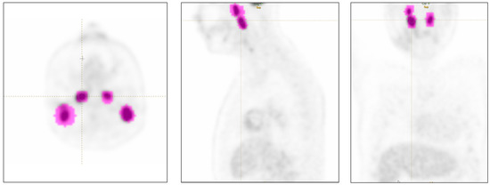
In this study, we investigated whether radiomics features from pre-treatment positron emission tomography (PET) images could be used to predict disease progression in patients with HPV-positive oropharyngeal cancer treated with definitive proton or x-ray radiotherapy. Machine learning models were built using a dataset [...] Read more.
In this study, we investigated whether radiomics features from pre-treatment positron emission tomography (PET) images could be used to predict disease progression in patients with HPV-positive oropharyngeal cancer treated with definitive proton or x-ray radiotherapy. Machine learning models were built using a dataset from Mayo Clinic, Rochester, Minnesota (n = 72) and tested on a dataset from Mayo Clinic, Phoenix, Arizona (n = 22). A total of 71 clinical and radiomics features were considered. The Mann–Whitney U test was used to identify the top 2 clinical and top 20 radiomics features that were significantly different between progression and progression-free patients. Two dimensionality reduction methods were used to define two feature sets (manually filtered or machine-driven). A forward feature selection scheme was conducted on each feature set to build models of increased complexity (number of input features from 1 to 6) and evaluate model robustness and overfitting. The machine-driven features had superior performance and were less prone to overfitting compared to the manually filtered features. The four-variable Gaussian Naïve Bayes model using the ‘Radiation Type’ clinical feature and three machine-driven features achieved a training accuracy of 79% and testing accuracy of 77%. These results demonstrate that radiomics features can provide risk stratification beyond HPV-status to formulate individualized treatment and follow-up strategies. Full article
Show Figures

Figure 1

14 pages, 3458 KiB  
Article
Radiomics of Tumor Heterogeneity in 18F-FDG-PET-CT for Predicting Response to Immune Checkpoint Inhibition in Therapy-Naïve Patients with Advanced Non-Small-Cell Lung Cancer
by David Ventura, Philipp Schindler, Max Masthoff, Dennis Görlich, Matthias Dittmann, Walter Heindel, Michael Schäfers, Georg Lenz, Eva Wardelmann, Michael Mohr, Peter Kies, Annalen Bleckmann, Wolfgang Roll and Georg Evers
Cancers 2023, 15(8), 2297; https://doi.org/10.3390/cancers15082297 - 14 Apr 2023
Cited by 5 | Viewed by 2843
Abstract
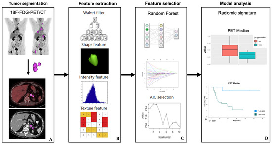
We aimed to evaluate the predictive and prognostic value of baseline 18F-FDG-PET-CT (PET-CT) radiomic features (RFs) for immune checkpoint-inhibitor (CKI)-based first-line therapy in advanced non-small-cell lung cancer (NSCLC) patients. In this retrospective study 44 patients were included. Patients were treated with either [...] Read more.
We aimed to evaluate the predictive and prognostic value of baseline 18F-FDG-PET-CT (PET-CT) radiomic features (RFs) for immune checkpoint-inhibitor (CKI)-based first-line therapy in advanced non-small-cell lung cancer (NSCLC) patients. In this retrospective study 44 patients were included. Patients were treated with either CKI-monotherapy or combined CKI-based immunotherapy–chemotherapy as first-line treatment. Treatment response was assessed by the Response Evaluation Criteria in Solid Tumors (RECIST). After a median follow-up of 6.4 months patients were stratified into “responder” (n = 33) and “non-responder” (n = 11). RFs were extracted from baseline PET and CT data after segmenting PET-positive tumor volume of all lesions. A Radiomics-based model was developed based on a Radiomics signature consisting of reliable RFs that allow classification of response and overall progression using multivariate logistic regression. These RF were additionally tested for their prognostic value in all patients by applying a model-derived threshold. Two independent PET-based RFs differentiated well between responders and non-responders. For predicting response, the area under the curve (AUC) was 0.69 for “PET-Skewness” and 0.75 predicting overall progression for “PET-Median”. In terms of progression-free survival analysis, patients with a lower value of PET-Skewness (threshold < 0.2014; hazard ratio (HR) 0.17, 95% CI 0.06–0.46; p < 0.001) and higher value of PET-Median (threshold > 0.5233; HR 0.23, 95% CI 0.11–0.49; p < 0.001) had a significantly lower probability of disease progression or death. Our Radiomics-based model might be able to predict response in advanced NSCLC patients treated with CKI-based first-line therapy. Full article
Show Figures

Figure 1

16 pages, 1619 KiB  
Article
Combination of FDG PET/CT Radiomics and Clinical Parameters for Outcome Prediction in Patients with Hodgkin’s Lymphoma
by Claudia Ortega, Yael Eshet, Anca Prica, Reut Anconina, Sarah Johnson, Danny Constantini, Sareh Keshavarzi, Roshini Kulanthaivelu, Ur Metser and Patrick Veit-Haibach
Cancers 2023, 15(7), 2056; https://doi.org/10.3390/cancers15072056 - 30 Mar 2023
Cited by 8 | Viewed by 2061
Abstract
Purpose: The aim of the study is to evaluate the prognostic value of a joint evaluation of PET and CT radiomics combined with standard clinical parameters in patients with HL. Methods: Overall, 88 patients (42 female and 46 male) with a median age [...] Read more.
Purpose: The aim of the study is to evaluate the prognostic value of a joint evaluation of PET and CT radiomics combined with standard clinical parameters in patients with HL. Methods: Overall, 88 patients (42 female and 46 male) with a median age of 43.3 (range 21–85 years) were included. Textural analysis of the PET/CT images was performed using freely available software (LIFE X). 65 radiomic features (RF) were evaluated. Univariate and multivariate models were used to determine the value of clinical characteristics and FDG PET/CT radiomics in outcome prediction. In addition, a binary logistic regression model was used to determine potential predictors for radiotherapy treatment and odds ratios (OR), with 95% confidence intervals (CI) reported. Features relevant to survival outcomes were assessed using Cox proportional hazards to calculate hazard ratios with 95% CI. Results: albumin (p = 0.034) + ALP (p = 0.028) + CT radiomic feature GLRLM GLNU mean (p = 0.012) (Area under the curve (AUC): 95% CI (86.9; 100.0)—Brier score: 3.9, 95% CI (0.1; 7.8) remained significant independent predictors for PFS outcome. PET-SHAPE Sphericity (p = 0.033); CT grey-level zone length matrix with high gray-level zone emphasis (GLZLM SZHGE mean (p = 0.028)); PARAMS XSpatial Resampling (p = 0.0091) as well as hemoglobin results (p = 0.016) remained as independent factors in the final model for a binary outcome as predictors of the need for radiotherapy (AUC = 0.79). Conclusion: We evaluated the value of baseline clinical parameters as well as combined PET and CT radiomics in HL patients for survival and the prediction of the need for radiotherapy treatment. We found that different combinations of all three factors/features were independently predictive of the here evaluated endpoints. Full article
Show Figures

Figure 1

15 pages, 2865 KiB  
Article
Quantitative MRI to Characterize Hypoxic Tumors in Comparison to FMISO PET/CT for Radiotherapy in Oropharynx Cancers
by Pierrick Gouel, Françoise Callonnec, Franchel-Raïs Obongo-Anga, Pierre Bohn, Emilie Lévêque, David Gensanne, Sébastien Hapdey, Romain Modzelewski, Pierre Vera and Sébastien Thureau
Cancers 2023, 15(6), 1918; https://doi.org/10.3390/cancers15061918 - 22 Mar 2023
Cited by 3 | Viewed by 2373
Abstract
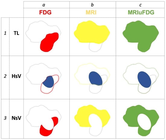
Intratumoral hypoxia is associated with a poor prognosis and poor response to treatment in head and neck cancers. Its identification would allow for increasing the radiation dose to hypoxic tumor subvolumes. 18F-FMISO PET imaging is the gold standard; however, quantitative multiparametric MRI could [...] Read more.
Intratumoral hypoxia is associated with a poor prognosis and poor response to treatment in head and neck cancers. Its identification would allow for increasing the radiation dose to hypoxic tumor subvolumes. 18F-FMISO PET imaging is the gold standard; however, quantitative multiparametric MRI could show the presence of intratumoral hypoxia. Thus, 16 patients were prospectively included and underwent 18F-FDG PET/CT, 18F-FMISO PET/CT, and multiparametric quantitative MRI (DCE, diffusion and relaxometry T1 and T2 techniques) in the same position before treatment. PET and MRI sub-volumes were segmented and classified as hypoxic or non-hypoxic volumes to compare quantitative MRI parameters between normoxic and hypoxic volumes. In total, 13 patients had hypoxic lesions. The Dice, Jaccard, and overlap fraction similarity indices were 0.43, 0.28, and 0.71, respectively, between the FDG PET and MRI-measured lesion volumes, showing that the FDG PET tumor volume is partially contained within the MRI tumor volume. The results showed significant differences in the parameters of SUV in FDG and FMISO PET between patients with and without measurable hypoxic lesions. The quantitative MRI parameters of ADC, T1 max mapping and T2 max mapping were different between hypoxic and normoxic subvolumes. Quantitative MRI, based on free water diffusion and T1 and T2 mapping, seems to be able to identify intra-tumoral hypoxic sub-volumes for additional radiotherapy doses. Full article
Show Figures

Figure 1

16 pages, 3515 KiB  
Article
Deep Learning Applied to Raman Spectroscopy for the Detection of Microsatellite Instability/MMR Deficient Colorectal Cancer
by Nathan Blake, Riana Gaifulina, Lewis D. Griffin, Ian M. Bell, Manuel Rodriguez-Justo and Geraint M. H. Thomas
Cancers 2023, 15(6), 1720; https://doi.org/10.3390/cancers15061720 - 11 Mar 2023
Cited by 4 | Viewed by 2182
Abstract
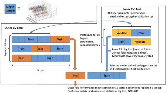
Defective DNA mismatch repair is one pathogenic pathway to colorectal cancer. It is characterised by microsatellite instability which provides a molecular biomarker for its detection. Clinical guidelines for universal testing of this biomarker are not met due to resource limitations; thus, there is [...] Read more.
Defective DNA mismatch repair is one pathogenic pathway to colorectal cancer. It is characterised by microsatellite instability which provides a molecular biomarker for its detection. Clinical guidelines for universal testing of this biomarker are not met due to resource limitations; thus, there is interest in developing novel methods for its detection. Raman spectroscopy (RS) is an analytical tool able to interrogate the molecular vibrations of a sample to provide a unique biochemical fingerprint. The resulting datasets are complex and high-dimensional, making them an ideal candidate for deep learning, though this may be limited by small sample sizes. This study investigates the potential of using RS to distinguish between normal, microsatellite stable (MSS) and microsatellite unstable (MSI-H) adenocarcinoma in human colorectal samples and whether deep learning provides any benefit to this end over traditional machine learning models. A 1D convolutional neural network (CNN) was developed to discriminate between healthy, MSI-H and MSS in human tissue and compared to a principal component analysis–linear discriminant analysis (PCA–LDA) and a support vector machine (SVM) model. A nested cross-validation strategy was used to train 30 samples, 10 from each group, with a total of 1490 Raman spectra. The CNN achieved a sensitivity and specificity of 83% and 45% compared to PCA–LDA, which achieved a sensitivity and specificity of 82% and 51%, respectively. These are competitive with existing guidelines, despite the low sample size, speaking to the molecular discriminative power of RS combined with deep learning. A number of biochemical antecedents responsible for this discrimination are also explored, with Raman peaks associated with nucleic acids and collagen being implicated. Full article
Show Figures

Figure 1

3 pages, 171 KiB  
Editorial
Editorial for Special Issue on Imaging Biomarker in Oncology
by Michela Polici, Andrea Laghi and Damiano Caruso
Cancers 2023, 15(4), 1071; https://doi.org/10.3390/cancers15041071 - 8 Feb 2023
Cited by 2 | Viewed by 1452
Abstract
Imaging biomarkers are the expression of quantitative imaging and have become central in the management of cancers, proving consistent and objective information to outline an appropriate workflow for oncologic patients [...] Full article

2022

Jump to: 2024, 2023, 2021

10 pages, 1619 KiB  
Article
Correlation between ADC Histogram-Derived Metrics and the Time to Metastases in Resectable Pancreatic Adenocarcinoma
by Riccardo De Robertis, Luisa Tomaiuolo, Francesca Pasquazzo, Luca Geraci, Giuseppe Malleo, Roberto Salvia and Mirko D’Onofrio
Cancers 2022, 14(24), 6050; https://doi.org/10.3390/cancers14246050 - 8 Dec 2022
Cited by 3 | Viewed by 1314
Abstract
Background: A non-invasive method to improve the prognostic stratification would be clinically beneficial in patients with resectable pancreatic adenocarcinoma (PDAC). The aim of this study was to correlate conventional magnetic resonance (MR) features and the metrics derived from the histogram analysis of apparent [...] Read more.
Background: A non-invasive method to improve the prognostic stratification would be clinically beneficial in patients with resectable pancreatic adenocarcinoma (PDAC). The aim of this study was to correlate conventional magnetic resonance (MR) features and the metrics derived from the histogram analysis of apparent diffusion coefficient (ADC) maps, with the risk and the time to metastases (TTM) after surgery in patients with PDAC. Methods: pre-operative MR examinations of 120 patients were retrospectively analyzed. Patients were grouped according to the presence (M+) or absence (M−) of metastases during follow-up. Conventional MR features and histogram-derived metrics were compared between M+ and M− patients using the Fisher’s or Mann–Whitney tests; receiver operating characteristic (ROC) curves were constructed for the features that showed a significant difference between groups. A Cox regression analysis was performed to identify the features with a significant effect on the TTM, and Kaplan–Meier curves were constructed for significant features. Results: 68.3% patients developed metastases over a mean follow-up time of 29 months (range, 3–54 months). ADC skewness and kurtosis were significantly higher in M+ than in M− patients (p < 0.001). Skewness had a significant effect on the risk of metastases (hazard ratio—HR = 5.22, p < 0.001). Patients with an ADC skewness ≥0.23 had a significantly shorter TTM than those with a skewness <0.22 (11.7 vs. 30.8 months, p < 0.001). Conclusions: pre-operative histogram analysis of ADC maps provides parameters correlated to the metastatic potential of PDAC. Higher ADC skewness seems to be associated with a significantly shorter TTM in patients with resectable PDAC. Full article
Show Figures

Figure 1

13 pages, 2690 KiB  
Article
Two-Stage Deep Learning Model for Automated Segmentation and Classification of Splenomegaly
by Aymen Meddeb, Tabea Kossen, Keno K. Bressem, Noah Molinski, Bernd Hamm and Sebastian N. Nagel
Cancers 2022, 14(22), 5476; https://doi.org/10.3390/cancers14225476 - 8 Nov 2022
Cited by 4 | Viewed by 3107
Abstract
Splenomegaly is a common cross-sectional imaging finding with a variety of differential diagnoses. This study aimed to evaluate whether a deep learning model could automatically segment the spleen and identify the cause of splenomegaly in patients with cirrhotic portal hypertension versus patients with [...] Read more.
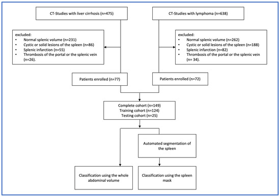
Splenomegaly is a common cross-sectional imaging finding with a variety of differential diagnoses. This study aimed to evaluate whether a deep learning model could automatically segment the spleen and identify the cause of splenomegaly in patients with cirrhotic portal hypertension versus patients with lymphoma disease. This retrospective study included 149 patients with splenomegaly on computed tomography (CT) images (77 patients with cirrhotic portal hypertension, 72 patients with lymphoma) who underwent a CT scan between October 2020 and July 2021. The dataset was divided into a training (n = 99), a validation (n = 25) and a test cohort (n = 25). In the first stage, the spleen was automatically segmented using a modified U-Net architecture. In the second stage, the CT images were classified into two groups using a 3D DenseNet to discriminate between the causes of splenomegaly, first using the whole abdominal CT, and second using only the spleen segmentation mask. The classification performances were evaluated using the area under the receiver operating characteristic curve (AUC), accuracy (ACC), sensitivity (SEN), and specificity (SPE). Occlusion sensitivity maps were applied to the whole abdominal CT images, to illustrate which regions were important for the prediction. When trained on the whole abdominal CT volume, the DenseNet was able to differentiate between the lymphoma and liver cirrhosis in the test cohort with an AUC of 0.88 and an ACC of 0.88. When the model was trained on the spleen segmentation mask, the performance decreased (AUC = 0.81, ACC = 0.76). Our model was able to accurately segment splenomegaly and recognize the underlying cause. Training on whole abdomen scans outperformed training using the segmentation mask. Nonetheless, considering the performance, a broader and more general application to differentiate other causes for splenomegaly is also conceivable. Full article
Show Figures

Figure 1

9 pages, 860 KiB  
Article
Bone Mineral Density as an Individual Prognostic Biomarker in Patients with Surgically-Treated Brain Metastasis from Lung Cancer (NSCLC)
by Inja Ilic, Anna-Laura Potthoff, Valeri Borger, Muriel Heimann, Daniel Paech, Frank Anton Giordano, Leonard Christopher Schmeel, Alexander Radbruch, Patrick Schuss, Niklas Schäfer, Ulrich Herrlinger, Hartmut Vatter, Asadeh Lakghomi and Matthias Schneider
Cancers 2022, 14(19), 4633; https://doi.org/10.3390/cancers14194633 - 24 Sep 2022
Cited by 4 | Viewed by 1996
Abstract
Patients with BM are in advanced stages of systemic cancer, which may translate into significant alterations of body composition biomarkers, such as BMD. The present study investigated the prognostic value of BMD on overall survival (OS) of 95 patients with surgically-treated BM related [...] Read more.
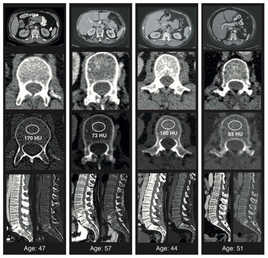
Patients with BM are in advanced stages of systemic cancer, which may translate into significant alterations of body composition biomarkers, such as BMD. The present study investigated the prognostic value of BMD on overall survival (OS) of 95 patients with surgically-treated BM related to NSCLC. All patients were treated in a large tertiary care neuro-oncological center between 2013 and 2018. Preoperative BMD was determined from the first lumbar vertebrae (L1) from routine preoperative staging computed tomography (CT) scans. Results were stratified into pathologic and physiologic values according to recently published normative reference ranges and correlated with survival parameters. Median preoperative L1-BMD was 99 Hounsfield units (HU) (IQR 74–195) compared to 140 HU (IQR 113–159) for patients with pathological and physiologic BMD (p = 0.03), with a median OS of 6 versus 15 months (p = 0.002). Multivariable analysis revealed pathologic BMD as an independent prognostic predictor for increased 1-year mortality (p = 0.03, OR 0.5, 95% CI 0.2–1.0). The present study suggests that decreased preoperative BMD values may represent a previously unrecognized negative prognostic factor in patients of BM requiring surgery for NSCLC. Based on guideline-adherent preoperative staging, BMD may prove to be a highly individualized, readily available biomarker for prognostic assessment and treatment guidance in affected patients. Full article
Show Figures

Figure 1

12 pages, 1815 KiB  
Article
Body Composition as a Predictor of the Survival in Anal Cancer
by Ahmed Allam Mohamed, Kathrin Risse, Jennifer Stock, Alexander Heinzel, Felix M. Mottaghy, Philipp Bruners and Michael J. Eble
Cancers 2022, 14(18), 4521; https://doi.org/10.3390/cancers14184521 - 18 Sep 2022
Cited by 3 | Viewed by 2043
Abstract
Background and aim: Sarcopenia and body composition parameters such as visceral and subcutaneous adipose tissue and visceral-to-subcutaneous adipose tissue ratio have been shown to be relevant biomarkers for prognosis in patients with different types of cancer. However, these findings have not been well [...] Read more.
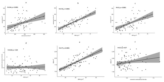
Background and aim: Sarcopenia and body composition parameters such as visceral and subcutaneous adipose tissue and visceral-to-subcutaneous adipose tissue ratio have been shown to be relevant biomarkers for prognosis in patients with different types of cancer. However, these findings have not been well studied in anal cancer to date. Therefore, the aim of this study was to evaluate the prognostic value of different body composition parameters in patients undergoing radiation therapy for the treatment of anal cancer with curative intent. Material and Methods: After approval by the institutional ethical committee, we retrospectively identified 81 patients in our local registry, who received radical intensity-modulated radiotherapy for the management of anal squamous cell cancer (ASCC). Clinical information, including body mass index (BMI), survival, and toxicities outcome, were retrieved from the local hospital registry. Based on the pre-therapeutic computer tomography (CT), we measured the total psoas muscle area, visceral adipose tissue area (VAT), subcutaneous adipose tissue area (SAT), and visceral-to-subcutaneous adipose tissue area ratio (VSR). In addition to the classical prognostic factors as T-stage, N-stage, gender, and treatment duration, we analyzed the impact of body composition on the prognosis in univariate and multivariate analyses. Results: Sarcopenia was not associated with increased mortality in anal cancer patients, whereas increased BMI (≥27 kg/m2) and VSR (≥0.45) were significantly associated with worsened overall survival and cancer-specific survival in both univariate and multivariate analyses. VSR—not BMI—was statistically higher in males. Sarcopenia and VSR ≥ 0.45 were associated with advanced T-stages. None of the body composition parameters resulted in a significant increase in treatment-related toxicities. Conclusion: BMI and visceral adiposity are independent prognostic factors for the survival of patients with anal cancer. Measurements to treat adiposity at the time of diagnosis may be needed to improve the survival outcomes for the affected patients. Full article
Show Figures

Figure 1

14 pages, 13907 KiB  
Article
Detection of Experimental Colorectal Peritoneal Metastases by a Novel PDGFRβ-Targeting Nanobody
by Esther Strating, Sjoerd Elias, Guus van Scharrenburg, Kaisa Luoto, André Verheem, Inne Borel Rinkes, Herman Steen and Onno Kranenburg
Cancers 2022, 14(18), 4348; https://doi.org/10.3390/cancers14184348 - 6 Sep 2022
Cited by 3 | Viewed by 2108
Abstract
Peritoneal metastases in colorectal cancer (CRC) belong to Consensus Molecular Subtype 4 (CMS4) and are associated with poor prognosis. Conventional imaging modalities, such as Computed Tomography (CT) and Fluorodeoxyglucose-Positron Emission Tomography (FDG-PET), perform very poorly in the detection of peritoneal metastases. However, the [...] Read more.
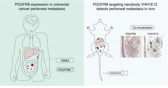
Peritoneal metastases in colorectal cancer (CRC) belong to Consensus Molecular Subtype 4 (CMS4) and are associated with poor prognosis. Conventional imaging modalities, such as Computed Tomography (CT) and Fluorodeoxyglucose-Positron Emission Tomography (FDG-PET), perform very poorly in the detection of peritoneal metastases. However, the stroma-rich nature of these lesions provides a basis for developing molecular imaging strategies. In this study, conducted from 2019 to 2021, we aimed to generate a Platelet-Derived Growth Factor Receptor beta (PDGFRB)-binding molecular imaging tracer for the detection of CMS4 CRC, including peritoneal metastases. The expression of PDGFRB mRNA discriminated CMS4 from CMS1-3 (AUROC = 0.86 (95% CI 0.85–0.88)) and was associated with poor relapse-free survival. PDGFRB mRNA and protein levels were very high in all human peritoneal metastases examined (n = 66). Therefore, we generated a PDGFRB-targeting llama nanobody (VHH1E12). Biotin-labelled VHH1E12 bound to immobilized human and mouse PDGFRB with high affinity (EC50 human PDGFRB = 7 nM; EC50 murine PDGFRB = 0.8 nM), and to PDGFRB-expressing HEK293 cells grown in vitro. A pharmacokinetic analysis of IRDye-800CW-conjugated VHH1E12 in mice showed that the plasma half-life was 6 min. IRDye-800CW-conjugated VHH1E12 specifically accumulated in experimentally induced colorectal cancer peritoneal metastases in mice. A tissue analysis subsequently demonstrated co-localization of the nanobody with PDGFRB expression in the tumour stroma. Our results demonstrate the potential value of PDGFRB-targeted molecular imaging as a novel strategy for the non-invasive detection of CMS4 CRC, in particular, peritoneal metastases. Full article
Show Figures

Graphical abstract

16 pages, 763 KiB  
Review
Challenges in Glioblastoma Radiomics and the Path to Clinical Implementation
by Philip Martin, Lois Holloway, Peter Metcalfe, Eng-Siew Koh and Caterina Brighi
Cancers 2022, 14(16), 3897; https://doi.org/10.3390/cancers14163897 - 12 Aug 2022
Cited by 9 | Viewed by 3342
Abstract
Radiomics is a field of medical imaging analysis that focuses on the extraction of many quantitative imaging features related to shape, intensity and texture. These features are incorporated into models designed to predict important clinical or biological endpoints for patients. Attention for radiomics [...] Read more.
Radiomics is a field of medical imaging analysis that focuses on the extraction of many quantitative imaging features related to shape, intensity and texture. These features are incorporated into models designed to predict important clinical or biological endpoints for patients. Attention for radiomics research has recently grown dramatically due to the increased use of imaging and the availability of large, publicly available imaging datasets. Glioblastoma multiforme (GBM) patients stand to benefit from this emerging research field as radiomics has the potential to assess the biological heterogeneity of the tumour, which contributes significantly to the inefficacy of current standard of care therapy. Radiomics models still require further development before they are implemented clinically in GBM patient management. Challenges relating to the standardisation of the radiomics process and the validation of radiomic models impede the progress of research towards clinical implementation. In this manuscript, we review the current state of radiomics in GBM, and we highlight the barriers to clinical implementation and discuss future validation studies needed to advance radiomics models towards clinical application. Full article
Show Figures

Figure 1

14 pages, 5477 KiB  
Article
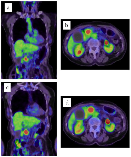
Clinical Impact of Dual Time Point 18F-Fluorodeoxyglucose Positron Emission Tomography/Computed Tomography Fusion Imaging in Pancreatic Cancer
by Takahiro Einama, Yoji Yamagishi, Yasuhiro Takihata, Fukumi Konno, Kazuki Kobayashi, Naoto Yonamine, Ibuki Fujinuma, Takazumi Tsunenari, Keita Kouzu, Akiko Nakazawa, Toshimitsu Iwasaki, Eiji Shinto, Jiro Ishida, Hideki Ueno and Yoji Kishi
Cancers 2022, 14(15), 3688; https://doi.org/10.3390/cancers14153688 - 28 Jul 2022
Cited by 5 | Viewed by 1847
Abstract
We examined the value of preoperative dual time point (DTP) 18F-fluorodeoxyglucose positron emission tomography/computed tomography fusion imaging (FDG PET/CT) as a predictor of early recurrence or the outcomes in patients with pancreatic cancer. Standardized uptake values (SUVs) in DTP FDG PET/CT were [...] Read more.
We examined the value of preoperative dual time point (DTP) 18F-fluorodeoxyglucose positron emission tomography/computed tomography fusion imaging (FDG PET/CT) as a predictor of early recurrence or the outcomes in patients with pancreatic cancer. Standardized uptake values (SUVs) in DTP FDG PET/CT were performed as preoperative staging. SUVmax1 and SUVmax2 were obtained in 60 min and 120 min, respectively. ΔSUVmax% was defined as (SUVmax2 − SUVmax1)/SUVmax1 × 100. The optimal cut-off values for SUVmax parameters were selected based on tumor relapse within 1 year of surgery. Optimal cut-off values for SUVmax1 and ΔSUVmax% were 7.18 and 24.25, respectively. The combination of SUVmax1 and ΔSUVmax% showed higher specificity and sensitivity, and higher positive and negative predictive values for tumor relapse within 1 year than SUVmax1 alone. Relapse-free survival (RFS) was significantly worse in the subgroups of high SUVmax1 and high ΔSUVmax% (median 7.0 months) than in the other subgroups (p < 0.0001). The multivariate Cox analysis of RFS identified high SUVmax1 and high ΔSUVmax% as independent prognostic factors (p = 0.0060). DTP FDG PET/CT may effectively predict relapse in patients with pancreatic cancer. The combination of SUVmax1 and ΔSUVmax% identified early recurrent patient groups more precisely than SUVmax1 alone. Full article
Show Figures

Figure 1

11 pages, 1101 KiB  
Article
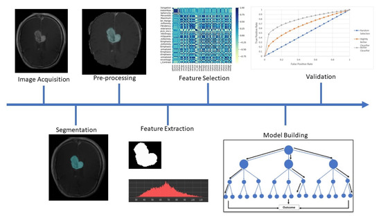
Radiomic Cancer Hallmarks to Identify High-Risk Patients in Non-Metastatic Colon Cancer
by Damiano Caruso, Michela Polici, Marta Zerunian, Antonella Del Gaudio, Emanuela Parri, Maria Agostina Giallorenzi, Domenico De Santis, Giulia Tarantino, Mariarita Tarallo, Filippo Maria Dentice di Accadia, Elsa Iannicelli, Giovanni Maria Garbarino, Giulia Canali, Paolo Mercantini, Enrico Fiori and Andrea Laghi
Cancers 2022, 14(14), 3438; https://doi.org/10.3390/cancers14143438 - 15 Jul 2022
Cited by 6 | Viewed by 1918
Abstract
The study was aimed to develop a radiomic model able to identify high-risk colon cancer by analyzing pre-operative CT scans. The study population comprised 148 patients: 108 with non-metastatic colon cancer were retrospectively enrolled from January 2015 to June 2020, and 40 patients [...] Read more.
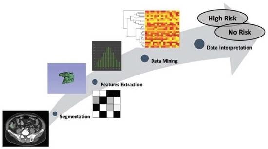
The study was aimed to develop a radiomic model able to identify high-risk colon cancer by analyzing pre-operative CT scans. The study population comprised 148 patients: 108 with non-metastatic colon cancer were retrospectively enrolled from January 2015 to June 2020, and 40 patients were used as the external validation cohort. The population was divided into two groups—High-risk and No-risk—following the presence of at least one high-risk clinical factor. All patients had baseline CT scans, and 3D cancer segmentation was performed on the portal phase by two expert radiologists using open-source software (3DSlicer v4.10.2). Among the 107 radiomic features extracted, stable features were selected to evaluate the inter-class correlation (ICC) (cut-off ICC > 0.8). Stable features were compared between the two groups (T-test or Mann–Whitney), and the significant features were selected for univariate and multivariate logistic regression to build a predictive radiomic model. The radiomic model was then validated with an external cohort. In total, 58/108 were classified as High-risk and 50/108 as No-risk. A total of 35 radiomic features were stable (0.81 ≤ ICC <  0.92). Among these, 28 features were significantly different between the two groups (p < 0.05), and only 9 features were selected to build the radiomic model. The radiomic model yielded an AUC of 0.73 in the internal cohort and 0.75 in the external cohort. In conclusion, the radiomic model could be seen as a performant, non-invasive imaging tool to properly stratify colon cancers with high-risk disease. Full article
Show Figures

Graphical abstract

15 pages, 1324 KiB  
Review
The Potential and Emerging Role of Quantitative Imaging Biomarkers for Cancer Characterization
by Hishan Tharmaseelan, Alexander Hertel, Shereen Rennebaum, Dominik Nörenberg, Verena Haselmann, Stefan O. Schoenberg and Matthias F. Froelich
Cancers 2022, 14(14), 3349; https://doi.org/10.3390/cancers14143349 - 9 Jul 2022
Cited by 3 | Viewed by 2938
Abstract
Similar to the transformation towards personalized oncology treatment, emerging techniques for evaluating oncologic imaging are fostering a transition from traditional response assessment towards more comprehensive cancer characterization via imaging. This development can be seen as key to the achievement of truly personalized and [...] Read more.
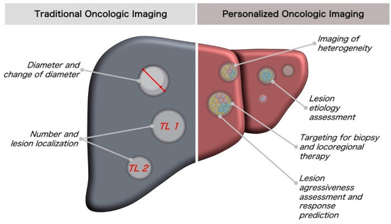
Similar to the transformation towards personalized oncology treatment, emerging techniques for evaluating oncologic imaging are fostering a transition from traditional response assessment towards more comprehensive cancer characterization via imaging. This development can be seen as key to the achievement of truly personalized and optimized cancer diagnosis and treatment. This review gives a methodological introduction for clinicians interested in the potential of quantitative imaging biomarkers, treating of radiomics models, texture visualization, convolutional neural networks and automated segmentation, in particular. Based on an introduction to these methods, clinical evidence for the corresponding imaging biomarkers—(i) dignity and etiology assessment; (ii) tumoral heterogeneity; (iii) aggressiveness and response; and (iv) targeting for biopsy and therapy—is summarized. Further requirements for the clinical implementation of these imaging biomarkers and the synergistic potential of personalized molecular cancer diagnostics and liquid profiling are discussed. Full article
Show Figures

Figure 1

21 pages, 3485 KiB  
Article
Analysis of Cross-Combinations of Feature Selection and Machine-Learning Classification Methods Based on [18F]F-FDG PET/CT Radiomic Features for Metabolic Response Prediction of Metastatic Breast Cancer Lesions
by Ober Van Gómez, Joaquin L. Herraiz, José Manuel Udías, Alexander Haug, Laszlo Papp, Dania Cioni and Emanuele Neri
Cancers 2022, 14(12), 2922; https://doi.org/10.3390/cancers14122922 - 14 Jun 2022
Cited by 12 | Viewed by 2712
Abstract
Background: This study aimed to identify optimal combinations between feature selection methods and machine-learning classifiers for predicting the metabolic response of individual metastatic breast cancer lesions, based on clinical variables and radiomic features extracted from pretreatment [18F]F-FDG PET/CT images. Methods: A [...] Read more.
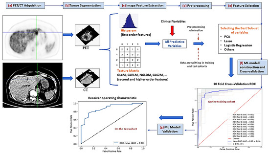
Background: This study aimed to identify optimal combinations between feature selection methods and machine-learning classifiers for predicting the metabolic response of individual metastatic breast cancer lesions, based on clinical variables and radiomic features extracted from pretreatment [18F]F-FDG PET/CT images. Methods: A total of 48 patients with confirmed metastatic breast cancer, who received different treatments, were included. All patients had an [18F]F-FDG PET/CT scan before and after the treatment. From 228 metastatic lesions identified, 127 were categorized as responders (complete or partial metabolic response) and 101 as non-responders (stable or progressive metabolic response), by using the percentage changes in SULpeak (peak standardized uptake values normalized for body lean body mass). The lesion pool was divided into training (n = 182) and testing cohorts (n = 46); for each lesion, 101 image features from both PET and CT were extracted (202 features per lesion). These features, along with clinical and pathological information, allowed the prediction model’s construction by using seven popular feature selection methods in cross-combination with another seven machine-learning (ML) classifiers. The performance of the different models was investigated with the receiver-operating characteristic curve (ROC) analysis, using the area under the curve (AUC) and accuracy (ACC) metrics. Results: The combinations, least absolute shrinkage and selection operator (Lasso) + support vector machines (SVM), or random forest (RF) had the highest AUC in the cross-validation, with 0.93 ± 0.06 and 0.92 ± 0.03, respectively, whereas Lasso + neural network (NN) or SVM, and mutual information (MI) + RF, had the higher AUC and ACC in the validation cohort, with 0.90/0.72, 0.86/0.76, and 87/85, respectively. On average, the models with Lasso and models with SVM had the best mean performance for both AUC and ACC in both training and validation cohorts. Conclusions: Image features obtained from a pretreatment [18F]F-FDG PET/CT along with clinical vaiables could predict the metabolic response of metastatic breast cancer lesions, by their incorporation into predictive models, whose performance depends on the selected combination between feature selection and ML classifier methods. Full article
Show Figures

Figure 1

14 pages, 1742 KiB  
Article
Simultaneous and Spatially-Resolved Analysis of T-Lymphocytes, Macrophages and PD-L1 Immune Checkpoint in Rare Cancers
by Karina Cereceda, Nicolas Bravo, Roddy Jorquera, Roxana González-Stegmaier and Franz Villarroel-Espíndola
Cancers 2022, 14(11), 2815; https://doi.org/10.3390/cancers14112815 - 6 Jun 2022
Cited by 2 | Viewed by 2467
Abstract
Penile, vulvar and anal neoplasms show an incidence lower than 0.5% of the population per year and therefore can be considered as rare cancers but with a dramatic impact on quality of life and survival. This work describes the experience of a Chilean [...] Read more.
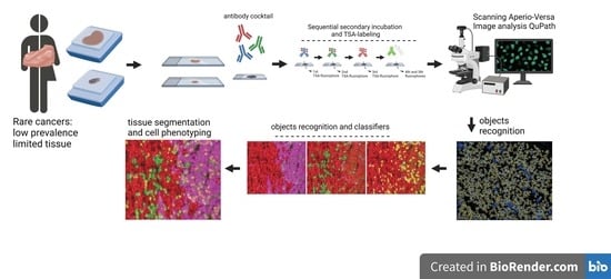
Penile, vulvar and anal neoplasms show an incidence lower than 0.5% of the population per year and therefore can be considered as rare cancers but with a dramatic impact on quality of life and survival. This work describes the experience of a Chilean cancer center using multiplexed immunofluorescence to study a case series of four penile cancers, two anal cancers and one vulvar cancer and simultaneous detection of CD8, CD68, PD-L1, Cytokeratin and Ki-67 in FFPE samples. Fluorescent image analyses were performed using open sources for automated tissue segmentation and cell phenotyping. Our results showed an objective and reliable counting of objects with a single or combined labeling or within a specific tissue compartment. The variability was below 10%, and the correlation between analytical events was 0.92–0.97. Critical cell phenotypes, such as TILs, PD-L1+ or proliferative tumor cells were detected in a supervised and unsupervised manner with a limit of detection of less than 1% of relative abundance. Finally, the observed diversity and abundance of the different cell phenotypes within the tumor microenvironment for the three studied tumor types confirmed that our methodology is useful and robust to be applicable for many other solid tumors. Full article
Show Figures

Graphical abstract

13 pages, 1674 KiB  
Article
Automated Prediction of the Response to Neoadjuvant Chemoradiotherapy in Patients Affected by Rectal Cancer
by Giuseppe Filitto, Francesca Coppola, Nico Curti, Enrico Giampieri, Daniele Dall'Olio, Alessandra Merlotti, Arrigo Cattabriga, Maria Adriana Cocozza, Makoto Taninokuchi Tomassoni, Daniel Remondini, Luisa Pierotti, Lidia Strigari, Dajana Cuicchi, Alessandra Guido, Karim Rihawi, Antonietta D'Errico, Francesca Di Fabio, Gilberto Poggioli, Alessio Giuseppe Morganti, Luigi Ricciardiello, Rita Golfieri and Gastone Castellaniadd Show full author list remove Hide full author list
Cancers 2022, 14(9), 2231; https://doi.org/10.3390/cancers14092231 - 29 Apr 2022
Cited by 7 | Viewed by 2888
Abstract
Background: Rectal cancer is a malignant neoplasm of the large intestine resulting from the uncontrolled proliferation of the rectal tract. Predicting the pathologic response of neoadjuvant chemoradiotherapy at an MRI primary staging scan in patients affected by locally advanced rectal cancer (LARC) could [...] Read more.
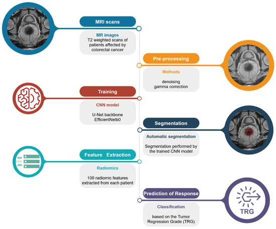
Background: Rectal cancer is a malignant neoplasm of the large intestine resulting from the uncontrolled proliferation of the rectal tract. Predicting the pathologic response of neoadjuvant chemoradiotherapy at an MRI primary staging scan in patients affected by locally advanced rectal cancer (LARC) could lead to significant improvement in the survival and quality of life of the patients. In this study, the possibility of automatizing this estimation from a primary staging MRI scan, using a fully automated artificial intelligence-based model for the segmentation and consequent characterization of the tumor areas using radiomic features was evaluated. The TRG score was used to evaluate the clinical outcome. Methods: Forty-three patients under treatment in the IRCCS Sant’Orsola-Malpighi Polyclinic were retrospectively selected for the study; a U-Net model was trained for the automated segmentation of the tumor areas; the radiomic features were collected and used to predict the tumor regression grade (TRG) score. Results: The segmentation of tumor areas outperformed the state-of-the-art results in terms of the Dice score coefficient or was comparable to them but with the advantage of considering mucinous cases. Analysis of the radiomic features extracted from the lesion areas allowed us to predict the TRG score, with the results agreeing with the state-of-the-art results. Conclusions: The results obtained regarding TRG prediction using the proposed fully automated pipeline prove its possible usage as a viable decision support system for radiologists in clinical practice. Full article
Show Figures

Figure 1

15 pages, 1933 KiB  
Article
Adjusted CT Image-Based Radiomic Features Combined with Immune Genomic Expression Achieve Accurate Prognostic Classification and Identification of Therapeutic Targets in Stage III Colorectal Cancer
by Yi-Ching Huang, Yi-Shan Tsai, Chung-I Li, Ren-Hao Chan, Yu-Min Yeh, Po-Chuan Chen, Meng-Ru Shen and Peng-Chan Lin
Cancers 2022, 14(8), 1895; https://doi.org/10.3390/cancers14081895 - 8 Apr 2022
Cited by 7 | Viewed by 2427
Abstract
To evaluate whether adjusted computed tomography (CT) scan image-based radiomics combined with immune genomic expression can achieve accurate stratification of cancer recurrence and identify potential therapeutic targets in stage III colorectal cancer (CRC), this cohort study enrolled 71 patients with postoperative stage III [...] Read more.
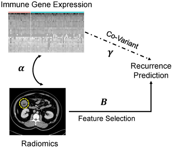
To evaluate whether adjusted computed tomography (CT) scan image-based radiomics combined with immune genomic expression can achieve accurate stratification of cancer recurrence and identify potential therapeutic targets in stage III colorectal cancer (CRC), this cohort study enrolled 71 patients with postoperative stage III CRC. Based on preoperative CT scans, radiomic features were extracted and selected to build pixel image data using covariate-adjusted tensor classification in the high-dimension (CATCH) model. The differentially expressed RNA genes, as radiomic covariates, were identified by cancer recurrence. Predictive models were built using the pixel image and immune genomic expression factors, and the area under the curve (AUC) and F1 score were used to evaluate their performance. Significantly adjusted radiomic features were selected to predict recurrence. The association between the significantly adjusted radiomic features and immune gene expression was also investigated. Overall, 1037 radiomic features were converted into 33 × 32-pixel image data. Thirty differentially expressed genes were identified. We performed 100 iterations of 3-fold cross-validation to evaluate the performance of the CATCH model, which showed a high sensitivity of 0.66 and an F1 score of 0.69. The area under the curve (AUC) was 0.56. Overall, ten adjusted radiomic features were significantly associated with cancer recurrence in the CATCH model. All of these methods are texture-associated radiomics. Compared with non-adjusted radiomics, 7 out of 10 adjusted radiomic features influenced recurrence-free survival. The adjusted radiomic features were positively associated with PECAM1, PRDM1, AIF1, IL10, ISG20, and TLR8 expression. We provide individualized cancer therapeutic strategies based on adjusted radiomic features in recurrent stage III CRC. Adjusted CT scan image-based radiomics with immune genomic expression covariates using the CATCH model can efficiently predict cancer recurrence. The correlation between adjusted radiomic features and immune genomic expression can provide biological relevance and individualized therapeutic targets. Full article
Show Figures

Figure 1

18 pages, 2382 KiB  
Article
Automatically Extracted Machine Learning Features from Preoperative CT to Early Predict Microvascular Invasion in HCC: The Role of the Zone of Transition (ZOT)
by Matteo Renzulli, Margherita Mottola, Francesca Coppola, Maria Adriana Cocozza, Silvia Malavasi, Arrigo Cattabriga, Giulio Vara, Matteo Ravaioli, Matteo Cescon, Francesco Vasuri, Rita Golfieri and Alessandro Bevilacqua
Cancers 2022, 14(7), 1816; https://doi.org/10.3390/cancers14071816 - 3 Apr 2022
Cited by 18 | Viewed by 3068
Abstract
Background: Microvascular invasion (MVI) is a consolidated predictor of hepatocellular carcinoma (HCC) recurrence after treatments. No reliable radiological imaging findings are available for preoperatively diagnosing MVI, despite some progresses of radiomic analysis. Furthermore, current MVI radiomic studies have not been designed for small [...] Read more.
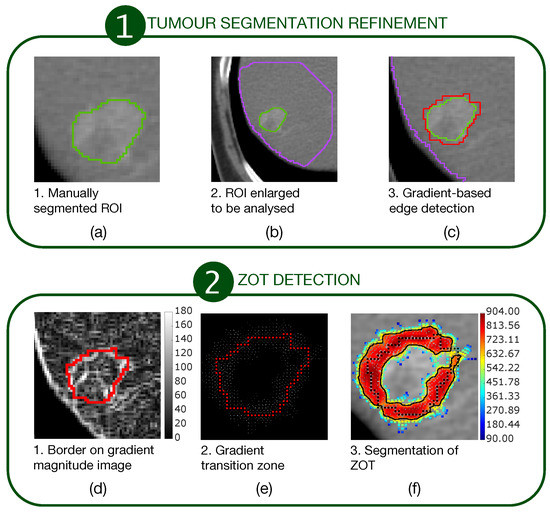
Background: Microvascular invasion (MVI) is a consolidated predictor of hepatocellular carcinoma (HCC) recurrence after treatments. No reliable radiological imaging findings are available for preoperatively diagnosing MVI, despite some progresses of radiomic analysis. Furthermore, current MVI radiomic studies have not been designed for small HCC nodules, for which a plethora of treatments exists. This study aimed to identify radiomic MVI predictors in nodules ≤3.0 cm by analysing the zone of transition (ZOT), crossing tumour and peritumour, automatically detected to face the uncertainties of radiologist’s tumour segmentation. Methods: The study considered 117 patients imaged by contrast-enhanced computed tomography; 78 patients were finally enrolled in the radiomic analysis. Radiomic features were extracted from the tumour and the ZOT, detected using an adaptive procedure based on local image contrast variations. After data oversampling, a support vector machine classifier was developed and validated. Classifier performance was assessed using receiver operating characteristic (ROC) curve analysis and related metrics. Results: The original 89 HCC nodules (32 MVI+ and 57 MVI−) became 169 (62 MVI+ and 107 MVI−) after oversampling. Of the four features within the signature, three are ZOT heterogeneity measures regarding both arterial and venous phases. On the test set (19MVI+ and 33MVI−), the classifier predicts MVI+ with area under the curve of 0.86 (95%CI (0.70–0.93), p∼105), sensitivity = 79% and specificity = 82%. The classifier showed negative and positive predictive values of 87% and 71%, respectively. Conclusions: The classifier showed the highest diagnostic performance in the literature, disclosing the role of ZOT heterogeneity in predicting the MVI+ status. Full article
Show Figures

Figure 1

13 pages, 7311 KiB  
Article
Identification of CT Imaging Phenotypes of Colorectal Liver Metastases from Radiomics Signatures—Towards Assessment of Interlesional Tumor Heterogeneity
by Hishan Tharmaseelan, Alexander Hertel, Fabian Tollens, Johann Rink, Piotr Woźnicki, Verena Haselmann, Isabelle Ayx, Dominik Nörenberg, Stefan O. Schoenberg and Matthias F. Froelich
Cancers 2022, 14(7), 1646; https://doi.org/10.3390/cancers14071646 - 24 Mar 2022
Cited by 19 | Viewed by 2889
Abstract
(1) Background: Tumoral heterogeneity (TH) is a major challenge in the treatment of metastatic colorectal cancer (mCRC) and is associated with inferior response. Therefore, the identification of TH would be beneficial for treatment planning. TH can be assessed by identifying genetic alterations. In [...] Read more.
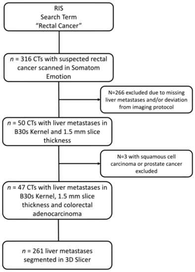
(1) Background: Tumoral heterogeneity (TH) is a major challenge in the treatment of metastatic colorectal cancer (mCRC) and is associated with inferior response. Therefore, the identification of TH would be beneficial for treatment planning. TH can be assessed by identifying genetic alterations. In this work, a radiomics-based approach for assessment of TH in colorectal liver metastases (CRLM) in CT scans is demonstrated. (2) Methods: In this retrospective study, CRLM of mCRC were segmented and radiomics features extracted using pyradiomics. Unsupervised k-means clustering was applied to features and lesions. Feature redundancy was evaluated by principal component analysis and reduced by Pearson correlation coefficient cutoff. Feature selection was conducted by LASSO regression and visual analysis of the clusters by radiologists. (3) Results: A total of 47 patients’ (36% female, median age 64) CTs with 261 lesions were included. Five clusters were identified, and the categories small disseminated (n = 31), heterogeneous (n = 105), homogeneous (n = 64), mixed (n = 59), and very large type (n = 2) were assigned based on visual characteristics. Further statistical analysis showed correlation (p < 0.01) of clusters with sex, primary location, T- and N-status, and mutational status. Feature reduction and selection resulted in the identification of four features as a final set for cluster definition. (4) Conclusions: Radiomics features can characterize TH in liver metastases of mCRC in CT scans, and may be suitable for a better pretherapeutic classification of liver lesion phenotypes. Full article
Show Figures

Figure 1

14 pages, 927 KiB  
Article
18F-Fluorodeoxyglucose PET/CT for Early Prediction of Outcomes in Patients with Advanced Lung Adenocarcinomas and EGFR Mutations Treated with First-Line EGFR-TKIs
by Yu-Erh Huang, Ying-Huang Tsai, Yu-Jie Huang, Jr-Hau Lung, Kuo-Wei Ho, Tzu-Chen Yen, Sheng-Chieh Chan, Shu-Tian Chen, Ming-Feng Tsai and Ming-Szu Hung
Cancers 2022, 14(6), 1507; https://doi.org/10.3390/cancers14061507 - 15 Mar 2022
Cited by 5 | Viewed by 2981
Abstract
This study aims to investigate the role of 18F-fluorodeoxyglucose positron emission tomography/computed tomography (18F-FDG PET/CT) in early prediction of response and survival following epithelial growth factor receptor (EGFR)–tyrosine kinase inhibitor (TKI) therapy in patients with advanced lung adenocarcinomas and EGFR [...] Read more.
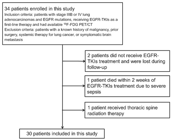
This study aims to investigate the role of 18F-fluorodeoxyglucose positron emission tomography/computed tomography (18F-FDG PET/CT) in early prediction of response and survival following epithelial growth factor receptor (EGFR)–tyrosine kinase inhibitor (TKI) therapy in patients with advanced lung adenocarcinomas and EGFR mutations. Thirty patients with stage IIIB/IV lung adenocarcinomas and EGFR mutations receiving first-line EGFR-TKIs were prospectively evaluated between November 2012 and May 2015. EGFR mutations were quantified by delta cycle threshold (dCt). 18F-FDG PET/CT was performed before and 2 weeks after treatment initiation. PET response was assessed based on PET Response Criteria in Solid Tumors (PERCIST). Baseline and percentage changes in the summed standardized uptake value, metabolic tumor volume (bsumMTV and ΔsumMTV, respectively), and total lesion glycolysis of ≤5 target lesions/patient were calculated. The association between parameters (clinical and PET) and non-progression disease after 3 months of treatment in CT based on the Response Evaluation Criteria in Solid Tumors Version 1.1 (nPD3mo), progression-free survival (PFS), and overall survival (OS) were tested. The median follow-up time was 19.6 months. The median PFS and OS were 12.0 and 25.3 months, respectively. The PERCIST criteria was an independent predictor of nPD3mo (p = 0.009), dCt (p = 0.014) and bsumMTV (p = 0.014) were independent predictors of PFS, and dCt (p = 0.014) and ΔsumMTV (p = 0.005) were independent predictors of OS. 18F-FDG PET/CT achieved early prediction of outcomes in patients with advanced lung adenocarcinomas and EGFR mutations receiving EGFR-TKIs. Full article
Show Figures

Figure 1

13 pages, 3424 KiB  
Article
Prediction of Early Response to Immunotherapy: DCE-US as a New Biomarker
by Raphael Naccache, Younes Belkouchi, Littisha Lawrance, Baya Benatsou, Joya Hadchiti, Paul-Henry Cournede, Samy Ammari, Hugues Talbot and Nathalie Lassau
Cancers 2022, 14(5), 1337; https://doi.org/10.3390/cancers14051337 - 4 Mar 2022
Cited by 1 | Viewed by 2715
Abstract
Purpose: The objective of our study is to propose fast, cost-effective, convenient, and effective biomarkers using the perfusion parameters from dynamic contrast-enhanced ultrasound (DCE-US) for the evaluation of immune checkpoint inhibitors (ICI) early response. Methods: The retrospective cohort used in this study included [...] Read more.
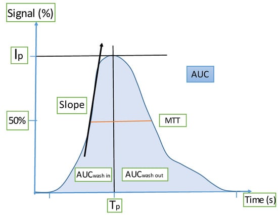
Purpose: The objective of our study is to propose fast, cost-effective, convenient, and effective biomarkers using the perfusion parameters from dynamic contrast-enhanced ultrasound (DCE-US) for the evaluation of immune checkpoint inhibitors (ICI) early response. Methods: The retrospective cohort used in this study included 63 patients with metastatic cancer eligible for immunotherapy. DCE-US was performed at baseline, day 8 (D8), and day 21 (D21) after treatment onset. A tumor perfusion curve was modeled on these three dates, and change in the seven perfusion parameters was measured between baseline, D8, and D21. These perfusion parameters were studied to show the impact of their variation on the overall survival (OS). Results: After the removal of missing or suboptimal DCE-US, the Baseline-D8, the Baseline-D21, and the D8-D21 groups included 37, 53, and 33 patients, respectively. A decrease of more than 45% in the area under the perfusion curve (AUC) between baseline and D21 was significantly associated with better OS (p = 0.0114). A decrease of any amount in the AUC between D8 and D21 was also significantly associated with better OS (p = 0.0370). Conclusion: AUC from DCE-US looks to be a promising new biomarker for fast, effective, and convenient immunotherapy response evaluation. Full article
Show Figures

Figure 1

12 pages, 1514 KiB  
Article
Imaging Kv1.3 Expressing Memory T Cells as a Marker of Immunotherapy Response
by Julian L. Goggi, Shivashankar Khanapur, Boominathan Ramasamy, Siddesh V. Hartimath, Tang Jun Rong, Peter Cheng, Yun Xuan Tan, Xin Yi Yeo, Sangyong Jung, Stephanie Shee Min Goay, Seow Theng Ong, You Yi Hwang, K. George Chandy and Edward G. Robins
Cancers 2022, 14(5), 1217; https://doi.org/10.3390/cancers14051217 - 26 Feb 2022
Cited by 10 | Viewed by 2951
Abstract
Immune checkpoint inhibitors have shown great promise, emerging as a new pillar of treatment for cancer; however, only a relatively small proportion of recipients show a durable response to treatment. Strategies that reliably differentiate durably-responding tumours from non-responsive tumours are a critical unmet [...] Read more.
Immune checkpoint inhibitors have shown great promise, emerging as a new pillar of treatment for cancer; however, only a relatively small proportion of recipients show a durable response to treatment. Strategies that reliably differentiate durably-responding tumours from non-responsive tumours are a critical unmet need. Persistent and durable immunological responses are associated with the generation of memory T cells. Effector memory T cells associated with tumour response to immune therapies are characterized by substantial upregulation of the potassium channel Kv1.3 after repeated antigen stimulation. We have developed a new Kv1.3 targeting radiopharmaceutical, [18F]AlF-NOTA-KCNA3P, and evaluated whether it can reliably differentiate tumours successfully responding to immune checkpoint inhibitor (ICI) therapy targeting PD-1 alone or combined with CLTA4. In a syngeneic colon cancer model, we compared tumour retention of [18F]AlF-NOTA-KCNA3P with changes in the tumour immune microenvironment determined by flow cytometry. Imaging with [18F]AlF-NOTA-KCNA3P reliably differentiated tumours responding to ICI therapy from non-responding tumours and was associated with substantial tumour infiltration of T cells, especially Kv1.3-expressing CD8+ effector memory T cells. Full article
Show Figures

Figure 1

2021

Jump to: 2024, 2023, 2022

13 pages, 1233 KiB  
Article
Preoperative Nodal US Features for Predicting Recurrence in N1b Papillary Thyroid Carcinoma
by Na Lae Eun, Jeong-Ah Kim, Hye Mi Gweon, Ji Hyun Youk and Eun Ju Son
Cancers 2022, 14(1), 174; https://doi.org/10.3390/cancers14010174 - 30 Dec 2021
Cited by 5 | Viewed by 1879
Abstract
This study aimed to investigate whether preoperative ultrasonographic (US) features of metastatic lymph nodes (LNs) are associated with tumor recurrence in patients with N1b papillary thyroid carcinoma (PTC). We enrolled 692 patients (mean age, 41.9 years; range, 6–80 years) who underwent total thyroidectomy [...] Read more.
This study aimed to investigate whether preoperative ultrasonographic (US) features of metastatic lymph nodes (LNs) are associated with tumor recurrence in patients with N1b papillary thyroid carcinoma (PTC). We enrolled 692 patients (mean age, 41.9 years; range, 6–80 years) who underwent total thyroidectomy and lateral compartment LN dissection between January 2009 and December 2015 and were followed-up for 12 months or longer. Clinicopathologic findings and US features of the index tumor and metastatic LNs in the lateral neck were reviewed. A Kaplan-Meier analysis and Cox proportion hazard model were used to analyze the recurrence-free survival rates and features associated with postoperative recurrence. Thirty-seven (5.3%) patients had developed recurrence at a median follow-up of 66.5 months. On multivariate Cox proportional hazard analysis, male sex (hazard ratio [HR], 2.277; 95% confidence interval [CI]: 1.131, 4.586; p = 0.021), age ≥55 years (HR, 3.216; 95% CI: 1.529, 6.766; p = 0.002), LN size (HR, 1.054; 95% CI: 1.024, 1.085; p < 0.001), and hyperechogenicity of LN (HR, 8.223; 95% CI: 1.689, 40.046; p = 0.009) on US were independently associated with recurrence. Preoperative US features of LNs, including size and hyperechogenicity, may be valuable for predicting recurrence in patients with N1b PTC. Full article
Show Figures

Figure 1

13 pages, 2353 KiB  
Article
Prognostic Impact of Sarcopenia in Patients with Metastatic Hormone-Sensitive Prostate Cancer
by Ji Hyun Lee, Byul A Jee, Jae-Hun Kim, Hoyoung Bae, Jae Hoon Chung, Wan Song, Hyun Hwan Sung, Hwang Gyun Jeon, Byong Chang Jeong, Seong Il Seo, Seong Soo Jeon, Hyun Moo Lee, Se Hoon Park and Minyong Kang
Cancers 2021, 13(24), 6345; https://doi.org/10.3390/cancers13246345 - 17 Dec 2021
Cited by 10 | Viewed by 2792
Abstract
The clinical value of sarcopenia has not been determined yet in metastatic hormone-sensitive prostate cancer (mHSPC). We retrospectively evaluated data of 70 consecutive patients with mHSPC receiving treatment with either early docetaxel (n = 42) or abiraterone acetate (n = 28) [...] Read more.
The clinical value of sarcopenia has not been determined yet in metastatic hormone-sensitive prostate cancer (mHSPC). We retrospectively evaluated data of 70 consecutive patients with mHSPC receiving treatment with either early docetaxel (n = 42) or abiraterone acetate (n = 28) between July 2018 and April 2021. Skeletal muscle index was calculated from cross-sectional areas of skeletal muscle on baseline computed tomography (CT), defining sarcopenia as a skeletal muscle index of ≤52.4 cm2/m2. Failure-free survival (FFS), radiographic progression-free survival, and time to prostate-specific antigen (PSA) progression were estimated using the Kaplan–Meier method, and differences in survival probability were compared using the log-rank test. Cox proportional hazards regression analysis was conducted to identify the predictors of clinical outcomes. Patients with sarcopenia (n = 47) had shorter FFS than those without sarcopenia (n = 23) (median, 20.1 months vs. not reached; log-rank p < 0.001). Sarcopenia was independently associated with shorter FFS (hazard ratio (HR), 6.69; 95% confidence interval (CI), 1.57–28.49; p = 0.010) and time to PSA progression (HR, 12.91; 95% CI, 1.08–153.85; p = 0.043). In conclusion, sarcopenia is an independent prognostic factor for poor FFS and time to PSA progression in patients with mHSPC who receive early docetaxel or abiraterone acetate treatment. Full article
Show Figures

Figure 1

Back to TopTop