Oncology: State-of-the-Art Research in the USA
A topical collection in Cancers (ISSN 2072-6694). This collection belongs to the section "Cancer Therapy".

Viewed by 65066

Editors


E-Mail Website
Guest Editor
Department of Translational Hematology and Oncology Research, Taussig Cancer Institute, Cleveland Clinic, Cleveland, OH 44195, USA
Interests: myeloid malignancies; bone marrow failure syndromes; RNA splicing
Special Issues, Collections and Topics in MDPI journals

E-Mail Website
Guest Editor
Department of Pathology, Department of Cell & Molecular Medicine, Rush University Medical Center, Jelke Southcenter, Suite 444, 1750 W. Harrison St., Chicago, IL 606012, USA
Interests: lung cancer; biomarkers; cancer cachexia; immunotherapy; recurrence; lung cancer screening; early detection; EMT; proteomics; biobanking

E-Mail Website
Guest Editor
The New York Eye and Ear Infirmary of Mount Sinai, New York, NY, USA
Interests: uveal melanoma; ocular surface neoplasia; retinoblastoma and other rare intraocular tumors

Topical Collection Information

Dear Colleagues,

Among many other countries, the USA is at the forefront of cancer research. There are commitments from stakeholders from academic institutions, health service organizations, industries, funding bodies and public and patient advocacy groups to support research into all types of cancer. We have a unique National Health Service, which emphasizes the needs of everyone and is free at the point of delivery. It believes that integrating research into the health service organization will improve outcomes and transform cancer care.  

In this Special Issue, we aim to showcase state-of-the-art research in oncology in the USA. We invite submissions looking at all kinds of research covering all cancer types and stages, from basic laboratory research to translational and clinical research, including cohort studies, randomized controlled trials and epidemiological studies. Narrated reviews describing the history and significant contributions of cancer research in the USA are also welcomed.

Dr. Valeria Visconte
Dr. Nelson S. Yee
Dr. Jeffrey A. Borgia
Dr. Eakaterina Semenova
Guest Editors

Manuscript Submission Information

Manuscripts should be submitted online at www.mdpi.com by registering and logging in to this website. Once you are registered, click here to go to the submission form. Manuscripts can be submitted until the deadline. All submissions that pass pre-check are peer-reviewed. Accepted papers will be published continuously in the journal (as soon as accepted) and will be listed together on the collection website. Research articles, review articles as well as communications are invited. For planned papers, a title and short abstract (about 100 words) can be sent to the Editorial Office for announcement on this website.

Submitted manuscripts should not have been published previously, nor be under consideration for publication elsewhere (except conference proceedings papers). All manuscripts are thoroughly refereed through a single-blind peer-review process. A guide for authors and other relevant information for submission of manuscripts is available on the Instructions for Authors page. Cancers is an international peer-reviewed open access semimonthly journal published by MDPI.

Please visit the Instructions for Authors page before submitting a manuscript. The Article Processing Charge (APC) for publication in this open access journal is 2900 CHF (Swiss Francs). Submitted papers should be well formatted and use good English. Authors may use MDPI's English editing service prior to publication or during author revisions.

Keywords

  • cancer
  • oncology
  • research
  • USA

Published Papers (33 papers)

2024

Jump to: 2023, 2022

17 pages, 743 KiB  
Review
Evaluating the Safety of Immune Checkpoint Inhibitors and Combination Therapies in the Management of Brain Metastases: A Comprehensive Review
by Vivek Podder, Tulika Ranjan, Kim Margolin, Arun Maharaj and Manmeet Singh Ahluwalia
Cancers 2024, 16(23), 3929; https://doi.org/10.3390/cancers16233929 - 23 Nov 2024
Viewed by 375
Abstract
Brain metastases (BM) are a frequent and severe complication in patients with lung cancer, breast cancer, and melanoma. Immune checkpoint inhibitors (ICIs) have become a crucial treatment option for BM, whether used alone or in combination with chemotherapy and stereotactic radiosurgery (SRS). However, [...] Read more.
Brain metastases (BM) are a frequent and severe complication in patients with lung cancer, breast cancer, and melanoma. Immune checkpoint inhibitors (ICIs) have become a crucial treatment option for BM, whether used alone or in combination with chemotherapy and stereotactic radiosurgery (SRS). However, ICIs are associated with immune-related adverse events (irAEs) that can affect multiple organ systems, complicating their use in BM patients. This review examines the mechanisms of irAEs and their effects on different organs and evaluates the safety of ICIs across various treatment strategies for BM. Our analysis indicates that ICIs significantly improve survival and disease control in BM patients, but their use increases the risk of irAEs, including dermatologic, gastrointestinal, endocrine, pulmonary, and neurologic toxicities. Neurotoxic events, particularly treatment-associated brain necrosis (TABN) and encephalitis, are more common in BM patients. While the overall incidence of irAEs is similar between patients with and without BM, the neurotoxicity risk is higher in the BM population. Combining ICIs with chemotherapy and SRS enhances efficacy but also heightens the risk of adverse events across organ systems. ICIs offer substantial benefits for BM patients but require careful management to mitigate the risks of irAEs. Close patient monitoring, individualized treatment protocols, and prompt intervention are essential for optimizing the outcomes. Future research should focus on refining combination strategies and improving the management of irAEs, particularly neurotoxicity, to maximize therapeutic benefits for BM patients. Full article
8 pages, 1216 KiB  
Communication
Adverse Events as a Function of Biological Sex in a Multicenter Clinical Trial of Melanoma Vaccines
by Catherine E. Lyons, Ruyun Jin, Aaron D. Smith, Hong Zhu and Craig L. Slingluff, Jr.
Cancers 2024, 16(22), 3882; https://doi.org/10.3390/cancers16223882 - 20 Nov 2024
Viewed by 362
Abstract
Background/Objective: Biological females experience more autoimmune disease than males and more treatment-related adverse events (TRAEs) after immune checkpoint blockade therapy. However, little is known about sex-related differences in TRAEs after cancer vaccines. Methods: The Mel44 clinical trial (NCT00118274) enrolled 167 eligible patients [...] Read more.
Background/Objective: Biological females experience more autoimmune disease than males and more treatment-related adverse events (TRAEs) after immune checkpoint blockade therapy. However, little is known about sex-related differences in TRAEs after cancer vaccines. Methods: The Mel44 clinical trial (NCT00118274) enrolled 167 eligible patients with high-risk melanoma to treatment with either of two melanoma multipeptide vaccines. We hypothesized that females would experience higher rates and grades of TRAEs. TRAE rates and grades were compared between sexes, with adjustment for multiple comparisons, and with mixed-effects models. Results: Multiple sex-related differences in TRAE rate and grade were observed in unadjusted comparisons, but only hyperglycemia and hypopigmentation were significantly higher-grade by sex after correcting for multiple comparisons: they were increased in males. In mixed-effect models, vaccination strategy, but not patient sex, was independently associated with TRAE rates and grades. Conclusions: These data do not support our hypothesis that TRAEs would be increased in females. Vaccine safety was supported for both males and females. Full article
Show Figures

Figure 1

28 pages, 1152 KiB  
Article
Lung and Colon Cancer Detection Using a Deep AI Model
by Nazmul Shahadat, Ritika Lama and Anna Nguyen
Cancers 2024, 16(22), 3879; https://doi.org/10.3390/cancers16223879 - 20 Nov 2024
Viewed by 330
Abstract
Lung and colon cancers are among the leading causes of cancer-related mortality worldwide. Early and accurate detection of these cancers is crucial for effective treatment and improved patient outcomes. False or incorrect detection is harmful. Accurately detecting cancer in a patient’s tissue is [...] Read more.
Lung and colon cancers are among the leading causes of cancer-related mortality worldwide. Early and accurate detection of these cancers is crucial for effective treatment and improved patient outcomes. False or incorrect detection is harmful. Accurately detecting cancer in a patient’s tissue is crucial to their effective treatment. While analyzing tissue samples is complicated and time-consuming, deep learning techniques have made it possible to complete this process more efficiently and accurately. As a result, researchers can study more patients in a shorter amount of time and at a lower cost. Much research has been conducted to investigate deep learning models that require great computational ability and resources. However, none of these have had a 100% accurate detection rate for these life-threatening malignancies. Misclassified or falsely detecting cancer can have very harmful consequences. This research proposes a new lightweight, parameter-efficient, and mobile-embedded deep learning model based on a 1D convolutional neural network with squeeze-and-excitation layers for efficient lung and colon cancer detection. This proposed model diagnoses and classifies lung squamous cell carcinomas and adenocarcinoma of the lung and colon from digital pathology images. Extensive experiment demonstrates that our proposed model achieves 100% accuracy for detecting lung, colon, and lung and colon cancers from the histopathological (LC25000) lung and colon datasets, which is considered the best accuracy for around 0.35 million trainable parameters and around 6.4 million flops. Compared with the existing results, our proposed architecture shows state-of-the-art performance in lung, colon, and lung and colon cancer detection. Full article
Show Figures

Figure 1

23 pages, 5248 KiB  
Article
Preclinical Multi-Omic Assessment of Pioglitazone in Skeletal Muscles of Mice Implanted with Human HER2/neu Overexpressing Breast Cancer Xenografts
by Stuart A. Clayton, Alan D. Mizener, Marcella A. Whetsell, Lauren E. Rentz, Ethan M. Meadows, Werner J. Geldenhuys and Emidio E. Pistilli
Cancers 2024, 16(21), 3640; https://doi.org/10.3390/cancers16213640 - 29 Oct 2024
Viewed by 647
Abstract
Background/Objectives: Breast cancer (BC) is the second most commonly diagnosed cancer worldwide and is accompanied by fatigue during both active disease and remission in the majority of cases. Our lab has measured fatigue in isolated muscles from treatment-naive BC patient-derived orthotopic xenograft [...] Read more.
Background/Objectives: Breast cancer (BC) is the second most commonly diagnosed cancer worldwide and is accompanied by fatigue during both active disease and remission in the majority of cases. Our lab has measured fatigue in isolated muscles from treatment-naive BC patient-derived orthotopic xenograft (BC-PDOX) mice. Here, we conducted a preclinical trial of pioglitazone in BC-PDOX mice to determine its efficacy in ameliorating BC-induced muscle fatigue, as well as its effects on transcriptomic, metabolomic, and lipidomic profiles in skeletal muscle. Methods: The pioglitazone and vehicle groups were treated orally for 4 weeks upon reaching a tumor volume of 600 mm3. Whole-animal indirect calorimetry was used to evaluate systemic metabolic states. The transcriptome was profiled using short-read bulk RNA sequencing (RNA-seq). Liquid chromatography-tandem mass spectrometry (LC-MS/MS) was used to profile the metabolome and lipidome. Fast and slow skeletal muscle function were evaluated using isolated ex vivo testing. Results: Pioglitazone was associated with a 16.634% lower average O2 consumption (mL∙h−1, p = 0.035), 16.309% lower average CO2 production (mL∙h−1, p = 0.022), and 16.4% lower cumulative energy expenditure (EE) (kcal∙h−1, p = 0.035), with no changes in substrate utilization. RNA-seq supported the downstream effects of pioglitazone on target genes and displayed considerable upregulation of mitochondrial bioenergetic pathways. K-means cluster 5 showed enrichment of the PPAR signaling pathway (adj. p < 0.05, Log2FC = 2.58). Skeletal muscle metabolomic and lipidomic profiles exhibited dysregulation in response to BC, which was partially restored in pioglitazone-treated mice compared to vehicle-treated BC-PDOX mice. In particular, the overall abundance of total ceramide levels was significantly lower in the PioTx group (−46.327%, p = 0.048). Despite molecular support for pioglitazone’s efficacy, isolated muscle function was not affected by pioglitazone treatment. No significant difference in the area under the fatigue curve (AUC) was found between the pioglitazone and vehicle groups (p = 0.596). Conclusions: BC induces multi-omic dysregulation in skeletal muscle, which pioglitazone partially ameliorates. Future research should focus on profiling systemic metabolic dysfunction, identifying molecular biomarkers of fatigue, and testing alternative pioglitazone treatment regimens. Full article
Show Figures

Figure 1

11 pages, 1328 KiB  
Article
PARP Inhibitors in Pancreatic Cancer with Homologous Recombination Repair Gene Mutations: A Single-Institution Experience
by Ruoyu Miao, Kirsten Blue, Katelyn Sommerer, Anand Shah, Sal Bottiglieri, Alex del Cueto, Darcy K. Berry, Teresa T. Ho, James Kevin Hicks and Dae Won Kim
Cancers 2024, 16(20), 3447; https://doi.org/10.3390/cancers16203447 - 11 Oct 2024
Viewed by 945
Abstract
Background: Limited data are available regarding the anticancer activity of PARP inhibitors (PARPis) in pancreatic cancer with mutations in HRR genes other than BRCA and PALB2. Methods: We retrospectively reviewed the clinical characteristics and outcomes of 48 patients with advanced pancreatic cancer [...] Read more.
Background: Limited data are available regarding the anticancer activity of PARP inhibitors (PARPis) in pancreatic cancer with mutations in HRR genes other than BRCA and PALB2. Methods: We retrospectively reviewed the clinical characteristics and outcomes of 48 patients with advanced pancreatic cancer harboring pathogenic germline and/or somatic HRR mutations who were treated with PARPis. Results: Thirty patients had germline (g)HRR mutations only, twelve had somatic (s)HRR mutations only, and six had concomitant gHRR and sHRR mutations. The objective response rate (ORR) was 22%. The median progression-free survival (mPFS) and overall survival (mOS) were 6.9 and 11.5 months, respectively. Five patients received olaparib in the front-line setting due to borderline performance status. Their ORR was 20%, and their mPFS and mOS were both 11.3 months. The ORR was higher in patients with BRCA or PALB2 mutations (germline or somatic) than in those with non-BRCA/PALB2 mutations. Patients with somatic non-BRCA/PALB2 variants had a shorter mPFS. Patients with concomitant gHRR/sHRR mutations or gHRR mutations alone had a significantly longer mPFS than those with sHRR mutations only. Conclusions: PARP inhibitors may be considered for patients with advanced pancreatic cancer harboring pathogenic alterations of BRCA who cannot tolerate standard chemotherapy. Maintenance PARPis can be considered in selected patients with non-BRCA/non-PALB2 HRR mutations. Full article
Show Figures

Figure 1

15 pages, 1899 KiB  
Article
Vascular Biomarkers for Pulmonary Nodule Malignancy: Arteries vs. Veins
by Tong Yu, Xiaoyan Zhao, Joseph K. Leader, Jing Wang, Xin Meng, James Herman, David Wilson and Jiantao Pu
Cancers 2024, 16(19), 3274; https://doi.org/10.3390/cancers16193274 - 26 Sep 2024
Viewed by 551
Abstract
Objective: This study aims to investigate the association between the arteries and veins surrounding a pulmonary nodule and its malignancy. Methods: A dataset of 146 subjects from a LDCT lung cancer screening program was used in this study. AI algorithms were used to [...] Read more.
Objective: This study aims to investigate the association between the arteries and veins surrounding a pulmonary nodule and its malignancy. Methods: A dataset of 146 subjects from a LDCT lung cancer screening program was used in this study. AI algorithms were used to automatically segment and quantify nodules and their surrounding macro-vasculature. The macro-vasculature was differentiated into arteries and veins. Vessel branch count, volume, and tortuosity were quantified for arteries and veins at different distances from the nodule surface. Univariate and multivariate logistic regression (LR) analyses were performed, with a special emphasis on the nodules with diameters ranging from 8 to 20 mm. ROC-AUC was used to assess the performance based on the k-fold cross-validation method. Average feature importance was evaluated in several machine learning models. Results: The LR models using macro-vasculature features achieved an AUC of 0.78 (95% CI: 0.71–0.86) for all nodules and an AUC of 0.67 (95% CI: 0.54–0.80) for nodules between 8–20 mm. Models including macro-vasculature features, demographics, and CT-derived nodule features yielded an AUC of 0.91 (95% CI: 0.87–0.96) for all nodules and an AUC of 0.82 (95% CI: 0.71–0.92) for nodules between 8–20 mm. In terms of feature importance, arteries within 5.0 mm from the nodule surface were the highest-ranked among macro-vasculature features and retained their significance even with the inclusion of demographics and CT-derived nodule features. Conclusions: Arteries within 5.0 mm from the nodule surface emerged as a potential biomarker for effectively discriminating between malignant and benign nodules. Full article
Show Figures

Figure 1

18 pages, 2806 KiB  
Article
Topological Structures in the Space of Treatment-Naïve Patients with Chronic Lymphocytic Leukemia
by Reginald L. McGee II, Jake Reed, Caitlin E. Coombes, Carmen D. Herling, Michael J. Keating, Lynne V. Abruzzo and Kevin R. Coombes
Cancers 2024, 16(15), 2662; https://doi.org/10.3390/cancers16152662 - 26 Jul 2024
Viewed by 641
Abstract
Patients are complex and heterogeneous; clinical data sets are complicated by noise, missing data, and the presence of mixed-type data. Using such data sets requires understanding the high-dimensional “space of patients”, composed of all measurements that define all relevant phenotypes. The current state-of-the-art [...] Read more.
Patients are complex and heterogeneous; clinical data sets are complicated by noise, missing data, and the presence of mixed-type data. Using such data sets requires understanding the high-dimensional “space of patients”, composed of all measurements that define all relevant phenotypes. The current state-of-the-art merely defines spatial groupings of patients using cluster analyses. Our goal is to apply topological data analysis (TDA), a new unsupervised technique, to obtain a more complete understanding of patient space. We applied TDA to a space of 266 previously untreated patients with Chronic Lymphocytic Leukemia (CLL), using the “daisy” metric to compute distances between clinical records. We found clear evidence for both loops and voids in the CLL data. To interpret these structures, we developed novel computational and graphical methods. The most persistent loop and the most persistent void can be explained using three dichotomized, prognostically important factors in CLL: IGHV somatic mutation status, beta-2 microglobulin, and Rai stage. In conclusion, patient space turns out to be richer and more complex than current models suggest. TDA could become a powerful tool in a researcher’s arsenal for interpreting high-dimensional data by providing novel insights into biological processes and improving our understanding of clinical and biological data sets. Full article
Show Figures

Figure 1

27 pages, 1420 KiB  
Review
Immune Microenvironment in Childhood Cancers: Characteristics and Therapeutic Challenges
by Anup Singh Pathania
Cancers 2024, 16(12), 2201; https://doi.org/10.3390/cancers16122201 - 12 Jun 2024
Viewed by 1502
Abstract
The tumor immune microenvironment is pivotal in cancer initiation, advancement, and regulation. Its molecular and cellular composition is critical throughout the disease, as it can influence the balance between suppressive and cytotoxic immune responses within the tumor’s vicinity. Studies on the tumor immune [...] Read more.
The tumor immune microenvironment is pivotal in cancer initiation, advancement, and regulation. Its molecular and cellular composition is critical throughout the disease, as it can influence the balance between suppressive and cytotoxic immune responses within the tumor’s vicinity. Studies on the tumor immune microenvironment have enriched our understanding of the intricate interplay between tumors and their immunological surroundings in various human cancers. These studies illuminate the role of significant components of the immune microenvironment, which have not been extensively explored in pediatric tumors before and may influence the responsiveness or resistance to therapeutic agents. Our deepening understanding of the pediatric tumor immune microenvironment is helping to overcome challenges related to the effectiveness of existing therapeutic strategies, including immunotherapies. Although in the early stages, targeted therapies that modulate the tumor immune microenvironment of pediatric solid tumors hold promise for improved outcomes. Focusing on various aspects of tumor immune biology in pediatric patients presents a therapeutic opportunity that could improve treatment outcomes. This review offers a comprehensive examination of recent literature concerning profiling the immune microenvironment in various pediatric tumors. It seeks to condense research findings on characterizing the immune microenvironment in pediatric tumors and its impact on tumor development, metastasis, and response to therapeutic modalities. It covers the immune microenvironment’s role in tumor development, interactions with tumor cells, and its impact on the tumor’s response to immunotherapy. The review also discusses challenges targeting the immune microenvironment for pediatric cancer therapies. Full article
Show Figures

Figure 1

12 pages, 2442 KiB  
Article
Molecular Characterization and Therapeutic Opportunities in KRAS Wildtype Pancreatic Ductal Adenocarcinoma
by Aakash Desai, Alexander H. Xiao, Daheui Choi, Merih D. Toruner, Daniel Walden, Thorvardur R. Halfdanarson, Steven Alberts, Robert R. McWilliams, Amit Mahipal, Daniel Ahn, Hani Babiker, Gulnaz Stybayeva, Alexander Revzin, Sani Kizilbash, Alex Adjei, Tanios Bekaii-Saab, Aaron S. Mansfield, Ryan M. Carr and Wen Wee Ma
Cancers 2024, 16(10), 1861; https://doi.org/10.3390/cancers16101861 - 13 May 2024
Viewed by 2559
Abstract
Purpose: To investigate the molecular characteristics of and potential for precision medicine in KRAS wildtype pancreatic ductal adenocarcinoma (PDAC). Patients and Methods: We investigated 27 patients with KRASWT PDAC at our institution. Clinical data were obtained via chart review. Tumor specimens for [...] Read more.
Purpose: To investigate the molecular characteristics of and potential for precision medicine in KRAS wildtype pancreatic ductal adenocarcinoma (PDAC). Patients and Methods: We investigated 27 patients with KRASWT PDAC at our institution. Clinical data were obtained via chart review. Tumor specimens for each subject were interrogated for somatic single nucleotide variants, insertion and deletions, and copy number variants by DNA sequencing. Gene fusions were detected from RNA-seq. A patient-derived organoid (PDO) was developed from a patient with a MET translocation and expanded ex vivo to predict therapeutic sensitivity prior to enrollment in a phase 2 clinical trial. Results: Transcriptomic analysis showed our cohort may be stratified by the relative gene expression of the KRAS signaling cascade. The PDO derived from our patient harboring a TFG-MET rearrangement was found to have in vitro sensitivity to the multi-tyrosine kinase inhibitor crizotinib. The patient was enrolled in the phase 2 SPARTA clinical trial and received monotherapy with vebrelitinib, a c-MET inhibitor, and achieved a partial and durable response. Conclusions: KRASWT PDAC is molecularly distinct from KRASMUT and enriched with potentially actionable genetic variants. In our study, transcriptomic profiling revealed that the KRAS signaling cascade may play a key role in KRASWT PDAC. Our report of a KRASWT PDAC patient with TFG-MET rearrangement who responded to a cMET inhibitor further supports the pursuit of precision oncology in this sub-population. Identification of targetable mutations, perhaps through approaches like RNA-seq, can help enable precision-driven approaches to select optimal treatment based on tumor characteristics. Full article
Show Figures

Figure 1

16 pages, 3953 KiB  
Article
Amplification of Hippo Signaling Pathway Genes Is Governed and Implicated in the Serous Subtype-Specific Ovarian Carcino-Genesis
by Karthik Balakrishnan, Yuanhong Chen and Jixin Dong
Cancers 2024, 16(9), 1781; https://doi.org/10.3390/cancers16091781 - 5 May 2024
Cited by 2 | Viewed by 1496
Abstract
Among women, ovarian cancer ranks as the fifth most common cause of cancer-related deaths. This study examined the impact of Hippo signaling pathway on ovarian carcinogenesis. Therefore, the signatures related to Hippo signaling pathway were derived from the molecular signatures database (MSigDB) and [...] Read more.
Among women, ovarian cancer ranks as the fifth most common cause of cancer-related deaths. This study examined the impact of Hippo signaling pathway on ovarian carcinogenesis. Therefore, the signatures related to Hippo signaling pathway were derived from the molecular signatures database (MSigDB) and were used for further analysis. The Z score-based pathway activation scoring method was employed to investigate the expression patterns of these signatures in the mRNA expression profiles of ovarian cancer cohorts. Compared to other subtype tumors, the results of this study show that the Hippo signaling pathway signatures are dysregulated prominently in serous subtype-specific ovarian carcinogenesis. A receiver operating characteristic (ROC) curve-based results of the Hippo gene set, yes-associated protein 1 (YAP1), and mammalian sterile 20-like kinases 1 (MST1) genes can predict the serous subtype tumors by higher specificity and sensitivity with significant areas under the curve values also further reconfirmed these signaling dysregulations. Moreover, these gene sets were studied further for mutation analysis in the profile of high-grade serous ovarian adenocarcinoma in the cBioPortal database. The OncoPrint results reveal that these Hippo signaling pathway genes are amplified highly during the grade three and stage third or fourth of serous type ovarian tumors. In addition, the results of the Dependency Map (DepMap) plot also clearly show that these genes are amplified significantly across the ovarian cancer cell lines. Finally, overall survival (OS) curve plot investigations also revealed that these gene expressions show poor survival patterns linked to highly expressed conditions in serous subtypes of ovarian cancer patients with significant p-values (p < 0.05). Thus, the current finding would help to develop the targeted therapies treatment for serous subtype ovarian carcinogenesis. Full article
Show Figures

Figure 1

13 pages, 1369 KiB  
Article
Integration of Computational Pipeline to Streamline Efficacious Drug Nomination and Biomarker Discovery in Glioblastoma
by Danielle Maeser, Robert F. Gruener, Robert Galvin, Adam Lee, Tomoyuki Koga, Florina-Nicoleta Grigore, Yuta Suzuki, Frank B. Furnari, Clark Chen and R. Stephanie Huang
Cancers 2024, 16(9), 1723; https://doi.org/10.3390/cancers16091723 - 28 Apr 2024
Viewed by 1254
Abstract
Glioblastoma multiforme (GBM) is the deadliest, most heterogeneous, and most common brain cancer in adults. Not only is there an urgent need to identify efficacious therapeutics, but there is also a great need to pair these therapeutics with biomarkers that can help tailor [...] Read more.
Glioblastoma multiforme (GBM) is the deadliest, most heterogeneous, and most common brain cancer in adults. Not only is there an urgent need to identify efficacious therapeutics, but there is also a great need to pair these therapeutics with biomarkers that can help tailor treatment to the right patient populations. We built patient drug response models by integrating patient tumor transcriptome data with high-throughput cell line drug screening data as well as Bayesian networks to infer relationships between patient gene expression and drug response. Through these discovery pipelines, we identified agents of interest for GBM to be effective across five independent patient cohorts and in a mouse avatar model: among them are a number of MEK inhibitors (MEKis). We also predicted phosphoglycerate dehydrogenase enzyme (PHGDH) gene expression levels to be causally associated with MEKi efficacy, where knockdown of this gene increased tumor sensitivity to MEKi and overexpression led to MEKi resistance. Overall, our work demonstrated the power of integrating computational approaches. In doing so, we quickly nominated several drugs with varying known mechanisms of action that can efficaciously target GBM. By simultaneously identifying biomarkers with these drugs, we also provide tools to select the right patient populations for subsequent evaluation. Full article
Show Figures

Figure 1

2023

Jump to: 2024, 2022

28 pages, 2229 KiB  
Article
Clonal Neoantigen: Emerging “Mechanism-based” Biomarker of Immunotherapy Response
by John Nemunaitis, Laura Stanbery, David Willoughby, Ernest Bognar, Scott Brun, Adam Walter, Bradley J. Monk, Rodney P. Rocconi, Khalil Choucair and Robert L. Coleman
Cancers 2023, 15(23), 5616; https://doi.org/10.3390/cancers15235616 - 28 Nov 2023
Cited by 2 | Viewed by 2784
Abstract
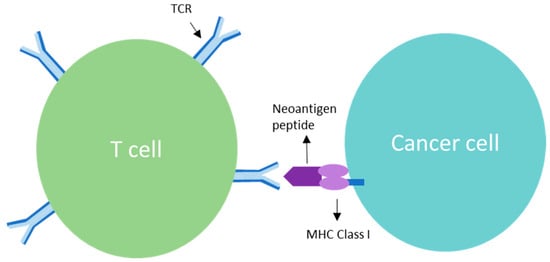
Clonal mutations represent the initiating molecular defects related to cellular transition of a normal phenotype to a malignant phenotype. Molecular genomic assessment utilizing next generation and whole exome sequencing is now being increasingly applied to biomarker determination to refine the use of targeted [...] Read more.
Clonal mutations represent the initiating molecular defects related to cellular transition of a normal phenotype to a malignant phenotype. Molecular genomic assessment utilizing next generation and whole exome sequencing is now being increasingly applied to biomarker determination to refine the use of targeted immune therapies. Case examples followed by retrospective study assessment have convincingly demonstrated clonal neoantigens provide a relevant predictor of response to checkpoint inhibition. A meta-analysis, by Litchfield et al., of over 1000 cancer patients from 12 landmark trials demonstrated no clinical benefit to checkpoint inhibitor (CPI) therapy in correlation to high subclonal tumor mutational burden (TMB), whereas high clonal TMB was found to be significantly correlated with better overall survival (p = 0.000000029). We discuss the mechanism of clonal vs. subclonal neoantigen targeting relationship to homologous recombination proficient (HRP) profile, evidence of preclinical and clinical benefit related to clonal neoantigens, and review a novel developing therapy called Vigil®, designed to expand the clonal neoantigen targeting effector cell populations. Vigil® is an autologous cellular immunotherapy which is designed to carry the full set of personal clonal neoantigens. Phase 2b results demonstrate a durable recurrence-free survival (RFS) and overall survival (OS) advantage for Vigil® in a subset ovarian cancer population with an HRP cancer profile. Full article
Show Figures

Figure 1

16 pages, 3723 KiB  
Article
Ring Chromosomes in Hematological Malignancies Are Associated with TP53 Gene Mutations and Characteristic Copy Number Variants
by Rachel J. Boyd, Jaclyn B. Murry, Laura A. Morsberger, Melanie Klausner, Suping Chen, Christopher D. Gocke, Andrew S. McCallion and Ying S. Zou
Cancers 2023, 15(22), 5439; https://doi.org/10.3390/cancers15225439 - 16 Nov 2023
Cited by 3 | Viewed by 1382
Abstract
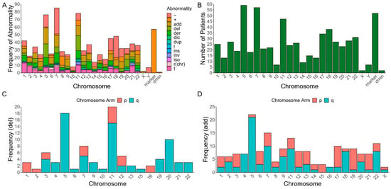
Ring chromosomes (RC) are present in <10% of patients with hematological malignancies and are associated with poor prognosis. Until now, only small cohorts of patients with hematological neoplasms and concomitant RCs have been cytogenetically characterized. Here, we performed a conventional chromosome analysis on [...] Read more.
Ring chromosomes (RC) are present in <10% of patients with hematological malignancies and are associated with poor prognosis. Until now, only small cohorts of patients with hematological neoplasms and concomitant RCs have been cytogenetically characterized. Here, we performed a conventional chromosome analysis on metaphase spreads from >13,000 patients diagnosed with hematological malignancies at the Johns Hopkins University Hospital and identified 98 patients with RCs—90 with myeloid malignancies and 8 with lymphoid malignancies. We also performed a targeted Next-Generation Sequencing (NGS) assay, using a panel of 642 cancer genes, to identify whether these patients harbor relevant pathogenic variants. Cytogenetic analyses revealed that RCs and marker chromosomes of unknown origin are concurrently present in most patients by karyotyping, and 93% of patients with NGS data have complex karyotypes. A total of 72% of these individuals have pathogenic mutations in TP53, most of whom also possess cytogenetic abnormalities resulting in the loss of 17p, including the loss of TP53. All patients with a detected RC and without complex karyotypes also lack TP53 mutations but have pathogenic mutations in TET2. Further, 70% of RCs that map to a known chromosome are detected in individuals without TP53 mutations. Our data suggest that RCs in hematological malignancies may arise through different mechanisms, but ultimately promote widespread chromosomal instability. Full article
Show Figures

Figure 1

19 pages, 6308 KiB  
Article
ASPP2 Is Phosphorylated by CDK1 during Mitosis and Required for Pancreatic Cancer Cell Proliferation
by Yi Xiao, Yuanhong Chen, Jianan Chen and Jixin Dong
Cancers 2023, 15(22), 5424; https://doi.org/10.3390/cancers15225424 - 15 Nov 2023
Cited by 2 | Viewed by 1529
Abstract
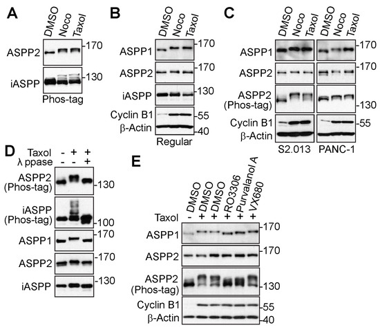
(1) Background: pancreatic cancer is highly lethal. The role of apoptosis-stimulating protein of p53-2 (ASPP2) in this lethal disease remains unclear. This protein belongs to the ASPP family of p53 interacting proteins. Previous studies in this lab used phosphate-binding tag (Phos-tag) sodium dodecyl [...] Read more.
(1) Background: pancreatic cancer is highly lethal. The role of apoptosis-stimulating protein of p53-2 (ASPP2) in this lethal disease remains unclear. This protein belongs to the ASPP family of p53 interacting proteins. Previous studies in this lab used phosphate-binding tag (Phos-tag) sodium dodecyl sulfate (SDS) polyacrylamide gels and identified a motility upshift of the ASPP family of proteins during mitosis. (2) Purpose: this study expands on previous findings to identify the detailed phosphorylation regulation of ASPP2 during mitosis, as well as the function of ASPP2 in pancreatic cancer. (3) Methods: the Phos-tag technique was used to investigate the phosphorylation mechanism of ASPP2 during mitosis. Phospho-specific antibodies were generated to validate the phosphorylation of ASPP2, and ASPP2-inducible expression cell lines were established to determine the role of ASPP2 in pancreatic cancer. RNA sequencing (RNA-Seq) was used to uncover the downstream targets of ASPP2. (4) Results: results demonstrate that ASPP2 is phosphorylated during mitosis by cyclin-dependent kinase 1 (CDK1) at sites S562 and S704. In vitro and in vivo results show that ASPP2 is required for pancreatic cancer growth. Furthermore, the expressions of yes-associated protein (YAP)-related genes are found to be dramatically altered by ASPP2 depletion. Together, these findings reveal the phosphorylation mechanism of ASPP2 during mitosis. Collectively, results strongly indicate that ASPP2 is a potential target for abating tumor cell growth in pancreatic cancer. Full article
Show Figures

Figure 1

17 pages, 3797 KiB  
Article
Dpep Inhibits Cancer Cell Growth and Survival via Shared and Context-Dependent Transcriptome Perturbations
by Qing Zhou and Lloyd A. Greene
Cancers 2023, 15(22), 5318; https://doi.org/10.3390/cancers15225318 - 7 Nov 2023
Cited by 1 | Viewed by 1440
Abstract
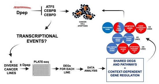
Dpep is a cell-penetrating peptide targeting transcription factors ATF5, CEBPB, and CEBPD, and that selectively promotes the apoptotic death of multiple tumor cell types in vitro and in vivo. As such, it is a potential therapeutic. To better understand its mechanism of action, [...] Read more.
Dpep is a cell-penetrating peptide targeting transcription factors ATF5, CEBPB, and CEBPD, and that selectively promotes the apoptotic death of multiple tumor cell types in vitro and in vivo. As such, it is a potential therapeutic. To better understand its mechanism of action, we used PLATE-seq to compare the transcriptomes of six cancer cell lines of diverse origins before and after Dpep exposure. This revealed a context-dependent pattern of regulated genes that was unique to each line, but that exhibited a number of elements that were shared with other lines. This included the upregulation of pro-apoptotic genes and tumor suppressors as well as the enrichment of genes associated with responses to hypoxia and interferons. Downregulated transcripts included oncogenes and dependency genes, as well as enriched genes associated with different phases of the cell cycle and with DNA repair. In each case, such changes have the potential to lie upstream of apoptotic cell death. We also detected the regulation of unique as well as shared sets of transcription factors in each line, suggesting that Dpep may initiate a cascade of transcriptional responses that culminate in cancer cell death. Such death thus appears to reflect context-dependent, yet shared, disruption of multiple cellular pathways as well as of individual survival-relevant genes. Full article
Show Figures

Graphical abstract

12 pages, 1946 KiB  
Article
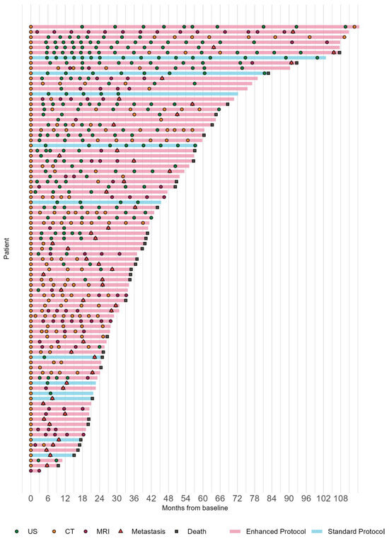
Surveillance for Metastasis in High-Risk Uveal Melanoma Patients: Standard versus Enhanced Protocols
by Yağmur Seda Yeşiltaş, Emily C. Zabor, Jacquelyn Wrenn, Zackery Oakey and Arun D. Singh
Cancers 2023, 15(20), 5025; https://doi.org/10.3390/cancers15205025 - 17 Oct 2023
Cited by 2 | Viewed by 1299
Abstract
Purpose: to evaluate the effectiveness of enhanced surveillance protocols (EP) utilizing high frequency (HF) or enhanced modality (EM) compared to the standard protocol (SP) in detecting metastasis and determining their impact on overall survival (OS) in high-risk uveal melanoma (UM) patients. Methods: A [...] Read more.
Purpose: to evaluate the effectiveness of enhanced surveillance protocols (EP) utilizing high frequency (HF) or enhanced modality (EM) compared to the standard protocol (SP) in detecting metastasis and determining their impact on overall survival (OS) in high-risk uveal melanoma (UM) patients. Methods: A total of 87 consecutive patients with Class 2 (high risk) primary UM were enrolled, with negative baseline systemic staging. The patients underwent systemic surveillance with either SP (hepatic ultrasonography [US] every 6 months) or EP (either HF [US every 3 months] or EM [incorporation hepatic computed tomography/magnetic resonance imaging]) following informed discussion. The main outcome measures were largest diameter of largest hepatic metastasis (LDLM), number of hepatic metastatic lesions, time to detection of metastasis (TDM), and OS. Results: This study revealed significant differences in LDLM between surveillance protocols, with the use of EP detecting smaller metastatic lesions (HF, EM, and SP were 1.5 cm, 1.6 cm, and 6.1 cm, respectively). Patients on the EM protocol had a lower 24-month cumulative incidence of >3 cm metastasis (3.5% EM vs. 39% SP; p = 0.021), while those on the HF protocol had a higher 24-month cumulative incidence of ≤3 cm metastasis compared to SP (31% HF vs. 10% SP; p = 0.017). Hazard of death following metastasis was significantly reduced in the EP (HR: 0.25; 95% CI: 0.07, 0.84), HF (HR: 0.23; 95% CI: 0.06, 0.84), and EM (HR: 0.11; 95% CI: 0.02, 0.5) groups compared to SP. However, TDM and OS did not significantly differ between protocols. Conclusions: Enhanced surveillance protocols improved early detection of hepatic metastasis in UM patients but did not translate into a survival advantage in our study cohort. However, early detection of metastasis in patients receiving liver-directed therapies may lead to improved overall survival. Full article
Show Figures

Figure 1

16 pages, 1745 KiB  
Review
Early-Onset Colorectal Cancer: Current Insights
by Fauzia Ullah, Ashwathy Balachandran Pillai, Najiullah Omar, Danai Dima and Seema Harichand
Cancers 2023, 15(12), 3202; https://doi.org/10.3390/cancers15123202 - 15 Jun 2023
Cited by 12 | Viewed by 4589
Abstract
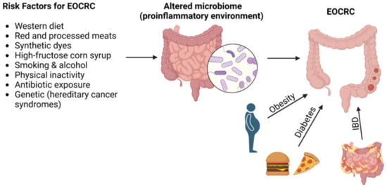
Over the past decade, the incidence of colorectal cancer has increased in individuals under the age of 50 years. Meanwhile, the incidence has gradually decreased in the older population. As described herein, we reviewed the available literature to summarize the current landscape of [...] Read more.
Over the past decade, the incidence of colorectal cancer has increased in individuals under the age of 50 years. Meanwhile, the incidence has gradually decreased in the older population. As described herein, we reviewed the available literature to summarize the current landscape of early-onset colorectal cancer, including risk factors, clinicopathological presentation, genetic makeup of patients, and management. Currently, early-onset colorectal cancer is treated similarly as late-onset colorectal cancer, yet the available literature shows that early-onset colorectal cancer is more aggressive and different, and this remains a significant unmet need. A detailed understanding of early-onset colorectal cancer is needed to identify risk factors for the increased incidence and tailor treatments accordingly. Full article
Show Figures

Figure 1

13 pages, 530 KiB  
Review
Regulations of Tumor Microenvironment by Prostaglandins
by Jeffrey Z. Nie, Man-Tzu Wang and Daotai Nie
Cancers 2023, 15(12), 3090; https://doi.org/10.3390/cancers15123090 - 7 Jun 2023
Cited by 8 | Viewed by 1996
Abstract
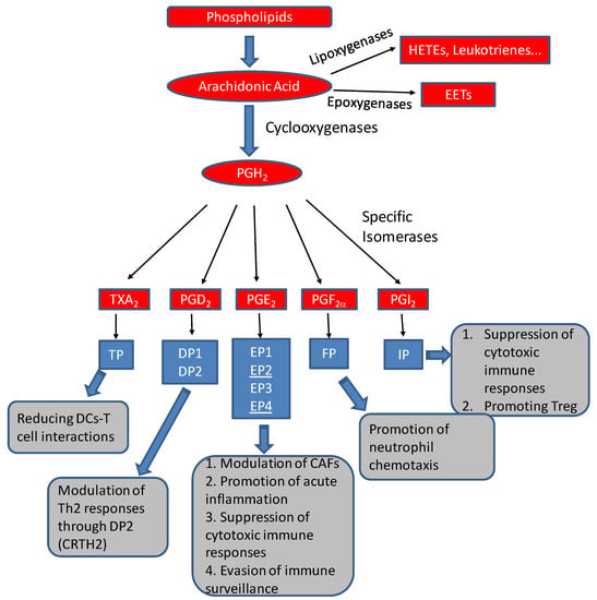
Prostaglandins, the bioactive lipids generated from the metabolism of arachidonic acid through cyclooxygenases, have potent effects on many constituents of tumor microenvironments. In this review, we will describe the formation and activities of prostaglandins in the context of the tumor microenvironment. We will [...] Read more.
Prostaglandins, the bioactive lipids generated from the metabolism of arachidonic acid through cyclooxygenases, have potent effects on many constituents of tumor microenvironments. In this review, we will describe the formation and activities of prostaglandins in the context of the tumor microenvironment. We will discuss the regulation of cancer-associated fibroblasts and immune constituents by prostaglandins and their roles in immune escapes during tumor progression. The review concludes with future perspectives on improving the efficacy of immunotherapy through repurposing non-steroid anti-inflammatory drugs and other prostaglandin modulators. Full article
Show Figures

Figure 1

29 pages, 3670 KiB  
Review
Crosstalk between Noncoding RNAs and the Epigenetics Machinery in Pediatric Tumors and Their Microenvironment
by Anup S. Pathania
Cancers 2023, 15(10), 2833; https://doi.org/10.3390/cancers15102833 - 19 May 2023
Cited by 4 | Viewed by 1906
Abstract
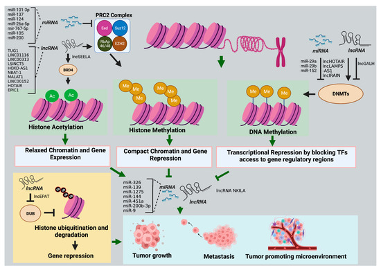
According to the World Health Organization, every year, an estimated 400,000+ new cancer cases affect children under the age of 20 worldwide. Unlike adult cancers, pediatric cancers develop very early in life due to alterations in signaling pathways that regulate embryonic development, and [...] Read more.
According to the World Health Organization, every year, an estimated 400,000+ new cancer cases affect children under the age of 20 worldwide. Unlike adult cancers, pediatric cancers develop very early in life due to alterations in signaling pathways that regulate embryonic development, and environmental factors do not contribute much to cancer development. The highly organized complex microenvironment controlled by synchronized gene expression patterns plays an essential role in the embryonic stages of development. Dysregulated development can lead to tumor initiation and growth. The low mutational burden in pediatric tumors suggests the predominant role of epigenetic changes in driving the cancer phenotype. However, one more upstream layer of regulation driven by ncRNAs regulates gene expression and signaling pathways involved in the development. Deregulation of ncRNAs can alter the epigenetic machinery of a cell, affecting the transcription and translation profiles of gene regulatory networks required for cellular proliferation and differentiation during embryonic development. Therefore, it is essential to understand the role of ncRNAs in pediatric tumor development to accelerate translational research to discover new treatments for childhood cancers. This review focuses on the role of ncRNA in regulating the epigenetics of pediatric tumors and their tumor microenvironment, the impact of their deregulation on driving pediatric tumor progress, and their potential as effective therapeutic targets. Full article
Show Figures

Figure 1

19 pages, 1126 KiB  
Review
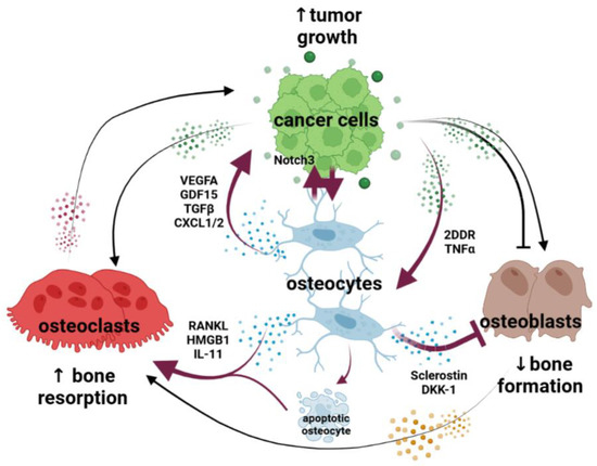
Osteocytes: New Kids on the Block for Cancer in Bone Therapy
by Aric Anloague and Jesus Delgado-Calle
Cancers 2023, 15(9), 2645; https://doi.org/10.3390/cancers15092645 - 7 May 2023
Cited by 4 | Viewed by 2810
Abstract
The tumor microenvironment plays a central role in the onset and progression of cancer in the bone. Cancer cells, either from tumors originating in the bone or from metastatic cancer cells from other body systems, are located in specialized niches where they interact [...] Read more.
The tumor microenvironment plays a central role in the onset and progression of cancer in the bone. Cancer cells, either from tumors originating in the bone or from metastatic cancer cells from other body systems, are located in specialized niches where they interact with different cells of the bone marrow. These interactions transform the bone into an ideal niche for cancer cell migration, proliferation, and survival and cause an imbalance in bone homeostasis that severely affects the integrity of the skeleton. During the last decade, preclinical studies have identified new cellular mechanisms responsible for the dependency between cancer cells and bone cells. In this review, we focus on osteocytes, long-lived cells residing in the mineral matrix that have recently been identified as key players in the spread of cancer in bone. We highlight the most recent discoveries on how osteocytes support tumor growth and promote bone disease. Additionally, we discuss how the reciprocal crosstalk between osteocytes and cancer cells provides the opportunity to develop new therapeutic strategies to treat cancer in the bone. Full article
Show Figures

Figure 1

14 pages, 1570 KiB  
Article
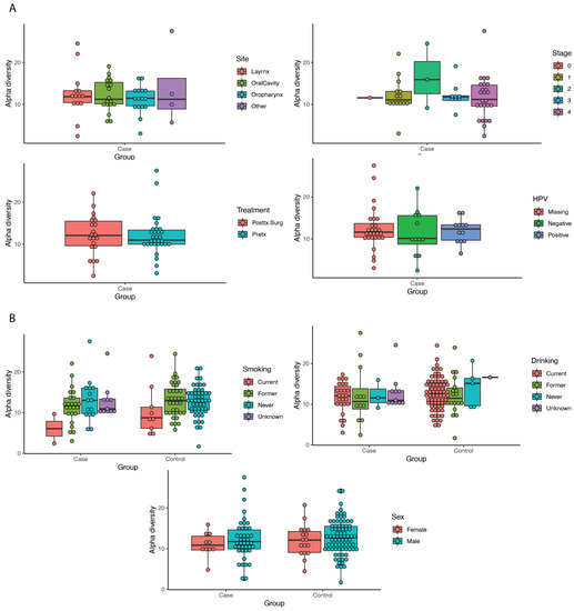
Oral Microbiome Community Composition in Head and Neck Squamous Cell Carcinoma
by William J. Benjamin, Kai Wang, Katherine Zarins, Emily Bellile, Freida Blostein, Ilona Argirion, Jeremy M. G. Taylor, Nisha J. D’Silva, Steven B. Chinn, Samara Rifkin, Maureen A. Sartor and Laura S. Rozek
Cancers 2023, 15(9), 2549; https://doi.org/10.3390/cancers15092549 - 29 Apr 2023
Cited by 13 | Viewed by 2732
Abstract
The impact of the oral microbiome on head and neck cancer pathogenesis and outcomes requires further study. 16s rRNA was isolated and amplified from pre-treatment oral wash samples for 52 cases and 102 controls. The sequences were binned into operational taxonomic units (OTUs) [...] Read more.
The impact of the oral microbiome on head and neck cancer pathogenesis and outcomes requires further study. 16s rRNA was isolated and amplified from pre-treatment oral wash samples for 52 cases and 102 controls. The sequences were binned into operational taxonomic units (OTUs) at the genus level. Diversity metrics and significant associations between OTUs and case status were assessed. The samples were binned into community types using Dirichlet multinomial models, and survival outcomes were assessed by community type. Twelve OTUs from the phyla Firmicutes, Proteobacteria, and Acinetobacter were found to differ significantly between the cases and the controls. Beta-diversity was significantly higher between the cases than between the controls (p < 0.01). Two community types were identified based on the predominant sets of OTUs within our study population. The community type with a higher abundance of periodontitis-associated bacteria was more likely to be present in the cases (p < 0.01), in older patients (p < 0.01), and in smokers (p < 0.01). Significant differences between the cases and the controls in community type, beta-diversity, and OTUs indicate that the oral microbiome may play a role in HNSCC. Full article
Show Figures

Figure 1

22 pages, 2897 KiB  
Article
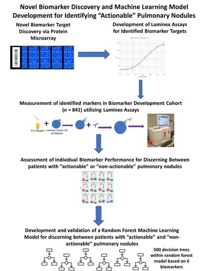
Development of a Novel Circulating Autoantibody Biomarker Panel for the Identification of Patients with ‘Actionable’ Pulmonary Nodules
by Claire Auger, Hita Moudgalya, Matthew R. Neely, Jeremy T. Stephan, Imad Tarhoni, David Gerard, Sanjib Basu, Cristina L. Fhied, Ahmed Abdelkader, Moises Vargas, Shaohui Hu, Tyler Hulett, Michael J. Liptay, Palmi Shah, Christopher W. Seder and Jeffrey A. Borgia
Cancers 2023, 15(8), 2259; https://doi.org/10.3390/cancers15082259 - 12 Apr 2023
Cited by 2 | Viewed by 2675
Abstract
Due to poor compliance and uptake of LDCT screening among high-risk populations, lung cancer is often diagnosed in advanced stages where treatment is rarely curative. Based upon the American College of Radiology’s Lung Imaging and Reporting Data System (Lung-RADS) 80–90% of patients screened [...] Read more.
Due to poor compliance and uptake of LDCT screening among high-risk populations, lung cancer is often diagnosed in advanced stages where treatment is rarely curative. Based upon the American College of Radiology’s Lung Imaging and Reporting Data System (Lung-RADS) 80–90% of patients screened will have clinically “non-actionable” nodules (Lung-RADS 1 or 2), and those harboring larger, clinically “actionable” nodules (Lung-RADS 3 or 4) have a significantly greater risk of lung cancer. The development of a companion diagnostic method capable of identifying patients likely to have a clinically actionable nodule identified during LDCT is anticipated to improve accessibility and uptake of the paradigm and improve early detection rates. Using protein microarrays, we identified 501 circulating targets with differential immunoreactivities against cohorts characterized as possessing either actionable (n = 42) or non-actionable (n = 20) solid pulmonary nodules, per Lung-RADS guidelines. Quantitative assays were assembled on the Luminex platform for the 26 most promising targets. These assays were used to measure serum autoantibody levels in 841 patients, consisting of benign (BN; n = 101), early-stage non-small cell lung cancer (NSCLC; n = 245), other early-stage malignancies within the lung (n = 29), and individuals meeting United States Preventative Screening Task Force (USPSTF) screening inclusion criteria with both actionable (n = 87) and non-actionable radiologic findings (n = 379). These 841 patients were randomly split into three cohorts: Training, Validation 1, and Validation 2. Of the 26 candidate biomarkers tested, 17 differentiated patients with actionable nodules from those with non-actionable nodules. A random forest model consisting of six autoantibody (Annexin 2, DCD, MID1IP1, PNMA1, TAF10, ZNF696) biomarkers was developed to optimize our classification performance; it possessed a positive predictive value (PPV) of 61.4%/61.0% and negative predictive value (NPV) of 95.7%/83.9% against Validation cohorts 1 and 2, respectively. This panel may improve patient selection methods for lung cancer screening, serving to greatly reduce the futile screening rate while also improving accessibility to the paradigm for underserved populations. Full article
Show Figures

Graphical abstract

20 pages, 4345 KiB  
Article
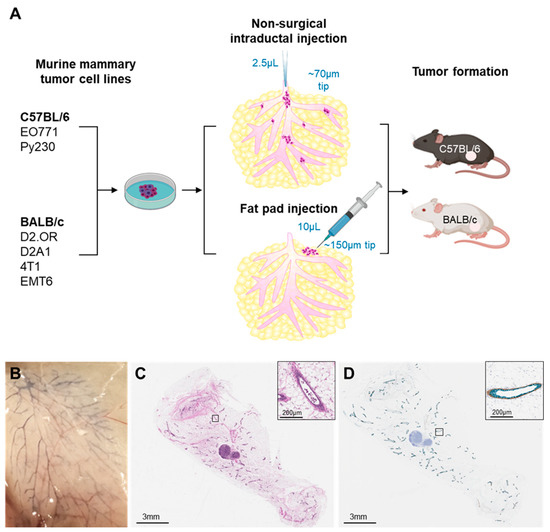
Isogenic Mammary Models of Intraductal Carcinoma Reveal Progression to Invasiveness in the Absence of a Non-Obligatory In Situ Stage
by Sarah M. Bernhardt, Elizabeth Mitchell, Stephanie Stamnes, Reuben J. Hoffmann, Andrea Calhoun, Alex Klug, Tanya D. Russell, Nathan D. Pennock, Joshua M. Walker and Pepper Schedin
Cancers 2023, 15(8), 2257; https://doi.org/10.3390/cancers15082257 - 12 Apr 2023
Cited by 3 | Viewed by 2703
Abstract
In breast cancer, progression to invasive ductal carcinoma (IDC) involves interactions between immune, myoepithelial, and tumor cells. Development of IDC can proceed through ductal carcinoma in situ (DCIS), a non-obligate, non-invasive stage, or IDC can develop without evidence of DCIS and these cases [...] Read more.
In breast cancer, progression to invasive ductal carcinoma (IDC) involves interactions between immune, myoepithelial, and tumor cells. Development of IDC can proceed through ductal carcinoma in situ (DCIS), a non-obligate, non-invasive stage, or IDC can develop without evidence of DCIS and these cases associate with poorer prognosis. Tractable, immune-competent mouse models are needed to help delineate distinct mechanisms of local tumor cell invasion and prognostic implications. To address these gaps, we delivered murine mammary carcinoma cell lines directly into the main mammary lactiferous duct of immune-competent mice. Using two strains of immune-competent mice (BALB/c, C57BL/6), one immune-compromised (severe combined immunodeficiency; SCID) C57BL/6 strain, and six different murine mammary cancer cell lines (D2.OR, D2A1, 4T1, EMT6, EO771, Py230), we found early loss of ductal myoepithelial cell differentiation markers p63, α-smooth muscle actin, and calponin, and rapid formation of IDC in the absence of DCIS. Rapid IDC formation also occurred in the absence of adaptive immunity. Combined, these studies demonstrate that loss of myoepithelial barrier function does not require an intact immune system, and suggest that these isogenic murine models may prove a useful tool to study IDC in the absence of a non-obligatory DCIS stage—an under-investigated subset of poor prognostic human breast cancer. Full article
Show Figures

Figure 1

15 pages, 729 KiB  
Review
Precision Oncology Targets in Biliary Tract Cancer
by Nicole Farha, Danai Dima, Fauzia Ullah and Suneel Kamath
Cancers 2023, 15(7), 2105; https://doi.org/10.3390/cancers15072105 - 31 Mar 2023
Cited by 11 | Viewed by 3994
Abstract
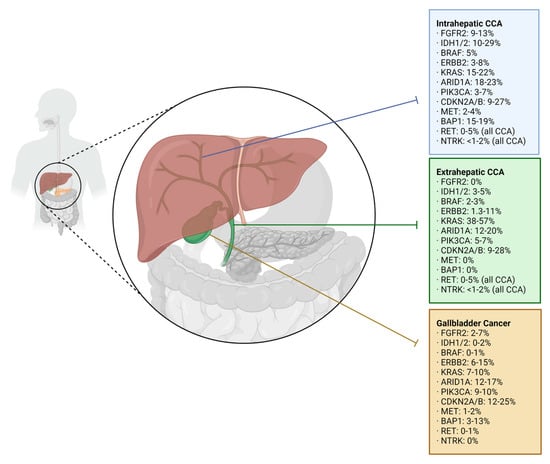
Targeted therapies in biliary tract cancer (BTC) are emerging as options for patients not who do not respond to first-line treatment. Agents acting on tumor-specific oncogenes in BTC may target fibroblast growth factor receptor 2 (FGFR2), isocitrate dehydrogenase (IDH), B-raf kinase (BRAF), and [...] Read more.
Targeted therapies in biliary tract cancer (BTC) are emerging as options for patients not who do not respond to first-line treatment. Agents acting on tumor-specific oncogenes in BTC may target fibroblast growth factor receptor 2 (FGFR2), isocitrate dehydrogenase (IDH), B-raf kinase (BRAF), and human epidermal growth factor receptor 2 (HER-2). Additionally, given the heterogeneous genetic landscape of advanced BTCs, many harbor genetic aberrations that are common among solid tumors, including RET fusions, tropomyosin receptor kinase (TRK) fusions, and high tumor mutational burden (TMB). This review aims to provide updates on the evolving array of therapeutics available, and to summarize promising works on the horizon. Full article
Show Figures

Figure 1

15 pages, 1152 KiB  
Article
Rural–Urban Disparities in Patient Care Experiences among Prostate Cancer Survivors: A SEER-CAHPS Study
by Ambrish A. Pandit, Nilesh N. Patil, Mostafa Mostafa, Mohamed Kamel, Michael T. Halpern and Chenghui Li
Cancers 2023, 15(7), 1939; https://doi.org/10.3390/cancers15071939 - 23 Mar 2023
Cited by 4 | Viewed by 1944
Abstract
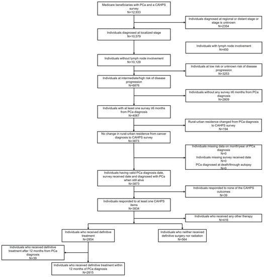
Background: We sought to evaluate rural–urban disparities in patient care experiences (PCEs) among localized prostate cancer (PCa) survivors at intermediate-to-high risk of disease progression. Methods: Using 2007–2015 Surveillance Epidemiology and End Results (SEER) data linked to Medicare Consumer Assessment of Healthcare Providers and [...] Read more.
Background: We sought to evaluate rural–urban disparities in patient care experiences (PCEs) among localized prostate cancer (PCa) survivors at intermediate-to-high risk of disease progression. Methods: Using 2007–2015 Surveillance Epidemiology and End Results (SEER) data linked to Medicare Consumer Assessment of Healthcare Providers and Systems (CAHPS) surveys, we analyzed survivors’ first survey ≥6 months post-diagnosis. Covariate adjusted linear regressions were used to estimate associations of treatment status (definitive treatment vs. none) and residence (large metro vs. metro vs. rural) with PCE composite and rating measures. Results: Among 3779 PCa survivors, 1798 (53.2%) and 370 (10.9%) resided in large metro and rural areas, respectively; more rural (vs. large metro) residents were untreated (21.9% vs. 16.7%; p = 0.017). Untreated (vs. treated) PCa survivors reported lower scores for doctor communication (ß = −2.0; p = 0.022), specialist rating (ß = −2.5; p = 0.008), and overall care rating (ß = −2.4; p = 0.006). While treated rural survivors gave higher (ß = 3.6; p = 0.022) scores for obtaining needed care, untreated rural survivors gave lower scores for obtaining needed care (ß = −7.0; p = 0.017) and a lower health plan rating (ß = −7.9; p = 0.003) compared to their respective counterparts in large metro areas. Conclusions: Rural PCa survivors are less likely to receive treatment. Rural–urban differences in PCEs varied by treatment status. Full article
Show Figures

Figure 1

16 pages, 2109 KiB  
Review
Inflammation, Infiltration, and Evasion—Tumor Promotion in the Aging Breast
by Nicole Cruz-Reyes and Derek C. Radisky
Cancers 2023, 15(6), 1836; https://doi.org/10.3390/cancers15061836 - 18 Mar 2023
Cited by 3 | Viewed by 2845
Abstract
Breast cancer is a significant cause of morbidity and mortality in women, with over two million new cases reported worldwide each year, the majority of which occur in post-menopausal women. Despite advances in early detection and treatment, approximately one-third of patients diagnosed with [...] Read more.
Breast cancer is a significant cause of morbidity and mortality in women, with over two million new cases reported worldwide each year, the majority of which occur in post-menopausal women. Despite advances in early detection and treatment, approximately one-third of patients diagnosed with breast cancer will develop metastatic disease. The pathogenesis and progression of breast cancer are influenced by a variety of biological and social risk factors, including age, ethnicity, pregnancy status, diet, and genomic alterations. Recent advancements in breast cancer research have focused on harnessing the power of the patient’s adaptive and innate immune systems for diagnostic and therapeutic purposes. The breast immune microenvironment plays a critical role in regulating tissue homeostasis and resistance to tumorigenesis. In this review, we explore the dynamic changes in the breast immune microenvironment that occur with age, how these changes impact breast cancer development and progression, and how targeted therapeutic interventions that leverage the immune system can be used to improve patient outcomes. Our review emphasizes the importance of understanding the complex interplay between aging, the immune system, and breast cancer, and highlights the potential of immune-based therapies in the fight against this devastating disease. Full article
Show Figures

Figure 1

15 pages, 1689 KiB  
Article
Unique Metabolic Contexts Sensitize Cancer Cells and Discriminate between Glycolytic Tumor Types
by Jonathan A. Chacon-Barahona, Jeffrey P. MacKeigan and Nathan J. Lanning
Cancers 2023, 15(4), 1158; https://doi.org/10.3390/cancers15041158 - 11 Feb 2023
Cited by 5 | Viewed by 2075
Abstract
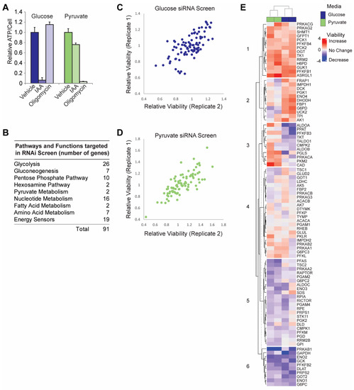
Cancer cells utilize variable metabolic programs in order to maintain homeostasis in response to environmental challenges. To interrogate cancer cell reliance on glycolytic programs under different nutrient availabilities, we analyzed a gene panel containing all glycolytic genes as well as pathways associated with [...] Read more.
Cancer cells utilize variable metabolic programs in order to maintain homeostasis in response to environmental challenges. To interrogate cancer cell reliance on glycolytic programs under different nutrient availabilities, we analyzed a gene panel containing all glycolytic genes as well as pathways associated with glycolysis. Using this gene panel, we analyzed the impact of an siRNA library on cellular viability in cells containing only glucose or only pyruvate as the major bioenergetic nutrient source. From these panels, we aimed to identify genes that elicited conserved and glycolysis-dependent changes in cellular bioenergetics across glycolysis-promoting and OXPHOS-promoting conditions. To further characterize gene sets within this panel and identify similarities and differences amongst glycolytic tumor RNA-seq profiles across a pan-cancer cohort, we then used unsupervised statistical classification of RNA-seq profiles for glycolytic cancers and non-glycolytic cancer types. Here, Kidney renal clear cell carcinoma (KIRC); Head and Neck squamous cell carcinoma (HNSC); and Lung squamous cell carcinoma (LUSC) defined the glycolytic cancer group, while Prostate adenocarcinoma (PRAD), Thyroid carcinoma (THCA), and Thymoma (THYM) defined the non-glycolytic cancer group. These groups were defined based on glycolysis scoring from previous studies, where KIRC, HNSC, and LUSC had the highest glycolysis scores, meanwhile, PRAD, THCA, and THYM had the lowest. Collectively, these results aimed to identify multi-omic profiles across cancer types with demonstrated variably glycolytic rates. Our analyses provide further support for strategies aiming to classify tumors by metabolic phenotypes in order to therapeutically target tumor-specific vulnerabilities. Full article
Show Figures

Figure 1

13 pages, 676 KiB  
Review
Changes in Expression of Tumor Suppressor Gene RKIP Impact How Cancers Interact with Their Complex Environment
by Christopher Figy, Anna Guo, Veani Roshale Fernando, Saori Furuta, Fahd Al-Mulla and Kam C. Yeung
Cancers 2023, 15(3), 958; https://doi.org/10.3390/cancers15030958 - 2 Feb 2023
Cited by 2 | Viewed by 2042
Abstract
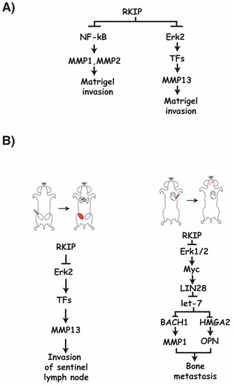
Tumor microenvironment (TME) is the immediate environment where cancer cells reside in a tumor. It is composed of multiple cell types and extracellular matrix. Microenvironments can be restrictive or conducive to the progression of cancer cells. Initially, microenvironments are suppressive in nature. Stepwise [...] Read more.
Tumor microenvironment (TME) is the immediate environment where cancer cells reside in a tumor. It is composed of multiple cell types and extracellular matrix. Microenvironments can be restrictive or conducive to the progression of cancer cells. Initially, microenvironments are suppressive in nature. Stepwise accumulation of mutations in oncogenes and tumor suppressor genes enables cancer cells to acquire the ability to reshape the microenvironment to advance their growth and metastasis. Among the many genetic events, the loss-of-function mutations in tumor suppressor genes play a pivotal role. In this review, we will discuss the changes in TME and the ramifications on metastasis upon altered expression of tumor metastasis suppressor gene RKIP in breast cancer cells. Full article
Show Figures

Figure 1

10 pages, 888 KiB  
Review
The Influence of the Normal Mammary Microenvironment on Breast Cancer Cells
by Caroline J. Campbell and Brian W. Booth
Cancers 2023, 15(3), 576; https://doi.org/10.3390/cancers15030576 - 18 Jan 2023
Cited by 1 | Viewed by 2017
Abstract
The tumor microenvironment is recognized as performing a critical role in tumor initiation, progression, and metastasis of many cancers, including breast cancer. The breast cancer microenvironment is a complex mixture of cells consisting of tumor cells, immune cells, fibroblasts, and vascular cells, as [...] Read more.
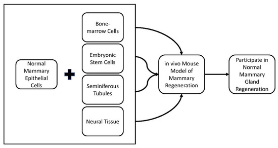
The tumor microenvironment is recognized as performing a critical role in tumor initiation, progression, and metastasis of many cancers, including breast cancer. The breast cancer microenvironment is a complex mixture of cells consisting of tumor cells, immune cells, fibroblasts, and vascular cells, as well as noncellular components, such as extracellular matrix and soluble products. The interactions between the tumor cells and the tumor microenvironment modulate tumor behavior and affect the responses of cancer patients to therapies. The interactions between tumor cells and the surrounding environment can include direct cell-to-cell contact or through intercellular signals over short and long distances. The intricate functions of the tumor microenvironment in breast cancer have led to increased research into the tumor microenvironment as a possible therapeutic target of breast cancer. Though expanded research has shown the clear importance of the tumor microenvironment, there is little focus on how normal mammary epithelial cells can affect breast cancer cells. Previous studies have shown the normal breast microenvironment can manipulate non-mammary stem cells and tumor-derived cancer stem cells to participate in normal mammary gland development. The tumorigenic cells lose their tumor-forming capacity and are “redirected” to divide into “normal”, non-tumorigenic cells. This cellular behavior is “cancer cell redirection”. This review will summarize the current literature on cancer cell redirection and the normal mammary microenvironment’s influence on breast cancer cells. Full article
Show Figures

Figure 1

2022

Jump to: 2024, 2023

23 pages, 6057 KiB  
Article
Compound C Inhibits Ovarian Cancer Progression via PI3K-AKT-mTOR-NFκB Pathway
by Alia Ghoneum, Daniela Gonzalez, Hesham Afify, Junjun Shu, Abigail Hegarty, Jemima Adisa, Michael Kelly, Samuel Lentz, Freddie Salsbury and Neveen Said
Cancers 2022, 14(20), 5099; https://doi.org/10.3390/cancers14205099 - 18 Oct 2022
Cited by 3 | Viewed by 2541
Abstract
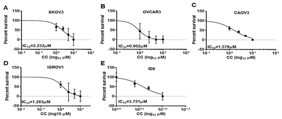
Epithelial Ovarian cancer (OvCa) is the leading cause of death from gynecologic malignancies in the United States, with most patients diagnosed at late stages. High-grade serous cancer (HGSC) is the most common and lethal subtype. Despite aggressive surgical debulking and chemotherapy, recurrence of [...] Read more.
Epithelial Ovarian cancer (OvCa) is the leading cause of death from gynecologic malignancies in the United States, with most patients diagnosed at late stages. High-grade serous cancer (HGSC) is the most common and lethal subtype. Despite aggressive surgical debulking and chemotherapy, recurrence of chemo-resistant disease occurs in ~80% of patients. Thus, developing therapeutics that not only targets OvCa cell survival, but also target their interactions within their unique peritoneal tumor microenvironment (TME) is warranted. Herein, we report therapeutic efficacy of compound C (also known as dorsomorphin) with a novel mechanism of action in OvCa. We found that CC not only inhibited OvCa growth and invasiveness, but also blunted their reciprocal crosstalk with macrophages, and mesothelial cells. Mechanistic studies indicated that compound C exerts its effects on OvCa cells through inhibition of PI3K-AKT-NFκB pathways, whereas in macrophages and mesothelial cells, CC inhibited cancer-cell-induced canonical NFκB activation. We further validated the specificity of the PI3K-AKT-NFκB as targets of compound C by overexpression of constitutively active subunits as well as computational modeling. In addition, real-time monitoring of OvCa cellular bioenergetics revealed that compound C inhibits ATP production, mitochondrial respiration, and non-mitochondrial oxygen consumption. Importantly, compound C significantly decreased tumor burden of OvCa xenografts in nude mice and increased their sensitivity to cisplatin-treatment. Moreover, compound C re-sensitized patient-derived resistant cells to cisplatin. Together, our findings highlight compound C as a potent multi-faceted therapeutic in OvCa. Full article
Show Figures

Figure 1

16 pages, 632 KiB  
Article
Omega-3 Polyunsaturated Fatty Acids, Gut Microbiota, Microbial Metabolites, and Risk of Colorectal Adenomas
by Tengteng Wang, Nicole M. Brown, Amber N. McCoy, Robert S. Sandler and Temitope O. Keku
Cancers 2022, 14(18), 4443; https://doi.org/10.3390/cancers14184443 - 13 Sep 2022
Cited by 6 | Viewed by 1731
Abstract
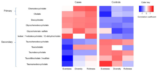
Omega-3 polyunsaturated fatty acids (ω-3 PUFAs) are thought to protect against colorectal adenoma (CRA) development. We aimed to further understand the underlying mechanisms by examining the relationships between ω-3 PUFAs and the gut microbiota on CRAs. We assessed the mucosal microbiota via bacterial [...] Read more.
Omega-3 polyunsaturated fatty acids (ω-3 PUFAs) are thought to protect against colorectal adenoma (CRA) development. We aimed to further understand the underlying mechanisms by examining the relationships between ω-3 PUFAs and the gut microbiota on CRAs. We assessed the mucosal microbiota via bacterial 16S rRNA sequencing among 217 CRA cases and 218 controls who completed PUFA intake questionnaires. The overall microbial composition was assessed by α-diversity measurements (diversity, richness, and evenness). Global metabolomics was conducted using a random subset of case–control pairs (n = 50). We compared microbiota and metabolite signatures between cases and controls according to fold change (FC). Odds ratios (OR) and confidence intervals (CI) were estimated from logistic regression for associations of ω-3 PUFAs and the microbiota with CRAs. We observed an inverse association between overall ω-3 PUFA intake and CRAs, especially for short-chain ω -3 PUFAs (OR = 0.45, 95% CI: 0.21, 0.97). Such inverse associations were modified by bacterial evenness (p-interaction = 0.03). Participants with higher levels (FC > 2) of bile acid-relevant metabolites were more likely to have CRAs than the controls, and the correlation between bile acids and bacterial diversity differed by case–control status. Our findings suggest that ω-3 PUFAs are inversely associated with CRA development, and the association may be modified by gut microbiota profiles. Full article
Show Figures

Figure 1

19 pages, 46308 KiB  
Article
Demonstrating Tumor Vascular Disrupting Activity of the Small-Molecule Dihydronaphthalene Tubulin-Binding Agent OXi6196 as a Potential Therapeutic for Cancer Treatment
by Li Liu, Regan Schuetze, Jeni L. Gerberich, Ramona Lopez, Samuel O. Odutola, Rajendra P. Tanpure, Amanda K. Charlton-Sevcik, Justin K. Tidmore, Emily A.-S. Taylor, Payal Kapur, Hans Hammers, Mary Lynn Trawick, Kevin G. Pinney and Ralph P. Mason
Cancers 2022, 14(17), 4208; https://doi.org/10.3390/cancers14174208 - 30 Aug 2022
Cited by 4 | Viewed by 2027
Abstract
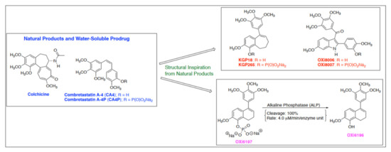
The vascular disrupting activity of a promising tubulin-binding agent (OXi6196) was demonstrated in mice in MDA-MB-231 human breast tumor xenografts growing orthotopically in mammary fat pad and syngeneic RENCA kidney tumors growing orthotopically in the kidney. To enhance water solubility, OXi6196, was derivatized [...] Read more.
The vascular disrupting activity of a promising tubulin-binding agent (OXi6196) was demonstrated in mice in MDA-MB-231 human breast tumor xenografts growing orthotopically in mammary fat pad and syngeneic RENCA kidney tumors growing orthotopically in the kidney. To enhance water solubility, OXi6196, was derivatized as its corresponding phosphate prodrug salt OXi6197, facilitating effective delivery. OXi6197 is stable in water, but rapidly releases OXi6196 in the presence of alkaline phosphatase. At low nanomolar concentrations OXi6196 caused G2/M cell cycle arrest and apoptosis in MDA-MB-231 breast cancer cells and monolayers of rapidly growing HUVECs underwent concentration-dependent changes in their morphology. Loss of the microtubule structure and increased bundling of filamentous actin into stress fibers followed by cell collapse, rounding and blebbing was observed. OXi6196 (100 nM) disrupted capillary-like endothelial networks pre-established with HUVECs on Matrigel®. When prodrug OXi6197 was administered to mice bearing orthotopic MDA-MB-231-luc tumors, dynamic bioluminescence imaging (BLI) revealed dose-dependent vascular shutdown with >80% signal loss within 2 h at doses ≥30 mg/kg and >90% shutdown after 6 h for doses ≥35 mg/kg, which remained depressed by at least 70% after 24 h. Twice weekly treatment with prodrug OXi6197 (20 mg/kg) caused a significant tumor growth delay, but no overall survival benefit. Similar efficacy was observed for the first time in orthotopic RENCA-luc tumors, which showed massive hemorrhage and necrosis after 24 h. Twice weekly dosing with prodrug OXi6197 (35 mg/kg) caused tumor growth delay in most orthotopic RENCA tumors. Immunohistochemistry revealed extensive necrosis, though with surviving peripheral tissues. These results demonstrate effective vascular disruption at doses comparable to the most effective vascular-disrupting agents (VDAs) suggesting opportunities for further development. Full article
Show Figures

Figure 1

15 pages, 12114 KiB  
Review
The Extracellular Matrix Environment of Clear Cell Renal Cell Carcinoma
by Leif Oxburgh
Cancers 2022, 14(17), 4072; https://doi.org/10.3390/cancers14174072 - 23 Aug 2022
Cited by 6 | Viewed by 2839
Abstract
The extracellular matrix (ECM) of tumors is a complex mix of components characteristic of the tissue of origin. In the majority of clear cell renal cell carcinomas (ccRCCs), the tumor suppressor VHL is inactivated. VHL controls matrix organization and its loss promotes a [...] Read more.
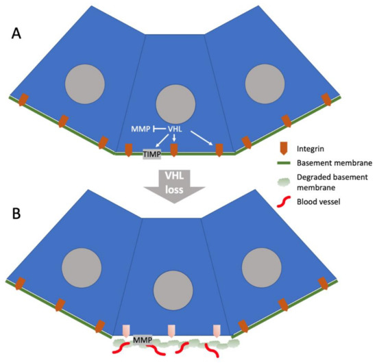
The extracellular matrix (ECM) of tumors is a complex mix of components characteristic of the tissue of origin. In the majority of clear cell renal cell carcinomas (ccRCCs), the tumor suppressor VHL is inactivated. VHL controls matrix organization and its loss promotes a loosely organized and angiogenic matrix, predicted to be an early step in tumor formation. During tumor evolution, cancer-associated fibroblasts (CAFs) accumulate, and they are predicted to produce abundant ECM. The ccRCC ECM composition qualitatively resembles that of the healthy kidney cortex in which the tumor arises, but there are important differences. One is the quantitative difference between a healthy cortex ECM and a tumor ECM; a tumor ECM contains a higher proportion of interstitial matrix components and a lower proportion of basement membrane components. Another is the breakdown of tissue compartments in the tumor with mixing of ECM components that are physically separated in healthy kidney cortex. Numerous studies reviewed in this work reveal effects of specific ECM components on the growth and invasive behaviors of ccRCCs, and extrapolation from other work suggests an important role for ECM in controlling ccRCC tumor rigidity, which is predicted to be a key determinant of invasive behavior. Full article
Show Figures

Figure 1

Back to TopTop