Molecular and Cellular Mechanisms of Synaptic Function: Neurotransmitter Release, Signal Transduction and Plasticity

A special issue of Cells (ISSN 2073-4409). This special issue belongs to the section "Cells of the Nervous System".

Deadline for manuscript submissions: closed (15 March 2023) | Viewed by 8514
Special Issue Editors


E-Mail Website
Guest Editor
1. Leibniz Institute for Neurobiology, RG Neuroplasticity, Brenneckestr. 6, 39118 Magdeburg, Germany
2. Center for Behavioral Brain Sciences, Otto-von-Guericke University Magdeburg, 39120 Magdeburg, Germany
Interests: synapse biology; NMDA receptors; long-distance protein transport; activity-dependent gene expression; membrane trafficking; activity-regulated autophagy
Special Issues, Collections and Topics in MDPI journals

E-Mail Website
Guest Editor
Leibniz Institute for Neurobiology, RG Cognition and Emotion, Brenneckestr. 6, 39118 Magdeburg, Germany
Interests: neural circuits; electrophysiology; calcium imaging techniques; behavioural neuroscience

E-Mail Website
Guest Editor
1. Institute for Biology/Genetics, Freie Universität Berlin, Berlin, Germany
2. Institute for Chemistry and Biochemistry/SupraFAB, Berlin, Germany
3. NeuroCure Cluster of Excellence, Charité Universitätsmedizin, Takustr. 6, 14195 Berlin, Germany
Interests: regulation of neurotransmitter release; presynaptic plasticity; super-resolution microscopy of synaptic scaffolds; aging; neuronal autophagy

Special Issue Information

Dear Colleagues, 

Brain information processing and storage, the basis of memory function, rely on the intercellular communication between neurons, polarized cells with complex cellular architecture. They predominantly communicate with each other via highly specialized contact sites, so-called chemical synapses. Synapses connecting two neurons, as well as neuro-muscular junctions (NMJs), represent the major structures crucial for signal transduction mediated by neurotransmitters. The effective conversion of electrical signals to chemical and back to electrical allows the nervous system to maintain adaptive responses to environmental stimuli. Synapses can change their signalling strength based on inputs from other neurons, a feature termed ‘synaptic plasticity’, widely considered as the cellular mechanism underlying learning and memory.

Recent technological advancements, such as the development of optogenetic tools and indicators to study neuronal transmission and connectivity within neuronal circuits in vivo, CRISPR/Cas technology, progress in computer simulations, and the development of advanced imaging techniques, are crucial for broadening our understanding of brain function and plasticity.

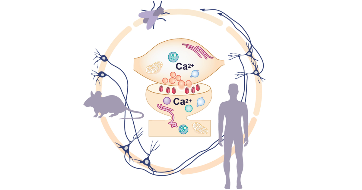
This Special Issue, entitled “Molecular and cellular mechanisms of synaptic function: neurotransmitter release, signal transduction and plasticity", aims to bridge recent multi-scale advances in the research of synaptic transmission and plasticity, ranging from Drosophila to the mammalian brain, in both health and disease. We plan to address the cellular and molecular aspects of neuronal excitability, calcium homeostasis, and the mechanisms of synaptic plasticity underlying behavioural change caused by experience. Additionally, this Special Issue aims to summarize the knowledge in this area of research and provide insights into the most recent developments in the field of synaptic plasticity. Therefore, we invite authors to contribute original research and review articles on each of these aspects. 

We welcome the submission of work covering, but not limited to, the following topics: 

  • Synaptic processes of neurotransmitter release;
  • Synaptic plasticity, including structural plasticity at the pre- and postsynaptic scaffold;
  • Molecular dynamics at the synapse;
  • Synapse and active zone assembly/maintenance;
  • Synapto-dendritic plasticity in development and learning;
  • Network connectivity;
  • Cognitive and emotional responses to multisensory environmental stimuli.

Dr. Anna V. Karpova
Dr. Sanja Bauer Mikulovic
Dr. Marta Maglione
Guest Editors

Manuscript Submission Information

Manuscripts should be submitted online at www.mdpi.com by registering and logging in to this website. Once you are registered, click here to go to the submission form. Manuscripts can be submitted until the deadline. All submissions that pass pre-check are peer-reviewed. Accepted papers will be published continuously in the journal (as soon as accepted) and will be listed together on the special issue website. Research articles, review articles as well as short communications are invited. For planned papers, a title and short abstract (about 100 words) can be sent to the Editorial Office for announcement on this website.

Submitted manuscripts should not have been published previously, nor be under consideration for publication elsewhere (except conference proceedings papers). All manuscripts are thoroughly refereed through a single-blind peer-review process. A guide for authors and other relevant information for submission of manuscripts is available on the Instructions for Authors page. Cells is an international peer-reviewed open access semimonthly journal published by MDPI.

Please visit the Instructions for Authors page before submitting a manuscript. The Article Processing Charge (APC) for publication in this open access journal is 2700 CHF (Swiss Francs). Submitted papers should be well formatted and use good English. Authors may use MDPI's English editing service prior to publication or during author revisions.

Keywords

  • synaptic plasticity
  • signal transduction
  • active zone
  • post-synaptic density
  • synaptic vesicle release
  • extracellular matrix
  • network connectivity
  • cognitive and emotional responses to multisensory environmental stimuli

Benefits of Publishing in a Special Issue

  • Ease of navigation: Grouping papers by topic helps scholars navigate broad scope journals more efficiently.
  • Greater discoverability: Special Issues support the reach and impact of scientific research. Articles in Special Issues are more discoverable and cited more frequently.
  • Expansion of research network: Special Issues facilitate connections among authors, fostering scientific collaborations.
  • External promotion: Articles in Special Issues are often promoted through the journal's social media, increasing their visibility.
  • e-Book format: Special Issues with more than 10 articles can be published as dedicated e-books, ensuring wide and rapid dissemination.

Further information on MDPI's Special Issue polices can be found here.

Published Papers (3 papers)

Order results
Result details
Select all
Export citation of selected articles as:

Research

Jump to: Review

19 pages, 2766 KiB  
Article
Medium-Chain Fatty Acids Rescue Motor Function and Neuromuscular Junction Degeneration in a Drosophila Model of Amyotrophic Lateral Sclerosis
by Ella Dunn, Joern R. Steinert, Aelfwin Stone, Virender Sahota, Robin S. B. Williams, Stuart Snowden and Hrvoje Augustin
Cells 2023, 12(17), 2163; https://doi.org/10.3390/cells12172163 - 28 Aug 2023
Cited by 2 | Viewed by 2367
Abstract
Amyotrophic lateral sclerosis (ALS) is an adult-onset neurodegenerative disease characterised by progressive degeneration of the motor neurones. An expanded GGGGCC (G4C2) hexanucleotide repeat in C9orf72 is the most common genetic cause of ALS and frontotemporal dementia (FTD); therefore, the resulting disease is known [...] Read more.
Amyotrophic lateral sclerosis (ALS) is an adult-onset neurodegenerative disease characterised by progressive degeneration of the motor neurones. An expanded GGGGCC (G4C2) hexanucleotide repeat in C9orf72 is the most common genetic cause of ALS and frontotemporal dementia (FTD); therefore, the resulting disease is known as C9ALS/FTD. Here, we employ a Drosophila melanogaster model of C9ALS/FTD (C9 model) to investigate a role for specific medium-chain fatty acids (MCFAs) in reversing pathogenic outcomes. Drosophila larvae overexpressing the ALS-associated dipeptide repeats (DPRs) in the nervous system exhibit reduced motor function and neuromuscular junction (NMJ) defects. We show that two MCFAs, nonanoic acid (NA) and 4-methyloctanoic acid (4-MOA), can ameliorate impaired motor function in C9 larvae and improve NMJ degeneration, although their mechanisms of action are not identical. NA modified postsynaptic glutamate receptor density, whereas 4-MOA restored defects in the presynaptic vesicular release. We also demonstrate the effects of NA and 4-MOA on metabolism in C9 larvae and implicate various metabolic pathways as dysregulated in our ALS model. Our findings pave the way to identifying novel therapeutic targets and potential treatments for ALS. Full article
Show Figures

Figure 1

22 pages, 14529 KiB  
Article
α-Synuclein Preformed Fibrils Bind to β-Neurexins and Impair β-Neurexin-Mediated Presynaptic Organization
by Benjamin Feller, Aurélie Fallon, Wen Luo, Phuong Trang Nguyen, Irina Shlaifer, Alfred Kihoon Lee, Nicolas Chofflet, Nayoung Yi, Husam Khaled, Samer Karkout, Steve Bourgault, Thomas M. Durcan and Hideto Takahashi
Cells 2023, 12(7), 1083; https://doi.org/10.3390/cells12071083 - 4 Apr 2023
Cited by 6 | Viewed by 2523
Abstract
Synucleinopathies form a group of neurodegenerative diseases defined by the misfolding and aggregation of α-synuclein (α-syn). Abnormal accumulation and spreading of α-syn aggregates lead to synapse dysfunction and neuronal cell death. Yet, little is known about the synaptic mechanisms underlying the α-syn pathology. [...] Read more.
Synucleinopathies form a group of neurodegenerative diseases defined by the misfolding and aggregation of α-synuclein (α-syn). Abnormal accumulation and spreading of α-syn aggregates lead to synapse dysfunction and neuronal cell death. Yet, little is known about the synaptic mechanisms underlying the α-syn pathology. Here we identified β-isoforms of neurexins (β-NRXs) as presynaptic organizing proteins that interact with α-syn preformed fibrils (α-syn PFFs), toxic α-syn aggregates, but not α-syn monomers. Our cell surface protein binding assays and surface plasmon resonance assays reveal that α-syn PFFs bind directly to β-NRXs through their N-terminal histidine-rich domain (HRD) at the nanomolar range (KD: ~500 nM monomer equivalent). Furthermore, our artificial synapse formation assays show that α-syn PFFs diminish excitatory and inhibitory presynaptic organization induced by a specific isoform of neuroligin 1 that binds only β-NRXs, but not α-isoforms of neurexins. Thus, our data suggest that α-syn PFFs interact with β-NRXs to inhibit β-NRX-mediated presynaptic organization, providing novel molecular insight into how α-syn PFFs induce synaptic pathology in synucleinopathies such as Parkinson’s disease and dementia with Lewy bodies. Full article
Show Figures

Figure 1

Review

Jump to: Research

24 pages, 2038 KiB  
Review
Presynaptic Precursor Vesicles—Cargo, Biogenesis, and Kinesin-Based Transport across Species
by Astrid G. Petzoldt
Cells 2023, 12(18), 2248; https://doi.org/10.3390/cells12182248 - 11 Sep 2023
Cited by 2 | Viewed by 2960
Abstract
The faithful formation and, consequently, function of a synapse requires continuous and tightly controlled delivery of synaptic material. At the presynapse, a variety of proteins with unequal molecular properties are indispensable to compose and control the molecular machinery concerting neurotransmitter release through synaptic [...] Read more.
The faithful formation and, consequently, function of a synapse requires continuous and tightly controlled delivery of synaptic material. At the presynapse, a variety of proteins with unequal molecular properties are indispensable to compose and control the molecular machinery concerting neurotransmitter release through synaptic vesicle fusion with the presynaptic membrane. As presynaptic proteins are produced mainly in the neuronal soma, they are obliged to traffic along microtubules through the axon to reach the consuming presynapse. This anterograde transport is performed by highly specialised and diverse presynaptic precursor vesicles, membranous organelles able to transport as different proteins such as synaptic vesicle membrane and membrane-associated proteins, cytosolic active zone proteins, ion-channels, and presynaptic membrane proteins, coordinating synaptic vesicle exo- and endocytosis. This review aims to summarise and categorise the diverse and numerous findings describing presynaptic precursor cargo, mode of trafficking, kinesin-based axonal transport and the molecular mechanisms of presynaptic precursor vesicles biogenesis in both vertebrate and invertebrate model systems. Full article
Show Figures

Figure 1

Back to TopTop