Oxidative Stress in Human Health and Disease
A topical collection in Cells (ISSN 2073-4409).
Viewed by 62491
Share This Topical Collection
Editors
 Prof. Dr. Carsten Theiss
 Prof. Dr. Carsten Theiss
E-Mail
Website
Collection Editor
Department of Cytology, Institute of Anatomy, Ruhr University Bochum, 44780 Bochu, Germany
Interests: neurodegeneration/-regeneration; neuroinflammation; amyotrophic lateral sclerosis; schizophrenia
 Dr. Veronika Matschke
 Dr. Veronika Matschke
E-Mail
Website
Collection Editor
Department of Cytology, Institute of Anatomy, Ruhr University Bochum, 44801 Bochum, Germany
Interests: neurodegeneration; mitochondrial dysfunction; oxidative stress; redox-molecules; antioxidant defense; amyotrophic lateral sclerosis
Topical Collection Information
Dear Colleagues,
Oxygen is essential for human life. The complete oxidation of nutrients for biological energy supply is one of the most important prerequisites for the formation of higher life forms. However, since adaptation to oxygen as an energy source, the cells that benefit from oxidative respiration are also stressed by the reactive oxygen species (ROS). Oxygen radicals are constantly produced in aerobic organisms and are involved in hormone synthesis, signaling pathways, transcriptional regulation, among others. Therefore, it can be assumed that ROS in limited concentrations are part of a healthy metabolism. Healthy cells balance the formation and elimination of ROS, creating and maintaining ROS-homeostasis. When the concentration of free radicals exceeds a critical level, and the homeostasis is disturbed oxidative stress occurs. Upon oxidative stress, free radicals of oxygen can cause damage to cells, cell organelles, and components, such as lipids, proteins, and DNA. This damage, if not repaired, can lead to cell death. Oxidative changes play a role in the pathogenesis of many diseases, such as diabetes, cardiovascular and neurodegenerative diseases, but also autoimmune processes and cancer, to name a few. To counteract this potential threat of oxidative stress, eukaryotic cells have developed protective systems that attempt to prevent the generation of free radicals in parallel with the evolutionary adaptation to the oxygen-rich atmosphere through the uptake of the oxidative metabolizing mitochondria. These oxidation protection systems have in common that they can prevent or delay oxidative damage and thus help to maintain the balance between the malignant or healthy stage in various diseases. Thus, the oxidative system plays a major role in both the physiology and pathology of various diseases.
This Topical Collection entitled “Oxidative Stress in Human Health and Disease” welcomes manuscripts (original papers as well as comprehensive reviews) providing insights into mechanisms of oxidative stress development leading to pathophysiology as well as physiological mechanisms to overcome these harmful events.
We look forward to receiving your contributions.
Prof. Dr. Carsten Theiss
Dr. Veronika Matschke
Collection Editors
Manuscript Submission Information
Manuscripts should be submitted online at www.mdpi.com by registering and logging in to this website. Once you are registered, click here to go to the submission form. Manuscripts can be submitted until the deadline. All submissions that pass pre-check are peer-reviewed. Accepted papers will be published continuously in the journal (as soon as accepted) and will be listed together on the collection website. Research articles, review articles as well as short communications are invited. For planned papers, a title and short abstract (about 100 words) can be sent to the Editorial Office for announcement on this website.
Submitted manuscripts should not have been published previously, nor be under consideration for publication elsewhere (except conference proceedings papers). All manuscripts are thoroughly refereed through a single-blind peer-review process. A guide for authors and other relevant information for submission of manuscripts is available on the Instructions for Authors page. Cells is an international peer-reviewed open access semimonthly journal published by MDPI.
Please visit the Instructions for Authors page before submitting a manuscript.
The Article Processing Charge (APC) for publication in this open access journal is 2700 CHF (Swiss Francs).
Submitted papers should be well formatted and use good English. Authors may use MDPI's
English editing service prior to publication or during author revisions.
Keywords
- ROS metabolism
- oxidative stress
- reductive stress
- neurodegenerative disease
- cancer
- inflammation
- psychotic disorders
- antioxidant defense mechanisms
- mitochondria
Published Papers (13 papers)
Open AccessArticle
Are There Associations between Seminal Plasma Advanced Oxidation Protein Products and Selected Redox-Associated Biochemical Parameters in Infertile Male Patients? A Preliminary Report
by
Ewa Janiszewska, Izabela Kokot, Agnieszka Kmieciak, Iwona Gilowska, Ricardo Faundez and Ewa Maria Kratz
Cited by 2 | Viewed by 1659
Abstract
Oxidative stress (OS) is one of the reasons for male infertility. Seminal plasma contains a multitude of enzymes and ions which influence OS and thus may affect male fertility. The aim of the study was to check for associations between seminal plasma advanced
[...] Read more.
Oxidative stress (OS) is one of the reasons for male infertility. Seminal plasma contains a multitude of enzymes and ions which influence OS and thus may affect male fertility. The aim of the study was to check for associations between seminal plasma advanced oxidation protein products (AOPP) concentrations and levels of selected biochemical parameters (total protein, iron, uric acid, magnesium, calcium) in infertile men, and establish whether they are associated with sperm disorders. Seminal plasma AOPP, as well as total protein, iron, uric acid, calcium, and magnesium concentrations, were determined for the following patient groups: normozoospermic (N;
n = 33), teratozoospermic (T;
n = 30), asthenoteratozoospermic (AT;
n = 18), and oligoasthenoteratozoospermic (OAT;
n = 28). AOPP concentrations were significantly higher in N and T groups in comparison to AT and OAT groups. Total protein concentrations were significantly lower in the T group in comparison to the AT and OAT groups, whereas iron concentrations significantly decreased in the OAT group in comparison to the T and N patients. AOPP differentiates AT patients from men with other sperm disorders. Our results suggest that asthenozoospermia may be connected with total protein levels. Insufficient iron levels may reflect a decrease in sperm count.
Full article
►▼
Show Figures
Open AccessArticle
Vitamin B12 Attenuates Changes in Phospholipid Levels Related to Oxidative Stress in SH-SY5Y Cells
by
Elena Leoni Theiss, Lea Victoria Griebsch, Anna Andrea Lauer, Daniel Janitschke, Vincent Konrad Johannes Erhardt, Elodie Christiane Haas, Konstantin Nicolas Kuppler, Juliane Radermacher, Oliver Walzer, Dorothea Portius, Heike Sabine Grimm, Tobias Hartmann and Marcus Otto Walter Grimm
Cited by 8 | Viewed by 4342
Abstract
Oxidative stress is closely linked to Alzheimer’s disease (AD), and is detected peripherally as well as in AD-vulnerable brain regions. Oxidative stress results from an imbalance between the generation and degradation of reactive oxidative species (ROS), leading to the oxidation of proteins, nucleic
[...] Read more.
Oxidative stress is closely linked to Alzheimer’s disease (AD), and is detected peripherally as well as in AD-vulnerable brain regions. Oxidative stress results from an imbalance between the generation and degradation of reactive oxidative species (ROS), leading to the oxidation of proteins, nucleic acids, and lipids. Extensive lipid changes have been found in post mortem AD brain tissue; these changes include the levels of total phospholipids, sphingomyelin, and ceramide, as well as plasmalogens, which are highly susceptible to oxidation because of their vinyl ether bond at the sn-1 position of the glycerol-backbone. Several lines of evidence indicate that a deficiency in the neurotropic vitamin B12 is linked with AD. In the present study, treatment of the neuroblastoma cell line SH-SY5Y with vitamin B12 resulted in elevated levels of phosphatidylcholine, phosphatidylethanolamine, sphingomyelin, and plasmalogens. Vitamin B12 also protected plasmalogens from hydrogen peroxide (H
2O
2)-induced oxidative stress due to an elevated expression of the ROS-degrading enzymes superoxide-dismutase (SOD) and catalase (CAT). Furthermore, vitamin B12 elevates plasmalogen synthesis by increasing the expression of alkylglycerone phosphate synthase (AGPS) and choline phosphotransferase 1 (CHPT1) in SH-SY5Y cells exposed to H
2O
2-induced oxidative stress.
Full article
►▼
Show Figures
Open AccessArticle
Switching of Redox Signaling by Prdx6 Expression Decides Cellular Fate by Hormetic Phenomena Involving Nrf2 and Reactive Oxygen Species
by
Bhavana Chhunchha, Eri Kubo and Dhirendra P. Singh
Cited by 13 | Viewed by 3208
Abstract
Changes in intracellular reactive oxygen species (ROS) levels due to remodeling of antioxidant defense can affect the status of biological homeostasis in aging/oxidative stress. Peroxiredoxin 6 (Prdx6), an antioxidant gene downstream target for the Nrf2 pathway, plays a role in regulating ROS homeostasis.
[...] Read more.
Changes in intracellular reactive oxygen species (ROS) levels due to remodeling of antioxidant defense can affect the status of biological homeostasis in aging/oxidative stress. Peroxiredoxin 6 (Prdx6), an antioxidant gene downstream target for the Nrf2 pathway, plays a role in regulating ROS homeostasis. Using aging human (h) lens epithelial cells (LECs) or
Prdx6-deficient (
Prdx6−/−) mouse (m) LECs, here we showed that dichlorofluorescein (DCF) oxidation or H
2O
2 were strictly controlled by Prdx6. We observed that a moderate degree of oxidative stress augmented Nrf2-mediated Prdx6 expression, while higher doses of H
2O
2 (≥100 µM) caused a dramatic loss of Prdx6 expression, resulting in increased DCF oxidation and H
2O
2 amplification and cell death. Mechanistically, at increased oxidative stress, Nrf2 upregulated transcriptional factor Klf9, and that Klf9 bound to the promoter and repressed the Prdx6 gene. Similarly, cells overexpressing Klf9 displayed Klf9-dependent Prdx6 suppression and DCF oxidation with H
2O
2 amplification, while
ShKlf9 reversed the process. Our data revealed that H
2O
2 and DCF oxidation levels play a hormetical role, and the Nrf2-Klf9-Prdx6 pathway is pivotal for the phenomena under the conditions of oxidative load/aging. On the whole, the results demonstrate that oxidative hormetical response is essentially based on levels of oxidative triggering and the status of Klf9-Prdx6 pathway activation; thus, Klf9 can be considered as a therapeutic target for hormetic shifting of cellular defense to improve protective resilience to oxidative stress.
Full article
►▼
Show Figures
Open AccessArticle
Psoralen Suppresses Lipid Deposition by Alleviating Insulin Resistance and Promoting Autophagy in Oleate-Induced L02 Cells
by
Yuhao Wang, Yonglun Wang, Fang Li, Jie Zou, Xiaoqian Li, Mengxia Xu, Daojiang Yu, Yijia Ma, Wei Huang, Xiaodong Sun and Yuanyuan Zhang
Cited by 11 | Viewed by 2896
Abstract
Non-alcoholic fatty liver disease (NAFLD) held a high global prevalence in recent decades. Hepatic lipid deposition is the major characteristic of NAFLD. We aim to explore the mechanisms of psoralen on lipid deposition in NAFLD. The effects of psoralen on insulin resistance, lipid
[...] Read more.
Non-alcoholic fatty liver disease (NAFLD) held a high global prevalence in recent decades. Hepatic lipid deposition is the major characteristic of NAFLD. We aim to explore the mechanisms of psoralen on lipid deposition in NAFLD. The effects of psoralen on insulin resistance, lipid deposition, the expression and membrane translocation of glucose transporter type 4 (GLUT4), autophagy, and lipogenesis enzymes were determined on sodium oleate-induced L02 cells. Chloroquine and 3-MA were employed. The AMP-activated protein kinase alpha (AMPKα) was knocked down by siRNA. Psoralen alleviated insulin resistance in sodium oleate-induced L02 hepatocytes by upregulating the expression and membrane translocation of GLUT4. Psoralen inhibited lipid accumulation by decreasing the expression of key lipogenesis enzymes. Psoralen promotes autophagy and the autophagic flux to enhance lipolysis. Psoralen promoted the fusion of the autophagosome with the lysosome. Both chloroquine and 3-MA blocked the effects of psoralen on autophagy and lipid accumulation. The AMPKα deficiency attenuated the effects of psoralen on autophagy and lipid accumulation. Our study demonstrated that as an antioxidant, psoralen attenuates NAFLD by alleviating insulin resistance and promoting autophagy via AMPK, suggesting psoralen to be a promising candidate for NAFLD.
Full article
►▼
Show Figures
Open AccessArticle
Mitofusin-2 Negatively Regulates Melanogenesis by Modulating Mitochondrial ROS Generation
by
Jyoti Tanwar, Suman Saurav, Reelina Basu, Jaya Bharti Singh, Anshu Priya, Maitreyee Dutta, Uma Santhanam, Manoj Joshi, Stephen Madison, Archana Singh, Nirmala Nair, Rajesh S. Gokhale and Rajender K. Motiani
Cited by 13 | Viewed by 4801
Abstract
Inter-organellar communication is emerging as one of the most crucial regulators of cellular physiology. One of the key regulators of inter-organellar communication is Mitofusin-2 (MFN2). MFN2 is also involved in mediating mitochondrial fusion–fission dynamics. Further, it facilitates mitochondrial crosstalk with the endoplasmic reticulum,
[...] Read more.
Inter-organellar communication is emerging as one of the most crucial regulators of cellular physiology. One of the key regulators of inter-organellar communication is Mitofusin-2 (MFN2). MFN2 is also involved in mediating mitochondrial fusion–fission dynamics. Further, it facilitates mitochondrial crosstalk with the endoplasmic reticulum, lysosomes and melanosomes, which are lysosome-related organelles specialized in melanin synthesis within melanocytes. However, the role of MFN2 in regulating melanocyte-specific cellular function, i.e., melanogenesis, remains poorly understood. Here, using a B16 mouse melanoma cell line and primary human melanocytes, we report that MFN2 negatively regulates melanogenesis. Both the transient and stable knockdown of MFN2 leads to enhanced melanogenesis, which is associated with an increase in the number of mature (stage III and IV) melanosomes and the augmented expression of key melanogenic enzymes. Further, the ectopic expression of MFN2 in MFN2-silenced cells leads to the complete rescue of the phenotype at the cellular and molecular levels. Mechanistically, MFN2-silencing elevates mitochondrial reactive-oxygen-species (ROS) levels which in turn increases melanogenesis. ROS quenching with the antioxidant N-acetyl cysteine (NAC) reverses the MFN2-knockdown-mediated increase in melanogenesis. Moreover, MFN2 expression is significantly lower in the darkly pigmented primary human melanocytes in comparison to lightly pigmented melanocytes, highlighting a potential contribution of lower MFN2 levels to higher physiological pigmentation. Taken together, our work establishes MFN2 as a novel negative regulator of melanogenesis.
Full article
►▼
Show Figures
Open AccessArticle
Cryptocaryone Promotes ROS-Dependent Antiproliferation and Apoptosis in Ovarian Cancer Cells
by
Yu-Chieh Chen, Che-Wei Yang, Te-Fu Chan, Ammad Ahmad Farooqi, Hsun-Shuo Chang, Chia-Hung Yen, Ming-Yii Huang and Hsueh-Wei Chang
Cited by 9 | Viewed by 2277
Abstract
Cryptocaryone (CPC) is a bioactive dihydrochalcone derived from
Cryptocarya plants, and its antiproliferation was rarely reported, especially for ovarian cancer (OVCA). This study aimed to examine the regulation ability and mechanism of CPC on three histotypes of OVCA cells (SKOV3, TOV-21G, and TOV-112D).
[...] Read more.
Cryptocaryone (CPC) is a bioactive dihydrochalcone derived from
Cryptocarya plants, and its antiproliferation was rarely reported, especially for ovarian cancer (OVCA). This study aimed to examine the regulation ability and mechanism of CPC on three histotypes of OVCA cells (SKOV3, TOV-21G, and TOV-112D). In a 24 h MTS assay, CPC showed antiproliferation effects to OVCA cells, i.e., IC
50 values 1.5, 3, and 9.5 μM for TOV-21G, SKOV3, and TOV-112D cells. TOV-21G and SKOV3 cells showed hypersensitivity to CPC when applied for exposure time and concentration experiments. For biological processes, CPC stimulated the generation of reactive oxygen species and mitochondrial superoxide and promoted mitochondrial membrane potential dysfunction in TOV-21G and SKOV3 cells. Apoptosis was detected in OVCA cells through subG1 accumulation and annexin V staining. Apoptosis signaling such as caspase 3/7 activities, cleaved poly (ADP-ribose) polymerase, and caspase 3 expressions were upregulated by CPC. Specifically, the intrinsic and extrinsic apoptotic caspase 9 and caspase 8 were overexpressed in OVCA cells following CPC treatment. Moreover, CPC also stimulated DNA damages in terms of γH2AX expression and increased γH2AX foci. CPC also induced 8-hydroxy-2′-deoxyguanosine DNA damages. These CPC-associated principal biological processes were validated to be oxidative stress-dependent by
N-acetylcysteine. In conclusion, CPC is a potential anti-OVCA natural product showing oxidative stress-dependent antiproliferation, apoptosis, and DNA damaging functions.
Full article
►▼
Show Figures
Open AccessReview
Oxidative Stress in Human Pathology and Aging: Molecular Mechanisms and Perspectives
by
Younis Ahmad Hajam, Raksha Rani, Shahid Yousuf Ganie, Tariq Ahmad Sheikh, Darakhshan Javaid, Syed Sanober Qadri, Sreepoorna Pramodh, Ahmad Alsulimani, Mustfa F. Alkhanani, Steve Harakeh, Arif Hussain, Shafiul Haque and Mohd Salim Reshi
Cited by 321 | Viewed by 20232
Abstract
Reactive oxygen and nitrogen species (RONS) are generated through various endogenous and exogenous processes; however, they are neutralized by enzymatic and non-enzymatic antioxidants. An imbalance between the generation and neutralization of oxidants results in the progression to oxidative stress (OS), which in turn
[...] Read more.
Reactive oxygen and nitrogen species (RONS) are generated through various endogenous and exogenous processes; however, they are neutralized by enzymatic and non-enzymatic antioxidants. An imbalance between the generation and neutralization of oxidants results in the progression to oxidative stress (OS), which in turn gives rise to various diseases, disorders and aging. The characteristics of aging include the progressive loss of function in tissues and organs. The theory of aging explains that age-related functional losses are due to accumulation of reactive oxygen species (ROS), their subsequent damages and tissue deformities. Moreover, the diseases and disorders caused by OS include cardiovascular diseases [CVDs], chronic obstructive pulmonary disease, chronic kidney disease, neurodegenerative diseases and cancer. OS, induced by ROS, is neutralized by different enzymatic and non-enzymatic antioxidants and prevents cells, tissues and organs from damage. However, prolonged OS decreases the content of antioxidant status of cells by reducing the activities of reductants and antioxidative enzymes and gives rise to different pathological conditions. Therefore, the aim of the present review is to discuss the mechanism of ROS-induced OS signaling and their age-associated complications mediated through their toxic manifestations in order to devise effective preventive and curative natural therapeutic remedies.
Full article
►▼
Show Figures
Open AccessArticle
The Anti-Acne Potential and Chemical Composition of Two Cultivated Cotoneaster Species
by
Barbara Krzemińska, Michał P. Dybowski, Katarzyna Klimek, Rafał Typek, Małgorzata Miazga-Karska and Katarzyna Dos Santos Szewczyk
Cited by 11 | Viewed by 4253
Abstract
In light of current knowledge on the role of reactive oxygen species and other oxidants in skin diseases, it is clear that oxidative stress facilitates inflammation and is an important factor involved in skin diseases, i.e., acne. Taking into consideration the fact that
[...] Read more.
In light of current knowledge on the role of reactive oxygen species and other oxidants in skin diseases, it is clear that oxidative stress facilitates inflammation and is an important factor involved in skin diseases, i.e., acne. Taking into consideration the fact that some
Cotoneaster plants are valuable curatives in skin diseases in traditional Asian medicine, we assumed that thus far untested species
C. hsingshangensis and
C. hissaricus may be a source of substances used in skin diseases. The aim of this study was to evaluate the antioxidant, anti-inflammatory, antimicrobial, and cytotoxic activities of their various extracts. LC-MS analysis revealed the presence of 47 compounds (flavonoids, phenolic acids, coumarins, sphingolipids, carbohydrates), while GC-MS procedure allowed for the identification of 42 constituents (sugar derivatives, phytosterols, fatty acids, and their esters). The diethyl ether fraction of
C. hsingshangensis (CHs-2) exhibited great ability to scavenge free radicals and good capacity to inhibit cyclooxygenase-1, cyclooxygenase-2, lipoxygenase, and hyaluronidase. Moreover, it had the most promising power against microaerobic Gram-positive strains, and importantly, it was non-toxic toward normal skin fibroblasts. Taking into account the value of the calculated therapeutic index (>10), it is worth noting that CHs-2 can be subjected to in vivo study and constitutes a promising anti-acne agent.
Full article
►▼
Show Figures
Open AccessFeature PaperArticle
Organophosphorus Flame Retardant TDCPP Displays Genotoxic and Carcinogenic Risks in Human Liver Cells
by
Quaiser Saquib, Abdullah M. Al-Salem, Maqsood A. Siddiqui, Sabiha M. Ansari, Xiaowei Zhang and Abdulaziz A. Al-Khedhairy
Cited by 17 | Viewed by 3148
Abstract
Tris(1,3-Dichloro-2-propyl)phosphate (TDCPP) is an organophosphorus flame retardant (OPFR) widely used in a variety of consumer products (plastics, furniture, paints, foams, and electronics). Scientific evidence has affirmed the toxicological effects of TDCPP in in vitro and in vivo test models; however, its genotoxicity and
[...] Read more.
Tris(1,3-Dichloro-2-propyl)phosphate (TDCPP) is an organophosphorus flame retardant (OPFR) widely used in a variety of consumer products (plastics, furniture, paints, foams, and electronics). Scientific evidence has affirmed the toxicological effects of TDCPP in in vitro and in vivo test models; however, its genotoxicity and carcinogenic effects in human cells are still obscure. Herein, we present genotoxic and carcinogenic properties of TDCPP in human liver cells (HepG2). 3-(4,5-Dimethylthiazol-2-yl)-2,5-diphenyl-2H-tetrazolium bromide (MTT) and neutral red uptake (NRU) assays demonstrated survival reduction in HepG2 cells after 3 days of exposure at higher concentrations (100–400 μM) of TDCPP. Comet assay and flow cytometric cell cycle experiments showed DNA damage and apoptosis in HepG2 cells after 3 days of TDCPP exposure. TDCPP treatment incremented the intracellular reactive oxygen species (ROS), nitric oxide (NO), Ca
2+ influx, and esterase level in exposed cells. HepG2 mitochondrial membrane potential (
ΔΨm) significantly declined and cytoplasmic localization of P53, caspase 3, and caspase 9 increased after TDCPP exposure. qPCR array quantification of the human cancer pathway revealed the upregulation of 11 genes and downregulation of two genes in TDCPP-exposed HepG2 cells. Overall, this is the first study to explicitly validate the fact that TDCPP bears the genotoxic, hepatotoxic, and carcinogenic potential, which may jeopardize human health.
Full article
►▼
Show Figures
Open AccessArticle
Hypoxic Processes Induce Complement Activation via Classical Pathway in Porcine Neuroretinas
by
Ana M. Mueller-Buehl, Torsten Buehner, Christiane Pfarrer, Leonie Deppe, Laura Peters, Burkhard H. Dick and Stephanie C. Joachim
Cited by 8 | Viewed by 3061
Abstract
Considering the fact that many retinal diseases are yet to be cured, the pathomechanisms of these multifactorial diseases need to be investigated in more detail. Among others, oxidative stress and hypoxia are pathomechanisms that take place in retinal diseases, such as glaucoma, age-related
[...] Read more.
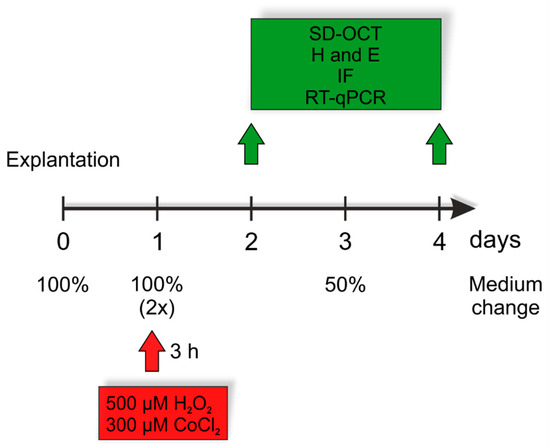
Considering the fact that many retinal diseases are yet to be cured, the pathomechanisms of these multifactorial diseases need to be investigated in more detail. Among others, oxidative stress and hypoxia are pathomechanisms that take place in retinal diseases, such as glaucoma, age-related macular degeneration, or diabetic retinopathy. In consideration of these diseases, it is also evidenced that the immune system, including the complement system and its activation, plays an important role. Suitable models to investigate neuroretinal diseases are organ cultures of porcine retina. Based on an established model, the role of the complement system was studied after the induction of oxidative stress or hypoxia. Both stressors led to a loss of retinal ganglion cells (RGCs) accompanied by apoptosis. Hypoxia activated the complement system as noted by higher C3
+ and MAC
+ cell numbers. In this model, activation of the complement cascade occurred via the classical pathway and the number of C1q
+ microglia was increased. In oxidative stressed retinas, the complement system had no consideration, but strong inflammation took place, with elevated
TNF,
IL6, and
IL8 mRNA expression levels. Together, this study shows that hypoxia and oxidative stress induce different mechanisms in the porcine retina inducing either the immune response or an inflammation. Our findings support the thesis that the immune system is involved in the development of retinal diseases. Furthermore, this study is evidence that both approaches seem suitable models to investigate undergoing pathomechanisms of several neuroretinal diseases.
Full article
►▼
Show Figures
Open AccessArticle
Adaptation to Chronic-Cycling Hypoxia Renders Cancer Cells Resistant to MTH1-Inhibitor Treatment Which Can Be Counteracted by Glutathione Depletion
by
Christine Hansel, Julian Hlouschek, Kexu Xiang, Margarita Melnikova, Juergen Thomale, Thomas Helleday, Verena Jendrossek and Johann Matschke
Cited by 10 | Viewed by 2975
Abstract
Tumor hypoxia and hypoxic adaptation of cancer cells represent major barriers to successful cancer treatment. We revealed that improved antioxidant capacity contributes to increased radioresistance of cancer cells with tolerance to chronic-cycling severe hypoxia/reoxygenation stress. We hypothesized, that the improved tolerance to oxidative
[...] Read more.
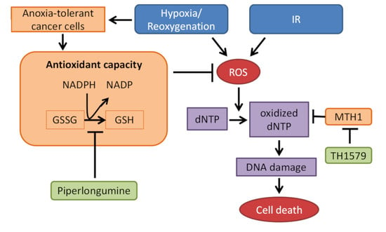
Tumor hypoxia and hypoxic adaptation of cancer cells represent major barriers to successful cancer treatment. We revealed that improved antioxidant capacity contributes to increased radioresistance of cancer cells with tolerance to chronic-cycling severe hypoxia/reoxygenation stress. We hypothesized, that the improved tolerance to oxidative stress will increase the ability of cancer cells to cope with ROS-induced damage to free deoxy-nucleotides (dNTPs) required for DNA replication and may thus contribute to acquired resistance of cancer cells in advanced tumors to antineoplastic agents inhibiting the nucleotide-sanitizing enzyme MutT Homologue-1 (MTH1), ionizing radiation (IR) or both. Therefore, we aimed to explore potential differences in the sensitivity of cancer cells exposed to acute and chronic-cycling hypoxia/reoxygenation stress to the clinically relevant MTH1-inhibitor TH1579 (Karonudib) and to test whether a multi-targeting approach combining the glutathione withdrawer piperlongumine (PLN) and TH1579 may be suited to increase cancer cell sensitivity to TH1579 alone and in combination with IR. Combination of TH1579 treatment with radiotherapy (RT) led to radiosensitization but was not able to counteract increased radioresistance induced by adaptation to chronic-cycling hypoxia/reoxygenation stress. Disruption of redox homeostasis using PLN sensitized anoxia-tolerant cancer cells to MTH1 inhibition by TH1579 under both normoxic and acute hypoxic treatment conditions. Thus, we uncover a glutathione-driven compensatory resistance mechanism towards MTH1-inhibition in form of increased antioxidant capacity as a consequence of microenvironmental or therapeutic stress.
Full article
►▼
Show Figures
Open AccessArticle
Auranofin and Cold Atmospheric Plasma Synergize to Trigger Distinct Cell Death Mechanisms and Immunogenic Responses in Glioblastoma
by
Jinthe Van Loenhout, Laurie Freire Boullosa, Delphine Quatannens, Jorrit De Waele, Céline Merlin, Hilde Lambrechts, Ho Wa Lau, Christophe Hermans, Abraham Lin, Filip Lardon, Marc Peeters, Annemie Bogaerts, Evelien Smits and Christophe Deben
Cited by 42 | Viewed by 4351
Abstract
Targeting the redox balance of malignant cells via the delivery of high oxidative stress unlocks a potential therapeutic strategy against glioblastoma (GBM). We investigated a novel reactive oxygen species (ROS)-inducing combination treatment strategy, by increasing exogenous ROS via cold atmospheric plasma and inhibiting
[...] Read more.
Targeting the redox balance of malignant cells via the delivery of high oxidative stress unlocks a potential therapeutic strategy against glioblastoma (GBM). We investigated a novel reactive oxygen species (ROS)-inducing combination treatment strategy, by increasing exogenous ROS via cold atmospheric plasma and inhibiting the endogenous protective antioxidant system via auranofin (AF), a thioredoxin reductase 1 (TrxR) inhibitor. The sequential combination treatment of AF and cold atmospheric plasma-treated PBS (pPBS), or AF and direct plasma application, resulted in a synergistic response in 2D and 3D GBM cell cultures, respectively. Differences in the baseline protein levels related to the antioxidant systems explained the cell-line-dependent sensitivity towards the combination treatment. The highest decrease of TrxR activity and GSH levels was observed after combination treatment of AF and pPBS when compared to AF and pPBS monotherapies. This combination also led to the highest accumulation of intracellular ROS. We confirmed a ROS-mediated response to the combination of AF and pPBS, which was able to induce distinct cell death mechanisms. On the one hand, an increase in caspase-3/7 activity, with an increase in the proportion of annexin V positive cells, indicates the induction of apoptosis in the GBM cells. On the other hand, lipid peroxidation and inhibition of cell death through an iron chelator suggest the involvement of ferroptosis in the GBM cell lines. Both cell death mechanisms induced by the combination of AF and pPBS resulted in a significant increase in danger signals (ecto-calreticulin, ATP and HMGB1) and dendritic cell maturation, indicating a potential increase in immunogenicity, although the phagocytotic capacity of dendritic cells was inhibited by AF. In vivo, sequential combination treatment of AF and cold atmospheric plasma both reduced tumor growth kinetics and prolonged survival in GBM-bearing mice. Thus, our study provides a novel therapeutic strategy for GBM to enhance the efficacy of oxidative stress-inducing therapy through a combination of AF and cold atmospheric plasma.
Full article
►▼
Show Figures
Open AccessArticle
Upregulation of Chemoresistance by Mg2+ Deficiency through Elevation of ATP Binding Cassette Subfamily B Member 1 Expression in Human Lung Adenocarcinoma A549 Cells
by
Saki Onuma, Aya Manabe, Yuta Yoshino, Toshiyuki Matsunaga, Tomohiro Asai and Akira Ikari
Cited by 3 | Viewed by 2448
Abstract
Several anticancer drugs including cisplatin (CDDP) induce hypomagnesemia. However, it remains fully uncertain whether Mg
2+ deficiency affects chemosensitivity of cancer cells. Here, we investigated the effect of low Mg
2+ concentration (LM) on proliferation and chemosensitivity using human lung adenocarcinoma A549 cells.
[...] Read more.
Several anticancer drugs including cisplatin (CDDP) induce hypomagnesemia. However, it remains fully uncertain whether Mg
2+ deficiency affects chemosensitivity of cancer cells. Here, we investigated the effect of low Mg
2+ concentration (LM) on proliferation and chemosensitivity using human lung adenocarcinoma A549 cells. Cell proliferation was reduced by continuous culture with LM accompanied with the elevation of G1 phase proportion. The amounts of reactive oxygen species (ROS) and stress makers such as phosphorylated-ataxia telangiectasia mutated and phosphorylated-p53 were increased by LM. Cell injury was dose-dependently increased by anticancer drugs such as CDDP and doxorubicin (DXR), which were suppressed by LM. Similar results were obtained by roscovitine, a cell cycle inhibitor. These results suggest that LM induces chemoresistance mediated by ROS production and G1 arrest. The mRNA and protein levels of ATP binding cassette subfamily B member 1 (ABCB1) were increased by LM and roscovitine. The LM-induced elevation of ABCB1 and nuclear p38 expression was suppressed by SB203580, a p38 MAPK inhibitor. PSC833, an ABCB1 inhibitor, and SB203580 rescued the sensitivity to anticancer drugs. In addition, cancer stemness properties were suppressed by SB203580. We suggest that Mg
2+ deficiency reduces the chemotherapy sensitivity of A549 cells, although it suppresses cell proliferation.
Full article
►▼
Show Figures
Planned Papers
The below list represents only planned manuscripts. Some of these
manuscripts have not been received by the Editorial Office yet. Papers
submitted to MDPI journals are subject to peer-review.
Title: Association between Vitamin B12 deficiency, oxidative stress and lipid homeostasis in neuroblastoma cells
Authors: Elena L. Theiss 1; Anna A. Lauer 1; Daniel Janitschke 1; Lea V. Griebsch 1; Sabrina M. Pilz 1, Heike S. Grimm 1; Tobias Hartmann 2; Marcus O. W. Grimm 1,2
Affiliation: 1 Experimental Neurology, Saarland University, Homburg / Saar, Germany
2 Deutsches Institut für DemenzPrävention, Saarland University, Homburg / Saar, Germany
Abstract: Vitamin B12, a water-soluble vitamin, is involved in many metabolic pathways, in particular amino acid metabolism, DNA synthesis and fatty acid metabolism. Due to age-related decline in acidity in stomach, Vitamin B 12 deficiency has a high prevalence in the elderly population besides vegetarian not sufficiently supplementing Vitamin B12. Several lines of evidence indicate that vitamin B12 hypovitaminosis is associated with oxidative stress. However, little is known whether Vitamin B12 deficiency associated oxidative stress results in changes in lipid homeostasis e.g. by increasing lipid-peroxidation. A shotgun lipidomics approach of SH-SY5Y neuroblastoma Vitamin B12 deficient cells revealed specific changes in the lipid-homeostasis, in particular in plasmalogens and phosphatidylcholines. In line with a decline in the oxidation susceptible vinyl-ether especially lipids with polyunsaturated fatty acids were decreased further indicating a potential involvement of reactive oxidative species in Vitamin B12 dependent lipid alterations. Indeed, lipid-peroxidation products were increased in absence of Vitamin B12. Our results show, that Vitamin B12 deficiency not only results in oxidative stress but also in changes in lipid-homeostasis. Importantly the found alteration in lipids is highly associated with many neurological disorders, like Alzheimer’s disease further underlining the importance of a balanced Vitamin B12 level in the elderly population.
Title: Non-enzymatic natural antioxidants with protective role in cardiovascular diseases: comparative and combination effects
Authors: Mahdi Garelnabi et al.
Affiliation: Department of Biomedical and Nutritional Sciences, University of Massachusetts Lowell, MA, USA
Abstract: Growing evidence suggest that non-enzymatic natural antioxidants provides protection against cardiovascular diseases. Oxidative stress is recognized as a principal event in the development of atherosclerosis and other metabolic disorder, which are proven risk factors for cardiovascular diseases. To reduce the harmful effect of oxidative stress, the cardiovascular system has been shown to have naturally-occurring antioxidant substances found in plants, animals, and human beings. Different natural antioxidants appear to act synergistically or additively, suggesting that supplementation with non-enzymatic natural antioxidants might be more effective if combined with other antioxidants or micronutrients. It is also shown that some antioxidants act antagonistically to other antioxidants. The objective of this review is to discuss the current evidence comparing the protective effects of various antioxidants, as well as the synergistic, additive or antagonistic effects produced by a combination of them in cardiovascular diseases. Additionally, the protective role and underlying mechanisms of action of non-enzymatic natural antioxidants in cardiovascular diseases will be summarized.


