Vascular Diseases Diagnostics

A topical collection in Diagnostics (ISSN 2075-4418). This collection belongs to the section "Medical Imaging and Theranostics".

Viewed by 125636

Editor


E-Mail Website
Guest Editor
Full Professor of Vascular Surgery, University of Ferrara, Ferrara, Italy
Interests: chronic venous disorders; venous leg ulcers; neurovascular disease; genetic polymorphism; hemodynamics; cerebral circulation; CCSVI; ultrasound; exercise; PAD
Special Issues, Collections and Topics in MDPI journals

Topical Collection Information

Dear Colleagues,

The arterial, venous and lymphatic components of the human circulation are a fascinating field of research. The physical parameters of blood flow, blood oxygenation, imaging patterns, electric signaling and, of course, circulating and wall molecules are all potential biomarkers to improve our understanding and treatment of vascular disease. The latter is a heterogeneous field of medicine, linked with metabolic and inflammatory disorders. In addition, there is a complex cross-talk between circulating cells, the coagulation cascade, endothelial cells, and inflammatory cytokines that needs to be further elucidated. Finally, vascular and endovascular surgery, pharmacological therapy, cell therapy and rehabilitative treatments urgently need predictive biomarkers to address indications and monitor the outcome of interventions during follow-up.

Prof. Paolo Zamboni
Guest Editor

Prof. Dr. Paolo ZAMBONI
Guest Editor

Manuscript Submission Information

Manuscripts should be submitted online at www.mdpi.com by registering and logging in to this website. Once you are registered, click here to go to the submission form. Manuscripts can be submitted until the deadline. All submissions that pass pre-check are peer-reviewed. Accepted papers will be published continuously in the journal (as soon as accepted) and will be listed together on the collection website. Research articles, review articles as well as short communications are invited. For planned papers, a title and short abstract (about 100 words) can be sent to the Editorial Office for announcement on this website.

Submitted manuscripts should not have been published previously, nor be under consideration for publication elsewhere (except conference proceedings papers). All manuscripts are thoroughly refereed through a single-blind peer-review process. A guide for authors and other relevant information for submission of manuscripts is available on the Instructions for Authors page. Diagnostics is an international peer-reviewed open access semimonthly journal published by MDPI.

Please visit the Instructions for Authors page before submitting a manuscript. The Article Processing Charge (APC) for publication in this open access journal is 2600 CHF (Swiss Francs). Submitted papers should be well formatted and use good English. Authors may use MDPI's English editing service prior to publication or during author revisions.

Published Papers (47 papers)

2023

Jump to: 2022

20 pages, 4419 KiB  
Review
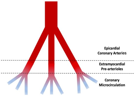
Invasive Assessment of Coronary Microcirculation: A State-of-the-Art Review
by Luca Ciaramella, Luigi Di Serafino, Lucia Mitrano, Maria Luisa De Rosa, Carlo Carbone, Francesco Saverio Rea, Salvatore Monaco, Maria Scalamogna, Plinio Cirillo and Giovanni Esposito
Diagnostics 2024, 14(1), 86; https://doi.org/10.3390/diagnostics14010086 - 30 Dec 2023
Viewed by 1779
Abstract
A significant proportion of patients presenting with signs and symptoms of myocardial ischemia have no “significant” epicardial disease; thereby, the assessment of coronary microcirculation gained an important role in improving diagnosis and guiding therapy. In fact, coronary microvascular dysfunction (CMD) could be found [...] Read more.
A significant proportion of patients presenting with signs and symptoms of myocardial ischemia have no “significant” epicardial disease; thereby, the assessment of coronary microcirculation gained an important role in improving diagnosis and guiding therapy. In fact, coronary microvascular dysfunction (CMD) could be found in a large proportion of these patients, supporting both symptoms and signs of myocardial ischemia. However, CMD represents a diagnostic challenge for two main reasons: (1) the small dimension of the coronary microvasculature prevents direct angiographic visualization, and (2) despite the availability of specific diagnostic tools, they remain invasive and underused in the current clinical practice. For these reasons, CMD remains underdiagnosed, and most of the patients remain with no specific treatment and quality-of-life-limiting symptoms. Of note, recent evidence suggests that a “full physiology” approach for the assessment of the whole coronary vasculature may offer a significant benefit in terms of symptom improvement among patients presenting with ischemia and non-obstructive coronary artery disease. We analyze the pathophysiology of coronary microvascular dysfunction, providing the readers with a guide for the invasive assessment of coronary microcirculation, together with the available evidence supporting its use in clinical practice. Full article
Show Figures

Figure 1

14 pages, 2209 KiB  
Article
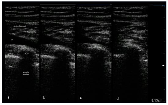
Feasibility of Ultrasound-Guided Axillary Vein Puncture under Valsalva Maneuver for Diagnostic and Cardiovascular Interventional Purposes: Pacemaker and Cardioverter-Defibrillator Implantation
by Biagio Sassone, Enrico Bertagnin, Santo Virzì, Giuseppe Simeti and Paolo Tolomeo
Diagnostics 2023, 13(20), 3274; https://doi.org/10.3390/diagnostics13203274 - 21 Oct 2023
Viewed by 1270
Abstract
Although ultrasound-guided axillary vein access (USGAVA) has proven to be a highly effective and safe method for cardiac electronic implantable device (CIED) lead placement, the collapsibility of the axillary vein (AV) during tidal breathing can lead to narrowing or complete collapse, posing a [...] Read more.
Although ultrasound-guided axillary vein access (USGAVA) has proven to be a highly effective and safe method for cardiac electronic implantable device (CIED) lead placement, the collapsibility of the axillary vein (AV) during tidal breathing can lead to narrowing or complete collapse, posing a challenge for successful vein puncture and cannulation. We investigated the potential of the Valsalva maneuver (Vm) as a facilitating technique for USGAVA in this context. Out of 148 patients undergoing CIED implantation via USGAVA, 41 were asked to perform the Vm, because they were considered unsuitable for venipuncture due to a narrower AV diameter, as assessed by ultrasound (2.7 ± 1.7 mm vs. 9.1 ± 3.3 mm, p < 0.0001). Among them, 37 patients were able to perform the Vm correctly. Overall, the Vm resulted in an average increase in the AV diameter of 4.9 ± 3.4 mm (p < 0.001). USGAVA performed during the Vm was successful in 30 patients (81%), and no Vm-related complications were observed during the 30-day follow-up. In patients with unsuccessful USGAVA, the Vm resulted in a notably smaller increase in AV diameter (0.5 ± 0.3 mm vs. 6.0 ± 2.8 mm, p < 0.0001) compared to patients who achieved successful USGAVA, while performing the Vm. Therefore, the Vm is a feasible maneuver to enhance AV diameter and the success rate of USGAVA in most patients undergoing CIED implantation while maintaining safety. Full article
Show Figures

Figure 1

20 pages, 6850 KiB  
Review
Focus on the Most Common Paucisymptomatic Vasculopathic Population, from Diagnosis to Secondary Prevention of Complications
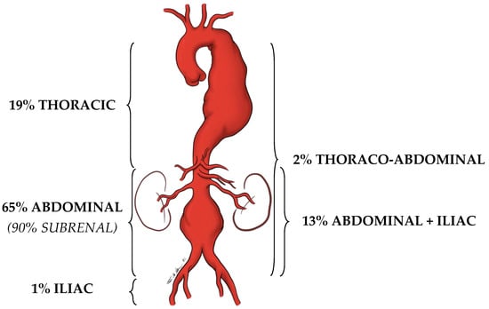
by Eugenio Martelli, Iolanda Enea, Matilde Zamboni, Massimo Federici, Umberto M. Bracale, Giuseppe Sangiorgi, Allegra R. Martelli, Teresa Messina and Alberto M. Settembrini
Diagnostics 2023, 13(14), 2356; https://doi.org/10.3390/diagnostics13142356 - 13 Jul 2023
Cited by 20 | Viewed by 1803
Abstract
Middle-aged adults can start to be affected by some arterial diseases (ADs), such as abdominal aortic or popliteal artery aneurysms, lower extremity arterial disease, internal carotid, or renal artery or subclavian artery stenosis. These vasculopathies are often asymptomatic or paucisymptomatic before manifesting themselves [...] Read more.
Middle-aged adults can start to be affected by some arterial diseases (ADs), such as abdominal aortic or popliteal artery aneurysms, lower extremity arterial disease, internal carotid, or renal artery or subclavian artery stenosis. These vasculopathies are often asymptomatic or paucisymptomatic before manifesting themselves with dramatic complications. Therefore, early detection of ADs is fundamental to reduce the risk of major adverse cardiovascular and limb events. Furthermore, ADs carry a high correlation with silent coronary artery disease (CAD). This study focuses on the most common ADs, in the attempt to summarize some key points which should selectively drive screening. Since the human and economic possibilities to instrumentally screen wide populations is not evident, deep knowledge of semeiotics and careful anamnesis must play a central role in our daily activity as physicians. The presence of some risk factors for atherosclerosis, or an already known history of CAD, can raise the clinical suspicion of ADs after a careful clinical history and a deep physical examination. The clinical suspicion must then be confirmed by a first-level ultrasound investigation and, if so, adequate treatments can be adopted to prevent dreadful complications. Full article
Show Figures

Figure 1

11 pages, 5101 KiB  
Article
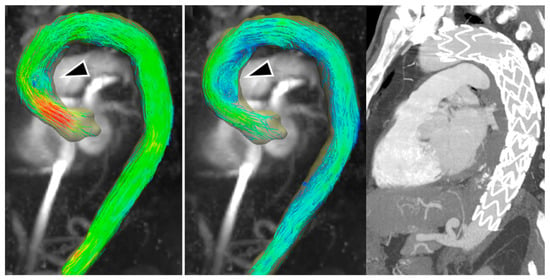
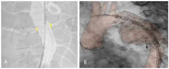
Four-Dimensional Flow MRI for the Evaluation of Aortic Endovascular Graft: A Pilot Study
by Paolo Righini, Francesco Secchi, Daniela Mazzaccaro, Daniel Giese, Marina Galligani, Dor Avishay, Davide Capra, Caterina Beatrice Monti and Giovanni Nano
Diagnostics 2023, 13(12), 2113; https://doi.org/10.3390/diagnostics13122113 - 19 Jun 2023
Viewed by 1959
Abstract
We aimed to explore the feasibility of 4D flow magnetic resonance imaging (MRI) for patients undergoing thoracic aorta endovascular repair (TEVAR). We retrospectively evaluated ten patients (two female), with a mean (±standard deviation) age of 61 ± 20 years, undergoing MRI for a [...] Read more.
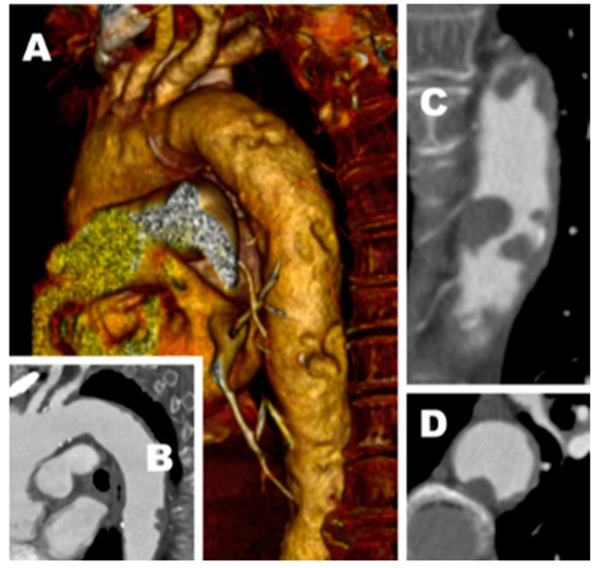
We aimed to explore the feasibility of 4D flow magnetic resonance imaging (MRI) for patients undergoing thoracic aorta endovascular repair (TEVAR). We retrospectively evaluated ten patients (two female), with a mean (±standard deviation) age of 61 ± 20 years, undergoing MRI for a follow-up after TEVAR. All 4D flow examinations were performed using a 1.5-T system (MAGNETOM Aera, Siemens Healthcare, Erlangen, Germany). In addition to the standard examination protocol, a 4D flow-sensitive 3D spatial-encoding, time-resolved, phase-contrast prototype sequence was acquired. Among our cases, flow evaluation was feasible in all patients, although we observed some artifacts in 3 out of 10 patients. Three individuals displayed a reduced signal within the vessel lumen where the endograft was placed, while others presented with turbulent or increased flow. An aortic endograft did not necessarily hinder the visualization of blood flow through 4D flow sequences, although the graft could generate flow artifacts in some cases. A 4D Flow MRI may represent the ideal tool to follow up on both healthy subjects deemed to be at an increased risk based on their anatomical characteristics or patients submitted to TEVAR for whom a surveillance protocol with computed tomography angiography would be cumbersome and unjustified. Full article
Show Figures

Figure 1

10 pages, 877 KiB  
Article
Post-Operative and Mid-Term Renal Function Impairment Following Elective Fenestrated Endovascular Aortic Repair for Complex Aortic Aneurysms: Incidence and Risk Factors Analysis
by Elda Chiara Colacchio, Mariagiovanna Berton, Franco Grego, Michele Piazza, Mirko Menegolo, Francesco Squizzato and Michele Antonello
Diagnostics 2023, 13(11), 1955; https://doi.org/10.3390/diagnostics13111955 - 3 Jun 2023
Viewed by 1306
Abstract
Background. The aim of this study was to assess the incidence of two post-operative acute kidney injury (AKI) stages according to the Risk, Injury, Failure, Loss of function, End-stage (RIFLE) criteria in patients undergoing fenestrated endovascular aortic repair (FEVAR) for complex aortic aneurysms. [...] Read more.
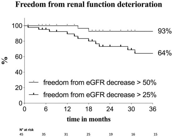
Background. The aim of this study was to assess the incidence of two post-operative acute kidney injury (AKI) stages according to the Risk, Injury, Failure, Loss of function, End-stage (RIFLE) criteria in patients undergoing fenestrated endovascular aortic repair (FEVAR) for complex aortic aneurysms. Furthermore, we analyzed predictors of post-operative AKI and mid-term renal function deterioration and mortality. Methods. We included all patients who underwent elective FEVAR for abdominal and thoracoabdominal aortic aneurysms between January 2014 and September 2021, independently from their preoperative renal function. We registered cases of post-operative acute kidney injury (AKI) both at risk (R-AKI) and injury stage (I-AKI) according to the RIFLE criteria. Estimated glomerular filtration rate (eGFR) was noted preoperatively, at the 48th post-operative hour, at the maximum post-operative peak, at discharge, and then during follow-up approximately every six months. Predictors of AKI were analyzed with univariate and multivariate logistic regression models. Predictors of mid-term chronic kidney disease (CKD) (stage ≥ 3) onset and mortality were analyzed using univariate and multivariate Cox proportional hazard models. Results. Forty-five patients were included in the present study. Mean age was 73.9 ± 6.1 years and 91% of patients were males. Thirteen patients (29%) presented with a preoperative CKD (stage ≥ 3). Post-operative I-AKI was detected in five patients (11.1%). The aneurysm diameter, thoracoabdominal aneurysms and chronic obstructive pulmonary disease were identified as predictors of AKI in univariate analysis (OR 1.05, 95% CI [1.005–1.20], p = 0.030; OR 6.25, 95% CI [1.03–43.97], p = 0.046; OR 7.43, 95% CI [1.20–53.36], p = 0.031; respectively), yet none of these factors were significative on multivariate analysis. Predictors of CKD onset (stage ≥3) during follow-up on multivariate analysis were age (HR 1.16, 95% CI [1.02–1.34], p = 0.023), post-operative I-AKI (HR 26.82, 95% CI [4.18–218.10], p < 0.001) and renal artery occlusion (HR 29.87, 95% CI [2.33–309.05], p = 0.013), while aortic-related reinterventions where not significantly associated with this outcome in univariate analysis (HR 0.66, 95% CI [0.07–2.77], p = 0.615). Mortality was influenced by preoperative CKD (stage ≥3) (HR 5.68, 95% CI [1.63–21.80], p = 0.006) and post-operative AKI (HR 11.60, 95% CI [1.70–97.51], p = 0.012). R-AKI did not represent a risk factor for CKD (stage ≥ 3) onset (HR 1.35, 95% CI [0.45–3.84], p = 0.569) or for mortality (HR 1.60, 95% CI [0.59–4.19], p = 0.339) during follow-up. Conclusions. In-hospital post-operative I-AKI represented the main major adverse event in our cohort, influencing CKD (≥ stage 3) onset and mortality during follow-up, which were not influenced by post-operative R-AKI and aortic-related reinterventions. Full article
Show Figures

Figure 1

11 pages, 4940 KiB  
Article
Cerebral Outflow Discrepancies in Recurrent Benign Paroxysmal Positional Vertigo: Focus on Ultrasonographic Examination
by Andrea Ciorba, Mirko Tessari, Erennio Natale, Fabio Buzzi, Giulia Baldazzi, Alessio Cosacco, Andrea Migliorelli, Virginia Corazzi, Chiara Bianchini, Francesco Stomeo, Stefano Pelucchi and Paolo Zamboni
Diagnostics 2023, 13(11), 1902; https://doi.org/10.3390/diagnostics13111902 - 29 May 2023
Cited by 1 | Viewed by 2109
Abstract
This prospective pilot study aimed to evaluate whether cerebral inflow and outflow abnormalities assessed by ultrasonographic examination could be associated with recurrent benign paroxysmal positional vertigo (BPPV). Twenty-four patients with recurrent BPPV, affected by at least two episodes, and diagnosed according to American [...] Read more.
This prospective pilot study aimed to evaluate whether cerebral inflow and outflow abnormalities assessed by ultrasonographic examination could be associated with recurrent benign paroxysmal positional vertigo (BPPV). Twenty-four patients with recurrent BPPV, affected by at least two episodes, and diagnosed according to American Academy of Otolaryngology–Head and Neck Surgery (AAO–HNS) criteria, evaluated at our University Hospital, between 1 February 2020 and 30 November 2021, have been included. At the ultrasonographic examination, 22 of 24 patients (92%) reported one or more alterations of the extracranial venous circulation, among those considered for the diagnosis of chronic cerebrospinal venous insufficiency (CCSVI), although none of the studied patients were found to have alterations in the arterial circulation. The present study confirms the presence of alterations of the extracranial venous circulation in recurrent BPPV; these anomalies (such as stenosis, blockages or regurgitation of flow, or abnormal valves, as per the CCSVI) could cause a disruption in the venous inner ear drainage, hampering the inner ear microcirculation and then possibly causing recurrent otolith detachment. Full article
Show Figures

Figure 1

10 pages, 1034 KiB  
Article
Single-Center Experience with Simultaneous Mural Aortic Thrombosis and Peripheral Obstructive Disease in Pre-COVID-19 and COVID-19 Era
by Filippo Benedetto, Francesco La Corte, Domenico Spinelli, Gabriele Piffaretti, Santi Trimarchi and Giovanni De Caridi
Diagnostics 2023, 13(6), 1208; https://doi.org/10.3390/diagnostics13061208 - 22 Mar 2023
Viewed by 2092
Abstract
Background: Mural aortic thrombosis associated with chronic peripheral obstruction of the lower limbs is an unusual event. Repeated embolism of instability aortic mural thrombosis caused acute limb ischemia (Rutherford 2 classification) in patients with peripheral arterial disease (PAD). We report a single-center experience [...] Read more.
Background: Mural aortic thrombosis associated with chronic peripheral obstruction of the lower limbs is an unusual event. Repeated embolism of instability aortic mural thrombosis caused acute limb ischemia (Rutherford 2 classification) in patients with peripheral arterial disease (PAD). We report a single-center experience for patients with transmural aortic thrombosis and peripheral artery disease. Methods: We retrospectively analyzed data of 54 patients with aortic mural thrombus disease with PAD presentation, treated at our center between 2013 and 2022. Results: Thirty patients (six with proven SARS-CoV-2 infection) underwent hybrid or staged treatment for an aortic lesion and for lower limb ischemia, by the placement of an endovascular aortic stent graft and a femoro-distal or a popliteal-distal bypass graft. The remaining 24 cases were only subjected to an intravascular treatment of the thoracic or abdominal aorta. Transient renal failure occurred in three patients. No embolic events were detected during the procedures. Aortic-related mortality was reported in just one patient who died from multiple organ failure. There was an embolic stroke in one patient with proven SARS-CoV-2 infection, three major amputations in patients with proven SARS-CoV-2 infection and no aortic-related mortality. Conclusions: Stent coverage of complex aortic lesions, alone or in association with a distal bypass graft, supports this approach in a variety of settings. The COVID-19 pandemic caused an increased mortality and amputation rate. Full article
Show Figures

Figure 1

12 pages, 1810 KiB  
Article
Role of Preoperative Ultrasound Shear-Wave Elastography and Radiofrequency-Based Arterial Wall Tracking in Assessing the Vulnerability of Carotid Plaques: Preliminary Results
by Daniela Mazzaccaro, Matteo Giannetta, Fabiana Fancoli, Giulia Matrone, Nicoletta Curcio, Michele Conti, Paolo Righini and Giovanni Nano
Diagnostics 2023, 13(4), 805; https://doi.org/10.3390/diagnostics13040805 - 20 Feb 2023
Viewed by 2017
Abstract
We aimed at evaluating the ability of point shear-wave elastography (pSWE) and of a radiofrequency (RF) echo-tracking-based method in preoperatively assessing the vulnerability of the carotid plaque in patients undergoing carotid endarterectomy (CEA) for significant asymptomatic stenosis. All patients who underwent CEA from [...] Read more.
We aimed at evaluating the ability of point shear-wave elastography (pSWE) and of a radiofrequency (RF) echo-tracking-based method in preoperatively assessing the vulnerability of the carotid plaque in patients undergoing carotid endarterectomy (CEA) for significant asymptomatic stenosis. All patients who underwent CEA from 03/2021 to 03/2022 performed a preoperative pSWE and an RF echo-based wall evaluation of arterial stiffness using an Esaote MyLab ultrasound system (EsaoteTM, Genova, Italy) with dedicated software. The data derived from these evaluations (Young’s modulus (YM), augmentation index (AIx), pulse-wave velocity (PWV)) were correlated with the outcome of the analysis of the plaque removed during the surgery. Data were analyzed on 63 patients (33 vulnerable and 30 stable plaques). In stable plaques, YM was significantly higher than in vulnerable plaques (49.6 + 8.1 kPa vs. 24.6 + 4.3 kPa, p = 0.009). AIx also tended to be slightly higher in stable plaques, even if it was not statistically significant (10.4 + 0.9% vs. 7.7 + 0.9%, p = 0.16). The PWV was similar (12.2 + 0.9 m/s for stable plaques vs. 10.6 + 0.5 m/s for vulnerable plaques, p = 0.16). For YM, values >34 kPa had a sensitivity of 50% and a specificity of 73.3% in predicting plaque nonvulnerability (area under the curve = 0.66). Preoperative measurement of YM by means of pSWE could be a noninvasive and easily applicable tool for assessing the preoperative risk of plaque vulnerability in asymptomatic patients who are candidates for CEA. Full article
Show Figures

Figure 1

11 pages, 2476 KiB  
Article
Integrated Assessment of Heart, Lung and Lower Extremity Veins Using Hand-Held Ultrasound Device in COVID-19 Patients: Feasibility and Clinical Application
by Dominika Filipiak-Strzecka, Jarosław D. Kasprzak and Piotr Lipiec
Diagnostics 2023, 13(4), 724; https://doi.org/10.3390/diagnostics13040724 - 14 Feb 2023
Cited by 1 | Viewed by 1831
Abstract
The emergence of the COVID-19 pandemic caused a significant shortage of medical personnel and the prioritization of life-saving procedures on internal medicine and cardiology wards. Thus, the cost- and time-effectiveness of each procedure proved vital. Implementing elements of imaging diagnostics into the physical [...] Read more.
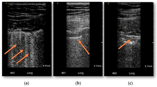
The emergence of the COVID-19 pandemic caused a significant shortage of medical personnel and the prioritization of life-saving procedures on internal medicine and cardiology wards. Thus, the cost- and time-effectiveness of each procedure proved vital. Implementing elements of imaging diagnostics into the physical examination of COVID-19 patients could prove beneficial to the treatment process, providing important clinical data at the moment of admission. Sixty-three patients with positive COVID-19 test results were enrolled into our study and underwent physical examination expanded with a handheld ultrasound device (HUD)—performed bedside assessment included: right ventricle measurement, visual and automated LVEF assessment, four-point compression ultrasound test (CUS) of lower extremities and lung ultrasound. Routine testing consisting of computed-tomography chest scanning, CT—pulmonary angiogram and full echocardiography performed on a high-end stationary device was completed in the following 24 h. Lung abnormalities characteristic for COVID-19 were detected in CT in 53 (84%) patients. The sensitivity and specificity of bedside HUD examination for detecting lung pathologies was 0.92 and 0.90, respectively. Increased number of B-lines had a sensitivity of 0.81, specificity 0.83 for the ground glass symptom in CT examination (AUC 0.82; p < 0.0001); pleural thickening sensitivity 0.95, specificity 0.88 (AUC 0.91, p < 0.0001); lung consolidations sensitivity 0.71, specificity 0.86 (AUC 0.79, p < 0.0001). In 20 patients (32%), pulmonary embolism was confirmed. RV was dilated in HUD examination in 27 patients (43%), CUS was positive in two patients. During HUD examination, software-derived LV function analysis failed to measure LVEF in 29 (46%) cases. HUD proved its potential as the first-line modality for the collection of heart–lung–vein imaging information among patients with severe COVID-19. HUD-derived diagnosis was especially effective for the initial assessment of lung involvement. Expectedly, in this group of patients with high prevalence of severe pneumonia, HUD-diagnosed RV enlargement had moderate predictive value and the option to simultaneously detect lower limb venous thrombosis was clinically attractive. Although most of the LV images were suitable for the visual assessment of LVEF, an AI-enhanced software algorithm failed in almost 50% of the study population. Full article
Show Figures

Figure 1

12 pages, 556 KiB  
Systematic Review
Carotid Plaque Vulnerability Diagnosis by CTA versus MRA: A Systematic Review
by Konstantinos Dakis, Petroula Nana, Chaidoulis Athanasios, Konstantinos Spanos, Batzalexis Konstantinos, Athanasios Giannoukas and George Kouvelos
Diagnostics 2023, 13(4), 646; https://doi.org/10.3390/diagnostics13040646 - 9 Feb 2023
Cited by 5 | Viewed by 2905
Abstract
Stenosis grade of the carotid arteries has been the primary indicator for risk stratification and surgical treatment of carotid artery disease. Certain characteristics of the carotid plaque render it vulnerable and have been associated with increased plaque rupture rates. Computed tomography angiography (CTA) [...] Read more.
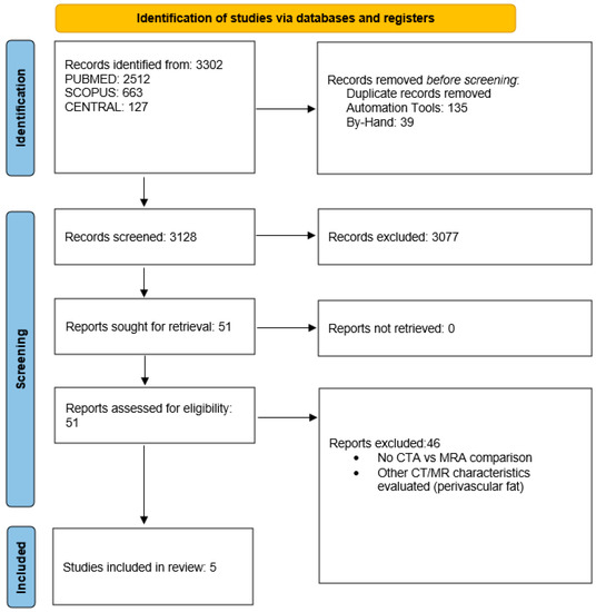
Stenosis grade of the carotid arteries has been the primary indicator for risk stratification and surgical treatment of carotid artery disease. Certain characteristics of the carotid plaque render it vulnerable and have been associated with increased plaque rupture rates. Computed tomography angiography (CTA) and magnetic resonance angiography (MRA) have been shown to detect these characteristics to a different degree. The aim of the current study was to report on the detection of vulnerable carotid plaque characteristics by CTA and MRA and their possible association. A systematic review of the medical literature was executed, utilizing PubMed, SCOPUS and CENTRAL databases, according to the Preferred Reporting Items for Systematic Reviews and Meta-Analysis (PRISMA) 2020 guidelines. The study protocol has been registered to PROSPERO (CRD42022381801). Comparative studies reporting on both CTA and MRA carotid artery studies were included in the analysis. The QUADAS tools were used for risk of bias diagnostic imaging studies. Outcomes included carotid plaque vulnerability characteristics described in CTA and MRA and their association. Five studies, incorporating 377 patients and 695 carotid plaques, were included. Four studies reported on symptomatic status (326 patients, 92.9%). MRA characteristics included intraplaque hemorrhage, plaque ulceration, type VI AHA plaque hallmarks and intra-plaque high-intensity signal. Intraplaque hemorrhage detected in MRA was the most described characteristic and was associated with increased plaque density, increased lumen stenosis, plaque ulceration and increased soft-plaque and hard-plaque thickness. Certain characteristics of vulnerable carotid plaques can be detected in carotid artery CTA imaging studies. Nevertheless, MRA continues to provide more detailed and thorough imaging. Both imaging modalities can be applied for comprehensive carotid artery work-up, each one complementing the other. Full article
Show Figures

Figure 1

21 pages, 5002 KiB  
Review
Photon-Counting Computed Tomography (PCCT): Technical Background and Cardio-Vascular Applications
by Antonella Meloni, Francesca Frijia, Daniele Panetta, Giulia Degiorgi, Carmelo De Gori, Erica Maffei, Alberto Clemente, Vincenzo Positano and Filippo Cademartiri
Diagnostics 2023, 13(4), 645; https://doi.org/10.3390/diagnostics13040645 - 9 Feb 2023
Cited by 29 | Viewed by 6150
Abstract
Photon-counting computed tomography (PCCT) is a new advanced imaging technique that is going to transform the standard clinical use of computed tomography (CT) imaging. Photon-counting detectors resolve the number of photons and the incident X-ray energy spectrum into multiple energy bins. Compared with [...] Read more.
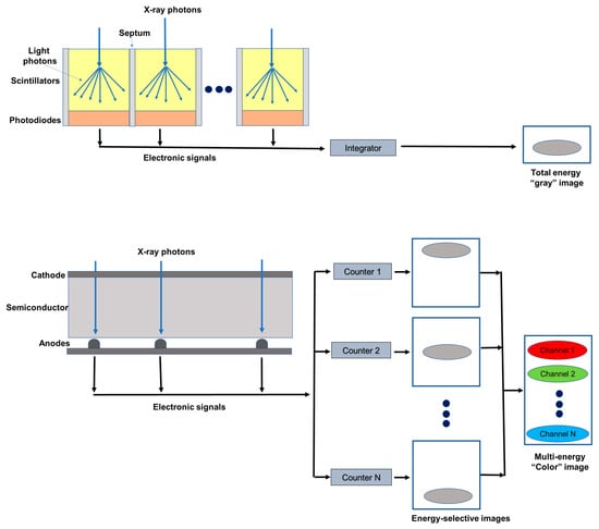
Photon-counting computed tomography (PCCT) is a new advanced imaging technique that is going to transform the standard clinical use of computed tomography (CT) imaging. Photon-counting detectors resolve the number of photons and the incident X-ray energy spectrum into multiple energy bins. Compared with conventional CT technology, PCCT offers the advantages of improved spatial and contrast resolution, reduction of image noise and artifacts, reduced radiation exposure, and multi-energy/multi-parametric imaging based on the atomic properties of tissues, with the consequent possibility to use different contrast agents and improve quantitative imaging. This narrative review first briefly describes the technical principles and the benefits of photon-counting CT and then provides a synthetic outline of the current literature on its use for vascular imaging. Full article
Show Figures

Figure 1

13 pages, 6614 KiB  
Article
Carotid Artery Disease in the Era of Biomarkers: A Pilot Study
by Ettore Dinoto, Domenico Mirabella, Francesca Ferlito, Graziella Tortomasi, Davide Turchino, Salvatore Evola, Massimiliano Zingales, Emanuela Bologna and Felice Pecoraro
Diagnostics 2023, 13(4), 644; https://doi.org/10.3390/diagnostics13040644 - 9 Feb 2023
Cited by 4 | Viewed by 2318
Abstract
The intima-media thickness (IMT) and its irregularities or ulcerations in the common carotid artery (CCA) are useful tools as sentinel biomarkers for the integrity of the cardiovascular system. Total homocysteine and lipoprotein levels are the most commonly used elements in cardiovascular risk stratification. [...] Read more.
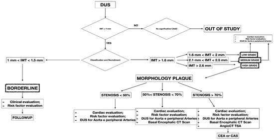
The intima-media thickness (IMT) and its irregularities or ulcerations in the common carotid artery (CCA) are useful tools as sentinel biomarkers for the integrity of the cardiovascular system. Total homocysteine and lipoprotein levels are the most commonly used elements in cardiovascular risk stratification. Duplex ultrasound (DUS), associated with serum biomarkers, can be used simply to assess the degree of atherosclerotic disease and cardiovascular risk. This study highlights the role of different kinds of biomarkers, showing their usefulness and potentiality in multi-district atherosclerotic patients, especially for early diagnosis and therapy effectiveness monitoring. A retrospective analysis performed from September 2021 to August 2022, of patients with carotid artery disease, was performed. A total of 341 patients with a mean age of 53.8 years were included in the study. The outcomes showed an increased risk of stroke in patients with significative carotid artery disease, nonresponsive to therapy, monitored through a series of serum biomarkers (homocysteine, C-reactive protein, and oxidized LDL). In this reported experience, the systematic use of DUS in association with the multiple biomarkers approach was effective for the early identification of patients at higher risk of disease progression or inefficient therapy. Full article
Show Figures

Figure 1

10 pages, 1153 KiB  
Article
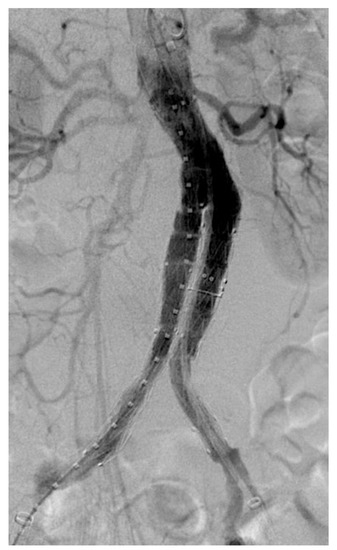
“Redo” 2D–3D Fusion Technique during Endovascular Redo Aortic Repair
by Fabrizio Minelli, Simona Sica, Fadia Salman, Federica Donato, May Dvir, Yamume Tshomba and Giovanni Tinelli
Diagnostics 2023, 13(4), 635; https://doi.org/10.3390/diagnostics13040635 - 8 Feb 2023
Cited by 1 | Viewed by 1516
Abstract
Purpose: The present study aims to describe a new 2D–3D fusion registration method in the case of endovascular redo aortic repair and compare the accuracy of the registration using the previously implanted devices vs. bones as landmarks. Materials and Methods: This single-center study [...] Read more.
Purpose: The present study aims to describe a new 2D–3D fusion registration method in the case of endovascular redo aortic repair and compare the accuracy of the registration using the previously implanted devices vs. bones as landmarks. Materials and Methods: This single-center study prospectively analyzed all the patients that underwent elective endovascular re-interventions using the Redo Fusion technique between January 2016 and December 2021 at the Vascular Surgery Unit of the Fondazione Policlinico Universitario A. Gemelli (FPUG)—IRCCS in Rome, Italy. The fusion overlay was performed twice, first using bone landmarks (bone fusion) and the second using radiopaque markers of a previous endovascular device (redo fusion). The pre-operative 3D model was fused with live fluoroscopy to create a roadmap. Longitudinal distances between the inferior margin of the target vessel in live fluoroscopy and the inferior margin of the target vessel in bone fusion and redo fusion were measured. Results: This single-center study prospectively analyzed 20 patients. There were 15 men and five women, with a median age of 69.7 (IQR 42) years. The median distance between the inferior margin of the target vessel ostium in digital subtraction angiography and the inferior margin of the target vessel ostium in bone fusion and redo fusion was 5.35 mm and 1.35 mm, respectively (p ≤ 0.0001). Conclusions: The redo fusion technique is accurate and allows the optimization of X-ray working views, supporting the endovascular navigation and vessel catheterization in case of endovascular redo aortic repair. Full article
Show Figures

Figure 1

14 pages, 1491 KiB  
Article
Retinal Blood Vessel Analysis Using Optical Coherence Tomography (OCT) in Multiple Sclerosis
by Nicholas Young, Robert Zivadinov, Michael G. Dwyer, Niels Bergsland, Bianca Weinstock-Guttman and Dejan Jakimovski
Diagnostics 2023, 13(4), 596; https://doi.org/10.3390/diagnostics13040596 - 6 Feb 2023
Cited by 2 | Viewed by 1991
Abstract
Background: Both greater retinal neurodegenerative pathology and greater cardiovascular burden are seen in people with multiple sclerosis (pwMS). Studies also describe multiple extracranial and intracranial vascular changes in pwMS. However, there have been few studies examining the neuroretinal vasculature in MS. Our aim [...] Read more.
Background: Both greater retinal neurodegenerative pathology and greater cardiovascular burden are seen in people with multiple sclerosis (pwMS). Studies also describe multiple extracranial and intracranial vascular changes in pwMS. However, there have been few studies examining the neuroretinal vasculature in MS. Our aim is to determine differences in retinal vasculature between pwMS and healthy controls (HCs) and to determine the relationship between retinal nerve fiber layer (RNFL) thickness and retinal vasculature characteristics. Methods: A total of 167 pwMS and 48 HCs were scanned using optical coherence tomography (OCT). Earlier OCT scans were available for 101 pwMS and 35 HCs for an additional longitudinal analysis. Segmentation of retinal vasculature was performed in a blinded manner in MATLAB’s optical coherence tomography segmentation and evaluation GUI (OCTSEG) software. Results: PwMS has fewer retinal blood vessels when compared to HCs (35.1 vs. 36.8, p = 0.017). Over the 5.4 year follow up, and when compared to HCs, pwMS has a significant decrease in number of retinal vessels (average loss of −3.7 p = 0.007). Moreover, the total vessel diameter in pwMS does not change when compared to the increase in vessel diameter in the HCs (0.06 vs. 0.3, p = 0.017). Only in pwMS is there an association between lower RNFL thickness and fewer retinal vessel number and smaller diameter (r = 0.191, p = 0.018 and r = 0.216, p = 0.007). Conclusions: Over 5 years, pwMS exhibit significant retinal vascular changes that are related to greater atrophy of the retinal layers. Full article
Show Figures

Figure 1

8 pages, 1073 KiB  
Article
Comparison of Doppler Ultrasound and Computerized Tomographic Angiography in Evaluation of Cervical Arteries Stenosis in Stroke Patients, a Retrospective Single-Center Study
by Naaem Simaan, Tamer Jubeh, Karine Beiruti Wiegler, Adi Sharabi-Nov, Asaf Honig and Radi Shahien
Diagnostics 2023, 13(3), 459; https://doi.org/10.3390/diagnostics13030459 - 26 Jan 2023
Cited by 3 | Viewed by 3225
Abstract
There are different diagnostic modalities to investigate atherosclerosis cervical artery disease in suspected stroke patients. We aimed to test the concordance of findings of the two most widely used diagnostic modalities in stroke patients: duplex ultrasound (DUS) and computerized tomographic angiography (CTA). A [...] Read more.
There are different diagnostic modalities to investigate atherosclerosis cervical artery disease in suspected stroke patients. We aimed to test the concordance of findings of the two most widely used diagnostic modalities in stroke patients: duplex ultrasound (DUS) and computerized tomographic angiography (CTA). A total of 100 stroke patients were retrospectively included in the study, all of them had DUS followed by CTA. Discrepancies of DUS compared to the CTA results in both the internal carotid and vertebral arteries were found in 44% of the patients. The patients with significant differences in diagnostic results were characterized by older age. Evaluation of the degree of carotid artery stenosis revealed vast differences in patients with 50–69% stenosis found by DUS, in which 45.5% of them had a different percentage of stenosis found by CTA. In studying the degree of stenosis of the vertebral artery, only 47.1% of the patients with more than 50% stenosis found by DUS had the same results with CTA, while the remaining revealed normal or less than 50% stenosis by CTA. The current study emphasizes that CTA is more accurate than DUS in the evaluation of stenosis of the cervical arteries including both the internal carotid and vertebral arteries. Full article
Show Figures

Figure 1

9 pages, 582 KiB  
Article
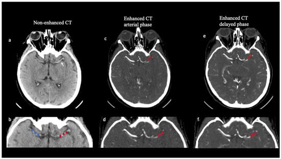
Correlation between Thrombus Perviousness and Distal Embolization during Mechanical Thrombectomy in Acute Stroke
by Fabio Pilato, Iacopo Valente, Andrea M. Alexandre, Rosalinda Calandrelli, Luca Scarcia, Francesco D’Argento, Emilio Lozupone, Vincenzo Arena and Alessandro Pedicelli
Diagnostics 2023, 13(3), 431; https://doi.org/10.3390/diagnostics13030431 - 25 Jan 2023
Cited by 4 | Viewed by 1994
Abstract
Purpose: Thrombus permeability has been related to clot composition and treatment outcomes in stroke patients undergoing reperfusion therapies. The aim of this study was to evaluate whether thrombus perviousness, evaluated by multiphase computed tomography angiography (mCTA), is associated with distal embolization risk. Methods: [...] Read more.
Purpose: Thrombus permeability has been related to clot composition and treatment outcomes in stroke patients undergoing reperfusion therapies. The aim of this study was to evaluate whether thrombus perviousness, evaluated by multiphase computed tomography angiography (mCTA), is associated with distal embolization risk. Methods: We interrogated our dataset of acute ischemic stroke (AIS) patients involving the M1 segment of the middle cerebral artery (MCA) who had undergone mechanical thrombectomy, and we calculated thrombus average attenuation measurement (dHU) on non-contrast CT (NCCT) and clot perviousness on mCTA. dHU was calculated as the difference between the thrombus HU average value (tHU) and the HU average value on the contralateral side (cHU), while perviousness was calculated as the difference in mean clot density on mCTA and NCCT both in arterial (Perviousness pre-post-1) and delayed (Perviousness pre-post 2) phases. Results: A total of 100 patients (53 females (53%), mean age 72.74 [± 2.31]) with M1 occlusion were available for analysis. Perviousness, calculated between baseline and arterial phase of mCTA (Perviousness pre-post1), was lower in patients with distal embolization (p = 0.05), revealing an association between reduced perviousness and distal embolization risk. Logistic regression showed that thrombus perviousness calculated on the arterial phase of mCTA (OR, 0.66; 95% CI, 0.44–0.99] (p = 0.04)) and the contact aspiration technique (OR, 0.39; 95% CI, 0.15–1.02] (p = 0.05)) were protecting factors against distal embolization. Conclusion: Our study showed an association between reduced perviousness and distal embolization, suggesting that perviousness evaluation may be a useful neuroimaging biomarker in predicting distal embolization risk during mechanical thrombectomy. Full article
Show Figures

Figure 1

18 pages, 2598 KiB  
Systematic Review
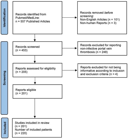
Pylephlebitis: A Systematic Review on Etiology, Diagnosis, and Treatment of Infective Portal Vein Thrombosis
by Lisa Fusaro, Stefano Di Bella, Paola Martingano, Lory Saveria Crocè and Mauro Giuffrè
Diagnostics 2023, 13(3), 429; https://doi.org/10.3390/diagnostics13030429 - 25 Jan 2023
Cited by 21 | Viewed by 7351
Abstract
Pylephlebitis, defined as infective thrombophlebitis of the portal vein, is a rare condition with an incidence of 0.37–2.7 cases per 100,000 person-years, which can virtually complicate any intra-abdominal or pelvic infections that develop within areas drained by the portal venous circulation. The current [...] Read more.
Pylephlebitis, defined as infective thrombophlebitis of the portal vein, is a rare condition with an incidence of 0.37–2.7 cases per 100,000 person-years, which can virtually complicate any intra-abdominal or pelvic infections that develop within areas drained by the portal venous circulation. The current systematic review aimed to investigate the etiology behind pylephlebitis in terms of pathogens involved and causative infective processes, and to report the most common symptoms at clinical presentation. We included 220 individuals derived from published cases between 1971 and 2022. Of these, 155 (70.5%) were male with a median age of 50 years. There were 27 (12.3%) patients under 18 years of age, 6 (2.7%) individuals younger than one year, and the youngest reported case was only 20 days old. The most frequently reported symptoms on admission were fever (75.5%) and abdominal pain (66.4%), with diverticulitis (26.5%) and acute appendicitis (22%) being the two most common causes. Pylephlebitis was caused by a single pathogen in 94 (42.8%) cases and polymicrobial in 60 (27.2%) cases. However, the responsible pathogen was not identified or not reported in 30% of the included patients. The most frequently isolated bacteria were Escherichia coli (25%), Bacteroides spp. (17%), and Streptococcus spp. (15%). The treatment of pylephlebitis consists initially of broad-spectrum antibiotics that should be tailored upon bacterial identification and continued for at least four to six weeks after symptom presentation. There is no recommendation for prescribing anticoagulants to all patients with pylephlebitis. However, they should be administered in patients with thrombosis progression on repeat imaging or persistent fever despite proper antibiotic therapy to increase the rates of thrombus resolution or decrease the overall mortality, which is approximately 14%. Full article
Show Figures

Figure 1

11 pages, 3061 KiB  
Article
[18F]FDG Uptake in Non-Infected Endovascular Grafts: A Retrospective Study
by Chiara Lauri, Alberto Signore, Giuseppe Campagna, Francesco Aloisi, Maurizio Taurino and Pasqualino Sirignano
Diagnostics 2023, 13(3), 409; https://doi.org/10.3390/diagnostics13030409 - 23 Jan 2023
Cited by 6 | Viewed by 1999
Abstract
Purpose: After endovascular aneurysm repair (EVAR), an increased [18F]FDG uptake may be observed at PET/CT, being common to both vascular graft/endograft infection (VGEI) and sterile post-surgical inflammation. Increased non-specific metabolic activity, due to foreign body reaction, can persist for several years [...] Read more.
Purpose: After endovascular aneurysm repair (EVAR), an increased [18F]FDG uptake may be observed at PET/CT, being common to both vascular graft/endograft infection (VGEI) and sterile post-surgical inflammation. Increased non-specific metabolic activity, due to foreign body reaction, can persist for several years after surgery, thus complicating the interpretation of PET/CT studies. In this paper, we aimed to assess [18F]FDG distribution at different time-points after the implant of abdominal Endurant® endografts in patients without suspicion of infection. Methods: We retrospectively evaluated [18F]FDG/CT in 16 oncological patients who underwent abdominal aortic aneurysm exclusion with Endurant® grafts. Patients had no clinical suspicion of infection and were followed up for at least 24 months after scan. [18F]FDG PET/CT scans were interpreted using both visual and semi-quantitative analyses. Results: The time between the EVAR procedure and [18F]FDG PET/CT ranged between 1 and 36 months. All grafts showed mild and diffuse [18F]FDG uptake without a focal pattern. Mean values of SUVmax were 2.63 ± 0.48 (95% CI 2.38–2.88); for SUVmean 1.90 ± 0.33 (95% CI 1.72–2.08); for T/B ratios 1.43 ± 0.41 (95% CI 1.21–1.65). SUVmax and SUVmean were not correlated to the time elapsed from the procedure, but we observed a declining trend in T/B ratio over time. Conclusions: Endovascular implant of Endurant® grafts does not cause a significant inflammatory reaction. The evidence of faint and diffuse [18F]FDG uptake along the graft can reliably exclude an infection, even in early post-procedural phases. Therefore, in patients with a low probability of VGEI, [18F]FDG PET/CT can also be performed immediately after EVAR. Full article
Show Figures

Figure 1

13 pages, 1684 KiB  
Article
The Presence of Ascending Aortic Dilatation in Patients Undergoing Transcatheter Aortic Valve Replacement Is Negatively Correlated with the Presence of Diabetes Mellitus and Does Not Impair Post-Procedural Outcomes
by Elke Boxhammer, Stefan Hecht, Reinhard Kaufmann, Jürgen Kammler, Jörg Kellermair, Christian Reiter, Kaveh Akbari, Hermann Blessberger, Clemens Steinwender, Michael Lichtenauer, Uta C. Hoppe, Klaus Hergan and Bernhard Scharinger
Diagnostics 2023, 13(3), 358; https://doi.org/10.3390/diagnostics13030358 - 18 Jan 2023
Viewed by 2423
Abstract
Both relevant aortic valve stenosis (AS) and aortic valve insufficiency significantly contribute to structural changes in the ascending aorta (AA) and thus to its dilatation. In patients with severe AS undergoing transcatheter aortic valve replacement (TAVR), survival data regarding aortic changes and laboratory [...] Read more.
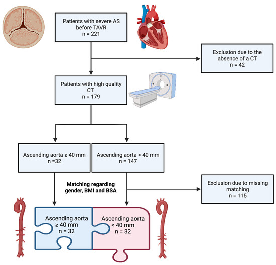
Both relevant aortic valve stenosis (AS) and aortic valve insufficiency significantly contribute to structural changes in the ascending aorta (AA) and thus to its dilatation. In patients with severe AS undergoing transcatheter aortic valve replacement (TAVR), survival data regarding aortic changes and laboratory biomarker analyses are scarce. Methods: A total of 179 patients with severe AS and an available computed tomography were included in this retrospective study. AA was measured, and dilatation was defined as a diameter ≥ 40 mm. Thirty-two patients had dilatation of the AA. A further 32 patients from the present population with a normal AA were matched to the aortic dilatation group with respect to gender, age, body mass index and body surface area, and the resulting study groups were compared with each other. In addition to echocardiographic and clinical characteristics, the expression of cardiovascular biomarkers such as brain natriuretic peptide (BNP), soluble suppression of tumorigenicity-2 (sST2), growth/differentiation of factor-15 (GDF-15), heart-type fatty-acid binding protein (H-FABP), insulin-like growth factor binding protein 2 (IGF-BP2) and soluble urokinase-type plasminogen activator receptor (suPAR) was analyzed. Kaplan–Meier curves for short- and long-term survival were obtained, and Pearson’s and Spearman’s correlations were calculated to identify the predictors between the diameter of the AA and clinical parameters. Results: A total of 19% of the total cohort had dilatation of the AA. The study group with an AA diameter ≥ 40 mm showed a significantly low comorbidity with respect to diabetes mellitus in contrast to the comparison cohort with an AA diameter < 40 mm (p = 0.010). This result continued in the correlation analyses performed, as the presence of diabetes mellitus correlated negatively not only with the diameter of the AA (r = −0.404; p = 0.001) but also with the presence of aortic dilatation (r = −0.320; p = 0.010). In addition, the presence of AA dilatation after TAVR was shown to have no differences in terms of patient survival at 1, 3 and 5 years. There were no relevant differences in the cardiovascular biomarkers studied between the patients with dilated and normal AAs. Conclusion: The presence of AA dilatation before successful TAVR was not associated with a survival disadvantage at the respective follow-up intervals of 1, 3 and 5 years. Diabetes mellitus in general seemed to have a protective effect against the development of AA dilatation or aneurysm in patients with severe AS. Full article
Show Figures

Figure 1

12 pages, 1739 KiB  
Article
Outcome Measures of In-Office Endovenous Radiofrequency Treatment of Varicose Vein Feasibility
by Alfonso Pannone, Alessia Di Girolamo, Matteo Orrico and Nicola Mangialardi
Diagnostics 2023, 13(2), 327; https://doi.org/10.3390/diagnostics13020327 - 16 Jan 2023
Cited by 1 | Viewed by 4383
Abstract
RFA is a relatively new treatment, approved by the FDA in 1999 and is a minimally invasive intervention that has become one of the most common alternatives due to its many advantages, including decreased pain, less morbidity, shorter hospital stay and faster return [...] Read more.
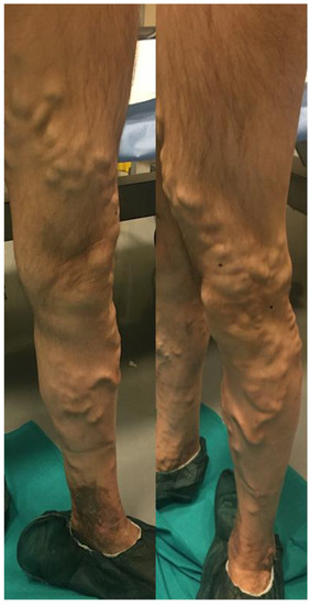
RFA is a relatively new treatment, approved by the FDA in 1999 and is a minimally invasive intervention that has become one of the most common alternatives due to its many advantages, including decreased pain, less morbidity, shorter hospital stay and faster return to work. We retrospectively analyzed a total of 503 limbs submitted for surgical interventions for VVs using the RFA, combined or not with surgical phlebectomies and sclerotherapy, in our institution between April 2012 and December 2020. The technical success was 99.8%, as in one case the RFA catheter arrested at the first third of the thigh due to the tortuosity of the vessel. On the first post-operative day, the mean VAS scale was 0.32 ± 0.56 (0–2). Perioperative complications occurred in 77 cases (15.3%): intraoperative pain in 24 cases, in nine cases associated with vagal syndrome, superficial hematoma in 30 cases, EHIT in seven cases, phlebitis in 14 cases and paresthesia in two cases. RFA procedures had been shown to be cost-effective therapeutic options in adult patients requiring treatment due to the incompetence of the GSV. In our study, we highlighted that this technique is feasible as an ambulatory procedure; it improves quality of life and symptoms in the majority of patients with varicose veins, with a rapid return to normal life and work activities. Full article
Show Figures

Figure 1

10 pages, 1244 KiB  
Article
Styloid Jugular Nutcracker: The Possible Role of the Styloid Process Spatial Orientation—A Preliminary Morphometric Computed Study
by Giorgio Mantovani, Pietro Zangrossi, Maria Elena Flacco, Giovanni Di Domenico, Enrico Nastro Siniscalchi, Francesco Saverio De Ponte, Rosario Maugeri, Pasquale De Bonis, Michele Alessandro Cavallo, Paolo Zamboni and Alba Scerrati
Diagnostics 2023, 13(2), 298; https://doi.org/10.3390/diagnostics13020298 - 13 Jan 2023
Cited by 2 | Viewed by 3701
Abstract
Styloid Jugular Nutcracker (SJN, also known as Eagle Jugular Syndrome EJS) derives from a jugular stenosis caused by an abnormal styloid process, compressing the vessel in its superior portion (J3) against the C1 anterior arch. It could be considered a venous vascular variant [...] Read more.
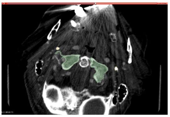
Styloid Jugular Nutcracker (SJN, also known as Eagle Jugular Syndrome EJS) derives from a jugular stenosis caused by an abnormal styloid process, compressing the vessel in its superior portion (J3) against the C1 anterior arch. It could be considered a venous vascular variant of Eagle Syndrome (ES). Main clinical features of this ES variant are headache, pulsatile tinnitus and dizziness, possibly related to venous hypertension and impaired cerebral parenchyma drainage. In our opinion, conceptually, it is not the absolute length of the styloid bone that defines its abnormality, but its spatial direction. An elongated bone pointing outward far away from the midline could not compress the vein; vice versa, a short styloid process tightly adherent to the cervical spine could be pathological. To prove this hypothesis, we developed a semi-automatic software that processes CT-Angio images, giving quantitative information about distance and direction of the styloid process in three-dimensional space. We compared eight patients with SJN to a sample of healthy subjects homogeneous for sex and age. Our results suggest that SJN patients have a more vertically directed styloid, and this feature is more important than the absolute distance between the two bony structures. More studies are needed to expand our sample, including patients with the classic and carotid variants of Eagle Syndrome. Full article
Show Figures

Figure 1

13 pages, 1564 KiB  
Article
Predictive Value of Echocardiographic Pulmonary to Left Atrial Ratio for In-Hospital Death in Patients with COVID-19
by Giulia Renda, Marco G. Mennuni, Giovanni Pizzoferrato, Daniele Esposto, Angela Alberani, Simona De Vecchi, Anna Degiovanni, Ailia Giubertoni, Enrico Guido Spinoni, Leonardo Grisafi, Emanuele Sagazio, Claudio Ucciferri, Katia Falasca, Jacopo Vecchiet, Sabina Gallina and Giuseppe Patti
Diagnostics 2023, 13(2), 224; https://doi.org/10.3390/diagnostics13020224 - 7 Jan 2023
Viewed by 1699
Abstract
Background: Echocardiographic Pulmonary to Left Atrial Ratio (ePLAR) represents an accurate and sensitive non-invasive tool to estimate the trans-pulmonary gradient. The prognostic value of ePLAR in hospitalized patients with COVID-19 remains unknown. We aimed to investigate the predictive value of ePLAR on in-hospital [...] Read more.
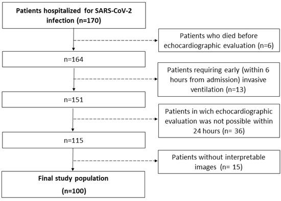
Background: Echocardiographic Pulmonary to Left Atrial Ratio (ePLAR) represents an accurate and sensitive non-invasive tool to estimate the trans-pulmonary gradient. The prognostic value of ePLAR in hospitalized patients with COVID-19 remains unknown. We aimed to investigate the predictive value of ePLAR on in-hospital mortality in patients with COVID-19. Methods: One hundred consecutive patients admitted to two Italian institutions for COVID-19 undergoing early (<24 h) echocardiographic examination were included; ePLAR was determined from the maximum tricuspid regurgitation continuous wave Doppler velocity (m/s) divided by the transmitral E-wave: septal mitral annular Doppler Tissue Imaging e′-wave ratio (TRVmax/E:e′). The primary outcome measure was in-hospital death. Results: patients who died during hospitalization had at baseline a higher prevalence of tricuspid regurgitation, higher ePLAR, right-side pressures, lower Tricuspid Annular Plane Systolic Excursion (TAPSE)/ systolic Pulmonary Artery Pressure (sPAP) ratio and reduced inferior vena cava collapse than survivors. Patients with ePLAR > 0.28 m/s at baseline showed non-significant but markedly increased in-hospital mortality compared to those having ePLAR ≤ 0.28 m/s (27% vs. 10.8%, p = 0.055). Multivariate Cox regression showed that an ePLAR > 0.28 m/s was independently associated with an increased risk of death (HR 5.07, 95% CI 1.04–24.50, p = 0.043), particularly when associated with increased sPAP (p for interaction = 0.043). Conclusions: A high ePLAR value at baseline predicts in-hospital death in patients with COVID-19, especially in those with elevated pulmonary arterial pressure. These results support an early ePLAR assessment in patients admitted for COVID-19 to identify those at higher risk and potentially guide strategies of diagnosis and care. Full article
Show Figures

Figure 1

10 pages, 1254 KiB  
Article
Subtypes of the Completely Reversed Flow Waveform in Vertebral Artery Can Help to Differentiate Subclavian Artery Occlusion from Severe Stenosis
by Shun-Ping Chen, Zhen Zhong and Dong-Pei Tu
Diagnostics 2023, 13(1), 146; https://doi.org/10.3390/diagnostics13010146 - 1 Jan 2023
Cited by 1 | Viewed by 2350
Abstract
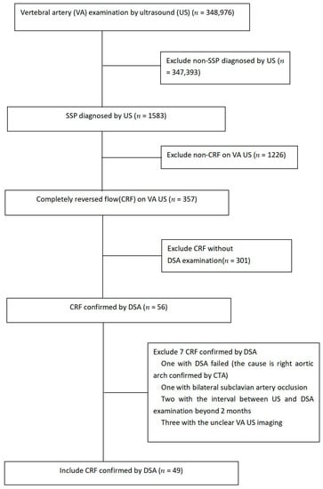
Objectives: To investigate the value of subtypes of completely reversed flow (CRF) waveform in vertebral artery (VA) on Doppler ultrasound in differentiation occlusion from severe stenosis of the ipsilateral proximal subclavian artery (SA). Methods: A total of 357 patients with CRF in the [...] Read more.
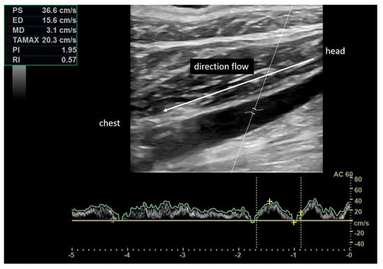
Objectives: To investigate the value of subtypes of completely reversed flow (CRF) waveform in vertebral artery (VA) on Doppler ultrasound in differentiation occlusion from severe stenosis of the ipsilateral proximal subclavian artery (SA). Methods: A total of 357 patients with CRF in the VA on Doppler US were reviewed retrospectively. Among them, 49 patients (mean age, 68.2 ± 7.8 years) confirmed by digital subtraction angiography (DSA) were included. According to the status of diastolic flow, the CRF was divided into continuous CRF (CCRF, n = 27) and intermittent CRF (ICRF, n = 22). The correlation of subtypes of CRF waveform and VA parameters with the severity of SA stenosis was evaluated. The severity of SA stenosis was determined by DSA. Results: Of those 49 patients, SA occlusion was observed in 33 patients (67%, occlusion group) and severe stenosis in 16 patients (33%, stenosis group). The subtypes of CRF waveforms showed a significant between-group difference (p = 0.005). CCRF exhibited an accuracy of 85.2% (23/27) in diagnosing SA occlusion. The diameter of the target VA with ICRF showed a significant between-group difference (p = 0.041). The target VA diameter ≥ 3.8 mm in ICRF achieved an accuracy of 81.8% (18/22), and its combination with CCRF achieved an accuracy of 83.7% (41/49) in the differentiation of SA occlusion from severe stenosis. Conclusions: Subtypes of CRF in VA can help to differentiate SA occlusion from severe stenosis. CCRF has higher accuracy in diagnosing SA occlusion. The CCRF waveform plus VA diameter in ICRF is more accurate for differentiating SA occlusion from severe stenosis. Full article
Show Figures

Figure 1

2022

Jump to: 2023

14 pages, 1017 KiB  
Systematic Review
Clinical Applications of Quantitative Perfusion Imaging with a C-Arm Flat-Panel Detector—A Systematic Review
by Abdallah H. A. Zaid Al-Kaylani, Richte C. L. Schuurmann, Wouter D. Maathuis, Riemer H. J. A. Slart, Jean-Paul P. M. De Vries and Reinoud P. H. Bokkers
Diagnostics 2023, 13(1), 128; https://doi.org/10.3390/diagnostics13010128 - 30 Dec 2022
Cited by 1 | Viewed by 2391
Abstract
C-arm systems with digital flat-panel detectors are used in interventional radiology and hybrid operating rooms for visualizing and performing interventions on three-dimensional structures. Advances in C-arm technology have enabled intraoperative quantitative perfusion imaging with these scanners. This systematic review provides an overview of [...] Read more.
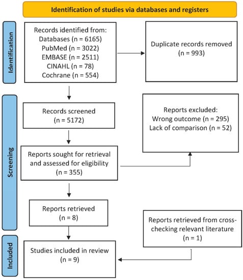
C-arm systems with digital flat-panel detectors are used in interventional radiology and hybrid operating rooms for visualizing and performing interventions on three-dimensional structures. Advances in C-arm technology have enabled intraoperative quantitative perfusion imaging with these scanners. This systematic review provides an overview of flat-panel detector C-arm techniques for quantifying perfusion, their clinical applications, and their validation. A systematic search was performed for articles published between January 2000 and October 2022 in which a flat-panel detector C-arm technique for quantifying perfusion was compared with a reference technique. Nine articles were retrieved describing two techniques: two-dimensional perfusion angiography (n = 5) and dual-phase cone beam computed tomography perfusion (n = 4). A quality assessment revealed no concerns about the applicability of the studies. The risk of bias was relatively high for the index and reference tests. Both techniques demonstrated potential for clinical application; however, weak-to-moderate correlations were reported between them and the reference techniques. In conclusion, both techniques could add new possibilities to treatment planning and follow-up; however, the available literature is relatively scarce and heterogeneous. Larger-scale randomized prospective studies focusing on clinical outcomes and standardization are required for the full understanding and clinical implementation of these techniques. Full article
Show Figures

Figure 1

7 pages, 2238 KiB  
Case Report
Differential Diagnosis of Thoracoacromial Artery Pseudoaneurysm from Shoulder Inflammatory Pseudotumor: A Case Report
by Tzu-Yen Huang, Pin-Chao Feng, Yao-Chang Wang and Chun-Yi Su
Diagnostics 2023, 13(1), 82; https://doi.org/10.3390/diagnostics13010082 - 28 Dec 2022
Cited by 1 | Viewed by 2455
Abstract
Diagnosing shoulder tumors is a challenge because the joint is very complex, and a static examination can misdiagnose some tumors. However, we found that a pseudoaneurysm provides a differential diagnosis of a tumor, and to that end, we present a case that mimics [...] Read more.
Diagnosing shoulder tumors is a challenge because the joint is very complex, and a static examination can misdiagnose some tumors. However, we found that a pseudoaneurysm provides a differential diagnosis of a tumor, and to that end, we present a case that mimics shoulder infection. The patient was an 80-year-old female who had a history of coronary artery disease and end-stage renal disease under regular hemodialysis and complained of right shoulder swelling and progression. A magnetic resonance imaging (MRI) scan revealed an abscess, inflammatory pseudotumor (IPT), and osteomyelitis of the humerus. Computed tomography (CT)-guided pigtail drainage was performed twice without significant improvement. An angiogram revealed a right shoulder pseudoaneurysm fed by the acromial branch of the thoracoacromial artery. After endovascular coiling, the patient was discharged and outpatient follow-up was arranged. If aspiration of the abscess leads to only mild improvement in shoulder swelling, further evaluation should be arranged. An angiogram examination is a good method for diagnosing and designing operations, and endovascular treatment is good for preventing injury to the muscle, tendon, nerve, or blood vessels. Full article
Show Figures

Figure 1

19 pages, 1221 KiB  
Review
Correlation between Microvascular Damage and Internal Organ Involvement in Scleroderma: Focus on Lung Damage and Endothelial Dysfunction
by Mario D’Oria, Ilaria Gandin, Pozzan Riccardo, Michael Hughes, Sandro Lepidi, Francesco Salton, Paola Confalonieri, Marco Confalonieri, Stefano Tavano and Barbara Ruaro
Diagnostics 2023, 13(1), 55; https://doi.org/10.3390/diagnostics13010055 - 25 Dec 2022
Cited by 14 | Viewed by 1979
Abstract
Background. Systemic sclerosis (SSc) is an incurable connective tissue disease characterized by decreased peripheral blood perfusion due to microvascular damage and skin thickening/hardening. The microcirculation deficit is typically secondary to structural vessel damage, which can be assessed morphologically and functionally in a variety [...] Read more.
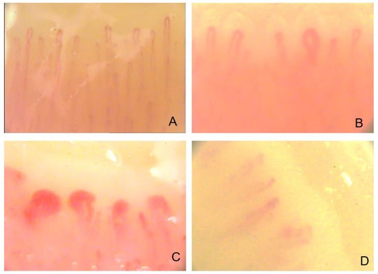
Background. Systemic sclerosis (SSc) is an incurable connective tissue disease characterized by decreased peripheral blood perfusion due to microvascular damage and skin thickening/hardening. The microcirculation deficit is typically secondary to structural vessel damage, which can be assessed morphologically and functionally in a variety of ways, exploiting different technologies. Objective. This paper focuses on reviewing new studies regarding the correlation between microvascular damage, endothelial dysfunction, and internal organ involvement, particularly pulmonary changes in SSc. Methods. We critically reviewed the most recent literature on the correlation between blood perfusion and organ involvement. Results. Many papers have demonstrated the link between structural microcirculatory damage and pulmonary involvement; however, studies that have investigated correlations between microvascular functional impairment and internal organ damage are scarce. Overall, the literature supports the correlation between organ involvement and functional microcirculatory impairment in SSc patients. Conclusions. Morphological and functional techniques appear to be emerging biomarkers in SSc, but obviously need further investigation. Full article
Show Figures

Figure 1

9 pages, 392 KiB  
Article
Quantitative EEG as a Biomarker in Evaluating Post-Stroke Depression
by Livia Livinț Popa, Diana Chira, Victor Dăbală, Elian Hapca, Bogdan Ovidiu Popescu, Constantin Dina, Răzvan Cherecheș, Ștefan Strilciuc and Dafin F. Mureșanu
Diagnostics 2023, 13(1), 49; https://doi.org/10.3390/diagnostics13010049 - 23 Dec 2022
Cited by 6 | Viewed by 2739
Abstract
Introduction: Post-stroke depression (PSD) has complex pathophysiology determined by various biological and psychological factors. Although it is a long-term complication of stroke, PSD is often underdiagnosed. Given the diagnostic role of quantitative electroencephalography (qEEG) in depression, it was investigated whether a possible [...] Read more.
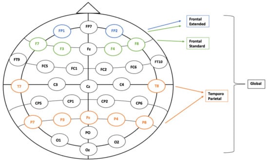
Introduction: Post-stroke depression (PSD) has complex pathophysiology determined by various biological and psychological factors. Although it is a long-term complication of stroke, PSD is often underdiagnosed. Given the diagnostic role of quantitative electroencephalography (qEEG) in depression, it was investigated whether a possible marker of PSD could be identified by observing the evolution of the (Delta + Theta)/(Alpha + Beta) Ratio (DTABR), respectively the Delta/Alpha Ratio (DAR) values in post-stroke depressed patients (evaluated through the HADS-D subscale). Methods: The current paper analyzed the data of 57 patients initially selected from a randomized control trial (RCT) that assessed the role of N-Pep 12 in stroke rehabilitation. EEG recordings from the original trial database were analyzed using signal processing techniques, respecting the conditions (eyes open, eyes closed), and several cognitive tasks. Results: We observed two significant associations between the DTABR values and the HADS-D scores of post-stroke depressed patients for each of the two visits (V1 and V2) of the N-Pep 12 trial. We recorded the relationships in the Global (V1 = 30 to 120 days after stroke) and Frontal Extended (V2 = 90 days after stroke) regions during cognitive tasks that trained attention and working memory. For the second visit, the association between the analyzed variables was negative. Conclusions: As both our relationships were described during the cognitive condition, we can state that the neural networks involved in processing attention and working memory might go through a reorganization process one to four months after the stroke onset. After a period longer than six months, the process could localize itself at the level of frontal regions, highlighting a possible divergence between the local frontal dynamics and the subjective well-being of stroke survivors. QEEG parameters linked to stroke progression evolution (like DAR or DTABR) can facilitate the identification of the most common neuropsychiatric complication in stroke survivors. Full article
Show Figures

Figure 1

10 pages, 2728 KiB  
Article
Safe Follow-Up after Endovascular Aortic Repair with Unenhanced MRI: The SAFEVAR Study
by Francesco Secchi, Davide Capra, Caterina Beatrice Monti, Nazanin Mobini, Maria Del Mar Galimberti Ortiz, Santi Trimarchi, Daniela Mazzaccaro, Paolo Righini, Giovanni Nano and Francesco Sardanelli
Diagnostics 2023, 13(1), 20; https://doi.org/10.3390/diagnostics13010020 - 21 Dec 2022
Cited by 3 | Viewed by 2434
Abstract
We aimed to investigate whether unenhanced magnetic resonance imaging (MRI) could represent a safe and highly sensitive tool for endoleak screening in patients treated with endovascular aneurysm repair (EVAR) using computed tomography angiography (CTA) as a reference standard. Patients who underwent CTA for [...] Read more.
We aimed to investigate whether unenhanced magnetic resonance imaging (MRI) could represent a safe and highly sensitive tool for endoleak screening in patients treated with endovascular aneurysm repair (EVAR) using computed tomography angiography (CTA) as a reference standard. Patients who underwent CTA for EVAR follow-up at our institution were prospectively enrolled. All MRI examinations were performed with a 1.5 T unit. The true-FISP and HASTE sequences of the MRI scans were assessed for the presence of hyperintensity within the aneurysm sac outside the graft, whereas phase-contrast through-plane sequences were used for blood flow quantification. We included 45 patients, 5 (11%) of whom were female. The median age was 73 years (IQR 68–78 years). Among our patients, 19 (42%) were positive for endoleaks at CTA, of whom 13 (68%) had type II endoleaks and 6 (32%) had type I endoleaks. There were no significant differences in age, sex, aneurysm type, prosthesis type, or contrast-to-noise ratio between hyperintensity and thrombus between patients with and without endoleaks (p > 0.300). The combined evaluation of true-FISP and HASTE yielded 100% sensitivity (95% CI: 79–100%) and 19% specificity (95% CI: 7–40%). Patients with a positive CTA had a median thrombus flow of 0.06 L/min (IQR 0.03–0.23 L/min), significantly greater than that of patients with a negative CTA (p = 0.007). Setting a threshold at 0.01 L/min, our MRI protocol yielded 100% sensitivity, 56% specificity, and an AUC of 0.76 (95% CI 0.60–0.91). In conclusion, unenhanced MRI has perfect sensitivity for endoleak detection, although with subpar specificity that could be improved with phase-contrast flow analysis. Full article
Show Figures

Figure 1

11 pages, 1255 KiB  
Article
Photoplethysmographic Measurement of Arterial Stiffness in Polish Patients with Long-COVID-19 Syndrome—The Results of a Cross-Sectional Study
by Izabela Szoltysek-Boldys, Wioleta Zielinska-Danch, Danuta Loboda, Jacek Wilczek, Michal Gibinski, Elzbieta Paradowska-Nowakowska, Krzysztof S. Golba and Beata Sarecka-Hujar
Diagnostics 2022, 12(12), 3189; https://doi.org/10.3390/diagnostics12123189 - 16 Dec 2022
Cited by 7 | Viewed by 1840
Abstract
The coronavirus disease 2019 (COVID-19) is associated with an increase in the incidence of cardiovascular diseases (CVD) that persists even several months after the onset of infection. COVID-19 may also have an impact on arterial stiffness, which is a risk factor for CVD. [...] Read more.
The coronavirus disease 2019 (COVID-19) is associated with an increase in the incidence of cardiovascular diseases (CVD) that persists even several months after the onset of infection. COVID-19 may also have an impact on arterial stiffness, which is a risk factor for CVD. We aimed to analyze if and to what extent arterial stiffness measured by photoplethysmography differed among COVID-19 convalescents depending on the acute phase severity and time elapsed since disease onset. A total of 225 patients (mean age 58.98 ± 8.57 years, 54.7% women) were analyzed after COVID-19 hospitalization at the Cardiac Rehabilitation Department of the Ustron Health Resort (Poland). In the entire study population, no differences were found in the mean values of stiffness index (SI) and reflection index (RI) depending on the severity of the acute COVID-19 and the time since the onset of the disease. There were no differences in the heart rate (HR) according to the severity of acute COVID-19; the mean HR was higher in patients who had COVID-19 less than 12 weeks before the study than in convalescents more than 24 weeks after the acute disease (p = 0.002). The mean values of SI and RI were higher in men than in women (p < 0.001), while the heart rate (HR) was similar in both sexes (p = 0.286). However, multiple linear regression analyses after adjusting for factors influencing arterial stiffness, i.e., sex, age, body mass index, smoking status, hypertension, diabetes, the severity of the acute COVID-19, and the time from the disease onset, confirmed that age, sex, time from disease onset, and diabetes are the most important determinants that could influence arterial stiffness. Full article
Show Figures

Figure 1

9 pages, 1677 KiB  
Article
Role of Contrast-Enhanced Ultrasound in the Follow-Up after Endovascular Abdominal Aortic Aneurysm Repair
by Filippo Benedetto, Domenico Spinelli, Francesco La Corte, Narayana Pipitò, Gabriele Passari and Giovanni De Caridi
Diagnostics 2022, 12(12), 3173; https://doi.org/10.3390/diagnostics12123173 - 15 Dec 2022
Cited by 5 | Viewed by 1784
Abstract
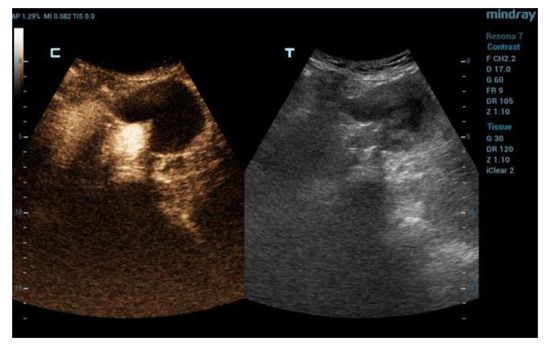
Background: The aim of this study was to assess whether contrast-enhanced ultrasound (CEUS) shows a false negative rate close to zero and therefore is suitable as the main non-invasive follow-up strategy for long-term monitoring after endovascular aortic repair (EVAR). Methods: We included all [...] Read more.
Background: The aim of this study was to assess whether contrast-enhanced ultrasound (CEUS) shows a false negative rate close to zero and therefore is suitable as the main non-invasive follow-up strategy for long-term monitoring after endovascular aortic repair (EVAR). Methods: We included all consecutive patients who underwent CEUS as follow-up after EVAR at our center between January 2017 and December 2021.The follow-up protocol consisted of Duplex ultrasound (DUS) with CEUS at 1, 3, 6 months postoperatively and every 6 months thereafter. Results: A total of 125 patients underwent 228 CEUS. The aneurysm sac showed shrinkage in 80 (64%) patients, stability in 32 (25.6%), and enlargement in 13 (10.4%). A total of 29 (23.2%) patients showed type 2 endoleak, 6 (4.8%) patients showed type 1 endoleak and 3 (2.4%) patients showed type 3 endoleak. Thirteen patients underwent one or more reinterventions. The sensitivity of CEUS vs. DUS was 100% vs. 75% (p > 0.0001). In classifying type 2 endoleak, CEUS compared to DUS showed a sensitivity of 93.2% vs. 59.4% and a specificity of 99.3% vs. 99.3%. CEUS showed a higher sensitivity compared to DUS in the detection of type 2 endoleak. CEUS permits the identification of a subset of patients requiring a stricter follow-up protocol. Full article
Show Figures

Figure 1

11 pages, 639 KiB  
Article
Overall Hemostatic Potential Assay Detects Risk of Progression to Post-Thrombotic Syndrome in Anticoagulated Patients following Deep Vein Thrombosis
by Blake McLeod, Hui Yin Lim, Harshal Nandurkar, Prahlad Ho and Julie Wang
Diagnostics 2022, 12(12), 3165; https://doi.org/10.3390/diagnostics12123165 - 14 Dec 2022
Cited by 6 | Viewed by 1598
Abstract
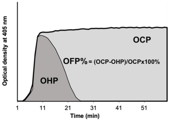
Deep vein thrombosis (DVT) frequently leads to post-thrombotic syndrome (PTS) which is challenging to predict and prevent. Identifying those at high risk of developing PTS may help to focus preventative strategies. Adults were recruited within 3 months of DVT diagnosis. Blood was sampled [...] Read more.
Deep vein thrombosis (DVT) frequently leads to post-thrombotic syndrome (PTS) which is challenging to predict and prevent. Identifying those at high risk of developing PTS may help to focus preventative strategies. Adults were recruited within 3 months of DVT diagnosis. Blood was sampled during the therapeutic anticoagulation phase. Overall hemostatic potential (OHP) assay, a spectrophotometric assay, was performed on platelet-poor plasma (PPP). In this assay, fibrin formation is triggered by small amounts of thrombin and termed the overall coagulation potential (OCP). Simultaneously, thrombin and tissue plasminogen activator are added to PPP and the resulting fibrin aggregation curve is the overall hemostatic potential (OHP). Fibrinolysis is expressed by the parameter overall fibrinolytic potential (OFP%). Patients were followed up at regular intervals. PTS was diagnosed if the Villalta score was ≥5 at least 3 months after the DVT diagnosis. Results were obtained from 190 patients (53.7% male, mean age 56.9 years). PTS developed in 62 (32.6%) patients. Patients with PTS displayed significantly higher median OCP (45.8 vs. 38.8 units, p = 0.010), OHP (12.8 vs. 9.2 units, p = 0.005) and significantly lower OFP (74.1 vs. 75.6%, p = 0.050). PTS patients had higher neutrophil/lymphocyte ratios (NLR) (2.3 vs. 1.9, p = 0.007). After multivariate analysis, proximal DVT location, history of varicose veins, NLR ≥ 2.6, OHP > 13.0 units and weight >108 kg were independent predictors for PTS. The c-statistic of the multivariate model was 0.77. This pilot study suggests that OHP testing while patients are still anticoagulated may assist in the prediction of PTS development and could assist in prognostication and targeting of preventative measures. However, larger prospective studies are needed to confirm these findings. Full article
Show Figures

Figure 1

10 pages, 928 KiB  
Article
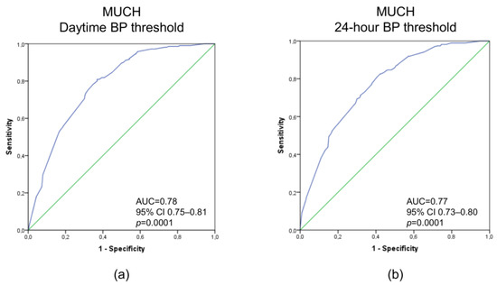
Prediction of Masked Uncontrolled Hypertension Detected by Ambulatory Blood Pressure Monitoring
by Francesca Coccina, Paola Borrelli, Anna M. Pierdomenico, Jacopo Pizzicannella, Maria T. Guagnano, Chiara Cuccurullo, Marta Di Nicola, Giulia Renda, Oriana Trubiani, Francesco Cipollone and Sante D. Pierdomenico
Diagnostics 2022, 12(12), 3156; https://doi.org/10.3390/diagnostics12123156 - 14 Dec 2022
Cited by 2 | Viewed by 1670
Abstract
The aim of this study was to provide prediction models for masked uncontrolled hypertension (MUCH) detected by ambulatory blood pressure (BP) monitoring in an Italian population. We studied 738 treated hypertensive patients with normal clinic BPs classified as having controlled hypertension (CH) or [...] Read more.
The aim of this study was to provide prediction models for masked uncontrolled hypertension (MUCH) detected by ambulatory blood pressure (BP) monitoring in an Italian population. We studied 738 treated hypertensive patients with normal clinic BPs classified as having controlled hypertension (CH) or MUCH if their daytime BP was < or ≥135/85 mmHg regardless of nighttime BP, respectively, or CH or MUCH if their 24-h BP was < or ≥130/80 mmHg regardless of daytime or nighttime BP, respectively. We detected 215 (29%) and 275 (37%) patients with MUCH using daytime and 24-h BP thresholds, respectively. Multivariate logistic regression analysis showed that males, those with a smoking habit, left ventricular hypertrophy (LVH), and a clinic systolic BP between 130–139 mmHg and/or clinic diastolic BP between 85–89 mmHg were associated with MUCH. The area under the receiver operating characteristic curve showed good accuracy at 0.78 (95% CI 0.75–0.81, p < 0.0001) and 0.77 (95% CI 0.73–0.80, p < 0.0001) for MUCH defined by daytime and 24 h BP, respectively. Internal validation suggested a good predictive performance of the models. Males, those with a smoking habit, LVH, and high-normal clinic BP are indicators of MUCH and models including these factors provide good diagnostic accuracy in identifying this ambulatory BP phenotype. Full article
Show Figures

Figure 1

11 pages, 1278 KiB  
Article
Glaucoma Is Associated with the Risk of Obstructive Sleep Apnea: A Population-Based Nationwide Cohort Study
by So Yeon Lee, Hyunjae Yu and Dong-Kyu Kim
Diagnostics 2022, 12(12), 2992; https://doi.org/10.3390/diagnostics12122992 - 29 Nov 2022
Cited by 4 | Viewed by 2209
Abstract
The association between glaucoma and the risk of obstructive sleep apnea (OSA) has not been fully evaluated. Therefore, this study aimed to investigate the prospective association between glaucoma and OSA. In total, 1437 patients with glaucoma and 5748 patients without glaucoma were enrolled [...] Read more.
The association between glaucoma and the risk of obstructive sleep apnea (OSA) has not been fully evaluated. Therefore, this study aimed to investigate the prospective association between glaucoma and OSA. In total, 1437 patients with glaucoma and 5748 patients without glaucoma were enrolled after 1:4 propensity score matching using a nationwide cohort sample. We investigated OSA events during a 10-year follow-up period. Survival analysis, the log-rank test, and Cox proportional hazards regression models were used to calculate the incidence, disease-free survival rate, and hazard ratio (HR). The incidence of OSA was 12,509.0 person-years among those with glaucoma. The adjusted HR for patients with glaucoma developing OSA events during the follow-up period was 1.52 (95% confidence interval [CI]: 0.64–3.621) after other covariates. In a subgroup analysis, primary angle-closure glaucoma (PACG) showed a significantly increased adjusted HR for OSA events (5.65, 95% CI: 1.65–19.41), whereas we could not find any significant association between primary open-angle glaucoma (POAG) and OSA. The adjusted HR of OSA events in POAG was considerably increased 4 years after POAG diagnosis. PACG may be associated with an increased incidence of OSA. Clinicians should pay attention to early detection of OSA in patients with PACG. Full article
Show Figures

Figure 1

10 pages, 255 KiB  
Review
Hypertension in Dialysis Patients: Diagnostic Approaches and Evaluation of Epidemiology
by Panagiotis I. Georgianos, Vasilios Vaios, Vasiliki Sgouropoulou, Theodoros Eleftheriadis, Dimitrios G. Tsalikakis and Vassilios Liakopoulos
Diagnostics 2022, 12(12), 2961; https://doi.org/10.3390/diagnostics12122961 - 26 Nov 2022
Cited by 6 | Viewed by 2267
Abstract
Whereas hypertension is an established cardiovascular risk factor in the general population, the contribution of increased blood pressure (BP) to the huge burden of cardiovascular morbidity and mortality in patients receiving dialysis continues to be debated. In a large part, this controversy is [...] Read more.
Whereas hypertension is an established cardiovascular risk factor in the general population, the contribution of increased blood pressure (BP) to the huge burden of cardiovascular morbidity and mortality in patients receiving dialysis continues to be debated. In a large part, this controversy is attributable to particular difficulties in the accurate diagnosis of hypertension. The reverse epidemiology of hypertension in dialysis patients is based on evidence from large cohort studies showing that routine predialysis or postdialysis BP measurements exhibit a U-shaped or J-shaped association with cardiovascular or all-cause mortality. However, substantial evidence supports the notion that home or ambulatory BP measurements are superior to dialysis-unit BP recordings in diagnosing hypertension, in detecting evidence of target-organ damage and in prognosticating the all-cause death risk. In the first part of this article, we explore the accuracy of different methods of BP measurement in diagnosing hypertension among patients on dialysis. In the second part, we describe how the epidemiology of hypertension is modified when the assessment of BP is based on dialysis-unit versus home or ambulatory recordings. Full article
12 pages, 670 KiB  
Article
Interplay between Myokine Profile and Glycemic Control in Type 2 Diabetes Mellitus Patients with Heart Failure
by Alexander A. Berezin, Zeljko Obradovic, Evgen V. Novikov, Elke Boxhammer, Michael Lichtenauer and Alexander E. Berezin
Diagnostics 2022, 12(12), 2940; https://doi.org/10.3390/diagnostics12122940 - 24 Nov 2022
Cited by 9 | Viewed by 2459
Abstract
Type 2 diabetes mellitus (T2DM) remains a powerful predictor of progressive heart failure (HF), but it is not clear whether altered glycemic control interferes with HF progression via an impaired profile of circulating myokines. The aim was to investigate plausible effects of glucose [...] Read more.
Type 2 diabetes mellitus (T2DM) remains a powerful predictor of progressive heart failure (HF), but it is not clear whether altered glycemic control interferes with HF progression via an impaired profile of circulating myokines. The aim was to investigate plausible effects of glucose control on a myokine signature in T2DM patients affected by chronic HF. We selected 372 T2DM patients from the local database and finally included 314 individuals suffering from chronic HF and subdivided them into two groups according to glycosylated hemoglobin (HbA1c) levels (<6.9% and ≥7.0%). Echocardiography and Doppler examinations along with biomarker measurements were performed at the baseline of the study. The results showed that irisin levels were significantly lower in patients with HbA1c ≥ 7.0% than in those with HbAc1 < 6.9%, whereas concentrations of apelin, myostatin and adropin did not significantly differ between these two groups. We also identified numerous predictors of poor glycemic control, but only N-terminal brain natriuretic propeptide (odds ratio [OR] = 1.07; 95% confidence interval [CI] = 1.02–1.10, p = 0.04) and irisin (OR = 1.09; 95% CI = 1.04–1.17, p = 0.001) remained independent predictors of the dependent variable. In conclusion, we found that decreased levels of irisin were associated with poor glycemic control in T2DM patients with HF regardless of clinical conditions and other biomarkers. Full article
Show Figures

Figure 1

11 pages, 521 KiB  
Systematic Review
Numb Chin Syndrome in Sickle Cell Disease: A Systematic Review and Recommendations for Investigation and Management
by Mahdi Bedrouni, Lahoud Touma, Caroline Sauvé, Stephan Botez, Denis Soulières and Stéphanie Forté
Diagnostics 2022, 12(12), 2933; https://doi.org/10.3390/diagnostics12122933 - 24 Nov 2022
Viewed by 4384
Abstract
Numb chin syndrome (NCS) is a rare sensory neuropathy resulting from inferior alveolar or mental nerve injury. It manifests as hypoesthesia, paraesthesia, or, rarely, as pain in the chin and lower lip. Several case reports suggest that sickle cell disease (SCD) could be [...] Read more.
Numb chin syndrome (NCS) is a rare sensory neuropathy resulting from inferior alveolar or mental nerve injury. It manifests as hypoesthesia, paraesthesia, or, rarely, as pain in the chin and lower lip. Several case reports suggest that sickle cell disease (SCD) could be a cause of NCS. However, information about NCS is scarce in this population. Our objectives were to synthesize all the available literature relevant to NCS in SCD and to propose recommendations for diagnosis and management based on the best available evidence. A systematic review was performed on several databases to identify all relevant publications on NCS in adults and children with SCD. We identified 73 publications; fourteen reports met the inclusion/exclusion criteria. These described 33 unique patients. Most episodes of NCS occurred in the context of typical veno-occlusive crises that involved the mandibular area. Radiological signs of bone infarction were found on some imaging, but not all. Neuropathy management was mostly directed toward the underlying cause. Overall, these observations suggest that vaso-occlusion and bone infarction could be important pathophysiological mechanisms of NCS. However, depending on the individual context, we recommend a careful evaluation to rule out differential causes, including infections, local tumors, metastatic disease, and stroke. Full article
Show Figures

Figure 1

14 pages, 1097 KiB  
Article
Analysis of Factors Determining Patient Survival after Receiving Free-Flap Reconstruction at a Single Center—A Retrospective Cohort Study
by Nicholas Moellhoff, Sara Taha, Nikolaus Wachtel, Maximilian Hirschmann, Marc Hellweg, Riccardo E. Giunta and Denis Ehrl
Diagnostics 2022, 12(11), 2877; https://doi.org/10.3390/diagnostics12112877 - 21 Nov 2022
Cited by 2 | Viewed by 2098
Abstract
Background: Microsurgical tissue transfer revolutionized reconstructive surgery after extensive trauma, oncological resections, and severe infections. Complex soft tissue reconstructions are increasingly performed in multimorbid and elderly patients. Therefore, it is crucial to investigate whether these patients benefit from these complex procedures. Objective: To [...] Read more.
Background: Microsurgical tissue transfer revolutionized reconstructive surgery after extensive trauma, oncological resections, and severe infections. Complex soft tissue reconstructions are increasingly performed in multimorbid and elderly patients. Therefore, it is crucial to investigate whether these patients benefit from these complex procedures. Objective: To evaluate the outcome for multimorbid patients who underwent microsurgical soft tissue reconstruction and to identify potential risk factors that may increase mortality. Methods: This single-center study retrospectively analyzed prospectively collected data of patients receiving free gracilis (GM) or latissimus dorsi muscle (LDM) flap reconstruction between September 2017 and December 2021. Cases were divided into two groups (dead vs. alive), depending on patient survival. Patient demographics, comorbidities and medication, perioperative details, free flap outcome, as well as microcirculation were determined. Results: A total of 151 flaps (LDM, n = 67; GM, n = 84) performed in 147 patients with a mean age of 61.15 ± 17.5 (range 19–94) years were included. A total of 33 patients (22.45%) passed away during the study period. Deceased patients were significantly older (Alive: 58.28 ± 17.91 vs. Dead: 71.39 ± 11.13; p = 0.001), were hospitalized significantly longer (Alive: 29.66 ± 26.97 vs. Dead: 36.88 ± 15.04 days; p = 0.046) and suffered from cardiovascular (Alive: 36.40% vs. Dead: 66.70%; p = 0.002) and metabolic diseases (Alive: 33.90% vs. Dead: 54.50%; p = 0.031) more frequently, which corresponded to a significantly higher ASA Score (p = 0.004). Revision rates (Alive: 11.00% vs. Dead: 18.20%; p = 0.371) and flap loss (Alive: 3.39% vs. Dead: 12.12%; p = 0.069) were higher in patients that died by the end of the study period. Conclusions: Free flap transfer is safe and effective, even in multimorbid patients. However, patient age, comorbidities, preoperative ASA status, and medication significantly impact postoperative patient survival in the short- and mid-term and must, therefore, be taken into account in preoperative decision-making and informed consent. Full article
Show Figures

Figure 1

14 pages, 6784 KiB  
Article
Results of Numerical Modeling of Blood Flow in the Internal Jugular Vein Exhibiting Different Types of Strictures
by Anas Rashid, Syed Atif Iqrar, Aiman Rashid and Marian Simka
Diagnostics 2022, 12(11), 2862; https://doi.org/10.3390/diagnostics12112862 - 18 Nov 2022
Cited by 6 | Viewed by 4055
Abstract
The clinical relevance of nozzle-like strictures in upper parts of the internal jugular veins remains unclear. This study was aimed at understanding flow disturbances caused by such stenoses. Computational fluid dynamics software, COMSOL Multiphysics, was used. Two-dimensional computational domain involved stenosis at the [...] Read more.
The clinical relevance of nozzle-like strictures in upper parts of the internal jugular veins remains unclear. This study was aimed at understanding flow disturbances caused by such stenoses. Computational fluid dynamics software, COMSOL Multiphysics, was used. Two-dimensional computational domain involved stenosis at the beginning of modeled veins, and a flexible valve downstream. The material of the venous valve was considered to be hyperelastic. In the vein models with symmetric 2-leaflets valve without upstream stenosis or with minor 30% stenosis, the flow was undisturbed. In the case of major 60% and 75% upstream stenosis, centerline velocity was positioned asymmetrically, and areas of reverse flow and flow separation developed. In the 2-leaflet models with major stenosis, vortices evoking flow asymmetry were present for the entire course of the model, while the valve leaflets were distorted by asymmetric flow. Our computational fluid dynamics modeling suggests that an impaired outflow from the brain through the internal jugular veins is likely to be primarily caused by pathological strictures in their upper parts. In addition, the jugular valve pathology can be exacerbated by strictures located in the upper segments of these veins. Full article
Show Figures

Figure 1

9 pages, 664 KiB  
Article
[18F]-Fludeoxyglucose Positron Emission Tomography/Computed Tomography with Radiomics Analysis in Patients Undergoing Aortic In-Situ Reconstruction with Cryopreserved Allografts
by Raffaella Berchiolli, Lorenzo Torri, Giulia Bertagna, Francesco Canovaro, Roberta Zanca, Francesco Bartoli, Davide Maria Mocellin, Mauro Ferrari, Paola Anna Erba and Nicola Troisi
Diagnostics 2022, 12(11), 2831; https://doi.org/10.3390/diagnostics12112831 - 17 Nov 2022
Cited by 1 | Viewed by 1602
Abstract
Background: The aim of this study was to evaluate the effectiveness of positron emission tomography/computed tomography with [18F]-fludeoxyglucose (FDG-PET/CT) and radiomics analysis in detecting differences between the native aorta and the abdominal aortic allograft after the total eradication of infection in [...] Read more.
Background: The aim of this study was to evaluate the effectiveness of positron emission tomography/computed tomography with [18F]-fludeoxyglucose (FDG-PET/CT) and radiomics analysis in detecting differences between the native aorta and the abdominal aortic allograft after the total eradication of infection in patients undergoing infected graft removal and in situ reconstruction with cryopreserved allografts. Methods: Between January 2008 and December 2018, 56 vascular reconstructions with allografts have been performed at our department. The present series included 12 patients undergoing abdominal aortic in situ reconstruction with cryopreserved allografts. During the follow-up, all patients underwent a total-body [18F]FDG PET/CT with subsequent radiomics analysis. In all patients, a comparative analysis between the data extracted from native aorta and cryopreserved graft for each patient was performed. Results: All patients were male with a mean age of 72.8 years (range 63–84). Mean duration of follow-up was 51.3 months (range 3–120). During the follow-up, 2 patients (16.7%) needed a redo allograft-related surgical intervention. Overall, the rate of allograft dilatation was 33.3%. No patient had a redo infection during the follow-up. Radiomics analysis showed a different signature of implanted allograft and native aorta. Comparative analysis between the native aortas and cryopreserved allografts (dilated or not) showed several statistical differences for many texture features. Conclusions: The higher metabolic activity of allografts could indicate a state of immune-mediated degeneration. This theory should be proven with prospective, multicentric studies with larger sample sizes. Full article
Show Figures

Figure 1

10 pages, 1648 KiB  
Article
Analysis of Spinal Ischemia after Frozen Elephant Trunk for Acute Aortic Dissection: An Observational, Single-Center Study
by Frederico Lomonaco Cuellar, Alexander Oberhuber, Sven Martens, Andreas Rukosujew, Elena Marchiori and Abdulhakim Ibrahim
Diagnostics 2022, 12(11), 2781; https://doi.org/10.3390/diagnostics12112781 - 14 Nov 2022
Cited by 3 | Viewed by 1849
Abstract
Background: This observational study aimed to evaluate the perioperative risk factors for spinal cord ischemia (SCI) in patients who underwent aortic repair with the frozen elephant trunk technique (FET) after acute aortic Stanford A dissection. Methods: From May 2015 to April 2019, 31 [...] Read more.
Background: This observational study aimed to evaluate the perioperative risk factors for spinal cord ischemia (SCI) in patients who underwent aortic repair with the frozen elephant trunk technique (FET) after acute aortic Stanford A dissection. Methods: From May 2015 to April 2019, 31 patients underwent aortic arch replacement with the FET technique, and spinal ischemia was observed in 4 patients. The risk factors for postoperative SCI were analyzed. Results: The mean age of patients with acute aortic dissection was 57.1 years, and 29.4% were female. Four patients developed SCI. There were no significant differences in characteristics such as age and body mass index. The female gender was associated with most of the SCI cases in the univariate analysis (75%, p = 0.016). Known perioperative and intraoperative risk factors were not related to postoperative SCI in our study. Patients who developed SCI had increased serum postoperative creatinine levels (p = 0.03). Twenty-four patients showed complete false lumen thrombosis up to zones 3–4, five patients up to zones 5–6 and two patients up to zones 7–9, which correlates with the postoperative development of SCI (p = 0.02). The total number of patent intercostal arteries was significantly reduced postoperatively in SCI patients (p = 0.044). Conclusions: Postoperative acute kidney injury, the reduction in patent intercostal arteries after surgery and the extension of false lumen thrombosis up to and beyond zone 5 may play a significant role in the development of clinically relevant spinal cord injury after FET. Full article
Show Figures

Figure 1

11 pages, 2574 KiB  
Systematic Review
Clinical and Imaging Predictors of Disease Progression in Type B Aortic Intramural Hematomas and Penetrating Aortic Ulcers: A Systematic Review
by Elda Chiara Colacchio, Francesco Squizzato, Michele Piazza, Mirko Menegolo, Franco Grego and Michele Antonello
Diagnostics 2022, 12(11), 2727; https://doi.org/10.3390/diagnostics12112727 - 8 Nov 2022
Cited by 2 | Viewed by 1697
Abstract
Background: This work aims to review recent literature on penetrating aortic ulcers (PAUs) and intramural hematomas (IMHs), in order to identify clinical and imaging factors connected to aortic-related adverse events (AAE). Methods: We performed a systematic review according to the Preferred Reporting Items [...] Read more.
Background: This work aims to review recent literature on penetrating aortic ulcers (PAUs) and intramural hematomas (IMHs), in order to identify clinical and imaging factors connected to aortic-related adverse events (AAE). Methods: We performed a systematic review according to the Preferred Reporting Items for Systematic review and Metanalyses (PRISMA) guidelines. An electronic search was conducted on Medline and Embase databases. We included articles reporting on PAUs and/or IMHs localized in the descending thoracic and/or abdominal aorta and analyzing clinical and/or radiological markers of AAE. Results: Of 964 records identified through database searching, 17 were incorporated in the present review, including 193 and 1298 patients with type B PAUs and IMHs, respectively. The 30-days aortic-related mortality (ARM) was 4.3% and 3.9% for PAUs and IMHs. A total of 21% of patients with IMHs underwent intervention during the follow-up period, and 32% experienced an AAE. PAU markers of AAE were minimum depth (ranging from 9.5 to 15 mm) and diameter (≥12.5 mm). Maximum aortic diameter (MAD) cut-off values ranging from 38 to 44.75 mm were related to AAE for IMHs, together with ulcer-like projection (ULP) of the aortic wall. Conclusions: Despite data heterogeneity in the literature, this PAU- and IMH-focused review has highlighted the imaging and clinical markers of disease progression, thus identifying patients that could benefit from an early intervention in order to reduce the AAE rate. Full article
Show Figures

Figure 1

10 pages, 1713 KiB  
Case Report
A Diagnostic of Acquired Hemophilia Following PD1/PDL1 Inhibitors in Advanced Melanoma: The Experience of Two Patients and a Literature Review
by Antonio Gidaro, Giuseppe Palmieri, Mattia Donadoni, Lucia A. Mameli, Leyla La Cava, Giuseppe Sanna, Dante Castro, Alessandro P. Delitala, Roberto Manetti and Roberto Castelli
Diagnostics 2022, 12(10), 2559; https://doi.org/10.3390/diagnostics12102559 - 21 Oct 2022
Cited by 10 | Viewed by 2369
Abstract
Acquired hemophilia A (AHA) is a rare bleeding disorder caused by the development of specific autoantibodies against factor VIII (FVIII). Immunotherapy is a recent therapeutic option that targets the patient’s self-tolerance against tumor cells. Because therapeutic effects of the immune checkpoint inhibitors (ICIs) [...] Read more.
Acquired hemophilia A (AHA) is a rare bleeding disorder caused by the development of specific autoantibodies against factor VIII (FVIII). Immunotherapy is a recent therapeutic option that targets the patient’s self-tolerance against tumor cells. Because therapeutic effects of the immune checkpoint inhibitors (ICIs) are mediated by enhancing the immune response to restore antitumor immunity, autoimmune-related adverse effects can be seen in up to 80% of patients during treatment and after treatment. A rare hematologic ICIs-related adverse event is AHA. Hereafter we report two cases of AHA developed during anti-PD-1 immunotherapy for advanced melanoma: one secondary to treatment with nivolumab and one secondary to pembrolizumab. Both patients were treated with activated FVII (Novoseven®, Novo Nordisk, Bagsværd, Denmark) as hemostatic treatment combined with the eradication of antibodies anti-FVIII obtained with rituximab. In the last few years these drugs have significantly improved the therapeutic armamentarium for the management of AHA. Indeed, while FVIIa has proven to be an effective and safe tool for the treatment of acute bleeding related to FVIII autoantibodies, rituximab is a promising alternative for the autoantibodies’ elimination and the restoration of normal hemostasis. Our finding supports the use of this combination even in AHA secondary to ICIs treatment. Full article
Show Figures

Figure 1

8 pages, 1218 KiB  
Review
Coronary Arteries Aneurysms: A Case-Based Literature Review
by Giuseppe Vadalà, Leandro Di Caccamo, Chiara Alaimo, Luca Di Fazio, Giovanni Ferraiuoli, Giancarlo Buccheri, Vincenzo Sucato and Alfredo Ruggero Galassi
Diagnostics 2022, 12(10), 2534; https://doi.org/10.3390/diagnostics12102534 - 19 Oct 2022
Cited by 4 | Viewed by 3866
Abstract
Coronary artery aneurysm (CAA) is an abnormal dilatation of a coronary artery segment; those coronary artery aneurysms that are very large in size are defined as giant. However, a standardized dimension cut-off to define giant CAAs is still missing. The reported prevalence of [...] Read more.
Coronary artery aneurysm (CAA) is an abnormal dilatation of a coronary artery segment; those coronary artery aneurysms that are very large in size are defined as giant. However, a standardized dimension cut-off to define giant CAAs is still missing. The reported prevalence of coronary aneurysms in the population who underwent coronary angiography ranges from 0.3% to 5%, and often CAAs are found in patient with aneurysms in other sites, such as the ascending or abdominal aorta. In half of the cases an atherosclerotic etiology could be recognized; often, CAA is found in the context of acute coronary syndrome. Seldomly, CAA is found at the autopsy of patients who died due to sudden cardiac death. Currently, very few data exist about CAA management and their prognostic relevance; moreover, CAA treatment is still not clearly codified, but rather case-based. Indeed, currently there are no published dedicated studies exploring the best medical therapy, i.e., with antiplatelets or anticoagulant agents rather than an interventional approach such as an endovascular or surgical technique. In this review, through two clinical cases, the current evidence regarding diagnostic tools and treatment options of CAAs will be described. Full article
Show Figures

Figure 1

9 pages, 1311 KiB  
Article
Volume Changes in the Descending Aorta after Frozen Elephant Trunk and Conventional Hemi-Arch Repair after Acute Type A Aortic Dissection
by Abdulhakim Ibrahim, Arash Motekallemi, Ahmed Yahia, Alexander Oberhuber, Thorsten Eierhoff, Sven Martens, Elena Marchiori and Andreas Rukosujew
Diagnostics 2022, 12(10), 2524; https://doi.org/10.3390/diagnostics12102524 - 18 Oct 2022
Cited by 2 | Viewed by 1907
Abstract
The aim of this study was to compare the mortality rates, re-intervention rates, and volumetric changes in aortas following surgery, in terms of the true lumen and false lumen changes, using conventional hemi-arch repair (CET) and frozen elephant trunk (FET) techniques. During the [...] Read more.
The aim of this study was to compare the mortality rates, re-intervention rates, and volumetric changes in aortas following surgery, in terms of the true lumen and false lumen changes, using conventional hemi-arch repair (CET) and frozen elephant trunk (FET) techniques. During the period from 2015 to 2018, 66 patients underwent surgical treatment for acute aortic dissection (Debakey type 1). Demographic and procedure-related data were evaluated. We measured volumetric change before surgical treatment, at discharge, and at 12- and 24-month time points based on computed tomography angiography. The study cohort was divided into two groups (FET vs. CET). The mean age of the patients was 56.9 ± 9.4 years in the FET group versus 63.6 ± 11 years in the CET group (p = 0.063). The mean follow-up time was 24 ± 6 and 25 ± 5 months for the FET and CET groups, respectively. There were no significant differences between the two groups in terms of the medical histories of the cohorts. The results showed a significant increase in true lumen volume after the FET procedure (within 24 months postoperatively; p = 0.005), and no significant changes in total (p = 0.392) or false lumen (p = 0.659) volumes were noted. After the CET procedure, there were significant increases in total and false lumen volumes (p = 0.013, p = 0.042), while no significant change in true lumen was observed (p = 0.219). The volume increase in true lumen after the FET procedure was higher compared to the CET group at all postoperative time points (at discharge, 12 months, and 24 months) without significant evidence (p = 0.416, p = 0.422, p = 0.268). At two years, the volume increase in false lumen was significantly higher among the CET group compared to the FET group (p = 0.02). The Kaplan–Meier curve analysis showed that patients who underwent the CET procedure underwent significantly more re-interventions due to false lumen expansion of the descending aorta (p = 0.047). Present study results indicate that the true and false lumen changes in the aorta following the FET and CET procedures were different. FET led to a significant increase in true lumen volume, while false lumen volume remained stable; however, after the CET procedure, significant false lumen enlargement was noted at mid-term follow-up time points. The re-intervention rate after CET was higher due to false lumen expansion. Full article
Show Figures

Figure 1

11 pages, 2288 KiB  
Article
The Effect of Submaximal Exercise on Jugular Venous Pulse Assessed by a Wearable Cervical Plethysmography System
by Erica Menegatti, Antonino Proto, Gianfranco Paternò, Giacomo Gadda, Sergio Gianesini, Andrea Raisi, Anselmo Pagani, Tommaso Piva, Valentina Zerbini, Gianni Mazzoni, Giovanni Grazzi, Angelo Taibi, Paolo Zamboni and Simona Mandini
Diagnostics 2022, 12(10), 2407; https://doi.org/10.3390/diagnostics12102407 - 4 Oct 2022
Cited by 2 | Viewed by 7466
Abstract
The jugular venous pulse (JVP) is a one of the crucial parameters of efficient cardiovascular function. Nowadays, limited data are available regarding the response of JVP to exercise because of its complex and/or invasive assessment procedure. The aim of the present work is [...] Read more.
The jugular venous pulse (JVP) is a one of the crucial parameters of efficient cardiovascular function. Nowadays, limited data are available regarding the response of JVP to exercise because of its complex and/or invasive assessment procedure. The aim of the present work is to test the feasibility of a non-invasive JVP plethysmography system to monitor different submaximal exercise condition. Twenty (20) healthy subjects (13M/7F mean age 25 ± 3, BMI 21 ± 2) underwent cervical strain-gauge plethysmography, acquired synchronously with the electrocardiogram, while they were carrying out different activities: stand supine, upright, and during the execution of aerobic exercise (2 km walking test) and leg-press machine exercise (submaximal 6 RM test). Peaks a and x of the JVP waveform were investigated since they reflect the volume of cardiac filling. To this aim, the Δax parameter was introduced, representing the amplitude differences between a and x peaks. Significant differences in the values of a, x, and Δax were found between static and exercise conditions (p < 0.0001, p < 0.0001, p < 0.0001), respectively. Particularly, the Δax value for the leg press was approximately three times higher than the supine, and during walking was even nine times higher. The exercise monitoring by means of the novel JVP plethysmography system is feasible during submaximal exercise, and it provides additional parameters on cardiac filling and cerebral venous drainage to the widely used heartbeat rate value. Full article
Show Figures

Figure 1

15 pages, 2227 KiB  
Article
Systemic Inflammatory Biomarkers and Chest CT Findings as Predictors of Acute Limb Ischemia Risk, Intensive Care Unit Admission, and Mortality in COVID-19 Patients
by Emil Marian Arbănași, Ioana Halmaciu, Réka Kaller, Adrian Vasile Mureșan, Eliza Mihaela Arbănași, Bogdan Andrei Suciu, Cătălin Mircea Coșarcă, Ioana Iulia Cojocaru, Razvan Marian Melinte and Eliza Russu
Diagnostics 2022, 12(10), 2379; https://doi.org/10.3390/diagnostics12102379 - 30 Sep 2022
Cited by 24 | Viewed by 2445
Abstract
Background: Numerous tools, including inflammatory biomarkers and lung injury severity scores, have been evaluated as predictors of thromboembolic events and the requirement for intensive therapy in COVID-19 patients. This study aims to verify the predictive role of inflammatory biomarkers [monocyte to lymphocyte ratio [...] Read more.
Background: Numerous tools, including inflammatory biomarkers and lung injury severity scores, have been evaluated as predictors of thromboembolic events and the requirement for intensive therapy in COVID-19 patients. This study aims to verify the predictive role of inflammatory biomarkers [monocyte to lymphocyte ratio (MLR), neutrophil to lymphocyte ratio (NLR), platelet to lymphocyte ratio (PLR), systemic inflammatory index (SII), Systemic Inflammation Response Index (SIRI), and Aggregate Index of Systemic Inflammation (AISI)] and the CT Severity Score in acute limb ischemia (ALI) risk, intensive unit care (ICU) admission, and mortality in COVID-19 patients.; Methods: The present study was designed as an observational, analytical, retrospective cohort study and included all patients older than 18 years of age with a diagnosis of COVID-19 infection, confirmed through real time-polymerase chain reaction (RT-PCR), and admitted to the County Emergency Clinical Hospital of Targu-Mureș, Romania, and Modular Intensive Care Unit of UMFST “George Emil Palade” of Targu Mures, Romania between January 2020 and December 2021. Results: Non-Survivors and “ALI” patients were associated with higher incidence of cardiovascular disease [atrial fibrillation (AF) p = 0.0006 and p = 0.0001; peripheral arterial disease (PAD) p = 0.006 and p < 0.0001], and higher pulmonary parenchyma involvement (p < 0.0001). Multivariate analysis showed a high baseline value for MLR, NLR, PLR, SII, SIRI, AISI, and the CT Severity Score independent predictor of adverse outcomes for all recruited patients (all p < 0.0001). Moreover, the presence of AF and PAD was an independent predictor of ALI risk and mortality. Conclusions: According to our findings, higher MLR, NLR, PLR, SII, SIRI, AISI, and CT Severity Score values at admission strongly predict ALI risk, ICU admission, and mortality. Moreover, patients with AF and PAD had highly predicted ALI risk and mortality but no ICU admission. Full article
Show Figures

Figure 1

13 pages, 3984 KiB  
Article
Comparability between Computed Tomography Morphological Vascular Parameters and Echocardiography for the Assessment of Pulmonary Hypertension in Patients with Severe Aortic Valve Stenosis—Results of a Multi-Center Study
by Elke Boxhammer, Bernhard Scharinger, Reinhard Kaufmann, Herwig Brandtner, Lukas Schmidbauer, Jürgen Kammler, Jörg Kellermair, Christian Reiter, Kaveh Akbari, Matthias Hammerer, Hermann Blessberger, Clemens Steinwender, Klaus Hergan, Uta C. Hoppe, Michael Lichtenauer and Stefan Hecht
Diagnostics 2022, 12(10), 2363; https://doi.org/10.3390/diagnostics12102363 - 29 Sep 2022
Cited by 2 | Viewed by 1836
Abstract
Background: Computed tomography (CT) of the aorta and cardiac vessels, which is performed in patients with severe aortic valve stenosis (AS) before transcatheter aortic valve replacement (TAVR), offers the possibility of non-invasive detection of pulmonary hypertension (PH), for example, by determining the diameter [...] Read more.
Background: Computed tomography (CT) of the aorta and cardiac vessels, which is performed in patients with severe aortic valve stenosis (AS) before transcatheter aortic valve replacement (TAVR), offers the possibility of non-invasive detection of pulmonary hypertension (PH), for example, by determining the diameter of the main pulmonary artery (PA), the right pulmonary artery (RPA) or the left pulmonary artery (LPA). An improvement of the significance of these radiological parameters is often achieved by indexing to the body surface area (BSA). The aim of this study was to compare different echocardiographic systolic pulmonary artery pressure (sPAP) values with radiological data in order to define potential clinical cut-off values for the presence or absence of PH. Methods: A total of 138 patients with severe AS undergoing TAVR underwent pre-interventional transthoracic echocardiography with determination of sPAP values and performance of CT angiography (CTA) of the aorta and femoral arteries. Radiologically, the PA, RPA, LPA, and ascending aorta (AA) diameters were obtained. Vascular diameters were not only indexed to BSA but also ratios were created with AA diameter (for example PA/AA-ratio). From these CT-derived vascular parameters, AUROC curves were obtained regarding the prediction of different sPAP values (sPAP 40–45–50 mmHg) and finally correlation analyses were calculated. Results: The best AUROC and correlation analyses were generally obtained at an sPAP ≥ 40 mmHg. When considering diameters alone, the PA diameter was superior to the RPA and LPA. Indexing to BSA generally increased the diagnostic quality of the parameters, and finally, in a synopsis of all results, PA/BSA had the best AUC 0.741 (95% CI 0.646–0. 836; p < 0.001; YI 0.39; sensitivity 0.87; specificity 0.52) and Spearman’s correlation coefficient (r = 0.408; p < 0.001) at an sPAP of ≥40 mmHg. Conclusions: Features related to pulmonary hypertension are fast and easily measurable on pre-TAVR CT and offer great potential regarding non-invasive detection of pulmonary hypertension in patients with severe AS and can support the echocardiographic diagnosis. In this study, the diameter of the main pulmonary artery with the additionally determined ratios were superior to the values of the right and left pulmonary artery. Additional indexing to body surface area and thus further individualization of the parameters with respect to height and weight can further improve the diagnostic quality. Full article
Show Figures

Figure 1

Back to TopTop