materials-logo

Journal Browser

Journal Browser

Bioactive Materials in Dentistry

A special issue of Materials (ISSN 1996-1944). This special issue belongs to the section "Biomaterials".

Deadline for manuscript submissions: closed (20 July 2022) | Viewed by 43376

Special Issue Editor


E-Mail Website
Guest Editor
Department of Endodontics and Restorative Dentistry, School of Dental Medicine University of Zagreb, 10000 Zagreb, Croatia
Interests: endodontics; restorative dentistry; dental materials

Special Issue Information

Dear Colleagues,

The evolution of dental materials and dentistry go hand in hand. Historically, the development of materials has evolved by mainly focusing on the improvement of physical and mechanical properties and enhancing their clinical performance and longevity. In recent times, there has been more emphasis on the development of bioactive materials that elicit a biological response. Bioactivity of the materials and a specific response at the interface between tissues and the material results in the formation of a bond and an apatite-like material by strong chemical interaction. Bioactive materials are produced in different forms and with different compositions. These materials are broadly used in all fields of dental medicine. Bioactive materials are promoted as dentin replacements, mimicking properties of hard dental tissues, and enabling biomineralization in dentin. Furthermore, in contact with pulp tissues or periodontal ligament, bioactive materials stimulate repair processes, and deposition of osseous tissue in injured bone.

In this Special Issue, modern trends of using bioactive materials in all fields of dentistry for regeneration, repair, and reconstruction are highlighted and discussed.

It is my pleasure to invite you to submit a manuscript for this Special Issue. Full papers, communications, and reviews are all welcome.

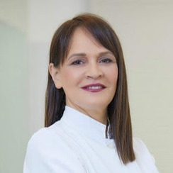
Prof. Dr. Ivana Miletić
Guest Editor

Manuscript Submission Information

Manuscripts should be submitted online at www.mdpi.com by registering and logging in to this website. Once you are registered, click here to go to the submission form. Manuscripts can be submitted until the deadline. All submissions that pass pre-check are peer-reviewed. Accepted papers will be published continuously in the journal (as soon as accepted) and will be listed together on the special issue website. Research articles, review articles as well as short communications are invited. For planned papers, a title and short abstract (about 100 words) can be sent to the Editorial Office for announcement on this website.

Submitted manuscripts should not have been published previously, nor be under consideration for publication elsewhere (except conference proceedings papers). All manuscripts are thoroughly refereed through a single-blind peer-review process. A guide for authors and other relevant information for submission of manuscripts is available on the Instructions for Authors page. Materials is an international peer-reviewed open access semimonthly journal published by MDPI.

Please visit the Instructions for Authors page before submitting a manuscript. The Article Processing Charge (APC) for publication in this open access journal is 2600 CHF (Swiss Francs). Submitted papers should be well formatted and use good English. Authors may use MDPI's English editing service prior to publication or during author revisions.

Keywords

  • bioactive materials
  • apatite-like material
  • bond
  • bioactivity
  • biomineralization
  • regeneration
  • repair

Benefits of Publishing in a Special Issue

  • Ease of navigation: Grouping papers by topic helps scholars navigate broad scope journals more efficiently.
  • Greater discoverability: Special Issues support the reach and impact of scientific research. Articles in Special Issues are more discoverable and cited more frequently.
  • Expansion of research network: Special Issues facilitate connections among authors, fostering scientific collaborations.
  • External promotion: Articles in Special Issues are often promoted through the journal's social media, increasing their visibility.
  • e-Book format: Special Issues with more than 10 articles can be published as dedicated e-books, ensuring wide and rapid dissemination.

Further information on MDPI's Special Issue polices can be found here.

Published Papers (12 papers)

Order results
Result details
Select all
Export citation of selected articles as:

Research

Jump to: Review

15 pages, 2088 KiB  
Article
Comparison of Different Types of Static Computer-Guided Implant Surgery in Varying Bone Inclinations
by Pisut Thangwarawut, Pokpong Amornvit, Dinesh Rokaya and Sirichai Kiattavorncharoen
Materials 2022, 15(9), 3004; https://doi.org/10.3390/ma15093004 - 20 Apr 2022
Cited by 7 | Viewed by 2923
Abstract
This research aimed to compare the accuracy of dental implant placement among three types of surgical guide: metal sleeve with key handle (Nobel guide, Nobel Biocare, Göteborg, Sweden), metal sleeve without key handle, and non-sleeve without key handle (Dentium full guide kit, Dentium [...] Read more.
This research aimed to compare the accuracy of dental implant placement among three types of surgical guide: metal sleeve with key handle (Nobel guide, Nobel Biocare, Göteborg, Sweden), metal sleeve without key handle, and non-sleeve without key handle (Dentium full guide kit, Dentium Co., Seoul, Korea) when placing the implant in different bone inclinations. A total of 72 polyurethane bone models were fabricated in different inclinations (0°, 45°, and 60°). The dental implants were placed in bone models following the company’s recommendations. After dental implants were installed, the digital scans were done by an extraoral scanner. The deviations of the dental implant position were evaluated by superimposition between post-implant placement and reference model by using GOM inspect software. The deviation measurement was shown in 5 parameters: angular deviation, 3D deviation at the crest, 3D deviation at the apex, lateral linear deviation, and vertical linear deviation. The data were analyzed using One-way ANOVA and post-hoc tests at a significance level of 0.05. The accuracy of the dental implant position was not significantly influenced by the difference in the surgical guide system (p > 0.05). There were significant differences between placed and planned implant positions in the different inclinations of the bone. A significant difference was found in all parameters of 0° and 60° bone inclinations (p < 0.05). At 0° and 45°, angulated bone showed significant differences except in 3D deviation at the apex. Between 45° and 60° were significant differences only in angular deviation. Within the limitations of this study, the accuracy of implant placement among three types of surgical guides (Non-sleeve without key handle, Metal sleeve without key handle, and Metal sleeve with key handle) from two companies (Dentium and Nobel Biocare) was similar. Hence, the operators can choose the surgical guide system according to their preference. The inclination of bone can influence the angulation of dental implants. Full article
(This article belongs to the Special Issue Bioactive Materials in Dentistry)
Show Figures

Figure 1

13 pages, 7257 KiB  
Article
The Effect of Y3+ Addition on Morphology, Structure, and Electrical Properties of Yttria-Stabilized Tetragonal Zirconia Dental Materials
by Marko Jakovac, Teodoro Klaser, Arijeta Bafti, Željko Skoko, Luka Pavić and Mark Žic
Materials 2022, 15(5), 1800; https://doi.org/10.3390/ma15051800 - 27 Feb 2022
Cited by 4 | Viewed by 2266
Abstract
Zirconia (ZrO2), a widely known material with an extensive range of applications, is especially suitable for dental applications. This kind of dental material is produced in the form of blocks or discs (mostly discs—depending on CAD/CAM machines) by cold isostatic pressing [...] Read more.
Zirconia (ZrO2), a widely known material with an extensive range of applications, is especially suitable for dental applications. This kind of dental material is produced in the form of blocks or discs (mostly discs—depending on CAD/CAM machines) by cold isostatic pressing (CIP). Such discs are subsequently milled by CAM/CAD technology into a desirable form. Due to the application of CIP, the resulting discs consist of different yttria-stabilized tetragonal zirconia polycrystal (Y-TZP) powders, additives and pigments. The diverse composition of the discs (e.g., different Y3+ content) governs material properties, such as hardness, toughness and translucence. The aim of this work was to investigate the impact of Y3+ addition on the grains and grain boundaries, on the ZrO2 phases lattice parameter values and on the electrical equivalent circuit parameters of the prepared Y-TZP samples. The disc-shaped samples were prepared by using CAM/CAD technology. It was observed that the grain size and the grain density were increased by Y3+ addition. The sample with the lowest Y3+ content was characterized by the highest portion of the tetragonal phase, whilst the disc with the highest Y3+ addition consisted mainly of the cubic phase. It was also observed that at the higher Y3+ ion concentrations, these ions mainly incorporated the tetragonal phase. Furthermore, conductivity investigations showed that the resistivity of the grains in the samples with the higher Y3+ concentrations was decreased as these ions were mainly trapped in the grain boundary. On the other hand, the Y3+ trapping increased the capacitance of the grain boundary. Full article
(This article belongs to the Special Issue Bioactive Materials in Dentistry)
Show Figures

Figure 1

10 pages, 988 KiB  
Article
Effectiveness of Two Different Fluoride-Based Agents in the Treatment of Dentin Hypersensitivity: A Prospective Clinical Trial
by Etleva Qeli, Çeljana Toti, Alessandra Odorici, Elisabetta Blasi, Emiljano Tragaj, Michele Tepedino, Francesco Masedu, Gerta Kaçani, Dorjan Hysi, Agron Meto, Luca Fiorillo and Aida Meto
Materials 2022, 15(3), 1266; https://doi.org/10.3390/ma15031266 - 8 Feb 2022
Cited by 12 | Viewed by 2944
Abstract
Hyperesthesia is related to increased sensitivity of dental tissues to mechanical, chemical and thermal stimuli. The aim of this prospective clinical trial was to compare the effectiveness of a calcium-fluoride-forming agent (Tiefenfluorid®, Humanchemie GmbH, Alfeld, Germany) with that of a fluoride [...] Read more.
Hyperesthesia is related to increased sensitivity of dental tissues to mechanical, chemical and thermal stimuli. The aim of this prospective clinical trial was to compare the effectiveness of a calcium-fluoride-forming agent (Tiefenfluorid®, Humanchemie GmbH, Alfeld, Germany) with that of a fluoride varnish (EnamelastTM, Ultradent Inc., Cologne, Germany) in the treatment of dental hyperesthesia in adult patients. In total, 176 individuals (106 females and 70 males, aged 18–59 years old) diagnosed with dental hyperesthesia (DH) were enrolled. The main clinical symptoms were hyperesthesia from coldness and sweetness during chewing; the types of clinical lesions were also determined and recorded. The patients were selected randomly and divided into two groups: (i) the first group of 96 patients was treated with Tiefenfluorid® applied in three appointments at 7-day intervals; (ii) the second group of 80 patients was treated with EnamelastTM, applied seven times at 7-day intervals. All the patients were recalled 7 days, 14 days, 1 month, 3 months, and 6 months from the last application. At the baseline and during every follow-up visit, the DH was measured with a pulp tester. A random intercept/random slope model was used to evaluate the effect of the treatment, at various times with respect to the initial diagnosis. Within the limits of the present study, Tiefenfluorid® was more effective than EnamelastTM against DH in that it provided long-lasting results, with a significant improvement still detected at the latest 6-month follow-up. Full article
(This article belongs to the Special Issue Bioactive Materials in Dentistry)
Show Figures

Figure 1

10 pages, 1375 KiB  
Article
Additively Manufactured Commercial Co-Cr Dental Alloys: Comparison of Microstructure and Mechanical Properties
by Dalibor Viderščak, Zdravko Schauperl, Sanja Šolić, Amir Ćatić, Matjaž Godec, Aleksandra Kocijan, Irena Paulin and Črtomir Donik
Materials 2021, 14(23), 7350; https://doi.org/10.3390/ma14237350 - 30 Nov 2021
Cited by 20 | Viewed by 3045
Abstract
Laser-powder bed fusion (LPBF) is one of the preferred techniques for producing Co-Cr metal structures for dental prosthodontic appliances. However, there is generally insufficient information about material properties related to the production process and parameters. This study was conducted on samples produced from [...] Read more.
Laser-powder bed fusion (LPBF) is one of the preferred techniques for producing Co-Cr metal structures for dental prosthodontic appliances. However, there is generally insufficient information about material properties related to the production process and parameters. This study was conducted on samples produced from three different commercially available Co-Cr dental alloys produced on three different LPBF machines. Identically prepared samples were used for tensile, three-point bending, and toughness tests. Light microscopy (LM), scanning electron microscopy (SEM), and electron backscatter diffraction (EBSD) analyses of microstructure were performed after testing. Differences were observed in microstructures, which reflected statistically significant differences in mechanical properties (one-way analysis of variance (ANOVA) and Scheffé post hoc test (α = 0.05)). The material produced on the 3D Systems DMP Dental 100 had 24 times greater elongation ε than the material produced on the Sysma MySint 100 device and the EOS M100 machine. On the other hand, the material produced on the EOS M100 had significantly higher hardness (HV0.2) than the other two produced materials. However, the microstructure of the Sysma specimens with its morphology deviates considerably from the studied group. LPBF-prepared Co-Cr dental alloys demonstrated significant differences in their microstructures and, consequently, mechanical properties. Full article
(This article belongs to the Special Issue Bioactive Materials in Dentistry)
Show Figures

Figure 1

8 pages, 892 KiB  
Article
Efficacy of Reciprocating Instruments in the Removal of Bioceramic and Epoxy Resin-Based Sealers: Micro-CT Analysis
by Marko Rajda, Ivana Miletić, Gorana Baršić, Silvana Jukić Krmek, Damir Šnjarić and Anja Baraba
Materials 2021, 14(21), 6670; https://doi.org/10.3390/ma14216670 - 5 Nov 2021
Cited by 7 | Viewed by 2210
Abstract
The objective of this study was to evaluate the efficacy of reciprocating instruments in the removal of bioceramic and epoxy resin-based sealers using micro-CT analysis. Root canals of 40 extracted human teeth were instrumented with a size R25 Reciproc instrument. Specimens were randomly [...] Read more.
The objective of this study was to evaluate the efficacy of reciprocating instruments in the removal of bioceramic and epoxy resin-based sealers using micro-CT analysis. Root canals of 40 extracted human teeth were instrumented with a size R25 Reciproc instrument. Specimens were randomly divided into two groups (n = 20) according to the root canal filling material. In the first group, root canals were obturated with AH Plus sealer and Reciproc R25 gutta-percha. In the second group, a combination of bioceramic gutta-percha (TotalFill BC) and bioceramic sealer (TotalFill BC) was used. After one week, the retreatment of all specimens was performed using Reciproc instruments. To analyze the differences in the filling remnants, specimens were scanned in a micro-CT device after obturation and after the retreatment procedure. Statistical analysis was performed using the Mann–Whitney U test (p < 0.05). A combination of bioceramic sealer and bioceramic gutta-percha was more effectively removed from canals using a reciprocating instrument, with a filling remnants volume of 4.01 ± 3.13 mm3, in comparison to the combination of epoxy resin-based sealer and gutta-percha (6.96 ± 2.70 mm3) (p < 0.05). A reciprocating instrument was more effective in removing bioceramic sealers than epoxy resin-based sealers, although none of the root canal filling materials were completely removed from the root canals. Full article
(This article belongs to the Special Issue Bioactive Materials in Dentistry)
Show Figures

Figure 1

9 pages, 1767 KiB  
Article
Compressive Strength of Conventional Glass Ionomer Cement Modified with TiO2 Nano-Powder and Marine-Derived HAp Micro-Powder
by Ana Ivanišević, Valentina Brzović Rajić, Ana Pilipović, Matej Par, Hrvoje Ivanković and Anja Baraba
Materials 2021, 14(17), 4964; https://doi.org/10.3390/ma14174964 - 31 Aug 2021
Cited by 9 | Viewed by 2293
Abstract
The aim of this research was to investigate the compressive strength (CS), breaking strength (BS), and compressive modulus (CM) of conventional glass ionomer cement (GIC) modified with TiO2 nano particles, marine-derived hydroxyapatite (md-HAp) microparticles (<45 µm), and a combination of TiO2 [...] Read more.
The aim of this research was to investigate the compressive strength (CS), breaking strength (BS), and compressive modulus (CM) of conventional glass ionomer cement (GIC) modified with TiO2 nano particles, marine-derived hydroxyapatite (md-HAp) microparticles (<45 µm), and a combination of TiO2 NP and md-HAp particles. The materials used in the study were conventional GIC Fuji IX GP Extra (GC Corporation, Tokyo, Japan), TiO2 powder P25 (Degussa, Essen, Germany), and HAp synthesized from cuttlefish bone and ground in a mortar to obtain md-HAp powder. md-HAp was characterized using FTIR and SEM analysis. There were four groups of GIC samples: (i) Fuji IX control group, (ii) powder modified with 3 wt% TiO2, (iii) powder modified with 3 wt% HAp, and (iv) powder modified with 1.5 wt% TiO2 + 1.5 wt% HAp. Measurements were performed in a universal testing machine, and CS, BS, and CM were calculated. Statistical analysis was performed using ANOVA and Tukey’s tests. CS, BS, and CM differed significantly between the Fuji IX control group and all experimental groups while differences between the experimental groups were not statistically significant. The addition of TiO2 NP, md-HAp micro-sized particles, and a combination of TiO2 and md-HAp reduced the CS, BS, and CM of conventional GICs when mixed at the powder/liquid (p/l) ratio recommended by the manufacturer. Full article
(This article belongs to the Special Issue Bioactive Materials in Dentistry)
Show Figures

Figure 1

11 pages, 4500 KiB  
Article
Efficient Design of a Clear Aligner Attachment to Induce Bodily Tooth Movement in Orthodontic Treatment Using Finite Element Analysis
by Kyungjae Hong, Won-Hyeon Kim, Emmanuel Eghan-Acquah, Jong-Ho Lee, Bu-Kyu Lee and Bongju Kim
Materials 2021, 14(17), 4926; https://doi.org/10.3390/ma14174926 - 30 Aug 2021
Cited by 34 | Viewed by 6610
Abstract
Clear aligner technology has become the preferred choice of orthodontic treatment for malocclusions for most adult patients due to their esthetic appeal and comfortability. However, limitations exist for aligner technology, such as corrections involving complex force systems. Composite attachments on the tooth surface [...] Read more.
Clear aligner technology has become the preferred choice of orthodontic treatment for malocclusions for most adult patients due to their esthetic appeal and comfortability. However, limitations exist for aligner technology, such as corrections involving complex force systems. Composite attachments on the tooth surface are intended to enable active control of tooth movements. However, unintended tooth movements still occur. In this study, we present an effective attachment design of an attachment that can efficiently induce tooth movement by comparing and analyzing the movement and rotation of teeth between a general attachment and an overhanging attachment. The 3D finite element modes were constructed from CBCT data and used to analyze the distal displacement of the central incisor using 0.5- and 0.75-mm-thick aligners without an attachment, and with general and overhanging attachments. The results show that the aligner with the overhanging attachment can effectively reduce crown tipping and prevent axial rotation for an intended distal displacement of the central incisor. In all models, an aligner with or without attachments was not capable of preventing the lingual inclination of the tooth. Full article
(This article belongs to the Special Issue Bioactive Materials in Dentistry)
Show Figures

Figure 1

12 pages, 3142 KiB  
Article
Impact of Sandblasting on Morphology, Structure and Conductivity of Zirconia Dental Ceramics Material
by Marko Jakovac, Teodoro Klaser, Borna Radatović, Arijeta Bafti, Željko Skoko, Luka Pavić and Mark Žic
Materials 2021, 14(11), 2834; https://doi.org/10.3390/ma14112834 - 25 May 2021
Cited by 15 | Viewed by 3246
Abstract
Over the last decade, zirconia (ZrO2)-based ceramic materials have become more applicable to modern dental medicine due to the sustained development of diverse computer-aided design/computer-aided manufacturing (CAD/CAM) systems. However, before the cementation and clinical application, the freshly prepared zirconia material (e.g., [...] Read more.
Over the last decade, zirconia (ZrO2)-based ceramic materials have become more applicable to modern dental medicine due to the sustained development of diverse computer-aided design/computer-aided manufacturing (CAD/CAM) systems. However, before the cementation and clinical application, the freshly prepared zirconia material (e.g., crowns) has to be processed by sandblasting in the dental laboratory. In this work, the impact of the sandblasting on the zirconia is monitored as changes in morphology (i.e., grains and cracks), and the presence of impurities might result in a poor adhesive bonding with cement. The sandblasting is conducted by using Al2O3 powder (25, 50, 110 and 125 µm) under various amounts of air-abrasion pressure (0.1, 0.2, 0.4 and 0.6 MPa). There has been much interest in both the determination of the impact of the sandblasting on the zirconia phase transformations and conductivity. Morphology changes are observed by using Scanning Electron Microscope (SEM), the conductivity is measured by Impedance Spectroscopy (IS), and the phase transformation is observed by using Powder X-Ray Diffraction (PXRD). The results imply that even the application of the lowest amount of air-abrasion pressure and the smallest Al2O3 powder size yields a morphology change, a phase transformation and a material contamination. Full article
(This article belongs to the Special Issue Bioactive Materials in Dentistry)
Show Figures

Figure 1

10 pages, 554 KiB  
Article
Effect of Nano-Filled Protective Coating and Different pH Enviroment on Wear Resistance of New Glass Hybrid Restorative Material
by Sandra Brkanović, Ana Ivanišević, Ivana Miletić, Dražen Mezdić and Silvana Jukić Krmek
Materials 2021, 14(4), 755; https://doi.org/10.3390/ma14040755 - 5 Feb 2021
Cited by 16 | Viewed by 3227
Abstract
The purpose of the study was to determine the wear rate of Equia Forte HT Fil with Equia Forte Coat or without coating and compare it with Fuji IX GP high-viscosity glass ionomer cement (GIC) in conditions with acid load or at neutral [...] Read more.
The purpose of the study was to determine the wear rate of Equia Forte HT Fil with Equia Forte Coat or without coating and compare it with Fuji IX GP high-viscosity glass ionomer cement (GIC) in conditions with acid load or at neutral pH. The samples were stored for 7 days: (1) in artificial saliva, (2) in artificial saliva and cyclically exposed to low pH, and (3) in distilled water and cyclically exposed to low pH. Wear was determined by measuring the difference in mass before and after brushing in an abrasion testing device. The wear of Fuji IX GP was significantly higher than that of Equia Forte HT Fil with or without coating (p = 0.000). The difference between Equia Forte HT Fil with and without Coat was not statistically significant (p < 0.803). The differences in wear resistance between samples stored in saliva and in distilled water were not significant (p = 0.588). Periodic exposure to the low pH solution significantly affected the wear resistance of all materials (p = 0.000). Equia Forte HT Fil was more resistant to wear than Fuji IX GP in all storage conditions. A resinous coat did not significantly increase wear resistance. Full article
(This article belongs to the Special Issue Bioactive Materials in Dentistry)
Show Figures

Figure 1

11 pages, 3141 KiB  
Article
Surface Characterization and Conductivity of Two Types of Lithium-Based Glass Ceramics after Accelerating Ageing
by Marko Jakovac, Teodoro Klaser, Borna Radatović, Željko Skoko, Luka Pavić and Mark Žic
Materials 2020, 13(24), 5632; https://doi.org/10.3390/ma13245632 - 10 Dec 2020
Cited by 8 | Viewed by 2681
Abstract
In this study, two different dental ceramics, based on zirconia-reinforced lithium-silicate (LS1) glass-ceramics (Celtra Duo, Dentsply Sirona, Bensheim, Germany) and lithium disilicate (LS2) ceramics (IPS e.max CAD, Ivoclar, Vivadent, Schaan, Liechtenstein) were examined. They were tested prior to and after the crystallization by [...] Read more.
In this study, two different dental ceramics, based on zirconia-reinforced lithium-silicate (LS1) glass-ceramics (Celtra Duo, Dentsply Sirona, Bensheim, Germany) and lithium disilicate (LS2) ceramics (IPS e.max CAD, Ivoclar, Vivadent, Schaan, Liechtenstein) were examined. They were tested prior to and after the crystallization by sintering in the dental furnace. Additionally, the impact of ageing on ceramic degradability was investigated by immersing it in 4% acetic acid at 80 °C for 16 h. The degradability of the materials was monitored by Impedance Spectroscopy (IS), X-Ray Powder Diffraction (XRPD), and Field Emission Scanning Electron Microscope (FE-SEM) techniques. It was detected that LS2 (vs. LS1) samples had a lower conductivity, which can be explained by reduced portions of structural defects. XRPD analyses also showed that the ageing increased the portion of defects in ceramics, which facilitated the ion diffusion and degradation of samples. To summarize, this study suggests that the non-destructive IS technique can be employed to probe the ageing properties of the investigated LS1 and LS2 ceramics materials. Full article
(This article belongs to the Special Issue Bioactive Materials in Dentistry)
Show Figures

Figure 1

Review

Jump to: Research

14 pages, 1386 KiB  
Review
Effectiveness of Direct Pulp Capping Bioactive Materials in Dentin Regeneration: A Systematic Review
by Ermin Nie, Jiali Yu, Rui Jiang, Xiangzhen Liu, Xiang Li, Rafiqul Islam and Mohammad Khursheed Alam
Materials 2021, 14(22), 6811; https://doi.org/10.3390/ma14226811 - 11 Nov 2021
Cited by 18 | Viewed by 6551
Abstract
Background: Regenerative endodontics aims to restore normal pulp function in necrotic and infected teeth, restoring protective functions, such as innate pulp immunity, pulp repair through mineralization, and pulp sensibility. The aim of this systematic review was to assess the dentin regeneration efficacy of [...] Read more.
Background: Regenerative endodontics aims to restore normal pulp function in necrotic and infected teeth, restoring protective functions, such as innate pulp immunity, pulp repair through mineralization, and pulp sensibility. The aim of this systematic review was to assess the dentin regeneration efficacy of direct pulp capping (DPC) biomaterials. Methods: The literature published between 2005 and 2021 was searched by using PubMed, Web of Science, Science Direct, Google Scholar, and Scopus databases. Clinical controlled trials, randomized controlled trials, and animal studies investigating DPC outcomes or comparing different capping materials after pulp exposure were included in this systematic review. Three independent authors performed the searches, and information was extracted by using a structured data format. Results: A total of forty studies (21 from humans and 19 from animals) were included in this systemic review. Histological examinations showed complete/partial/incomplete dentin bridge/reparative dentin formation during the pulp healing process at different follow-up periods, using different capping materials. Conclusions: Mineral trioxide aggregate (MTA) and Biodentine can induce dentin regeneration when applied over exposed pulp. This systematic review can conclude that MTA and its variants have better efficacy in the DPC procedure for dentin regeneration. Full article
(This article belongs to the Special Issue Bioactive Materials in Dentistry)
Show Figures

Figure 1

12 pages, 9207 KiB  
Review
Commercially Available Ion-Releasing Dental Materials and Cavitated Carious Lesions: Clinical Treatment Options
by Amel Slimani, Salvatore Sauro, Patricia Gatón Hernández, Sevil Gurgan, Lezize Sebnem Turkun, Ivana Miletic, Avijit Banerjee and Hervé Tassery
Materials 2021, 14(21), 6272; https://doi.org/10.3390/ma14216272 - 21 Oct 2021
Cited by 9 | Viewed by 3101
Abstract
The contemporary approach for operative caries management emphasizes personalized interventions for each patient, dependent upon the individual’s caries susceptibility/risk, the stage of the carious lesion and its activity. The clinician’s challenge is to optimize the extent of cavity preparation and the choice of [...] Read more.
The contemporary approach for operative caries management emphasizes personalized interventions for each patient, dependent upon the individual’s caries susceptibility/risk, the stage of the carious lesion and its activity. The clinician’s challenge is to optimize the extent of cavity preparation and the choice of dental restorative biomaterials, appreciating the benefits offered by ion-releasing restorative materials. There is a growing application of bioactive/bio-interactive materials in minimally invasive operative dentistry, as they may help with tissue recovery by ion release. In case of moderate or extensive occlusal cavitation, the clinical criteria include the individual caries susceptibility and carious lesion activity. In high caries risk cases, ion-releasing biomaterials (IRB) can be used, as well as for active carious lesions. In proximal lesions, the clinical criteria include the individual caries susceptibility, the lesion activity and presence of cavities with little or no enamel at the gingival margin. This article aims to discuss the restorative ion-releasing options, according to different clinical situations, and the caries susceptibility to manage cavitated carious lesions in permanent adult teeth. Full article
(This article belongs to the Special Issue Bioactive Materials in Dentistry)
Show Figures

Figure 1

Back to TopTop