Article

Powder Synthesized from Aqueous Solution of Calcium Nitrate and Mixed-Anionic Solution of Orthophosphate and Silicate Anions for Bioceramics Production

by
Daniil Golubchikov
1,*,
Tatiana V. Safronova
1,2,*,
Elizaveta Nemygina
1,
Tatiana B. Shatalova
1,2,
Irina N. Tikhomirova
3,
Ilya V. Roslyakov
1,4,
Dinara Khayrutdinova
5,
Vadim Platonov
2,
Olga Boytsova
2,
Maksim Kaimonov
1,
Denis A. Firsov
2 and
Konstantin A. Lyssenko
2
1
Department of Materials Science, Lomonosov Moscow State University, Building, 73, Leninskie Gory, 1, 119991 Moscow, Russia
2
Department of Chemistry, Lomonosov Moscow State University, Building, 3, Leninskie Gory, 1, 119991 Moscow, Russia
3
Department of General Technology of Silicates, Mendeleev University of Chemical Technology, Building, 1, Geroyev Panfilovtsev, 20, 125480 Moscow, Russia
4
Kurnakov Institute of General and Inorganic Chemistry, Russian Academy of Sciences, Leninskii Prosp., 31, 119071 Moscow, Russia
5
A.A. Baikov Institute of Metallurgy and Materials Science, Russian Academy of Sciences, Leninskii Prosp., 49, 119334 Moscow, Russia
*
Authors to whom correspondence should be addressed.
Coatings 2023, 13(2), 374; https://doi.org/10.3390/coatings13020374
Submission received: 31 December 2022 / Revised: 1 February 2023 / Accepted: 2 February 2023 / Published: 7 February 2023
(This article belongs to the Special Issue Advanced Biomaterials and Coatings)

Abstract

:
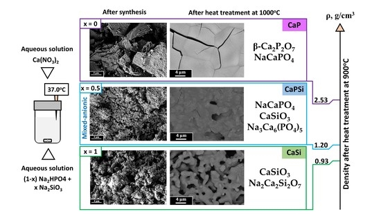
Synthesis from mixed-anionic aqueous solutions is a novel approach to obtain active powders for bioceramics production in the CaO-SiO2-P2O5-Na2O system. In this work, powders were prepared using precipitation from aqueous solutions of the following precursors: Ca(NO3)2 and Na2HPO4 (CaP); Ca(NO3)2 and Na2SiO3 (CaSi); and Ca(NO3)2, Na2HPO4 and Na2SiO3 (CaPSi). Phase composition of the CaP powder included brushite CaHPO4‧2H2O and the CaSi powder included calcium silicate hydrate. Phase composition of the CaPSi powder consisted of the amorphous phase (presumably containing hydrated quasi-amorphous calcium phosphate and calcium silicate phase). All synthesized powders contained NaNO3 as a by-product. The total weight loss after heating up to 1000 °C for the CaP sample—28.3%, for the CaSi sample—38.8% and for the CaPSi sample was 29%. Phase composition of the ceramic samples after the heat treatment at 1000 °C based on the CaP powder contained β-NaCaPO4 and β-Ca2P2O7, the ceramic samples based on the CaSi powder contained α-CaSiO3 and Na2Ca2Si2O7, while the ceramics obtained from the CaPSi powder contained sodium rhenanite β-NaCaPO4, wollastonite α-CaSiO3 and Na3Ca6(PO4)5. The densest ceramic sample was obtained in CaO-SiO2-P2O5-Na2O system at 900 °C from the CaP powder (ρ = 2.53 g/cm3), while the other samples had densities of 0.93 g/cm3 (CaSi) and 1.22 (CaPSi) at the same temperature. The ceramics prepared in this system contain biocompatible and bioresorbable phases, and can be recommended for use in medicine for bone-defect treatment.

Graphical Abstract

1. Introduction

Modern science faces a problem of increasing levels of bone diseases, traumas and cancers. Thereby, the development of bone repair orthopedic approaches as well as enhancement of implemented biomaterials is crucial [1,2]. Bone tissues possess a great regenerative potential and suitable biomaterials are required to support natural regeneration process [3,4,5,6]. One of the crucial processes of native bone forming is angiogenesis [7], which is vital for bone healing [8]. The most commonly used biomaterials in the field of bone tissue engineering are polymers [9,10,11], composites [12], as well as glass and ceramics, obtained in systems containing calcium phosphates and silicates [13,14,15,16]. Such materials are also required to provide a sufficient mechanical support and appropriate environment for cell attachment, proliferation, and differentiation [17,18].
Among the first steps in this area were materials in the Na2O-CaO-SiO2-P2O5 system, suggested by L. L. Hench [19]. This study marked the beginning of calcium-sodium phosphate-silicate glass-ceramics development as well as application of such materials in bone implant production. L.L. Hench et al. [20] demonstrated that synthesized surface-active bioglass-ceramics can be used in vivo without inflammation.
Materials synthesized in the considered system tend to be osteoinductive, since the presence of Si-containing ions is suggested to stimulate the proliferation of human aortic endothelial cells as well as to support the expression of genes encoding the proangiogenic downstream cytokines [21], which is necessary for successful angiogenesis processes [22].
A special part of the Na2O-CaO-SiO2-P2O5 system is the CaO-SiO2 system, in which two congruently melting compounds, Ca2SiO4 and CaSiO3 (wollastonite), and two incongruently melting compounds, Ca3SiO5 (stable from 1250 to 2050 °C), and Ca3Si2O7 (stable from low temperature to 1464 °C), can be obtained [23]. Congruently melting compounds are more promising for bone tissue engineering: for example, 3D-printed β-Ca2SiO4 scaffolds sintered at a higher temperature stimulated the adhesion, proliferation, ALP activity, and osteogenic-related gene expression of rBMSCs [24]. CaSiO3 ceramics have been extensively researched as biomaterial to replace other materials due to their superior biological activity, for example, compared to hydroxyapatite (HA) [25] showing their potential prospects in in vivo trials [26]. In addition, calcium silicate ceramics can be used for skin healing and cartage regeneration [27]. In [28], silicate ceramics are obtained by solid-phase synthesis from a mixture of powders.
In the CaO-P2O5, which is a part of the Na2O-CaO-SiO2-P2O5 system, the following biocompatible and bioresorbable phases of tricalcium phosphate (Ca3(PO4)2) and calcium pyrophosphate (Ca2P2O7) [29] can be obtained.
In Na2O-CaO-P2O5 sodium-substituted tricalcium phosphate Ca10Na(PO4)7, sodium rhenanite β-NaCaPO4, mixed sodium-calcium pyrophosphate (CaNa2P2O7) [29] and mixed sodium-calcium phosphate (Na3Ca6(PO4)5) can be obtained.
In the CaO-SiO2-P2O5, as a part of the Na2O-CaO-SiO2-P2O5 system, two main phases can be obtained: silicocarnotite (Ca5(PO4)2SiO4), with a structure type carnotite and a wide range of solid solutions [30,31], and nagelshmidtite (Ca7Si2P2O16) [32]. Both phases provide satisfactory biocompatibility. The addition of sodium to this system leads to the appearance of Na2Ca2Si3O9 and Na2CaSi3O8 after the sintering of material above 600 °C [33].
There are several approaches for obtaining ceramics or glass-ceramics in the CaO-SiO2-P2O5-Na2O system. First of all, the samples can be obtained in the form of glasses with subsequent crystallization during the heat treatment at an appropriate temperature [34]; nevertheless, this method has final phase composition limitations. In this case, the bioactive glass decomposition occurs, which leads to the appearance of additional crystalline phases. The composition of this phases directly depends on the composition of the initial mixture. For example, using the mixture of the initial components with the ratio of SiO2:NaO2:CaO = 41.8:26.7:31.5 (mol) makes it possible to obtain the Na2Ca2Si2O7 phase [35]. This phase was also synthesized in [36] from the mixture of reagent grade Na2CO3, CaO, bovine bone (P2O5 source), and rice husk (SiO2 source), as additional to the Na6Ca3Si6O18 main phase. Nevertheless, the sample containing this phase showed good results in mechanical properties and in in vitro tests.
Another approach to obtain ceramics in the considered CaO-SiO2-P2O5-Na2O system is based on the use of hydroxyapatite or tricalcium phosphate powders as a filler and an aqueous solution of sodium silicate as a binder [37]. This method leads to the formation of two main phases, which are β-rhenanite (β-NaCaPO4) and sodium calcium phosphate (Na3Ca6(PO4)5) after heat treatment. These phases are biocompatible and are used for the restoration of bone-tissue defects [38,39].
It is also possible to prepare ceramic materials using intermediate phases precipitated from the solution (such as hydroxyapatite and amorphous sodium silicate), which can be converted into final phases (sodium rhenanite, calcium-sodium silicate, and wollastonite) during the heat treatment process [37,40].
In addition, it is possible to print scaffolds from a composite of calcium phosphate powders and 45S5 Bioglass using a cementation reaction during printing [41,42]. In [42], calcium hydrogen phosphate dihydrate (CaHPO4⸱2H2O) was formed as a result of the cementing reaction. And the final phase composition of material after the heat treatment at 1000 °C contained sodium rhenanite (NaCaPO4) and wollastonite (CaSiO3).
Ceramics in the CaO-SiO2-P2O5-Na2O system can be obtained from powders synthesized from solutions. To obtain such ceramics, active powders with a highly homogeneous distribution of components are required. Amorphous powders synthesized from mixed-anionic solutions have the necessary homogeneity.
There are some examples of powders synthesized from mix-anionic HPO4/P2O7 [43] P2O7/CO3 [44], HPO4/CO3 [45,46], or mix-cationic K/Na [47] aqueous solutions with appropriate homogeneity that were used for the ceramic materials preparation. Preservation of reaction by-product as a component of the powder mixture also can be used as a method of preparation of powder mixtures with high homogeneity [25,48,49].
However, an approach involving synthesis from mixed-anionic solutions is more interesting, since this method leads to a more homogeneous distribution of components in the powder mixture [43,44,45,46]. In addition, a chemical approach to the synthesis of mixed-anionic powders makes it possible to obtain more dispersed powders with a larger specific surface area, which are more active, and the sintering process is more effective [48,49]. Additionally, the by-product of the synthesis from mixed-anionic solutions can be kept in the composition to form melts and promote the formation of new phases [48,49]. Thereby, this method can be the most promising approach to obtain ceramics in the CaO-SiO2-P2O5-Na2O system.
Thus, the aim of the present work consisted in the synthesis of powder, which contains precursors of calcium phosphate and silicate high-temperature phases, as well as the reaction by-product from the mixed-anionic solution, for preparation of composite ceramics in Na2O-CaO-SiO2-P2O5 system. The obtained powder mixture was implemented as a powder precursor with homogeneous distribution of the components for production of the composite material, while the by-product of synthesis was used both as a participant of the heterogeneous reactions and as a sintering aid.

2. Materials and Methods

2.1. Materials

Powders of calcium nitrate tetrahydrate Ca(NO3)2·4H2O (CAS no. 13477-34-4, ACS reagent, Sigma-Aldrich, Mumbai, India), sodium metasilicate pentahydrate Na2SiO3·5H2O (CAS no. 10213-79-3, RusKhim, Moscow, Russia), and sodium phosphate dibasic (CAS no. 7558-79-4, BioXtra, Sigma-Aldrich, Gillingham, UK) were used for powder mixture preparation.

2.2. Synthesis of Powders

The synthesis of powders was carried out using the Na2SiO3, Ca(NO3)2, Na2HPO4 (Table 1) according to the equations:
Ca(NO3)2 + Na2HPO4 + 2H2O CaHPO4‧2H2O + 2 NaNO3
2 Ca(NO3)2 + Na2HPO4 + Na2SiO3 + xH2O CaHPO4‧2H2O + CaSiO3‧xH2O + 4 NaNO3
Ca(NO3)2 + Na2SiO3 + xH2O CaSiO3‧xH2O + 2 NaNO3
The starting salts were dissolved in distilled water at a concentration of 0.5 M, then the calcium salt solution was slowly added to the sodium salt solution in the volume ratios corresponding to the reactions (1–3), and the suspension was stirred for 1 h. The synthesis was carried out at a temperature of 37 °C. Then, the precipitate was filtered using a vacuum filter and evenly distributed over a large surface area and left to dry for 1 week.
Further, the obtained powders were disaggregated in acetone medium using a planetary ball mill (Fritch Pulverisette, Bavaria, Germany) for 10 min in zirconia containers with grinding ZrO2-media (mpowder:mballs = 1:5). After that, when the acetone was completely removed each of the powders was sieved through a polyester sieve with a mesh size of 200 μm.

2.3. Preparation of Ceramic Samples

The obtained samples were pressed into the form of simple disks by uniaxial one-sided pressing on a manual press (Carver Laboratory Press model C, Fred S. Carver, Inc., Wabash, IN, USA) using steel die with a diameter of 12 mm. Pressing was carried out at a pressure of 100 MPa for 10 s.
The pressed samples in the form of disks were fired at 800, 900 and 1000 °C. Additionally, the powders were fired at 400 and 600 °C to control the phase transformations. Heating in the furnace was carried out at a speed of 5 °C/min. The holding time at these temperatures was 2 h. The heat treatment of the samples was carried out in order to study the effect of high temperatures on the initial composition, as well as to determine the mass losses and determine the thermal behavior of the materials in the temperature range mentioned. The linear shrinkage and density of the samples after the heat treatment were also calculated.

2.4. Methods of Analysis

The linear shrinkage after the heat treatment and the density of the samples before and after the heat treatment were calculated using Equations (4) and (5), respectively.
ΔDrel = (D0 − D)/D0 × 100, %,
where:
ΔDrel—linear shrinkage of the sample after the heat treatment, %;
D—diameter of the sample after the heat treatment, cm;
D0—diameter of the sample after pressing, cm.
ρ = m/(h × πD2/4), g/cm3,
where:
ρ—density of the sample, g/cm3;
m—weight of the sample, g;
h—thickness of the sample, cm;
D—diameter of the sample, cm.
The mass and the linear dimensions of the samples were measured with accuracy of ±0.001 g and ±0.01 mm, respectively, before and after the heat treatment.
Thermal analysis (TA) including thermogrvimetry (TG) and differential thermal analysis (DTA) was performed using an STA 409 PC Luxx thermal analyzer (NETZSCH, Selb, Germany) during heating in air (10 °C/min, 40–1000 °C), the specimen mass being at least 10 mg. The gas-phase composition was monitored by a Netzsch QMS 403C Aëolos quadrupole mass spectrometer (NETZSCH, Selb, Germany) coupled with a Netzsch STA 409 PC Luxx thermal analyzer (NETZSCH, Selb, Germany). The mass spectra were registered for the following m/Z values: 18 (H2O); 30 (NO).
The phase composition of the powders obtained after the synthesis was determined by X-ray powder diffraction (XRD) analysis using Rigaku D/Max-2500 diffractometer (Rigaku Corporation, Tokyo, Japan) with a rotating anode (Cu–Ka radiation), angle interval 2Ѳ: from 2° to 70° (step 2Ѳ − 0.02°). XRD analysis of the ceramic composition was also implemented using a Rigaku Miniflex 600 diffractometer (CuKα radiation, Kβ filter, and D/teX Ultra detector) in Bragg–Brentano geometry (Rigaku Corporation, Tokyo, Japan) with an angle interval 2Ѳ from 3° to 70° (step 2Ѳ − 0.02°). Phase analysis was performed using the ICDD PDF2 database and Match software (version https://www.crystalimpact.com/, 25 December 2022).
Scanning electron microscopy (SEM) images of the synthesized powder and powder mixtures were characterized by SEM on an NVision 40 microscope (Carl Zeiss, Jena, Germany), and SEM images of ceramic samples were taken with Tescan Vega II (Tescan, Brno, Czech Republic) at accelerating voltages from 1 to 20 kV in secondary electron imaging mode (SE2 detector). A chromium/gold layers (≤10 nm in thickness) on the surface of the ceramic sample was applied to the samples (Quorum Technologies spraying plant, Q150T ES, Great Britain, London, UK).

3. Results and Discussion

Table 2 shows weights of the prepared powders after synthesis and drying, weights of by-products calculated from the reactions (1–3) and weights of by-products isolated from the mother liquor by drying. The mass of prepared powders (column 3, Table 2) may include the mass of target minerals and the mass of by-product. In the row of powders CaP, CaPSi, CaSi the weight of the by-product collected from the mother liquor became lower. It was the lowest for CaSi, therefore, the amount of the by-product adsorbed and occluded by powder was the largest. The mass of adsorbed and occluded by-products increases from CaP to CaSi powder. The amount of the isolated reaction by-product apparently can be interpreted as an indirect confirmation of the presence of the largest active surface for CaSi and CaPSi powders. These values can apparently correlate with the surface area, which makes it possible to expect the highest activity for these powders.
According to XRD (Figure S1) the phase composition of by-products of all reactions (1–3) isolated from the mother liquors by drying contained only NaNO3.
The phase composition of all synthesized powders after disaggregation in acetone is presented in Figure 1. It should be noted that XRD patterns for powders after synthesis and after disaggregation were the same for each powder. The phase composition of the synthesized powders after the disaggregation process was not changed.
According to the XRD data (Figure 1), the powder synthesized from Ca(NO3)2 and Na2HPO4 solutions (CaP) after disaggregation contained brushite (dicalcium phosphate dihydrate, CaHPO4‧2H2O), as well as the reaction by-product of sodium nitrate (NaNO3).
According to the XRD analysis (Figure 1), the CaSi powder contained crystalline phases of calcium silicate hydrate (CSH) [50] and NaNO3. Under considered synthesis conditions (37 °C and maturation of precipitates during 1 h in the mother liquors), the formation of well-crystallized calcium silicate hydrates (CSH − Ca1.5SiO3.5‧xH2O [PDF card 33-306]) was not expected, since their formation occurs in the process of high-temperature synthesis under autoclave conditions as it is known from the literature [50]. Low-basic hydrosilicates (Ca/Si = 0.8–1.5) have similar peaks on the X-ray diffraction pattern, and weak-crystallized products have 3–4 peaks, which lead to the ambiguous interpretations of the chemical composition. Peaks with interplanar spacing of 3.02A (29.54°) and 1.81A (50.45°) possibly belong to calcium silicate hydrate (CSH), which has a variable Ca/Si ratio from 0.8 to 1.5. The main peaks of CSH are 29.54, 33.25 and 50.45° [50]. And peaks 29.54, 33.25 of CSH overlaps with sodium nitrate peaks. The formation of tobermorite-like gel is also possible [50]. Amorphous calcium phosphate can be stabilized by the presence of silicate ions, as well as calcium silicate hydrate with a Ca/Si ratio from 0.8 to 1.5 [16].
Apparently, the CaPSi powder contained the only crystalline phase of NaNO3. Other phases in the CaPSi powder were calcium silicate hydrate and amorphous calcium phosphate. Taking into account the presence of an amorphous halo near 30° on the XRD pattern of the CaPSi sample, this sample also possibly contained amorphous calcium phosphate (ACP), which could be effectively stabilized in the amorphous state by the presence of a silicate anion [16].
The morphology of the obtained powders after the synthesis and after the disaggregation process is presented in Figure 2.
Brushite crystallizes in the specific shape of large thin bars (longer than 10 μm) characteristic for brushite, which are observed in Figure 2a. This is consistent with the XRD data for CaP powder. After disaggregation, elongated lamellar particles with a size of 1–2 μm remained (Figure 2b).
Large spheroid aggregates with dimensions 4–8 μm are presented in the SEM images of the CaSi powder sample before and after disaggregation (Figure 2e,f). The dimensions of particles in aggregates before and after disaggregation were not bigger than 50 nm. Apparently, this morphology corresponds to the presence of amorphous calcium silicate hydrate.
The CaPSi sample of synthesized powder is also consisted of aggregates (4–8 μm) of spheroid particles (~50 nm), as it is shown by the SEM image (Figure 2c). These spheroid particles may consist of amorphous phase of both hydrated ACP and calcium silicate hydrate. After disaggregation, the powders contain mesoporous aggregates (2–4 μm) of spherical particles of submicron (~50 nm) size (Figure 2d). Thereby, implementation of synthesis via precipitation from mixed-anion solution (CaPSi sample) leads to the production of powders consisted of submicron particles, which are smaller than those reported in previous studies, where the powders were obtained using another method [51].
According to the TG data, the final mass loss of the CaP powder after heating to 1000 °C was 28.3% (Figure 3a). At the first stage (up to 160 °C), there was a gradual decomposition of structurally unbonded water, accompanied by a slight endoeffect. At the next stage (up to 200 °C), the transition of brushite to monetite (DCPD DCPA (dicalcium phosphate anhydrous)) occurred, accompanied by a significant endoeffect with a maximum at 190 °C (Figure 3b). The further process is the transformation of monetite to γ-pyrophosphate (DCPA γ-CPP), generally takes place at 400 °C. But the process of transformation of DCPA to CPP in powder CaP under investigation tooks place at lower temperature. The mass loss takes place with an endoeffect at 295 °C (Figure 3b). This endoeffect may also be associated with the melting of sodium nitrate (melting point – 308 °C). So, we can state that the presence of monetite can make the melting point of sodium nitrate lower and consequently the presence of the melt of sodium nitrate leads to the thermal transformation (DCPA CPP) at lower temperature. According to XRD data (Figure 4,a, Table 3) after heart treatment at 400 °C the phase composition of CaP powder included β-Ca2P2O7 and NaNO3. So, the transition of the γ-phase to the β-phase (γ-CPP β-CPP) also was facilitated and made a contribution in the appearance of endoeffect in interval 250–350 °C. The next step of mass loss with endoeffect at 503 °C can be explained by sodium rhenanite formation from calcium pyrophosphate and sodium nitrate as it was confirmed by XRD data for powder CaP after heat treatment at 600 °C (Figure 4a, Table 3). The maximum endoeffect at the temperature of 503 °C corresponds to the maximum at 500 °C in interval 400–560 °C at mass-spectroscopy graph (Figure S2) by NO ion current (NO (M = 30) release) and the formation of the sodium rhenanite phase according to the formal reaction (6), which is not reflect the complicity of the thermal decomposition of nitrates.
Ca2P2O7 + 2NaNO3  2NaCaPO4 + N2O5
For the CaSi sample, the final mass loss after heating to 1000 °C was 38.8% (Figure 3d). The first endoeffect (Figure 3e) at a temperature of 100–120 °C was apparently due to the elimination of structurally unbonded water. The endoeffect at the temperature of 300–310 °C was associated with the melting of sodium nitrate (melting point - 308 °C). Apparently, then there was a gradual crystallization of the amorphous phase with the formation of α-CaSiO3 (wollastonite), including the possible formation of the intermediate tobermarite phase at 400–500 °C, up to 650 °C (with a significant endoeffect on the DSC graph). Confirmation of the gradual formation of wollastonite is presented in Figure 4c. The formation of wollastonite apparently continues up to 800 °C (Table 3). The maximum endoeffect at a temperature of 700 °C can be associated with the complete decomposition of sodium nitrate, which was also observed at the corresponding mass-spectroscopy graphs (Figure S2) by NO ion current (NO (M = 30) release) and the formation of the Na2Ca2Si2O7 phase (Figure 3f) and the sodium rhenanite phase (Figure 3b,d). The temperature of NO release depends on the composition of the sample. This indirectly indicates the surface area of powder that possibly holds the by-product. Thus, CaSi has the largest surface area, the maximum content of the by-product (Table 2) and a higher temperature, which is required to detach the nitrate. The ratio of intensity values of ion current for m/Z = 30 (NO) also indicates an increase in the content of sodium nitrate in the samples in the series CaP-CaPSi-CaSi (correspondingly 0.70 × 10⁻¹², 1.06 × 10⁻¹², 2.57 × 10⁻¹²). The NO release peak for this series shifts to higher temperatures. Thus, the reactivity of CaP powder is higher than that of CaSi powder.
The total mass loss of the CaPSi sample after heating to 1000 °C was 29.0% (Figure 3b). Two steps of mass loss (17% and 12%) can be observed at the TG curve. According to MS data one step is connected with H2O release up to 420 °C. And the second step is observed due to the processes connected with the decomposition of sodium nitrate or with heterphase reactions with sodium nitrate in the interval 450–750 °C. DTA graph (Figure 3c) clearly shows that two significant endoeffects corresponds to these steps of mass loss. According to XRD data for CaPSi sample after heat treatment at 400 and 600 °C hydroxyapatite, sodium nitrate and wollastonite was found. Up to 250 °C, the formation of the hydroxyapatite phase occurs from the amorphous calcium phosphate [52], as well as the beginning of the wollastonite phase forming, which apparently lasts up to 800 °C and also causes a 2% mass change in the temperature range 250–500 °C [25]. After 500 °C, the formation of the Na3Ca6(PO4)5 phase, and the sodium rhenanite (β-NaCaPO4) phase occurs (Figure 3d).
As discussed above, heat treatment of the CaP powder at 600 °C and the ceramic samples at the range of temperatures from 800 to 1000 °C leads to the formation of a two-phase powder and ceramic samples containing calcium pyrophosphate (β-Ca2P2O7) and β-rhenanite (β-NaCaPO4). No other phases were found in the considered quasi-binary system [29].
In the case of the CaSi sample after the thermal treatment at 400 °C, there were no significant changes in phase composition, in comparison with the phase composition before the thermal treatment. After the thermal treatment at 600 °C, the formation of Ca5Si6O16(OH)2 and Na6Ca3Si6O18 occurred. Two-phase composite ceramics including wollastonite (α-CaSiO3) and Na2Ca2Si2O7 (Table 3, Figure 4; ICDD PDF2 database [53]) were also obtained after heat treatment of the CaSi samples at the range from 800 to 1000 °C.
After the heat treatment at 400 °C, the formation of hydroxyapatite and wollastonite occurred for the CaPSi sample. Presence of H2O at high temperature give us an opportunity to guess that hydroxyapatite was formed under hydrothermal conditions. At 600 °C, the formation of hydroxyapatite from the amorphous phase, accompanied with the formation of sodium rhenanite from the amorphous phase and NaNO3, occurred. The heat treatment at 800 °C led to the formation of tetraphasic ceramics, containing sodium rhenanite, hydroxyapatite, wollastonite and the Na3Ca6(PO4)5 phase. The phase composition of the CaPSi ceramic samples after the heat treatment in the range of temperatures from 900 to 1000 °C (Figure 4b, Table 3) remained almost unchanged. These samples after heat treatments at 900 and 1000 °C contained wollastonite (α-CaSiO3), sodium rhenanite (NaCaPO4), and mixed calcium-sodium phosphate (Na3Ca6(PO4)5). Nevertheless, the phase ratios varied depending on the heat treatment temperature. With an increase in the heat treatment temperature the phases tended to become more crystallized. The reflexes of the wollastonite (α-CaSiO3) phase increased in the range of temperatures from 800 to 1000 °C, while the hydroxyapatite phase disappeared. After this temperature, heterophase reactions between hydroxyapatite and sodium rhenanite occurred, leading to the formation of Na3Ca6(PO4)5.
The SEM images of the surface of ceramic samples sintered at 1000 °C are shown in Figure 5.
With an increase in the heat treatment temperature, the sintering of the samples proceeds more efficiently due to the elimination of pores and the growth of grains, the size of which is 8–10 μm when fired at 1000 °C (CaP), while the grain sizes for the other samples are 2–3 μm (CaSi) and 2–4 μm (CaPSi).
Apparently, the occurrence of heterophase reactions in the CaSi and CaPSi samples inhibits grain growth in ceramics. The formation of sodium rhenanite and pyrophosphate is completed already after the heat treatment at 600 °C. During the heat treatment at higher temperatures, grain growth is possible since there are no conditions for heterophase reactions in the quasi-binary calcium pyrophosphate-sodium rhenanite system.
With an increase in the heat treatment temperature (and in the content of wollastonite), the ceramics became denser, which was accompanied by a significant grain growth up to 2–4 μm from submicron size. Microporosity of CaPSi also disappeared and a small number of pores with a diameter of 2–3 μm were formed. The ceramics based on the CaSi powder were highly porous, which was consistent with the literature data [54].
The shrinkage density of the samples CaP, CaPSi, and CaSi during the heat treatment is presented in Figure 6. The initial density of all the powder pre-ceramic samples in the form of disks was approximately equal and was 1.30 g/cm3 for CaP and CaSi and 1.20 g/cm3 for CaPSi.
The CaP sample had the largest linear shrinkage (up to 26% at 900 and 1000 °C), and the CaSi sample had the smallest linear shrinkage (up to 5%) (Figure 6a). Linear shrinkage for the CaPSi sample ranged from 9 to 13% at 800 to 1000 °C, respectively. At the same time, fluctuations in density changes depending on the temperature for the CaPSi samples are minor, since the obtained components have approximately the same density (1.05–1.22 g/cm3).
The apparent densities of the obtained ceramics are consistent with the scanning electron microscopy data (Figure 6b). The CaP sample fired at 900 and 1000 °C had the highest density. The uniform distribution of cracks visible on the surface of the CaP ceramic sample was apparently caused by the phase transformation in sodium rhenanite.

4. Conclusions

A novel method of multi-component active powders production was proposed in this work. The powders were prepared using a synthesis from aqueous solutions of CaNO3 and a mixed-anion solution containing Na2HPO4 and Na2SiO3. For comparison, the powders were synthesized from solutions of CaNO3 and solutions containing these sodium salts separately.
Phase composition of the CaP powder included brushite CaHPO4‧2H2O and the CaSi powder included calcium silicate hydrate. Phase composition of the CaPSi powder consisted of the amorphous phase (presumably containing hydrated quasi-amorphous calcium phosphate and calcium silicate phase). All synthesized powders contained NaNO3 as a by-product. The quasi-amorphous phases of CaPSi powder, obtained by precipitation from mixed-anionic solution, as it can be assumed, stabilized each other. A significant amount of by-product NaNO3 was intentionally kept in the composition of all obtained powders for the synthesis of the ceramics of the Na2O-CaO-SiO2-P2O5 system during heat treatment of samples. This component acts both as a sintering aid and as a participant of the heterophase reactions of the formation of the ceramics based on the prepared powders (CaP. CaPSi, CaSi). The obtained powders were used to produce ceramics by the heat treatment at 800, 900, and 1000 °C. The main phases obtained for the ceramics based on the CaPSi powder were β-rhenanite β-NaCaPO4, wollastonite α-CaSiO3 and Na3Ca6(PO4)5. The density of the CaPSi ceramics increased with the heat treatment temperature from 1.47 g/cm3 (800 °C) to 1.72 g/cm3 (1000 °C). The ceramics based on the CaP powder had the highest density (2.53 g/cm3) after the heat treatment at 900 °C. The ceramics based on the CaSi powder were significantly porous and had the lowest density (1.52 g/cm3) after the heat treatment at 900 °C. The ceramics prepared in this work in the CaO-SiO2-P2O5-Na2O system and containing the biocompatible and bioresorbable phases can be recommended for use in medicine for bone defect treatment. The obtained materials are expected to support cell proliferation and differentiation. Osteoconductive 3D forms also can be manufactured from obtained powders.

Supplementary Materials

The following supporting information can be downloaded at: https://www.mdpi.com/article/10.3390/coatings13020374/s1, Figure S1: XRD data of by-product powders dried from solutions, collected after filtration (mother liquors) (PDF card 36-1474)–sodium nitrate, Figure S2: Mass-spectroscopy data of (a) CaP, (b) CaPSi, (c) CaSi powders.

Author Contributions

Conceptualization, D.G. and T.V.S.; methodology, D.G. and T.V.S.; validation, D.G., E.N. and T.V.S.; investigation, D.G., E.N., T.V.S., T.B.S., I.N.T., I.V.R., D.K., V.P., O.B., M.K., D.A.F. and K.A.L.; resources, T.B.S., I.V.R., O.B. and K.A.L.; data curation, D.G. and E.N.; writing—original draft preparation, D.G. and E.N.; writing—review and editing, D.G., E.N. and T.V.S.; visualization, D.G., E.N., T.B.S., I.V.R., D.K., V.P., O.B. and M.K.; supervision, T.V.S.; funding acquisition, T.V.S. All authors have read and agreed to the published version of the manuscript.

Funding

This work was carried out with financial support from the Russian Science Foundation (grant no. 22-19-00219).

Institutional Review Board Statement

Not applicable.

Informed Consent Statement

Not applicable.

Data Availability Statement

Not applicable.

Acknowledgments

This research was carried out using the equipment of the MSU Shared Research Equipment Center “Technologies for obtaining new nanostructured materials and their complex study” and purchased by MSU in the frame of the Equipment Renovation Program (National Project “Science”), and in the frame of the MSU Program of Development. The authors would like to thank Elena Golubchikova for the assistance with draft preparation. Some of the SEM images were recorded using scientific equipment at the Joint Research Center for Physical Methods of Research, located in the Kurnakov Institute of General and Inorganic Chemistry RAS.

Conflicts of Interest

The authors declare no conflict of interest.

References

  1. Stevens, M.M. Biomaterials for Bone Tissue Engineering. Mater. Today 2008, 11, 18–25. [Google Scholar] [CrossRef]
  2. Ansari, M. Bone Tissue Regeneration: Biology, Strategies and Interface Studies. Prog. Biomater. 2019, 8, 223–237. [Google Scholar] [CrossRef]
  3. Ashammakhi, N.; Hasan, A.; Kaarela, O.; Byambaa, B.; Sheikhi, A.; Gaharwar, A.K.; Khademhosseini, A. Advancing Frontiers in Bone Bioprinting. Adv. Healthc. Mater. 2019, 8, 1801048. [Google Scholar] [CrossRef] [PubMed]
  4. Amiryaghoubi, N.; Fathi, M.; Pesyan, N.N.; Samiei, M.; Barar, J.; Omidi, Y. Bioactive Polymeric Scaffolds for Osteogenic Repair and Bone Regenerative Medicine. Med. Res. Rev. 2020, 40, 1833–1870. [Google Scholar] [CrossRef] [PubMed]
  5. Shuai, C.; Yang, W.; Feng, P.; Peng, S.; Pan, H. Accelerated Degradation of HAP/PLLA Bone Scaffold by PGA Blending Facilitates Bioactivity and Osteoconductivity. Bioact. Mater. 2021, 6, 490–502. [Google Scholar] [CrossRef]
  6. Yamada, Y.; Inui, T.; Kinoshita, Y.; Shigemitsu, Y.; Honda, M.; Nakano, K.; Matsunari, H.; Nagaya, M.; Nagashima, H.; Aizawa, M. Silicon-Containing Apatite Fiber Scaffolds with Enhanced Mechanical Property Express Osteoinductivity and High Osteoconductivity. J. Asian Ceram. Soc. 2019, 7, 101–108. [Google Scholar] [CrossRef]
  7. Malhotra, A.; Habibovic, P. Calcium Phosphates and Angiogenesis: Implications and Advances for Bone Regeneration. Trends Biotechnol. 2016, 34, 983–992. [Google Scholar] [CrossRef] [PubMed]
  8. Schmidt-Bleek, K.; Schell, H.; Schulz, N.; Hoff, P.; Perka, C.; Buttgereit, F.; Volk, H.-D.; Lienau, J.; Duda, G.N. Inflammatory Phase of Bone Healing Initiates the Regenerative Healing Cascade. Cell Tissue Res. 2012, 347, 567–573. [Google Scholar] [CrossRef] [PubMed]
  9. Ligon, S.C.; Liska, R.; Stampfl, J.; Gurr, M.; Mülhaupt, R. Polymers for 3D Printing and Customized Additive Manufacturing. Chem. Rev. 2017, 117, 10212–10290. [Google Scholar] [CrossRef]
  10. Mao, M.; He, J.; Li, X.; Zhang, B.; Lei, Q.; Liu, Y.; Li, D. The Emerging Frontiers and Applications of High-Resolution 3D Printing. Micromachines 2017, 8, 113. [Google Scholar] [CrossRef] [Green Version]
  11. Pina, S.; Ribeiro, V.P.; Marques, C.F.; Maia, F.R.; Silva, T.H.; Reis, R.L.; Oliveira, J.M. Scaffolding Strategies for Tissue Engineering and Regenerative Medicine Applications. Materials 2019, 12, 1824. [Google Scholar] [CrossRef] [PubMed]
  12. Yunus Basha, R.; Kumar, T.S.S.; Doble, M. Design of Biocomposite Materials for Bone Tissue Regeneration. Mater. Sci. Eng. C 2015, 57, 452–463. [Google Scholar] [CrossRef] [PubMed]
  13. Guzzo, C.M.; Nychka, J.A. Bone ‘Spackling’ Paste: Mechanical Properties and In Vitro Response of a Porous Ceramic Composite Bone Tissue Scaffold. J. Mech. Behav. Biomed. Mater. 2020, 112, 103958. [Google Scholar] [CrossRef] [PubMed]
  14. Guzzo, C.M.; Nychka, J. Fabrication of a Porous and Formable Ceramic Composite Bone Tissue Scaffold at Ambient Temperature. Metall. Mater. Trans. A 2020, 51, 6110–6126. [Google Scholar] [CrossRef]
  15. Kazakova, G.; Safronova, T.; Golubchikov, D.; Shevtsova, O.; Rau, J.V. Resorbable Mg2+-Containing Phosphates for Bone Tissue Repair. Materials 2021, 14, 4857. [Google Scholar] [CrossRef]
  16. Zuev, D.M.; Golubchikov, D.O.; Evdokimov, P.V.; Putlyaev, V.I. Synthesis of Amorphous Calcium Phosphate Powders for Production of Bioceramics and Composites by 3D Printing. Russ. J. Inorg. Chem. 2022, 67, 940–951. [Google Scholar] [CrossRef]
  17. Koons, G.L.; Diba, M.; Mikos, A.G. Materials Design for Bone-Tissue Engineering. Nat. Rev. Mater. 2020, 5, 584–603. [Google Scholar] [CrossRef]
  18. Hu, X.; Wang, Y.; Tan, Y.; Wang, J.; Liu, H.; Wang, Y.; Yang, S.; Shi, M.; Zhao, S.; Zhang, Y.; et al. A Difunctional Regeneration Scaffold for Knee Repair Based on Aptamer-Directed Cell Recruitment. Adv. Mater. 2017, 29, 1605235. [Google Scholar] [CrossRef]
  19. Hench, L.L.; Splinter, R.J.; Allen, W.C.; Greenlee, T.K. Bonding Mechanisms at the Interface of Ceramic Prosthetic Materials. J. Biomed. Mater. Res. 1971, 5, 117–141. [Google Scholar] [CrossRef]
  20. Hench, L.L.; Paschall, H.A. Direct Chemical Bond of Bioactive Glass-Ceramic Materials to Bone and Muscle. J. Biomed. Mater. Res. 1973, 7, 25–42. [Google Scholar] [CrossRef]
  21. Li, H.; Xue, K.; Kong, N.; Liu, K.; Chang, J. Silicate Bioceramics Enhanced Vascularization and Osteogenesis through Stimulating Interactions between Endothelia Cells and Bone Marrow Stromal Cells. Biomaterials 2014, 35, 3803–3818. [Google Scholar] [CrossRef]
  22. Kaully, T.; Kaufman-Francis, K.; Lesman, A.; Levenberg, S. Vascularization—The Conduit to Viable Engineered Tissues. Tissue Eng. Part B Rev. 2009, 15, 159–169. [Google Scholar] [CrossRef] [PubMed]
  23. Hillert, M.; Sundman, B.; Wang, X. An Assessment of the CaO-SiO2 System. Metall. Mater. Trans. B 1990, 21, 303–312. [Google Scholar] [CrossRef]
  24. Fu, S.; Liu, W.; Liu, S.; Zhao, S.; Zhu, Y. 3D Printed Porous β-Ca2SiO4 Scaffolds Derived from Preceramic Resin and Their Physicochemical and Biological Properties. Sci. Technol. Adv. Mater. 2018, 19, 495–506. [Google Scholar] [CrossRef]
  25. Pan, Y.; Yin, J.; Yao, D.; Zuo, K.; Xia, Y.; Liang, H.; Zeng, Y. Effects of Silica Sol on the Microstructure and Mechanical Properties of CaSiO3 Bioceramics. Mater. Sci. Eng. C. 2016, 64, 336–340. [Google Scholar] [CrossRef]
  26. De Aza, P.N.; Luklinska, Z.B.; Martinez, A.; Anseau, M.R.; Guitian, F.; De Aza, S. Morphological and Structural Study of Pseudowollastonite Implants in Bone. J. Microsc. 2000, 197, 60–67. [Google Scholar] [CrossRef]
  27. Yu, Q.; Chang, J.; Wu, C. Silicate Bioceramics: From Soft Tissue Regeneration to Tumor Therapy. J. Mater. Chem. B 2019, 7, 5449–5460. [Google Scholar] [CrossRef]
  28. Hu, Y.; Xiao, Z.; Wang, H.; Ye, C.; Wu, Y.; Xu, S. Fabrication and Characterization of Porous CaSiO3 Ceramics. Ceram. Int. 2019, 45, 3710–3714. [Google Scholar] [CrossRef]
  29. Filippov, Y.; Murashko, A.; Evdokimov, P.; Safronova, T.; Putlayev, V. Stereolithography 3D printed calcium pyrophosphate macroporous ceramics for bone grafting. Open Ceram. 2021, 8, 100185. [Google Scholar] [CrossRef]
  30. Serena, S.; Sainz, M.; Caballero, A. Single-phase silicocarnotite synthesis in the subsystem Ca3(PO4)2–Ca2SiO4. Ceram. Int. 2014, 40, 8245–8252. [Google Scholar] [CrossRef]
  31. Ros-Tarraga, P.; Mazon, P.; Meseguer-Olmo, L.; De Aza, P. Revising the Subsystem Nurse’s A-Phase-Silicocarnotite within the System Ca3(PO4)2–Ca2SiO4. Materials 2016, 9, 322. [Google Scholar] [CrossRef] [PubMed]
  32. Xu, M.; Zhai, D.; Chang, J.; Wu, C. In Vitro assessment of three-dimensionally plotted nagelschmidtite bioceramic scaffolds with varied macropore morphologies. Acta Biomater. 2014, 10, 463–476. [Google Scholar] [CrossRef]
  33. Anand, V.; Singh, K.; Kaur, K. Investigation of Mg and Zn doped 45S5 bioactive materials by XRD, FTIR and SEM techniques. AIP Conf. Proc. 2014, 1591, 745. [Google Scholar] [CrossRef]
  34. Zhang, X.; Guo, X.; Zhang, J.; Fan, X.; Chen, M.; Yang, H. Nucleation, Crystallization and Biological Activity of Na2O-CaO-P2O5-SiO2 Bioactive Glass. J. Non Cryst. Solids 2021, 568, 120929. [Google Scholar] [CrossRef]
  35. Kahlenberg, V.; Hösch, A. The Crystal Structure of Na2Ca2Si2O7—A Mixed Anion Silicate with Defect Perovskite Characteristics. Z. Krist. Cryst. Mater. 2002, 217, 155–163. [Google Scholar] [CrossRef]
  36. Leenakul, W.; Pisitpipathsin, N.; Kantha, P.; Tawichai, N.; Tigunta, S.; Eitssayeam, S.; Rujijanagul, G.; Pengpat, K.; Munpakdee, A. Characteristics of 45S5 Bioglass-Ceramics Using Natural Raw Materials. AMR Adv. Mater. Res. 2012, 506, 174–177. [Google Scholar] [CrossRef]
  37. Kaimonov, M.; Safronova, T.; Shatalova, T.; Filippov, Y.; Tikhomirova, I.; Sergeev, N. Composite Ceramics in the Na2O–CaO–SiO2–P2O5 System Obtained from Pastes Including Hydroxyapatite and an Aqueous Solution of Sodium Silicate. Ceramics 2022, 5, 550–561. [Google Scholar] [CrossRef]
  38. Safronova, T.V. Inorganic Materials for Regenerative Medicine. Inorg. Mater. 2021, 57, 443–474. [Google Scholar] [CrossRef]
  39. Demirkiran, H.; Mohandas, A.; Dohi, M.; Fuentes, A.; Nguyen, K.; Aswath, P. Bioactivity and Mineralization of Hydroxyapatite with Bioglass as Sintering Aid and Bioceramics with Na3Ca6(PO4)5 and Ca5(PO4)2SiO4 in a Silicate Matrix. Mater. Sci. Eng. C 2010, 30, 263–272. [Google Scholar] [CrossRef]
  40. Lin, K.; Zhai, W.; Ni, S.; Chang, J.; Zeng, Y.; Qian, W. Study of the Mechanical Property and In Vitro Biocompatibility of CaSiO3 Ceramics. Ceram. Int. 2005, 31, 323–326. [Google Scholar] [CrossRef]
  41. Gmeiner, R.; Deisinger, U.; Schönherr, J.; Lechner, B.; Detsch, R.; Boccaccini, A.R.; Stampfl, J. Additive Manufacturing of Bioactive Glasses and Silicate Bioceramics. J. Ceram. Sci. Technol. 2015, 6, 75–86. [Google Scholar] [CrossRef]
  42. Bergmann, C.; Lindner, M.; Zhang, W.; Koczur, K.; Kirsten, A.; Telle, R.; Fischer, H. 3D Printing of Bone Substitute Implants Using Calcium Phosphate and Bioactive Glasses. J. Eur. Ceram. Soc. 2010, 30, 2563–2567. [Google Scholar] [CrossRef]
  43. Safronova, T.V.; Knot’ko, A.V.; Shatalova, T.B.; Evdokimov, P.V.; Putlyaev, V.I.; Kostin, M.S. Calcium phosphate ceramic based on powder synthesized from a mixed-anionic solution. Glass Ceram. 2016, 73, 25–31. [Google Scholar] [CrossRef]
  44. Peranidze, K.; Safronova, T.V.; Filippov, Y.; Kazakova, G.; Shatalova, T.; Rau, J.V. Powders Based on Ca2P2O7-CaCO3-H2O System as Model Objects for the Development of Bioceramics. Ceramics 2022, 5, 423–434. [Google Scholar] [CrossRef]
  45. Safronova, T.V.; Putlyaev, V.I.; Filippov, Y.Y.; Knot’Ko, A.V.; Klimashina, E.S.; Peranidze, K.K.; Evdokimov, P.V.; Vladimirova, S.A. Powders Synthesized from Calcium Acetate and Mixed-Anionic Solutions, Containing Orthophosphate and Carbonate Ions, for Obtaining Bioceramic. Glass Ceram. 2018, 75, 118–123. [Google Scholar] [CrossRef]
  46. Joksa, A.A.; Komarovska, L.; Ubele-Kalnina, D.; Viksna, A.; Gross, K.A. Role of carbonate on the crystallization and processing of amorphous calcium phosphates. Materialia 2023, 27, 101672. [Google Scholar] [CrossRef]
  47. Safronova, T.V.; Putlyaev, V.I.; Filippov, Y.; Shatalova, T.B.; Fatin, D.S. Ceramics based on brushite powder synthesized from calcium nitrate and disodium and dipotassium hydrogen phosphates. Inorg. Mater. 2018, 54, 195–207. [Google Scholar] [CrossRef]
  48. Safronova, T.V. Phase Composition of Ceramic Based on Calcium Hydroxyapatite Powders Containing Byproducts of the Synthesis Reaction. Glass Ceram. 2009, 66, 136–139. [Google Scholar] [CrossRef]
  49. Safronova, T.V.; Putlyaev, V.I.; Knot’ko, A.V.; Shatalova, T.B.; Artemov, M.V.; Filippov, Y.Y. Properties of Calcium Phosphate Powder Synthesized from Calcium Chloride and Potassium Pyrophosphate. Inorg. Mater. Appl. Res. 2020, 11, 44–49. [Google Scholar] [CrossRef]
  50. Gorshkov, V.; Timashev, V.; Saveliev, V. Identification characteristics of compounds containing water. In Methods for Physical and Chemical Analysis of Binders; Gaidzhurov, P., Nekrasov, K., Eds.; Vyshaya Shkola: Moscow, Russia, 1981; pp. 292–294. (In Russian) [Google Scholar]
  51. Solonenko, A.P.; Blesman, A.I.; Polonyankin, D.A.; Gorbunov, V.A. Synthesis of Calcium Phosphate and Calcium Silicate Composites. Russ. J. Inorg. Chem. 2018, 63, 993–1000. [Google Scholar] [CrossRef]
  52. Vecstaudza, J.; Gasik, M.; Locs, J. Amorphous Calcium Phosphate Materials: Formation, Structure and Thermal Behaviour. J. Eur. Ceram. Soc. 2019, 39, 1642–1649. [Google Scholar] [CrossRef]
  53. ICDD. PDF-4+ Database; Kabekkodu, S., Ed.; International Centre for Diffraction Data: Newtown Square, PA, USA, 2010; Available online: https://www.icdd.com/pdf-2/ (accessed on 20 February 2022).
  54. Li, L.; Hu, H.; Zhu, Y.; Zhu, M.; Liu, Z. 3D-Printed Ternary SiO2CaO P2O5 Bioglass-Ceramic Scaffolds with Tunable Compositions and Properties for Bone Regeneration. Ceram. Int. 2019, 45, 10997–11005. [Google Scholar] [CrossRef]
Figure 1. XRD data of powders after synthesis and disaggregation in acetone (CaP, CaPSi, and CaSi). (PDF card 9-77)—brushite, (PDF card 36-1474)—sodium nitrate, (PDF card 33-306)—calcium silicate hydrate.
Coatings 13 00374 g001
Figure 2. SEM images of powder samples CaP (a,b), CaPSi (c,d) and CaSi (e,f) after synthesis (a,c,e) and disaggregation in acetone (b,d,f).
Coatings 13 00374 g002
Figure 3. (a) step change in mass, and (b) TG/DTA curves obtained for the CaP synthesized powders; (c) step change in mass, and (d) TG/DTA curves obtained for the CaPSi synthesized powders, and (e) step change in mass, and (f) TG/DTA curves obtained for the CaSi synthesized powders.
Coatings 13 00374 g003aCoatings 13 00374 g003b
Figure 4. XRD patterns of CaP (a), CaSi (b), and CaPSi (c) samples after heat treatment at 1000 °C; CaPSi samples after heat treatment at 800, 900, and 1000 °C. (9-346)—Ca2P2O7—purple, (29-1193)—NaCaPO4—red, (76-186)—α-CaSiO3—green, (10-16)—Na2Ca2Si2O7—pink, (11-236)—Na3Ca6(PO4)5—dark red.
Coatings 13 00374 g004aCoatings 13 00374 g004b
Figure 5. SEM images of ceramics obtained from powders (a) CaP; (b) CaPSi; (c) CaSi after heat treatment at 1000 °C.
Coatings 13 00374 g005
Figure 6. (a) Shrinkage during heat treatment, and (b) density of samples CaP, CaSi, and CaSi.
Coatings 13 00374 g006
Table 1. Conditions used for the syntheses of powders from aqueous solutions Na2SiO3, Na2HPO4, and Ca(NO3)2.
No.LabelingConcentration × Volume
Na2SiO3Na2HPO4Ca(NO3)2
1CaP-0.5 M × 0.5 L0.5 M × 0.5 L
2CaPSi0.5 M × 0.25 L0.5 M × 0.25 L0.5 M × 0.5 L
3CaSi0.5 M × 0.5 L-0.5 M × 0.5 L
Table 2. Weights of prepared powders and by-products.
No.LabelingWeight of Prepared Powders, gCalculated Weight of By-Product, gWeight of Collected By-Product, gDifference in Weight of By-Product, g
1CaP41.9742.4440.012.44
2CaPSi47.8942.4432.899.55
3CaSi62.7642.4422.0520.39
Table 3. Transformation of the phase composition of preceramic and ceramic samples after heat treatment at specified temperature for 2 h.
SampleHeat Treatment Temperature, °C
4006008009001000
CaPβ-Ca2P2O7
NaNO3
β-Ca2P2O7
β-NaCaPO4
β-Ca2P2O7
β-NaCaPO4
β-Ca2P2O7
β-NaCaPO4
β-Ca2P2O7
β-NaCaPO4
CaPSiCa5(PO4)3OH
NaNO3
α-CaSiO3
β-NaCaPO4
Ca5(PO4)3OH
α-CaSiO3
β-NaCaPO4
Ca5(PO4)3OH
α-CaSiO3
Na3Ca6(PO4)5
β-NaCaPO4
α-CaSiO3
Na3Ca6(PO4)5
β-NaCaPO4
α-CaSiO3
Na3Ca6(PO4)5
CaSiCSH
NaNO3
CSH
NaNO3
Ca5Si6O16(OH)2
Na6Ca3Si6O18
α-CaSiO3
Na2Ca2Si2O7
α-CaSiO3
Na2Ca2Si2O7
α-CaSiO3
Na2Ca2Si2O7
Disclaimer/Publisher’s Note: The statements, opinions and data contained in all publications are solely those of the individual author(s) and contributor(s) and not of MDPI and/or the editor(s). MDPI and/or the editor(s) disclaim responsibility for any injury to people or property resulting from any ideas, methods, instructions or products referred to in the content.